
Development of Composite Scintillators Based on the LuAG: Pr Single Crystalline Films and LuAG:Sc Single Crystals

 ,
, 
Abstract
:1. Introduction
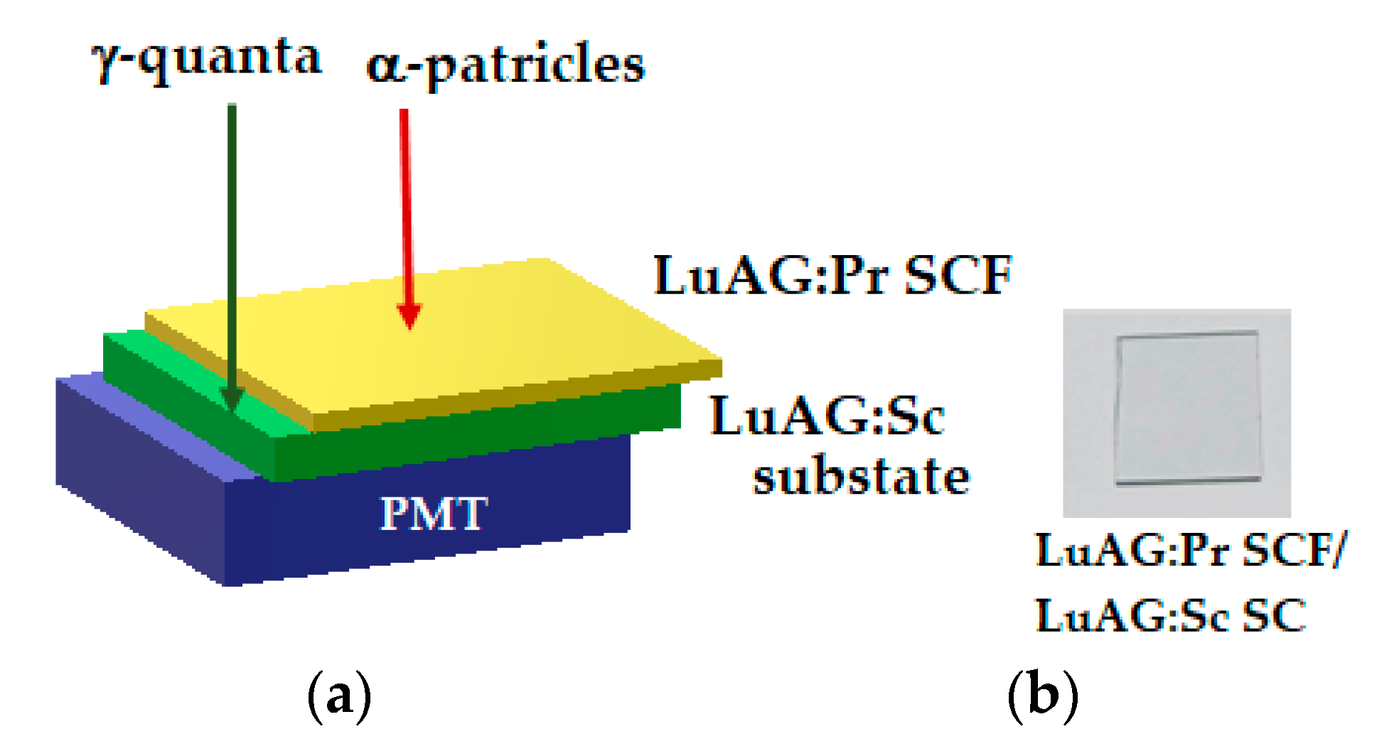
2. Growth of Composite Detectors
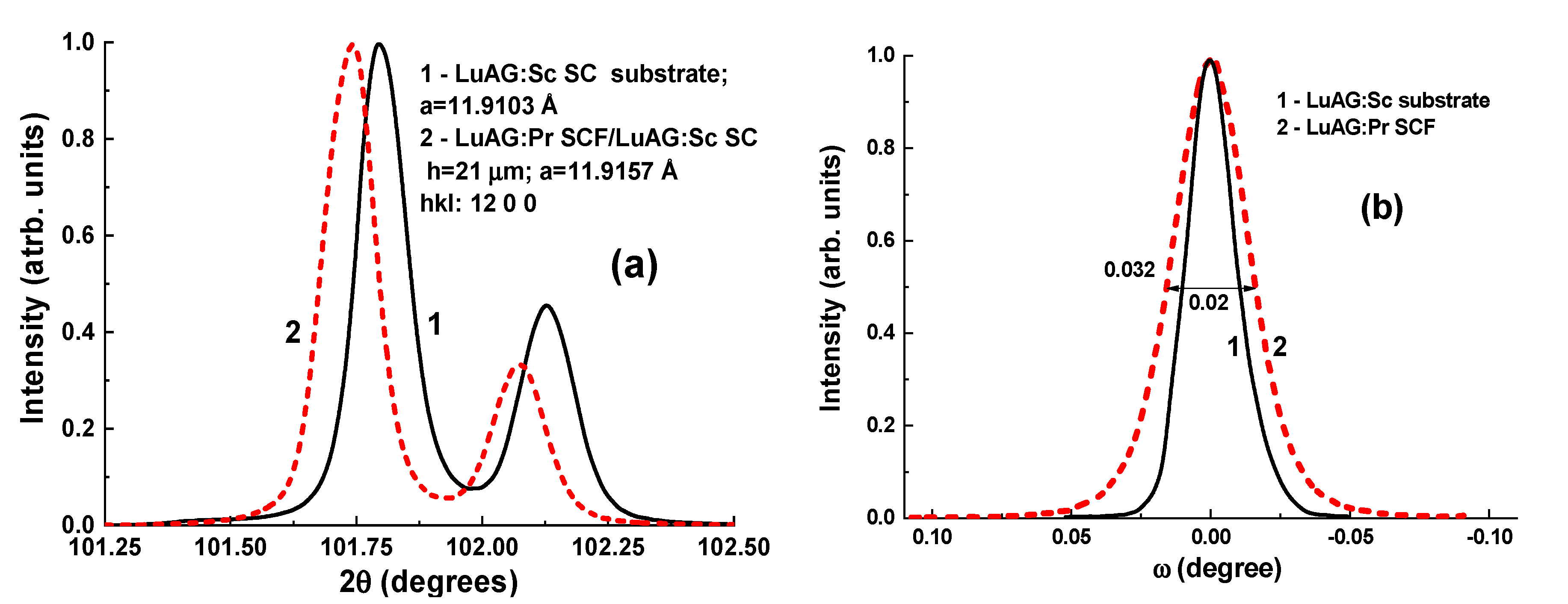
3. Experimental Results
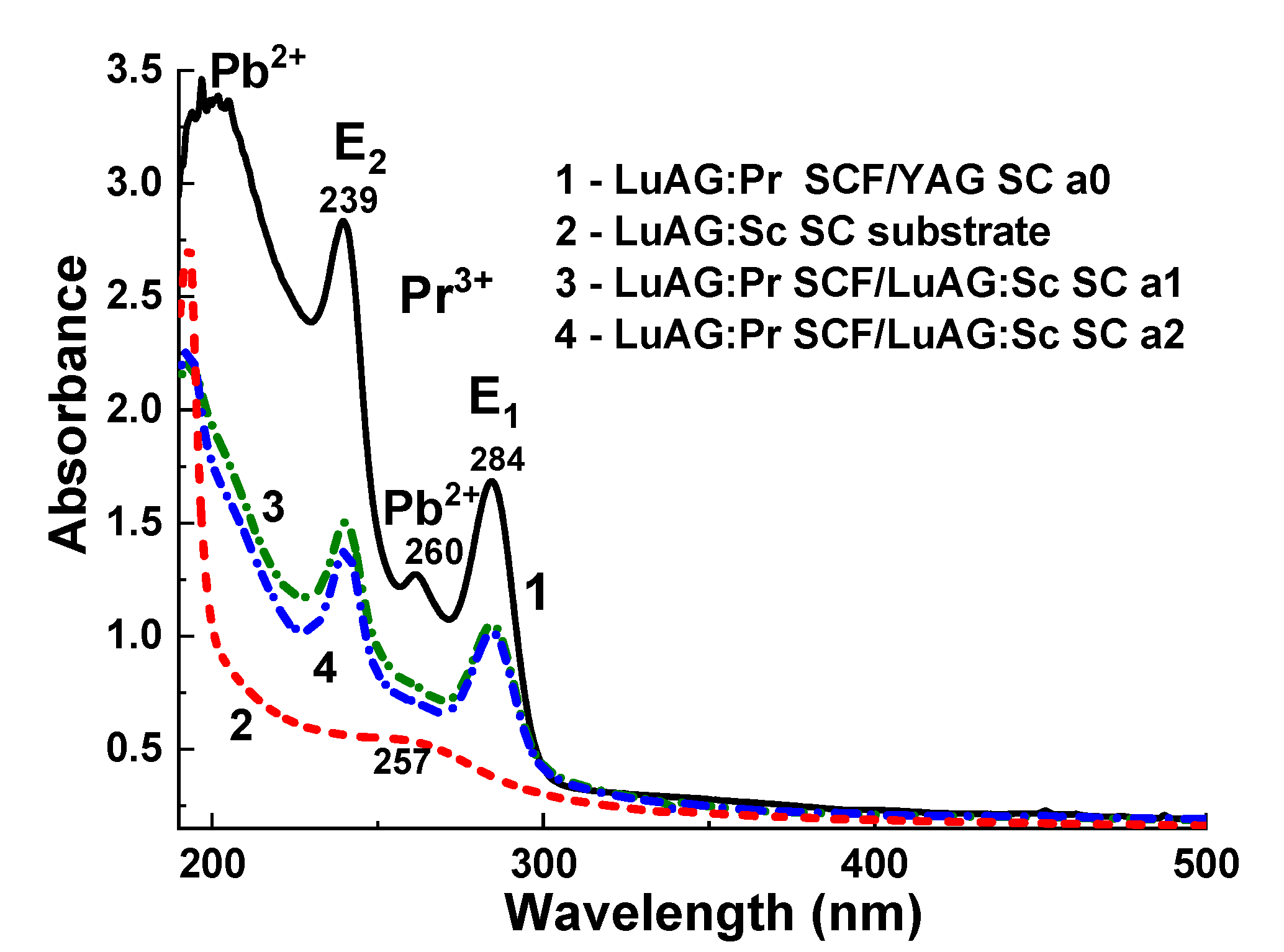
3.1. Absorption Spectra
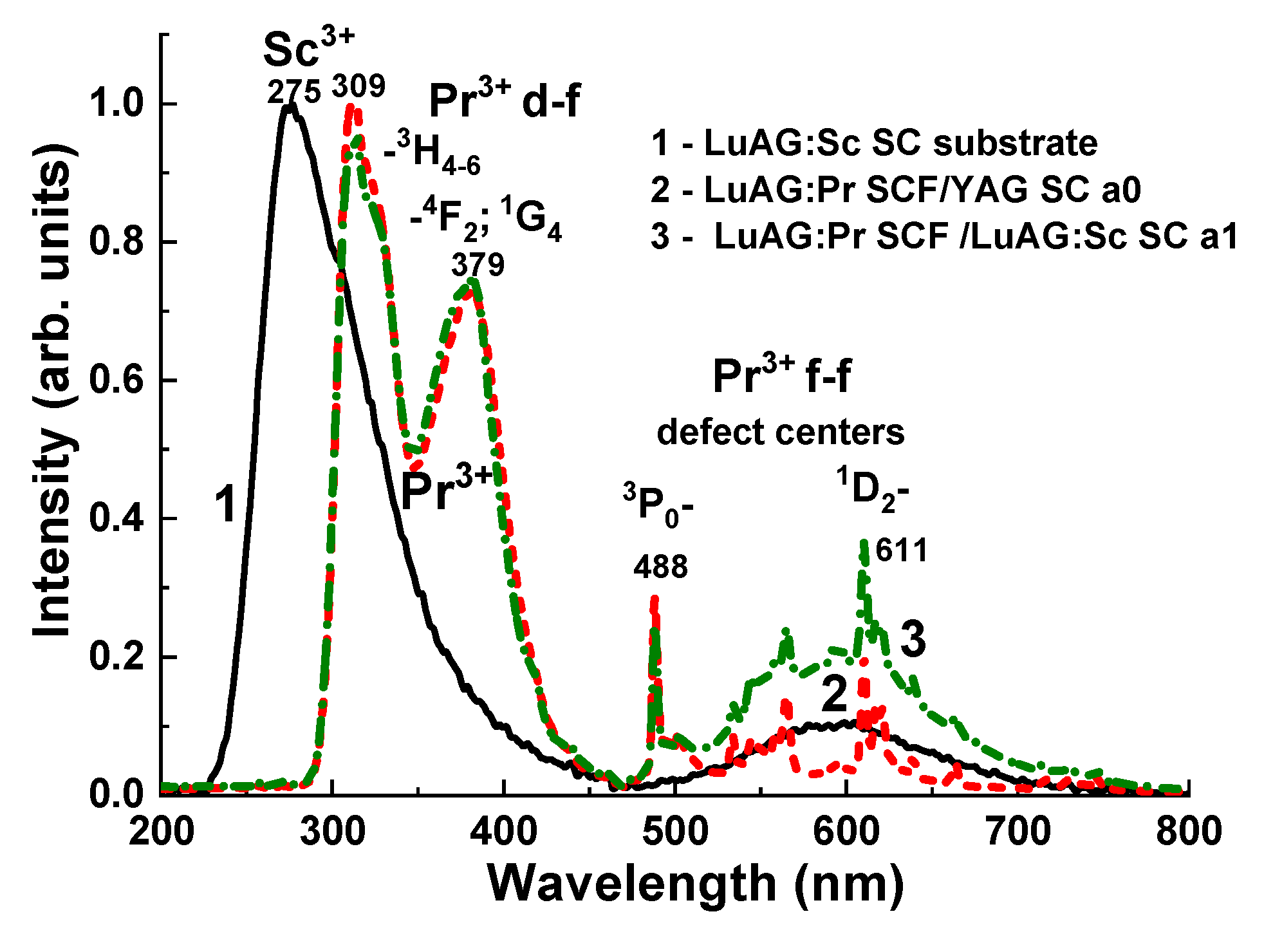
3.2. Cathodoluminescence Spectra (CL)
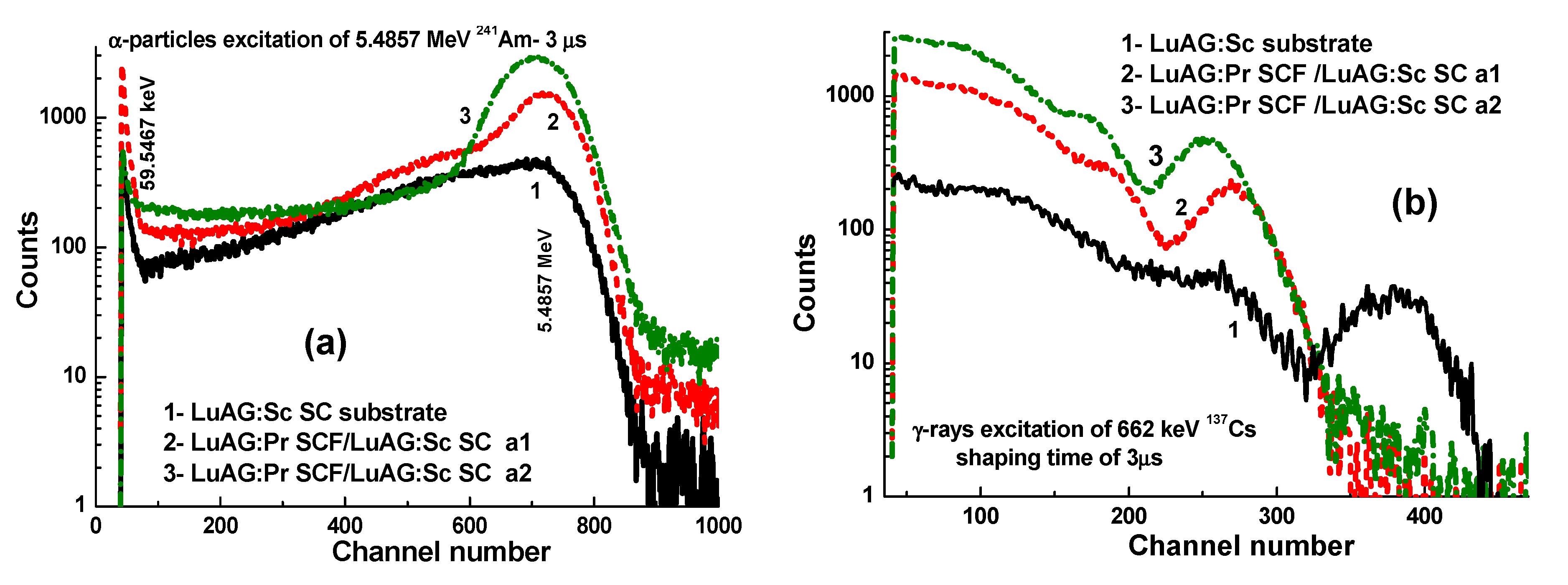
3.3. Pulse Height Spectra
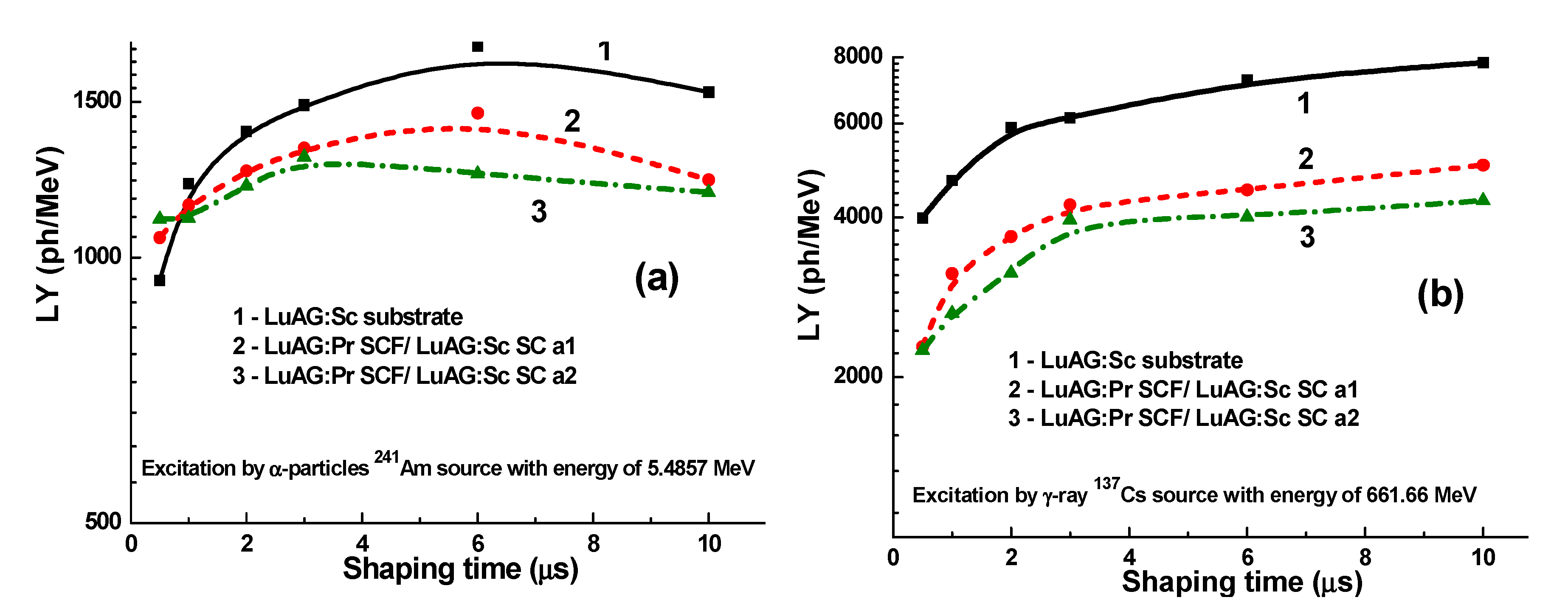
3.4. LY
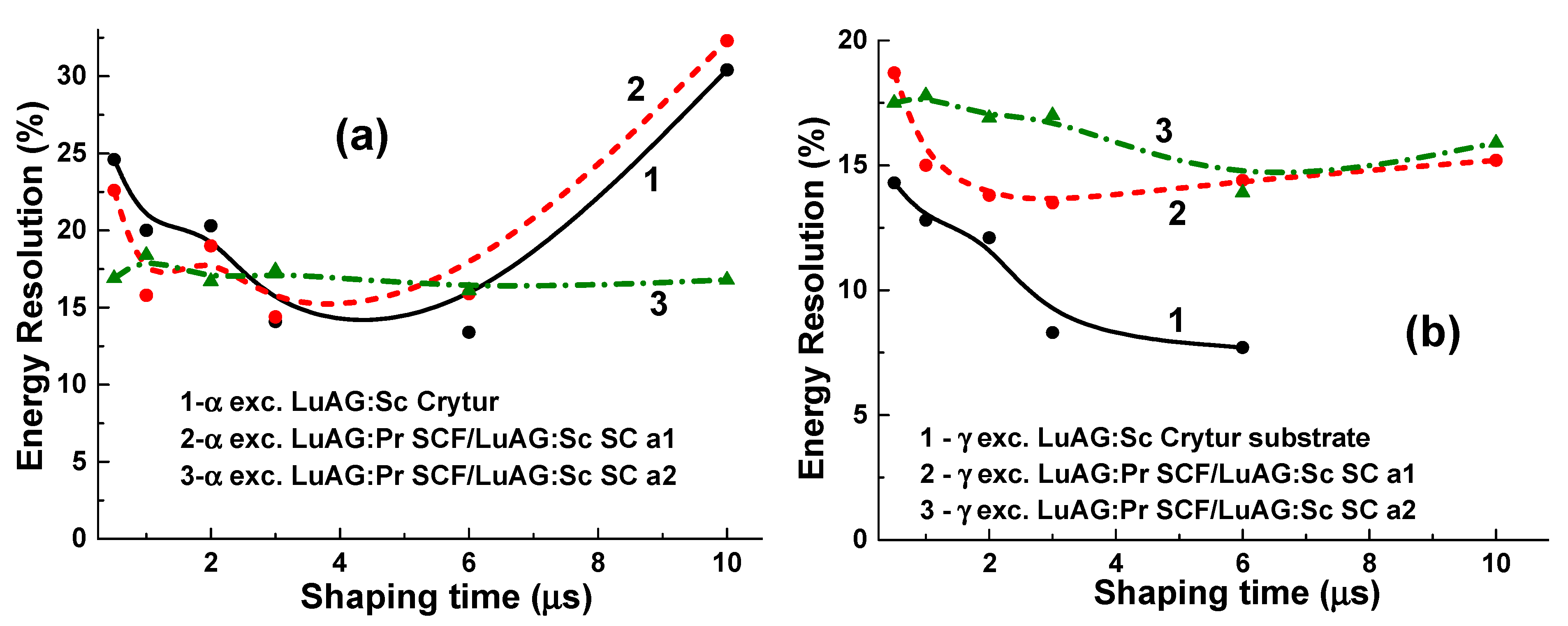
3.5. Energy Resolution
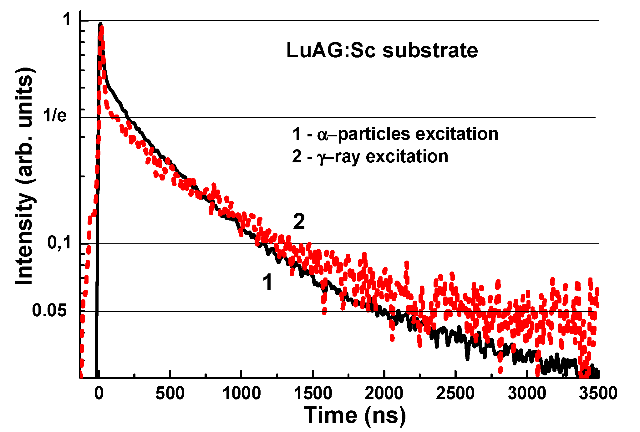
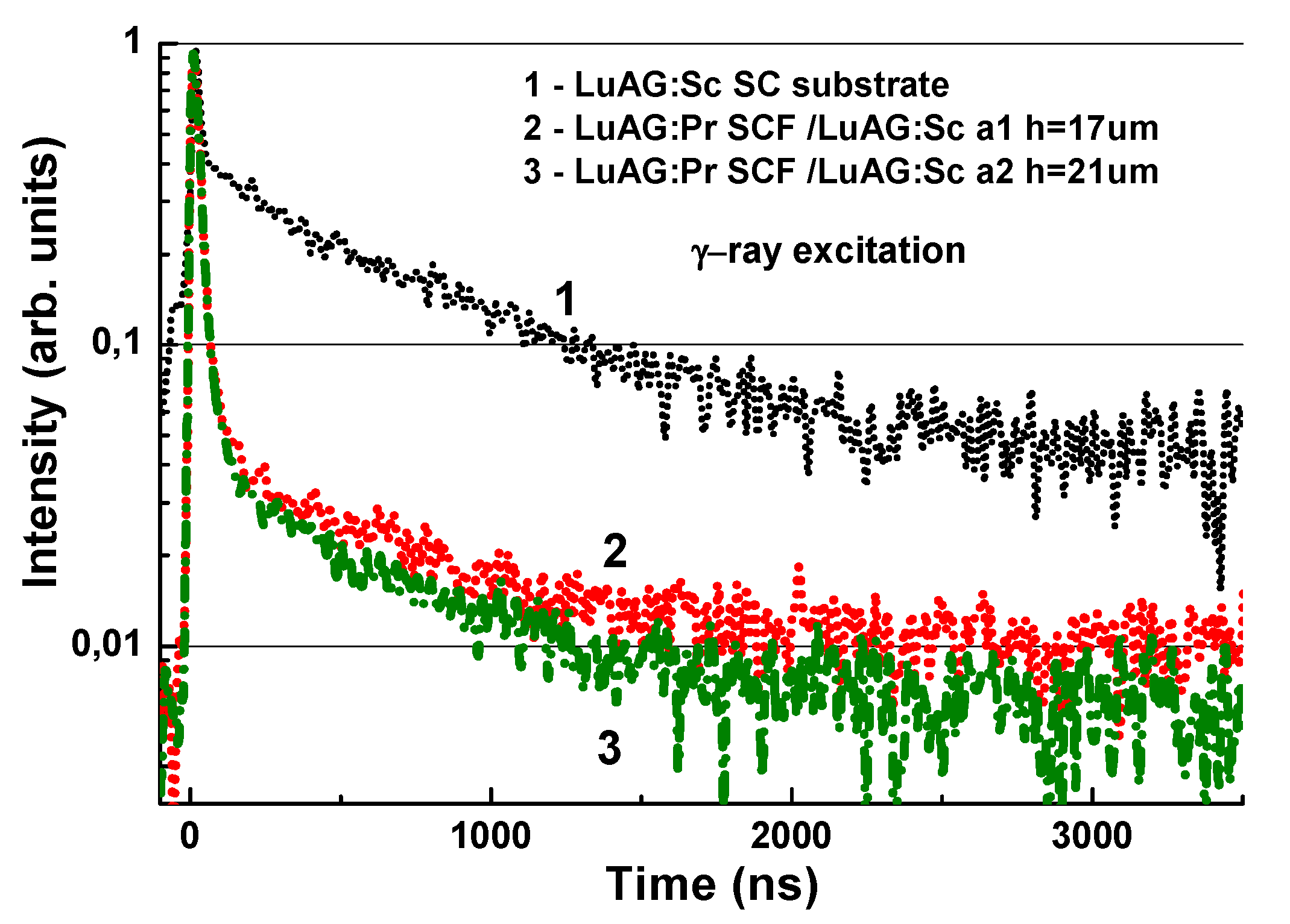
3.6. Scintillation Decay Kinetics
4. Discussion
5. Conclusions
Author Contributions
Funding
Acknowledgments
Conflicts of Interest
References
- Ferrand, B.; Chambaz, B.; Couchaud, M. Liquid phase epitaxy: A versatile technique for the development of miniature optical components in single crystal dielectric media. Opt. Mater. 1999, 11, 101–114. [Google Scholar] [CrossRef]
- Robertson, J.M.; Tol, V.M.V. Cathodoluminescent garnet layers. Thin Solid Film 1984, 114, 221–240. [Google Scholar] [CrossRef]
- Molva, E. Microchip lasers and their applications in optical microsystems. Opt. Mater. 1999, 11, 289–299. [Google Scholar] [CrossRef]
- Koch, A.; Peyrin, F.; Heurtier, P.; Chambaz, B.; Ferrand, B.; Ludwig, W.; Couchaud, M. X-ray camera for computed microtomography of biological samples with micrometer resolution using Lu3Al5O12 and Y3Al5O12 scintillators. Proc. SPIE 1999, 3659, 170. [Google Scholar]
- Martin, T.; Koch, A. Recent developments in X-ray imaging with micrometer spatial resolution. J. Synchrotron Radiat. 2006, 13, 180–194. [Google Scholar] [CrossRef] [PubMed] [Green Version]
- Zorenko, Y.; Gorbenko, V.; Zorenko, T.; Vasylkiv, Y. Luminescent properties of the Sc3+ doped single crystalline films of (Y, Lu, La)3(Al, Ga)5O12 multi-component garnets. Opt. Mater. 2014, 36, 1760–1764. [Google Scholar] [CrossRef]
- Prusa, P.; Kucera, M.; Mares, J.A.; Onderisinova, Z.; Hanus, M.; Babin, V.; Beitlerova, A.; Nikl, M. Composition tailoring in Ce-doped multicomponent garnet epitaxial film scintillators. Cryst. Growth Des. 2015, 15, 3715–3723. [Google Scholar] [CrossRef]
- Zorenko, Y.; Gorbenko, V.; Zorenko, T.; Popielarski, P.; Mosińska, L.; Fedorov, A. Luminescent and scintillation properties of the Ce3+ doped Y3−xLuxAl5O12:Ce single crystalline films. J. Lumin. 2016, 169, 822–827. [Google Scholar] [CrossRef]
- Gorbenko, V.; Zorenko, T.; Paprocki, K.; Mahlovanyi, B.; Mazalon, B.; Fedorov, A.; Zhydachevskyy, Y.; Suchocki, A.; Zorenko, Y. Epitaxial growth of single crystalline film scintillators based on the Pr3+ doped solid solution of Lu3Al5−xGaxO12 garnet. CrystEngComm 2017, 19, 7031–7040. [Google Scholar] [CrossRef]
- Rathaiah, M.; Kučera, M.; Průša, P.; Beitlerová, A.; Nikl, M. Effect of Si4+ co-doping on luminescence and scintillation properties of Lu3Al5O12: Ce, Ca epitaxial garnet films. Opt. Mater. 2019, 91, 321–325. [Google Scholar] [CrossRef]
- Průša, P.; Kučera, M.; Vedda, A.; Fasoli, M.; Moretti, F.; Hanuš, M.; Lučeničová, Z.; Vrba, T.; Nikl, M. Substantial reduction of trapping probability by Mg co-doping in LuAG: Ce, Mg epitaxial garnet films. J. Lumin. 2021, 238, 118230. [Google Scholar] [CrossRef]
- Buryi, M.; Laguta, V.V.; Nikl, M.; Zorenko, T.; Gorbenko, V.; Zorenko, Y. LPE growth and study of the Ce3+ incorporation in LuAlO3: Ce single crystalline film scintillators. CrystEngComm 2019, 21, 3313–3321. [Google Scholar] [CrossRef]
- Martin, T.; Douissard, P.-A.; Couchaud, M.; Cecilia, A.; Baumbach, T.; Dupré, K.; Rack, A. LSO-based single crystal film scintillator for synchrotron-based hard X-ray micro-imaging. IEEE Trans. Nucl. Sci. 2009, 56, 1412–1418. [Google Scholar] [CrossRef]
- Rack, A.; Zabler, S.; Mueller, B.R.; Riesemeier, H.; Weidemann, G.; Lange, A.; Goebbels, J.; Hentschel, M.; Goerner, W. High resolution synchrotron-based radiography and tomography using hard X-rays at the BAMline (BESSY II). Nucl. Instrum. Methods Phys. Res. A 2008, 586, 327–344. [Google Scholar] [CrossRef]
- Zorenko, Y.; Gorbenko, V.; Savchyn, V.; Zorenko, T.; Grinyov, B.; Sidletskiy, O.; Fedorov, A.; Mares, J.A.; Nikl, M.; Kucera, M. Lu2SiO5:Ce and Y2SiO5:Ce single crystals and single crystalline film scintillators: Comparison of the luminescent and scintillation properties. Radiat. Meas. 2013, 56, 85–89. [Google Scholar] [CrossRef]
- Kurosawa, S.; Yoshikawa, A.; Gorbenko, V.; Zorenko, T.; Witkiewicz-Lukaszek, S.; Zorenko, Y. Composite Scintillators Based on the Films and Crystals of (Lu,Gd,La)2Si2O7 Pyrosilicates. IEEE TNS 2020, 67, 994–998. [Google Scholar] [CrossRef]
- Zorenko, Y.; Gorbenko, V.; Voznyak, T.; Konstankevych, I.; Savchyn, V.; Batentschuk, M.; Winnacker, A.; Brabec, C.J. Scintillating screens based on the single crystalline films of multicomponent garnets: New achievements and possibilities. IEEE Trans. Nucl. Sci. 2012, 59, 2281–2285. [Google Scholar] [CrossRef]
- Zorenko, Y.; Zorenko, T.; Gorbenko, V.; Savchyn, V.; Voznyak, T.; Fabisiak, K.; Fedorov, A. Zhusupkalieva. Luminescent properties of Al2O3:Ce single crystalline films under synchrotron radiation excitation. Opt. Mater. 2016, 59, 141–144. [Google Scholar] [CrossRef]
- Zorenko, T.; Gorbenko, V.; Dulina, N.; Matveevskaya, N.; Yavetskiy, R.; Babayevskaya, N.; Zorenko, Y. Comparative study of the luminescent properties of oxide compounds under synchrotron radiation excitation: Lu2O3:Eu nanopowders, ceramics and films. J. Lumin. 2018, 199, 461–464. [Google Scholar] [CrossRef]
- Gorbenko, V.; Galązka, Z.; Zorenko, T.; Paprocki, K.; Witkiewicz, S.; Zorenko, Y. Growth and LPE luminescent and scintillation properties of Ga2O3 based monocrystalline films. CrystEngComm 2021, in press. [Google Scholar]
- Zorenko, Y.; Novosad, S.S.; Pashkovskii, M.V.; Lyskovich, A.B.; Savitskii, V.G.; Batenchuk, M.M.; Malyutenkov, P.S.; Patsagan, N.I.; Nazar, I.V.; Gorbenko, V.I. Epitaxial structures of garnets as scintillating detectors of ionizing radiations. J. Appl. Spectrosc. 1990, 52, 645. [Google Scholar] [CrossRef]
- Zorenko, Y.; Gorbenko, V.; Konstankevych, I.; Grinev, B.; Globus, M. Scintillation properties of Lu3Al5O12:Ce single-crystalline films. Nucl. Instrum. Methods Phys. Res. A 2002, 486, 309–314. [Google Scholar] [CrossRef]
- Nikl, M. (Ed.) Nanocomposite, Ceramic, and Thin Film Scintillators; Jenny Stanford Publishing: Singapore, 2017; ISBN 978-981-4745-22-2. [Google Scholar]
- Nikl, M.; Yoshikawa, A.; Kamada, K.; Nejezchleb, K.; Stanek, C.R.; Mares, J.A.; Blazek, K. Development of LuAG-based scintillator crystals—A review. Prog. Cryst. Growth Charact. Mater. 2013, 59, 47–72. [Google Scholar] [CrossRef]
- Witkiewicz-Lukaszek, S.; Gorbenko, V.; Zorenko, T.; Paprocki, K.; Sidletskiy, O.; Gerasymov, I.; Mares, J.A.; Kucerkova, R.; Nikl, M.; Zorenko, Y. Composite thermoluminescent detectors based on the Ce3+ doped LuAG/YAG and YAG/LuAG epitaxial structures. IEEE TNS 2018, 65, 2114–2119. [Google Scholar] [CrossRef]
- Witkiewicz-Lukaszek, S.; Gorbenko, V.; Zorenko, T.; Paprocki, K.; Sidletski, O.; Gerasymov, I.; Mares, J.A.; Kucerkova, R.; Nikl, M.; Zorenko, Y. Composite scintillators based on the crystals and single crystalline films of LuAG garnet doped with Ce3+, Pr3+ and Sc3+ ions. Opt. Mater. 2018, 84, 593–599. [Google Scholar] [CrossRef]
- Mares, J.A.; Witkiewicz-Lukaszek, S.; Gorbenko, V.; Zorenko, T.; Kucerkova, R.; Beitlerova, A.; D′Ambrosio, C.; Dlouhy, J.; Nikl, M.; Zorenko, Y. Alpha and gamma spectroscopy of composite scintillators based on the LuAG:Pr crystals and single crystalline films of LuAG:Ce and (Lu,Gd,Tb)AG:Ce garnets. Opt. Mater. 2019, 96, 109268. [Google Scholar] [CrossRef]
- Zorenko, Y.; Batentschuk, M.; Gorbenko, V.; Pashkovsky, M.; Konstankevych, I. Factors determining the energy yield of luminophors based on single-crystal films of Al2O3-Y2O3-R2O3 oxides. J. Appl. Spectrosc. 1999, 66, 953–959. [Google Scholar] [CrossRef]
- Zorenko, T.; Zorenko, Y.; Pavlyk, B.; Turchak, R.; Gorbenko, V.; Konstankevych, I.; Savchyn, V.; Voznyak, T. Influence of thermal treatment and γ-radiation on absorption, luminescence and scintillation properties of Lu3Al5O12:Ce single crystalline films. Radiat. Meas. 2007, 42, 557–560. [Google Scholar] [CrossRef]
- Zorenko, T.; Gorbenko, V.; Petrosyan, A.; Gieszczyk, W.; Bilski, P.; Zorenko, Y. Intrinsic and defect-related luminescence of YAlO3 and LuAlO3 single crystals and films. Opt. Mater. 2018, 86, 376–381. [Google Scholar] [CrossRef]
- Zorenko, Y.; Zorenko, T.; Voznyak, T.; Mandowski, A.; Qi, X.; Batentschuk, M.; Fridrich, J. Luminescence of F+ and F centers in AI2O3-Y2O3 oxide compounds. IOP Conf. Ser. Mater. Sci. Eng. 2010, 15, 012060. [Google Scholar] [CrossRef]
- Zorenko, Y.; Batenchuk, M.; Gorbenko, V.; Pashkovsky, M. Single-crystalline oxide films of the Al2O3-Y2O3-R2O3 system as optical sensors of various types of ionizing radiation: Significant advantages over volume analogs. Proc. SPIE 1997, 2967, 101. [Google Scholar]
- Zorenko, Y.; Gorbenko, V.; Voznyak, T.; Savchyn, V.; Nizhankovskiy, S.; Dan’ko, A.; Puzikov, V.; Laguta, V.; Mares, J.A.; Nikl, M.; et al. Luminescent and scintillation properties of Lu3Al5O12:Sc single crystal and single crystalline films. Opt. Mater. 2012, 34, 2080. [Google Scholar] [CrossRef]
- Gobiein, S. Phoswich Detectors for High Energy Backgrounds. Available online: http://www.detectors.saint-gobain.com (accessed on 29 December 2017).


| No. of Sample | Type of SCF | Type of Substrate | h, µm | Tg, °C | f, μm/min | LY, % |
|---|---|---|---|---|---|---|
| Crytur | LuAG:Sc | 500 | 95 | |||
| a0 | LuAG:Pr | YAG | 19 | 975 | 0.19 | 100 |
| a1 | LuAG:Pr | LuAG:Sc | 21 | 1007 | 0.3 | 80 |
| a2 | LuAG:Pr | LuAG:Sc | 17 | 995 | 0.66 | 69 |
| LY, ph/MeV | Shaping Time, μs | LuAG:Sc SC sub, μs | LuAG:Pr SCF/LuAG:Sc SC a1, μs | LuAG:Pr SCF/LuAG:Sc SC a2, μs |
|---|---|---|---|---|
| LYα | 0.5 | 942 | 1054 | 1071 |
| LYα | 10 | 1539 | 1225 | 1185 |
| LYɣ | 0.5 | 3988 | 2284 | 2246 |
| LYɣ | 10 | 7819 | 5012 | 4302 |
| LYα/LYɣ | 0.5 | 0.23 | 0.46 | 0.49 |
| LYα/LYɣ | 10 | 0.19 | 0.24 | 0.52 |
| Intensity | LuAG:Sc Substrate | ||
|---|---|---|---|
| tα, ns | tγ, ns | tα/tγ or tγ/tα Ratio | |
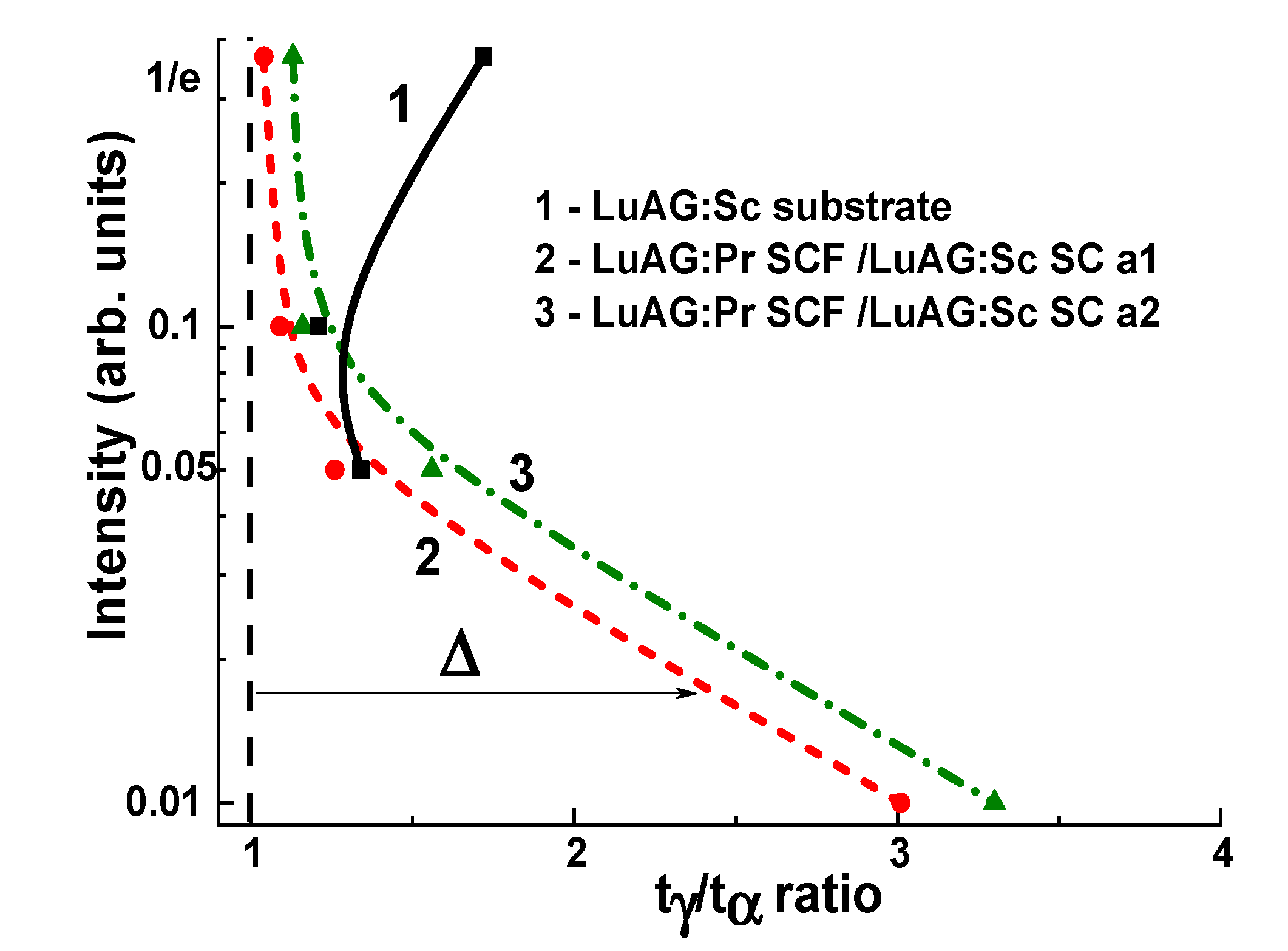
| 1/e | 208 | 121 | 1.72 |
| 0.1 | 1125 | 1360 | 1.21 |
| 0.05 | 1982 | 2646 | 1.34 |
| Intensity | a1 | a2 | ||||
|---|---|---|---|---|---|---|
| tα, ns | tγ, ns | tγ/tα Ratio | tα, ns | tγ, ns | tγ/tα Ratio | |
| 1/e | 34.8 | 36.5 | 1.04 | 32.7 | 37 | 1.13 |
| 0.1 | 60 | 65.5 | 1.09 | 58 | 67.4 | 1.16 |
| 0.05 | 85 | 107.6 | 1.26 | 86 | 133 | 1.55 |
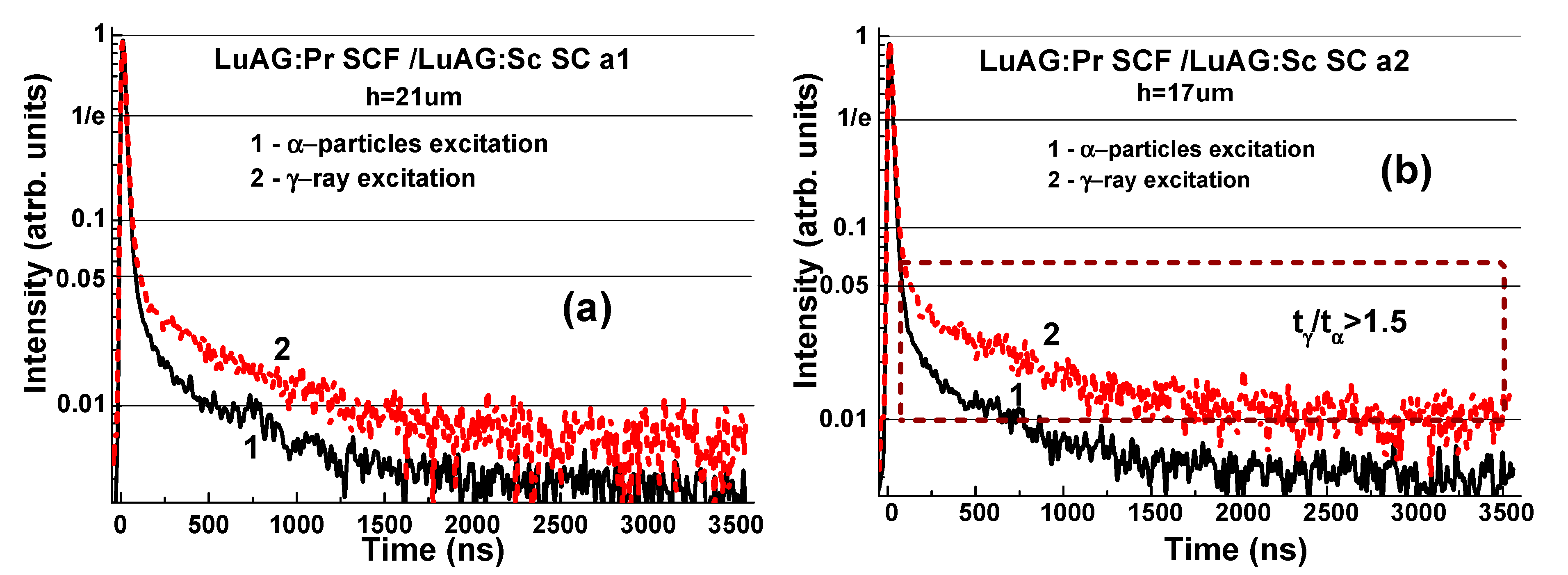
| 0.01 | 545 | 1641 | 3.01 | 830 | 2733 | 3.3 |
Publisher’s Note: MDPI stays neutral with regard to jurisdictional claims in published maps and institutional affiliations. |
© 2021 by the authors. Licensee MDPI, Basel, Switzerland. This article is an open access article distributed under the terms and conditions of the Creative Commons Attribution (CC BY) license (https://creativecommons.org/licenses/by/4.0/).
Share and Cite
Gorbenko, V.; Witkiewicz-Łukaszek, S.; Zorenko, T.; Syrotych, Y.; Mares, J.A.; Kucerkova, R.; Nikl, M.; Sidletskiy, O.; Fedorov, A.; Zorenko, Y. Development of Composite Scintillators Based on the LuAG: Pr Single Crystalline Films and LuAG:Sc Single Crystals. Crystals 2021, 11, 846. https://0-doi-org.brum.beds.ac.uk/10.3390/cryst11080846
Gorbenko V, Witkiewicz-Łukaszek S, Zorenko T, Syrotych Y, Mares JA, Kucerkova R, Nikl M, Sidletskiy O, Fedorov A, Zorenko Y. Development of Composite Scintillators Based on the LuAG: Pr Single Crystalline Films and LuAG:Sc Single Crystals. Crystals. 2021; 11(8):846. https://0-doi-org.brum.beds.ac.uk/10.3390/cryst11080846
Chicago/Turabian StyleGorbenko, Vitalii, Sandra Witkiewicz-Łukaszek, Tetiana Zorenko, Yuri Syrotych, Jiri A. Mares, Romana Kucerkova, Martin Nikl, Oleg Sidletskiy, Alexander Fedorov, and Yuriy Zorenko. 2021. "Development of Composite Scintillators Based on the LuAG: Pr Single Crystalline Films and LuAG:Sc Single Crystals" Crystals 11, no. 8: 846. https://0-doi-org.brum.beds.ac.uk/10.3390/cryst11080846