
Investigation of CoCr Dental Alloy: Example from a Casting Workflow Standpoint


1
School of Dental Medicine, University of Belgrade, Dr. Subotića 8, 11000 Belgrade, Serbia
2
Faculty of Mechanical Engineering, University of Maribor, Smetanova 17, 2000 Maribor, Slovenia
3
Zlatarna Celje d.o.o., Kersnikova Ulica 19, 3000 Celje, Slovenia
*
Author to whom correspondence should be addressed.
Crystals 2021, 11(8), 849; https://0-doi-org.brum.beds.ac.uk/10.3390/cryst11080849
Submission received: 24 May 2021
/
Revised: 16 July 2021
/
Accepted: 18 July 2021
/
Published: 22 July 2021
Abstract
:Cobalt-chromium (CoCr) alloys have been used in dentistry for dental bridges, crowns and implants for decades. When using CoCr alloys, a number of fractures have occurred in the Dental Laboratory, both when handling the castings and after they have been placed in the patient’s mouth. It is assumed that the key cause of the resulting fractures of CoCr dental bridges is the casting process, which includes the preparation and mixing of the basic components of the CoCr dental alloy, unstable solidification and the final treatment of the tooth casting surface. The aim of this study was, therefore, to examine three castings differently prepared from the CoCr alloy. For the initial CoCr alloy, we selected the one supplied directly from the manufacturer; three test samples were CoCr alloy remelted four times in the same crucible, while the fourth sample was the remaining solidified alloy from the crucible, taken at the last remelting. Characterisation of the microstructure of all four samples was performed by optical and scanning electron microscopy equipped with an energy dispersive X-ray spectroscope and X-ray diffractometry. Microhardness measurements were also performed. The investigation revealed that the microstructure of the castings is composed of a CoCr alloy matrix with a eutectic interdendritic composition and interdendritic precipitates, which were rich in W and Mo. The two oxides were identified as chromium oxide with silicon content and chromium oxide, which originated from the CoCr alloy as casting residue. The high content of silicon in the chromium oxide can be attributed to the silicon oxide from the ceramic melting crucible, mixed in with the remains from the CoCr alloy melting. The second oxide showed a more regular elemental content for chromium oxide, mixed with a small quantity of impurities and the casting CoCr alloy. Based on this research, some recommendations were made for working with CoCr alloys in the Dental Laboratory, with the aim of reducing the risk of dental bridge fractures in the future.
1. Introduction
Cobalt-chrome (Co-Cr) alloys are well known for their biocompatibility in addition to their good mechanical properties, high melting points and excellent corrosion resistance [1,2]. They have been used in dentistry for decades, for dental bridges, crowns and implants [3]. Biocompatibility is the most important property for such applications, which comes from the formation of a hard, passive, oxide layer on the surface of these alloys [4]. This prevents further corrosion of the alloy and the release of metal ions from the alloy into the mouth, which could cause systemic and local toxicity, allergies, or carcinogenicity [5]. Biocompatibility studies usually refer to noble metal alloys as the standard with which to compare other alloys, due to their anti-corrosive properties and acknowledging noble metal alloys as generally suitable for clinical use through their long history of use in dental restorations [3,6,7,8]. Studies investigating substance release from dental alloys in the mouth or saliva show different releases of metallic ions, depending on the alloy used [4,7,8,9,10]. From the usual elements present in dental alloys, in vitro cell viability tests showed that gold (Au), palladium (Pd), platinum (Pt) and indium (In) ions have no cytotoxic effect, while chromium (Cr), copper (Cu) and silver (Ag) ions were toxic and nickel (Ni), zinc (Zn) and cobalt (Co) ions were highly toxic [4].
CoCr alloys have a Face Centred Cubic (FCC) lattice—a γ phase at high temperatures, which affects the ductility—and the Hexagonal Close Packed (HCP) lattice—an ε phase at room temperature, which affects corrosion and wear resistance [11]. The chromium content of 22–28% in the CoCr alloy forms the passive oxide layer on the surface [11], and due to its presence, M23C6 type carbides are formed, which increase the hardness and wear resistance [12]. More stable M6C carbides are formed instead of M23C6 in CoCr alloys with tungsten and molybdenum concentrations over 4 wt%. The carbon content in CoCr alloys can reach up to 0.35%, forming carbide precipitates with sizes ranging from 50 to 300 nm [13], found in the interdendritic areas of the CoCr alloy microstructure [11,12]. Cobalt-based alloys go through a slow FCC to HCP martensitic transformation, keeping the FCC in a metastable state under normal cooling rates [13]. The properties of CoCr alloys are, thus, dependent on the γ phase and ε phase ratio, which depends on the alloy processing and the presence and distribution of carbides in the microstructure.
Corrosion and substance release are increased when the dental alloys are remelted and recast into new dental restorations, as this process introduces impurities and alloy composition inaccuracies, promoting corrosion. Usually, noble metal alloys are recast more frequently due to their high prices. Several studies have investigated the practice of reusing old dental alloys. One such systematic review included all the studies on dental alloy recasting from MEDLINE, Dentistry and Oral Science Source, Science Direct and the ISI Web of Science (up to July 2014) [14]. Thirty-four studies were included, published between 1983 and 2014. The number of recastings ranged from one to ten. The percentage of new alloys ranged from 0 to 100 wt%, although the mean value was 50 wt%. This study revealed that recasting up to four times seems acceptable only if at least 50% of new alloy is added during each recasting procedure [14]. Recasting with at least 50% of new alloy also does not affect the physico-chemical and mechanical properties of the alloys [15]. Some reports show that even 25% of new alloy could be used, when remelting only up to one or two times [16], while others state that using the contributions of scrap in castings deteriorates the strength properties substantially [17].
In the Prosthetics Clinic at the University of Belgrade School of Dentistry, practices with Co-Cr based alloys have shown numerous breakage of dental bridges when still working with them or even after they have been placed inside the patient’s mouth. Some 15–20 cases were seen. This resulted in the need for making new dental bridges and patient discomfort and dissatisfaction. The properties of these alloys are documented extensively with mechanical and clinical testing, in addition to their daily use in medical products. Based on this, the aim of this research was to explain the reason for the numerous breaks, in order to avoid problems when working with Co-Cr alloys and to minimise unnecessary dental work repairs in the future.
Producing dental products from CoCr alloys follows the conventional lost-wax casting approach [2,18]. Initially, the shape of the metal frame is made with a wax-up technique. The wax model is prepared with attached casting rods or sprues, invested into an investing material and heated for the wax to burn away. The metal should occupy the entire volume inside the void and is forced inside the mould with various devices while still molten. Finishing the product then involves cutting off the casting rods, polishing, and removing oxides, with minor final adjustments before sending to the dental office for fitting [19].
The reason for the failure of dental bridges from Co-Cr alloys is likely to be somewhere in the casting process, from the molten state to solidification and final treatment. In practice, a single ceramic melting crucible is used several times when casting the same alloy repeatedly. For this research, we analysed different states of the CoCr alloy in question: its initial state as received from the manufacturer, its cast state after several castings from the same crucible and the residual solidified alloy from the crucible.
2. Materials and Methods
2.1. Materials
The issue of dental metal frames breaking was with a CoCr metal based alloy I-BOND NF (Interdent d.o.o., Celje, Slovenia). The alloy conforms to Standards EN ISO 22674:2016 [20] and EN ISO 9693:2019 [21] for dental alloys. The chemical composition and properties of the alloy were obtained from the manufacturer and are given in Table 1.
2.2. Methods
2.2.1. Preparation of Samples
The melting of the CoCr dental alloy was performed at the School of Dental Medicine, Belgrade, Serbia, in an induction melting furnace at ambient pressure and temperature T = 1600 °C, using a ceramic melting crucible based on silicon oxides. The casting of the melted alloy into the form of tooth dentures was performed by applying centrifugal force. The samples of the tested dental alloy were as follows: CoCr alloy, which was melted once in the induction apparatus and cast in the conditions of the dental–technical laboratory and was used for further remelting and mixing with 50% new alloy (the mixing rate was always 50 mass.% of the new alloy, regardless of the previous remelting). Four-times remelting and casting was performed in the conditions of the dental–technical laboratory.
Three different material specimens concerning the CoCr alloy were examined, as shown in Figure 1: CoCr alloy as received from the manufacturer (Sample 1), cast CoCr after four-times casting from the same crucible (Samples 2 and 3) and the residual solidified CoCr alloy from the melting crucible (Sample 4) after the last casting.
2.2.2. Preparation of Metallographic Samples
Samples 1 and 2 were first cut and mounted with a compression mounting resin for easier metallographic examination. Sample 3 had casting sprues collected from the casting mould. Sample 4 was collected from the broken pieces at the bottom of the crucible (the residue of the CoCr alloy during remelting) and then mounted. All of the mounted samples were then ground using increasingly finer SiC grinding papers (P120, P180, P400, P600, P800, P1000, P2500 and P4000), followed by polishing with a 3 µm paste and 1 µm polishing suspension. The samples were finally etched using HNO3:HCl (1:3). The schematic presentation of the examined samples is shown in Figure 1.
2.3. Characterisation
The microstructure of the samples was investigated with an optical metallographic microscope, NIKON Epiphot 300 (Tokyo, Japan), with an Olympus DP12 camera (Boston, MA, USA). A Scanning Electron Microscope (SEM), Sirion 400NC (FEI, Hillsboro, OR, USA), with an Energy-Dispersive X-ray spectroscopy detector INCA 350 (Oxford Instruments, Abingdon, UK), was used for detailed microstructure observation and microchemical analyses of the different metallic phases found in the samples. The samples were analysed from secondary and backscattered electron images.
The microhardness of the samples was examined with Vickers hardness measurements HV1 on the ZWICK 3212 machine (Zwick Roell Group, Ulm, Germany). The measurement load of 1 kg was selected for the given sample dimensions.
The additional analysis of phases was performed on sample 4 with a Panalytical XPERT Pro PW 3040/60 goniometer (Panalytical, Almelo, The Netherlands) 2theta 10–90° with a step of 0.002° and a time of 100 ms per step. The anode was Cu (Kalfa = 0.154 nm) with a current of 40 mA and a voltage of 45 kV.
3. Results
3.1. Microstructure Observation
Optical micrographs of all the investigated samples are shown in Figure 2, where the characteristic dendritic microstructure is clearly visible. Sample 1 is the initial CoCr alloy, while samples 2 and 3 are an already used alloy mixed with certain percentages of new material. The microstructure of sample 3 shows larger dendrites, with various inclusions and impurities. From Figure 2, it is possible to infer the solidification directions: (i) the solidification direction was towards the centre of sample 1, as shown in the top left image, (ii) while, in samples 2 and 3, it can be deduced that the solidification was uniform in all directions. When comparing the samples, the images show a much finer grain size for sample 1 than for samples 2 and 3. The microstructure of the residual solidified CoCr alloy from the melting crucible did not show a typical dendritic morphology, but the presence of a large number of defects from voids and inclusions was evident.
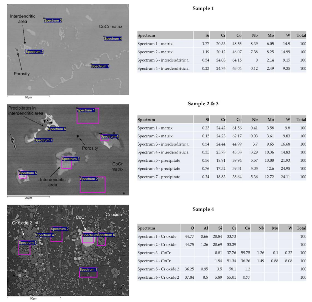
Figure 3 shows the SEM images of the samples with EDX elemental analysis. The backscatter electron image shows the atomic number contrast on the surface of the samples. In addition to some pores, two phases were found in Sample 1, the CoCr matrix and eutectic interdendritic areas. A similar structure was found in Sample 2, while the pores and the dendrites were somewhat larger in size. In addition, precipitates were found in the interdendritic areas, seen in the white colour of the BSE signal in the electron microscope. In sample 4, two types of compounds were found: chromium spinel with silicon content and a chromium compound with small remains of the CoCr alloy (Co spinel 2). The average compositional mass percentage values of the phases were as follows:
- CoCr matrix: 63Co, 24Cr, 9W, 3Mo, <1Nb, <1Si;
- Interdendritic area: 48Co, 23Cr, 15W, 8Mo, 5Nb, <1Si;
- Precipitates in interdendritic areas: 42Co, 18Cr, 21W, 11Mo, 7Nb, <1Si;
- Cr spinel: 33Cr, 45O, 21Si;
- Cr spinel 2: 59Cr, 37O, 3Si.
3.2. XRD Measurement
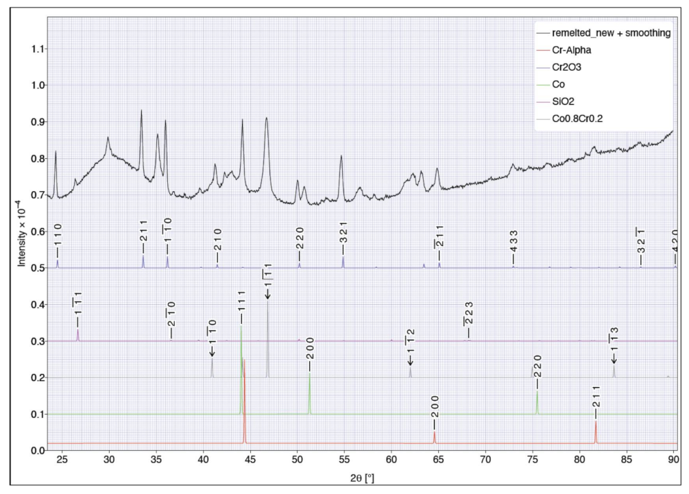
The oxide remains in the ceramic casting crucible from sample 4 were determined by XRD analysis, which indicated the presence of the Cr2O3 phase, as shown in Figure 4. The presence of α-Cr, Co phases was also identified, as well as SiO2 and Co0.8Cr0.2. All the phases are illustrated with characteristic dots that have been compared with literature data on structural factors [22]—see the Supplementary File. The obtained identification results for the Cr and Co phases are based on the origin in the Co-Cr phase diagram [23], which, in the equilibrium state, illustrates the possible presence of phases under ideal conditions with the selected chemical composition of the two-component system. A more detailed analysis of the recorded XRD spectrum did not confirm the presence of a typical sigma phase from the Co-Cr system [23,24], which would be expected at 63 mass% Co and 24 mass% Cr, but showed the existence of the Co0.8Cr0.2 phase, which indicates that the remelted alloy residue was metastable, as the presence of an atypical phase was confirmed. Thus, we also identified the SiO2 phase. Considering that the XRD analysis was performed on the oxide residue of the remelted CoCr dental alloy, we can conclude that the identified set of phases was diverse and atypical for CoCr dental alloy, which poses a high risk in dental practice if such a residue of dental alloys is used to make dental prosthetic products. The XRD analysis also recorded a high background, which originated for two reasons: (i) the sample was irregular in shape and partly also phosphorescent and (ii) the sample was a mixture of mass, metal particles and oxides.
3.3. Microhardness Measurement
The results of all the microhardness measurements are presented in Table 2. Samples 1, 2 and 3 had a measured hardness between 276 HV1 and 358 HV1, depending on the site of the measurement. The average HV1 values of the samples were 313, 305 and 321, as seen in Table 2. The declared hardness from the manufacturer was 285 HV10. The measured values were comparable, while it is important to mention that the measurement uncertainty increased with decreasing the measurement loads. The prepared samples were dimensionally small, not allowing for usage of greater measurement loads than HV1, as such loads are, typically, used for smaller samples, thin specimens, plated surfaces or thin films. Sample 4 had much higher hardness values, ranging from 958 HV1 to 1098 HV1.
4. Discussion
The revealed microstructures, determined elemental composition and microhardness results suggested that the CoCr alloy samples had different properties, which indicated that they were in different microstructures, which are not characteristic of the equilibrium state. Sample 1 had a finer grain size compared to Samples 2 and 3. A similar trend was seen in the SEM investigation, where the phases and pores were somewhat larger in Samples 2 and 3. The precipitates in the interdendritic areas had a higher tungsten and molybdenum content, constituting the intermetallic phases of the investigated alloy. The investigated CoCr alloy matrix, eutectic interdendritic composition and interdendritic precipitates were in agreement with the known microstructural investigations of CoCr alloys [12]. The W and Mo in the alloy diffused to the interdendritic areas or precipitated on the grain boundaries when the alloy solidified. It was shown that reducing the alloying element contents such as Mo in the initial solution increased the concentration of this alloying element in the interdendritic precipitates [12]. The conducted investigation showed that the cast specimen samples—Samples 2 and 3—were well suited for dental frames, as no major impurities were found in the samples.
Two types of oxides were observed by SEM/EDX in Sample 4, in addition to fragments of the CoCr alloy. The two oxides were resolved as chromium oxide with silicon content and chromium oxide with the remains of the CoCr alloy, as shown in Figure 3. The most stable form of chromium oxide is chromium (III) oxide, Cr2O3, while other chromium oxides may be present. The presence of this oxide was also confirmed by XRD analysis of the sample. The oxide shards in Sample 4 were also observed to be of a dark green colour, characteristic of chromium oxides. The high content of silicon in the first oxide can be attributed to silicon oxide from the ceramic melting crucible, mixed in with the remains from the alloy melting. The second oxide showed a more regular elemental content for chromium oxide, mixed with a small quantity of impurities and the casting alloy. The Cr2O3 had a hardness of 2955 HV [25] and raised the hardness of Sample 4 with an average value of 1030 HV1.
The instructions for working with the CoCr alloy state to use individual crucibles for preventing cross-contaminations between other materials and to clean the crucible after use [26]. In practice, the technician cleans the ceramic crucible with a knife, removing any residual material large enough for removal manually. As Cr2O3 has a melting temperature of 2279 °C [22], well above the casting temperature, it is possible that some oxide material was loosened when cleaning the crucible, while not being removed completely before the next dental frame was cast. These impurities in the castings may have caused the dental frames in question to become brittle. While the frames appeared to have been fine, the impurities resulted in the frames breaking after being in use for some time. The inclusion of residual oxide shards left over from a previous alloy melt in the new alloy construction also caused the frames to break shortly after casting. As the oxide melting point was not reached during remelting, the oxides did not have a strong enough bond to the new alloy, causing structural failure of the dental frames. Lower mechanical properties were also found in other investigations of using remelting of CoCrMo alloys [17], where inclusions of Al2O3 were found in the investigated samples.
5. Conclusions
Within the research performed on three types of samples, we came to the following conclusions:
- The microstructure of the cast CoCr alloy showed a characteristically dendritic microstructure with eutectic interdendritic precipitates.
- A key component for CoCr casting quality is the absence of oxides and other inclusions.
- SEM analyses have shown that the remelted casting alloys had appropriate microstructures for dental use.
- The ceramic crucible reused for casting had a high content of chromium oxides, which were removed manually. These oxide shards may have been incorporated into the final dental restorations, causing them to break shortly after casting.
- It is necessary to dispose of all residues from the casting of CoCr bridges from the crucible, as they represent complex oxides that are useless for dental technology and contaminate the process highly.
Supplementary Materials
The following are available online at https://0-www-mdpi-com.brum.beds.ac.uk/article/10.3390/cryst11080849/s1. Structure data for detected phases, output generated byCrystalDiffract6 for macOS.
Author Contributions
Conceptualisation, V.L. and R.R.; methodology, V.L. and R.R.; software, D.M. and P.M.; validation, V.L., A.M. and R.R.; formal analysis, D.M. and P.M.; investigation, D.M. and P.M.; resources, V.L. and R.R.; writing—original draft preparation, D.M., P.M. and R.R.; writing—review and editing, R.R.; supervision, R.R. All authors have read and agreed to the published version of the manuscript.
Funding
This research was funded by ERASMUS+ (STA) KA 107-2015/16 (Slovenia-Serbia) programme and by the Slovenian Research Agency—Research Core Funding Nos. P2-0120 and I0-0029.
Conflicts of Interest
The authors declare no conflict of interest.
References
- Carek, A.; Babic, J.Z.; Schauperl, Z.; Badel, T. Mechanical Properties of Co-Cr Alloys for Metal Base Framework. Int. J. Prosthodont. Restor. Dent. 2011, 1, 13–19. [Google Scholar] [CrossRef]
- Han, X.; Sawada, T.; Schille, C.; Schweizer, E.; Scheideler, L.; Geis-Gerstorfer, J.; Rupp, F.; Spintzyk, S. Comparative Analysis of Mechanical Properties and Metal-Ceramic Bond Strength of Co-Cr Dental Alloy Fabricated by Different Manufacturing Processes. Materials 2018, 11, 1801. [Google Scholar] [CrossRef] [Green Version]
- Braemer, W. Biocompatibility of Dental Alloys. Adv. Eng. Mater. 2001, 3, 753–761. [Google Scholar] [CrossRef]
- Elshahawy, W.; Watanabe, I. Biocompatibility of Dental Alloys Used in Dental Fixed Prosthodontics. Tanta Dent. J. 2014, 11, 150–159. [Google Scholar] [CrossRef] [Green Version]
- Wataha, J.C. Biocompatibility of Dental Casting Alloys: A Review. J. Prosthet. Dent. 2000, 83, 223–234. [Google Scholar] [CrossRef]
- Nierlich, J.; Papageorgiou, S.N.; Bourauel, C.; Hueltenschmidt, R.; Bayer, S.; Stark, H.; Keilig, L. Corrosion Behavior of Dental Alloys Used for Retention Elements in Prosthodontics. Eur. J. Oral Sci. 2016, 124, 287–294. [Google Scholar] [CrossRef] [Green Version]
- Richardson, G.M.; James, K.J.; Peters, R.E.; Clemow, S.R.; Siciliano, S.D. Assessment of Exposures and Potential Risks to the US Adult Population from the Leaching of Elements from Gold and Ceramic Dental Restorations. J. Expo. Sci. Environ. Epidemiol. 2016, 26, 309–314. [Google Scholar] [CrossRef] [PubMed]
- Holm, C.; Morisbak, E.; Kalfoss, T.; Dahl, J.E. In Vitro Element Release and Biological Aspects of Base–Metal Alloys for Metal-Ceramic Applications. Acta Biomater. Odontol. Scand. 2015, 1, 70–75. [Google Scholar] [CrossRef] [Green Version]
- Cortizo, M.C.; De Mele, M.F.L.; Cortizo, A.M. Metallic Dental Material Biocompatibility in Osteoblastlike Cells: Correlation with Metal Ion Release. Biol Trace Elem. Res. 2004, 100, 151–168. [Google Scholar] [CrossRef]
- Elshahawy, W.; Ajlouni, R.; James, W.; Abdellatif, H.; Watanabe, I. Elemental Ion Release from Fixed Restorative Materials into Patient Saliva. J. Oral Rehabil. 2013, 40, 381–388. [Google Scholar] [CrossRef] [PubMed]
- Dikova, T.; Dzhendov, D.; Simov, M. Microstructure and Hardness of Fixed Dental Prostheses Manufactured by Additive Technologies. J. Achiev. Mater. Manuf. Eng. 2015, 60, 69. [Google Scholar]
- Podrez-Radziszewska, M.; Haimann, K.; Dudziński, W.; Morawska-Sołtysik, M. Characteristic of Intermetallic Phases in Cast Dental CoCrMo Alloy. Arch. Foundry Eng. 2010, 10, 51–56. [Google Scholar]
- Barucca, G.; Santecchia, E.; Majni, G.; Girardin, E.; Bassoli, E.; Denti, L.; Gatto, A.; Iuliano, L.; Moskalewicz, T.; Mengucci, P. Structural Characterization of Biomedical Co–Cr–Mo Components Produced by Direct Metal Laser Sintering. Mater. Sci. Eng. C 2015, 48, 263–269. [Google Scholar] [CrossRef] [PubMed] [Green Version]
- Vaillant-Corroy, A.-S.; Corne, P.; De March, P.; Fleutot, S.; Cleymand, F. Influence of Recasting on the Quality of Dental Alloys: A Systematic Review. J. Prosthet Dent. 2015, 114, 205–211.e3. [Google Scholar] [CrossRef]
- Imirzalioglu, P.; Alaaddinoglu, E.; Yilmaz, Z.; Oduncuoglu, B.; Yilmaz, B.; Rosenstiel, S. Influence of Recasting Different Types of Dental Alloys on Gingival Fibroblast Cytotoxicity. J. Prosthet. Dent. 2012, 107, 24–33. [Google Scholar] [CrossRef]
- Gupta, S.; Mehta, A.S. The Effect of Remelting Various Combinations of New and Used Cobalt-Chromium Alloy on the Mechanical Properties and Microstructure of the Alloy. Indian J. Dent. Res. 2012, 23, 341. [Google Scholar] [CrossRef]
- Walczak, M.; Beer, K.; Surowska, B.; Borowicz, J. The Issue of Using Remelted CoCrMo Alloys in Dental Prosthetics. Arch. Civ. Mech. Eng. 2012, 12, 171–177. [Google Scholar] [CrossRef]
- Zhou, Y.; Li, N.; Yan, J.; Zeng, Q. Comparative Analysis of the Microstructures and Mechanical Properties of Co-Cr Dental Alloys Fabricated by Different Methods. J. Prosthet. Dent. 2018, 120, 617–623. [Google Scholar] [CrossRef]
- Ghidrai, G. How Are Dental Restorations Manufactured? Available online: https://www.infodentis.com/fixed-prosthodontics/wax-up-and-metal-casting.php (accessed on 15 October 2020).
- International Organization for Standardization ISO 22674:2016, Dentistry—Metallic Materials for Fixed and Removable Restorations and Appliances; International Organization for Standardization: Geneva, Switzerland, 2016.
- International Organization for Standardization ISO 9693:2019, Dentistry—Compatibility Testing for Metal-Ceramic and Ceramic-Ceramic Systems; International Organization for Standardization: Geneva, Switzerland, 2019.
- Copyright (c) 1996–2021 CrystalMaker Software Limited Centre for Innovation & Enterprise; Oxford University Begbroke Science Park: Yarnton, UK. Available online: crystalmaker.com (accessed on 15 October 2020).
- Nishizawa, T.; Ishida, K. The Co−Cu (Cobalt-Copper) system. Bull. Alloy. Phase Diagr. 1984, 5, 161–165. [Google Scholar] [CrossRef]
- Kasper, J.S.; Decker, B.F.; Belanger, J.R. The Crystal Structure of the Sigma-Phase in the Co-Cr System. J. Appl. Phys. 1951, 22, 361–362. [Google Scholar] [CrossRef]
- Shackelford, J.F.; Han, Y.-H.; Kim, S.; Kwon, S.-H.; Han, Y.-H.; Kim, S.; Kwon, S.-H. CRC Materials Science and Engineering Handbook; CRC Press: Boca Raton, FL, USA, 2016; ISBN 978-0-429-13068-7. [Google Scholar]
- Stamenković, D.; Obradović-Đuričić, K.; Rudolf, R.; Bobovnik, R.; Stamenković, D. Selective Laser Melting and Sintering Technique of the Cobalt-Chromium Dental Alloy. Srp. Arh. Za Celok. Lek. 2019, 147, 664–669. [Google Scholar] [CrossRef] [Green Version]
Figure 1.
Presentation of samples of CoCr alloy (samples 1–4), mounted and prepared metallographically.
Figure 1.
Presentation of samples of CoCr alloy (samples 1–4), mounted and prepared metallographically.

Figure 2.
Optical micrographs of all samples at 100× (left) and 200× (right) magnifications.

Figure 3.
SEM with EDX analysis of all samples.

Figure 4.
XRD analysis of the oxide remains in sample 4.


Table 1.
Chemical composition and properties of the CoCr alloy.
| Composition (Mass%) | Technical Data | ||
|---|---|---|---|
| Co | 63 | Density | 8.3 g/cm2 |
| Cr | 24 | Vickers hardness (HV10) | 285 |
| W | 8 | Coefficient of thermal expansion | |
| Mo | 3 | 25–500 °C | 13.9 × 10⁻⁶ K−1 |
| Si | 1.0 | 20–600 °C | 14.0 × 10⁻⁶ K−1 |
| Nb | 1.0 | Melting interval | 1304–1369 °C |
| 0.2% Elongation limit | 550 MPa (N/mm2) | ||
| E-module | approx. 210.000 MPa (N/mm2) | ||
| Ductile yield A5 | 10% | ||
| Casting temperature | 1480 °C | ||
Table 2.
Microhardness HV1 of the samples.
| Measurement | Sample 1 | Sample 2 | Sample 3 | Sample 4 |
|---|---|---|---|---|
| 1. | 276 | 330 | 297 | 1082 |
| 2. | 305 | 343 | 348 | 1017 |
| 3. | 343 | 290 | 358 | 958 |
| 4. | 334 | 293 | 297 | 1027 |
| 5. | 279 | 283 | 317 | 998 |
| 6. | 339 | 293 | 309 | 1098 |
| Mean | 313 | 305 | 321 | 1030 |
| St. Dev | 28 | 23 | 24 | 48 |
Publisher’s Note: MDPI stays neutral with regard to jurisdictional claims in published maps and institutional affiliations. |
© 2021 by the authors. Licensee MDPI, Basel, Switzerland. This article is an open access article distributed under the terms and conditions of the Creative Commons Attribution (CC BY) license (https://creativecommons.org/licenses/by/4.0/).
Share and Cite
MDPI and ACS Style
Majerič, D.; Lazić, V.; Majerič, P.; Marković, A.; Rudolf, R. Investigation of CoCr Dental Alloy: Example from a Casting Workflow Standpoint. Crystals 2021, 11, 849. https://0-doi-org.brum.beds.ac.uk/10.3390/cryst11080849
AMA Style
Majerič D, Lazić V, Majerič P, Marković A, Rudolf R. Investigation of CoCr Dental Alloy: Example from a Casting Workflow Standpoint. Crystals. 2021; 11(8):849. https://0-doi-org.brum.beds.ac.uk/10.3390/cryst11080849
Chicago/Turabian StyleMajerič, Dragana, Vojkan Lazić, Peter Majerič, Aleksa Marković, and Rebeka Rudolf. 2021. "Investigation of CoCr Dental Alloy: Example from a Casting Workflow Standpoint" Crystals 11, no. 8: 849. https://0-doi-org.brum.beds.ac.uk/10.3390/cryst11080849
Note that from the first issue of 2016, this journal uses article numbers instead of page numbers. See further details here.