
The Influence of Ion Beam Bombardment on the Properties of High Laser-Induced Damage Threshold HfO2 Thin Films


School of Optoelectronic Engineering, Xi’an Technological University, Xi’an 710021, China
*
Author to whom correspondence should be addressed.
Crystals 2022, 12(1), 117; https://0-doi-org.brum.beds.ac.uk/10.3390/cryst12010117
Submission received: 24 November 2021
/
Revised: 10 January 2022
/
Accepted: 12 January 2022
/
Published: 17 January 2022
(This article belongs to the Special Issue Photovoltaic Functional Crystals and Ceramics)
Abstract
:HfO2 thin films were deposited on BK-7 glass substrates using an electron beam evaporation deposition (EBD) technique and then post-treated with argon and oxygen ions at an ion energy ranging from 800 to 1200 eV. The optical properties, laser damage resistance, and surface morphology of the thin films exposed to Ar ions and O2 ions at various energies were studied. It was found that the two ion post-treatment methods after deposition were effective for improving the LIDT of HfO2 thin films, but the mechanism for the improvement differs. The dense thin films highly resistant to laser damage can be obtained through Ar ion post-treatment at a certain ion energy. The laser-induced damage threshold (LIDT) of thin films after O2 ion post-treatment was higher in comparison to those irradiated with Ar ion at the same ion energy.
1. Introduction
Optical films with laser damage resistant properties are in considerable demand in high energy laser applications [1]. Hafnium dioxide (HfO2) is one of the most important oxides materials with a high refractive index for the manufacture of interference multilayer films because of its excellent optical, thermal, and mechanical properties and is also known as a high laser damage threshold (LIDT) material [2]. It is well known that the laser damage resistance of HfO2 thin film is dependent on the parameters of the manufacturing procedure. Segregation can be introduced during the manufacturing process of the films, making the laser damage resistance of the HfO2 thin film deteriorate [3,4,5]. Additionally, dense morphology is also essential for a HfO2 thin film with high laser damage threshold.
Ion beam processing techniques have the advantage in the surface and physical properties’ modification of many optical films [6,7,8]. Irradiation through ionizing radiation has been developed to bring about changes in the oxide thin film’s structure and physical properties. Such changes may be in the form of thermal absorption or stoichiometry of oxidation hinging upon both the chemical and physical nature of the films. As a matter of fact, thermal absorption can inflict damage on optical films when exposed to the radiation of high-power lasers [9,10,11].
Extensive research has been carried out into the effect of ion post-treatment on the intrinsic properties of HfO2. Nevertheless, some reported results are contradictory, particularly those regarding ions and energy. Therefore, further clarifying studies are required. The current work comparatively studies various changes occurring in the intrinsic properties of HfO2 films when exposed to argon and oxygen ion radiation at different energies, respectively.
2. Experimental Details
HfO2 films were grown on BK-7 glass discs (diameter—25 mm) by means of electron-beam heating sources for the efficient evaporation of high purity particles. A standard RCA cleaning process was implemented to clean the specimens before the process of deposition, then the specimens were immediately laid into the coating chamber.
The preparation experiments of HfO2 films were performed using a ZZS500—2/G system from Chengdu Rankuum Machinery Limited, China. Prior to deposition. The vacuum pressure of the chamber was not less than 3.0 × 10⁻³ Pa, and the substrate temperature was maintained at 200 °C. High purity sintered HfO2 pellets were evaporated at the rate of 12.5 nm/min, and the thickness of the films was monitored by the turning point monitoring approach of photoelectricity. To compensate for the loss of oxygen, high purity oxygen (99.999% in purity) was emptied into the chamber through separate mass flow controllers. After the deposition, a cold-cathode ion source was installed into the chamber for ion post-treatment. Ion irradiation was carried out at various ion energies from 800 to 1200 eV. The ion flux was 20 μA/cm2, and the treatment duration was 15 min. To identify the influence of ion species, the ion beams used to bombard the surface of the as-deposited films samples discharged high purity argon and oxygen gases, respectively.
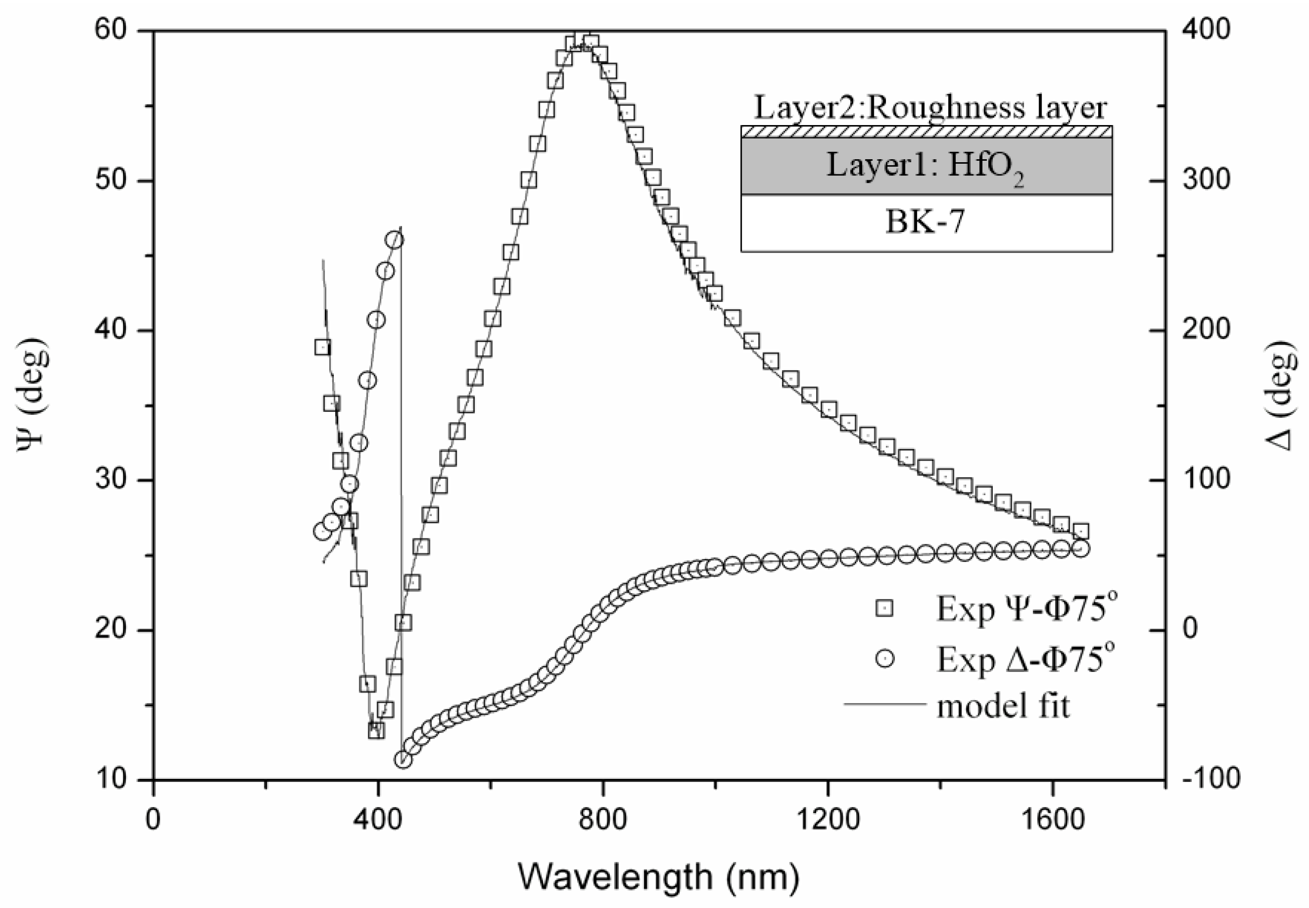
In order to investigate changes in the optical properties of irradiated HfO2 thin films, the refractive index was determined by using a M-2000UI spectroscopic ellipsometer (SE) manufactured by J.A.Woollam company of United States. Measured data were used to describe an optical model that can help to gather the thickness and optical properties by conducting regression analysis. The HfO2 film on glass substrate was initially assumed to have a four phase system (from top to bottom): the incident medium (air), the roughness layer, the HfO2 layer, and the glass substrate, as shown in Figure 1 (insert). An empirical formula of Cauchy’s model was applied to calculate n(λ), which was given by adjusting the fitting parameters according to the SE measured data of the thin HfO2 film due to its weak absorbance of light in the 400~900 nm wavelength range (i.e., k is negligible).
where λ is the wavelength of incident light, and A, B, and C are empirical constants. The typical experimental data, model fit to the data, and the fitted parameters are shown in Figure 1. The mean square error (MSE) of all samples in our experiment was less than 10, which indicates the measured and calculated results were in marked correspondence.
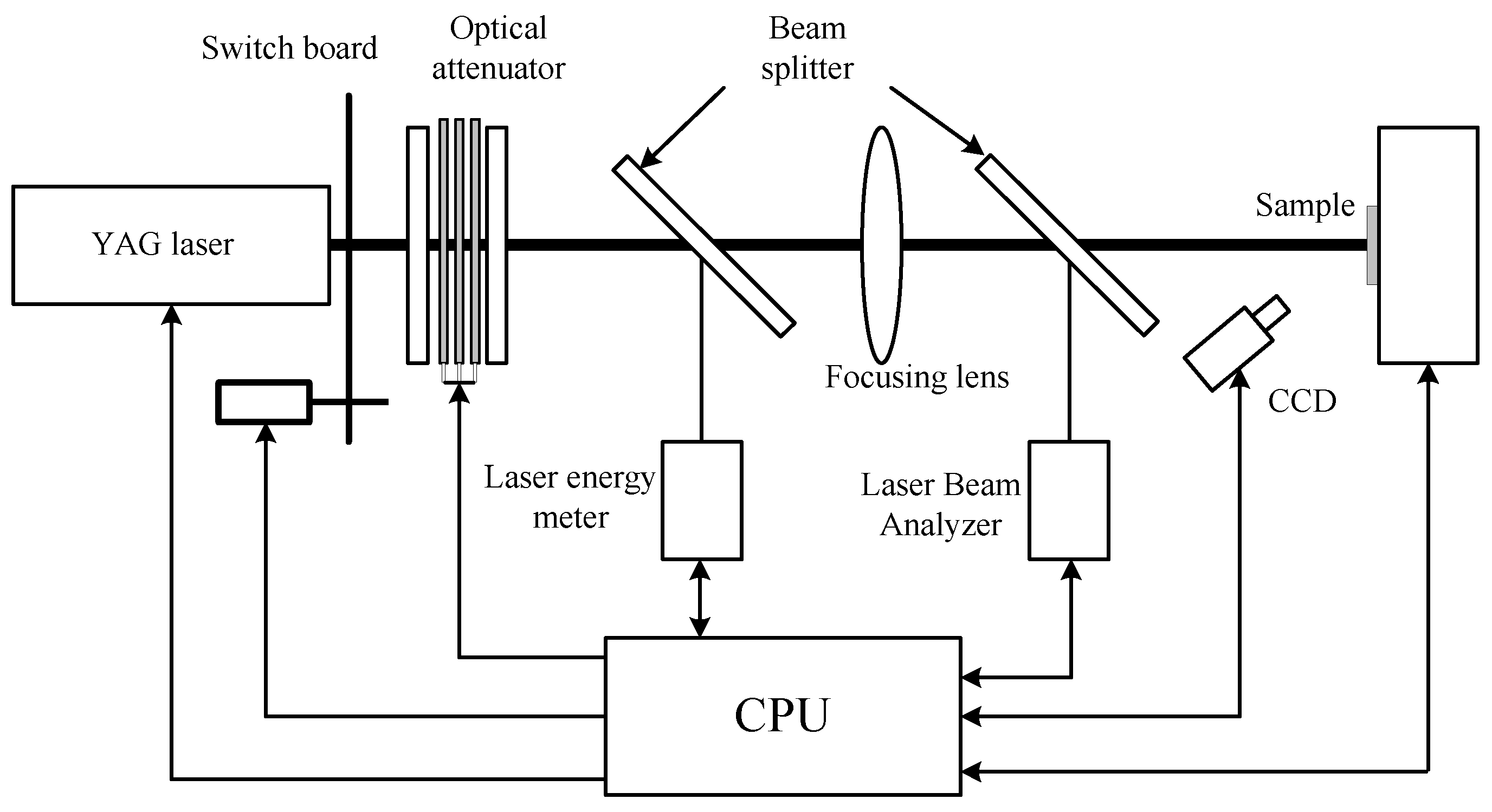
An LIDT test at 1064 nm was performed in the 1-on-1 mode in the light of ISO standard 11254-1. A new in situ laser damage image test apparatus was set up, as shown in Figure 2. The samples were exposed to a laser beam with 1064 nm wavelength and 12-ns effective pulse duration for the Nd: YAG laser system. The pulse energy was adjusted with an optical attenuator in a given fluence range and monitored in realtime with a laser energy meter. The morphology of laser damage on the surface of samples was investigated by means of a CCD camera-microscope device (magnification ×100), which ensured realtime testing and recording of the irradiated zone in situ [12].
The laser light was focused down to the small spot size of 0.8 mm onto the film sample surface. Using this device, the probability curves of damage were expressed, in the meantime, via tallying up the quantity of damaged regions at every fluence F, the damage probability P(F) was estimated. This test was performed via testing 100 points for laser radiation. The images of the test site before and after each shot was observed for a certain energy, which was divided into 10 different levels to identify a high-accuracy LIDT of the sample.
The films’ surface roughness was measured using a Talysurf CCI 2000 non-contact 3D profiler (Taylor Hobson Limited, Leicester, UK), and the root mean square roughness value (RMS) was presented.
3. Results and Discussion
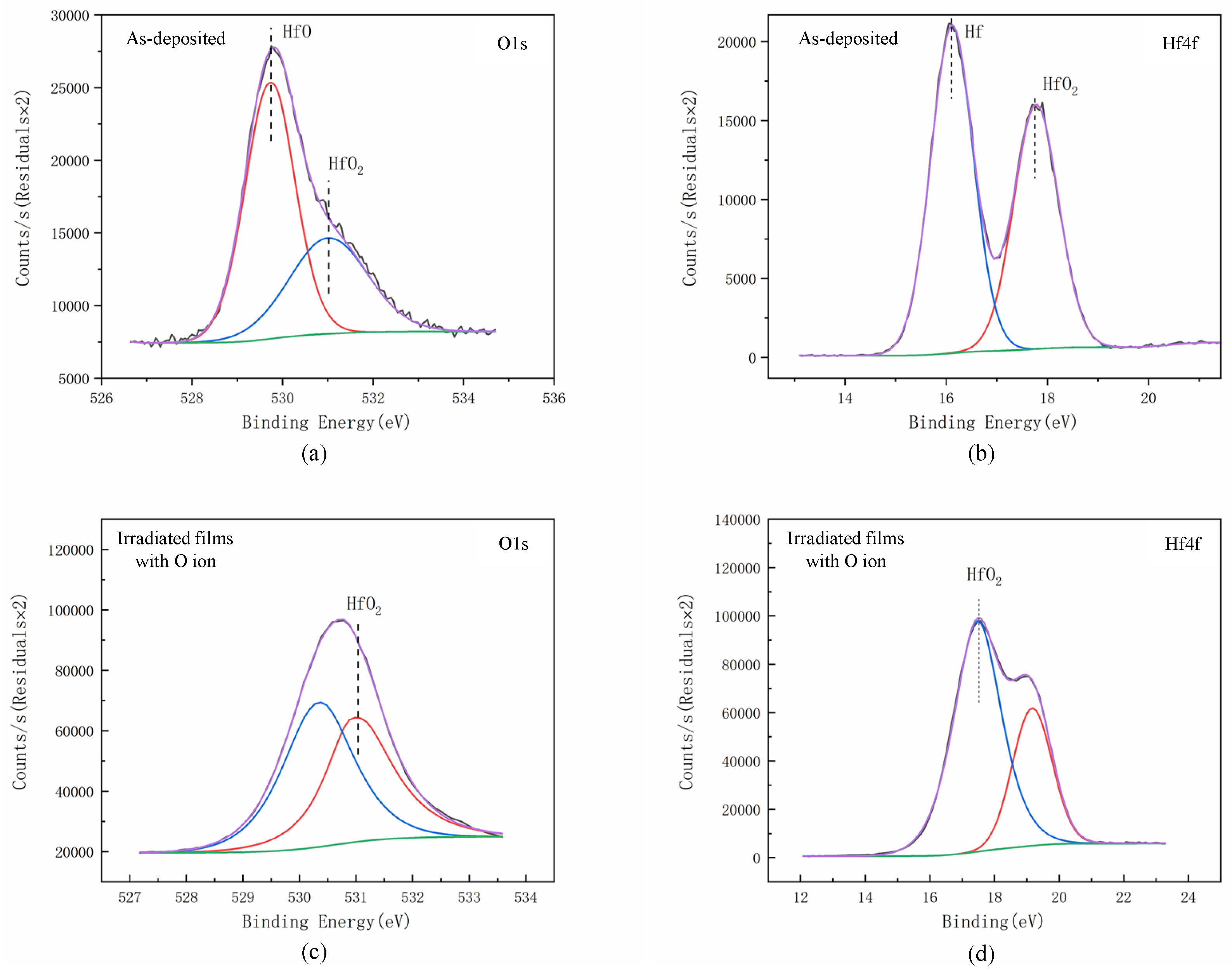
X-ray Photoelectron Spectroscopy (XPS) is one of the important ways to study the electronic and atomic structure of materials. In this experiment, the thin film samples were analyzed by a PHI-5400 X-ray photoelectron spectroscopy from the American PE company, and Cu-K were used as the X-ray source. Before depositions, all the film sample surfaces were subjected to argon ion etching for 90 s, in which the energy value of argon ion bombardment was 2 keV. In addition, the XPS spectra in this experiment were corrected by the binding energy of C1s orbital 284.8 eV. To obtain the accurate data of Hf and O elements in the film, the two elements were scanned by a fine spectrum, as shown in Figure 3.
According to the peak area of Hf4f and O1s, the stoichiometric ratio of Hf and O can be determined, and the expression is:
In the above formula, N1:N2 is the stoichiometric ratio of elements Hf and O; A1 and A2 are the spectral peak areas corresponding to the elements (A1 = 270,580 and A2 = 155,441 here); and S1 and S2 are sensitivity factors corresponding to elements [13] (here S1 = 0.71 and S2 = 2.221), The stoichiometric ratio of Hf and O is 1:1.78. After oxygen ion bombardment at ion energy of 1000 eV, the ratio can be increased to 1:1.86 (A1 = 257,609 and A2 = 154,454 here). The stoichiometric ratio of Hf and O irradiated film using an O Ion beam are closer to 1:2 than the as-deposited films without irradiation.
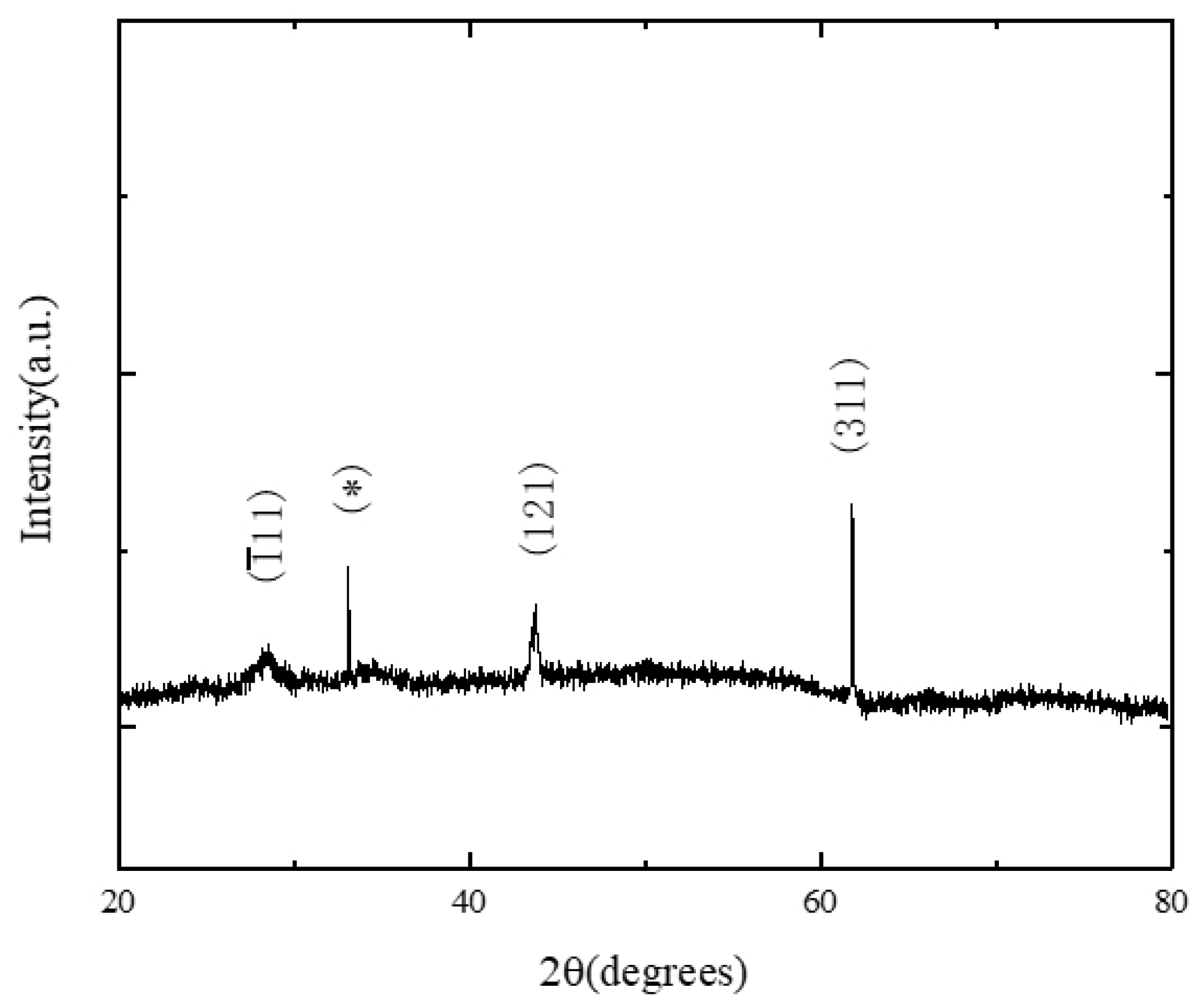
The crystal structure of HfO2 film was measured by Brooke-D2 X-ray diffractometer (XRD). During the test, the scanning step was 0.02°, the scanning area was 20°~100°, and Figure 4 shows the test results. It can be observed from the figure that a typical monoclinic HfO2 characteristic peak appeared in the diffraction pattern of the prepared film and had a preferred orientation in the (311) direction. The test results show that crystallization occurred in the HfO2 films deposited via electron beam evaporation deposition technology; moreover, the prepared films were polycrystalline films [14].
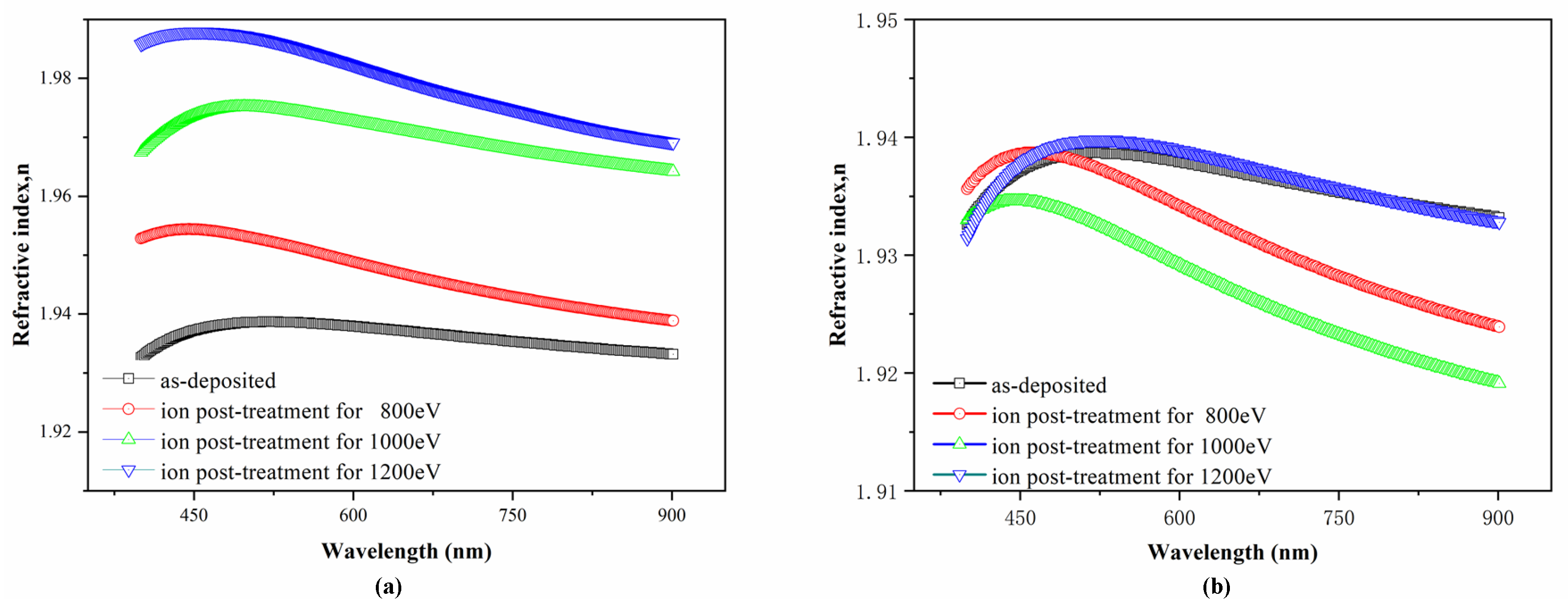
The changes in the optical properties of irradiated HfO2 films were analyzed by SE. Figure 5a,b, respectively, show the dispersion of the refractive index of HfO2 newly deposited film and irradiated films exposed to ion beam irradiation at different ion energies under Ar and O2 atmosphere. From Figure 5, it can be seen clearly that the refractive index for all samples investigated sharply increased with the wavelength between 400 and 450 nm, this anomalous dispersion is speculated to be caused by absorption in a shorter wavelength region for HfO2 thin films. When the energy of irradiation ion beam varied between 800 and 1200 eV, the refractive index of the irradiated films had significant and obvious changes in the given range of measurement wavelength, which was affected by the species of ion beam. It could also be observed that the n value increased along with the increase in the irradiation Ar ion energy; whereas, in comparison with the as-deposited films, the value of the refractive index decreased when it was beyond 1000 eV.
Many porous and void-rich structures were formed easily in the HfO2 thin film preparation by EBD. However, as shown in Figure 5a, the ion bombardment effect of argon plasma-treated at different energies from 800 to 1200 eV for as-deposited samples could make films become denser and solid, and more apparent ion bombardment effect in the aspect of optical properties was observed with the improvement of ion energy. On the other hand, oxygen was easily emitted from HfO2 during the deposition process, which was an imperative factor influencing the optical properties; in addition, hafnium was absorbed largely in the UV band, which increased the n value to a certain extent [15]. From Figure 5b, the n value was slightly lowered by ion bombardment with oxygen plasma. It obviously reveals that low energy oxygen ions could improve the stoichiometry in thin films.
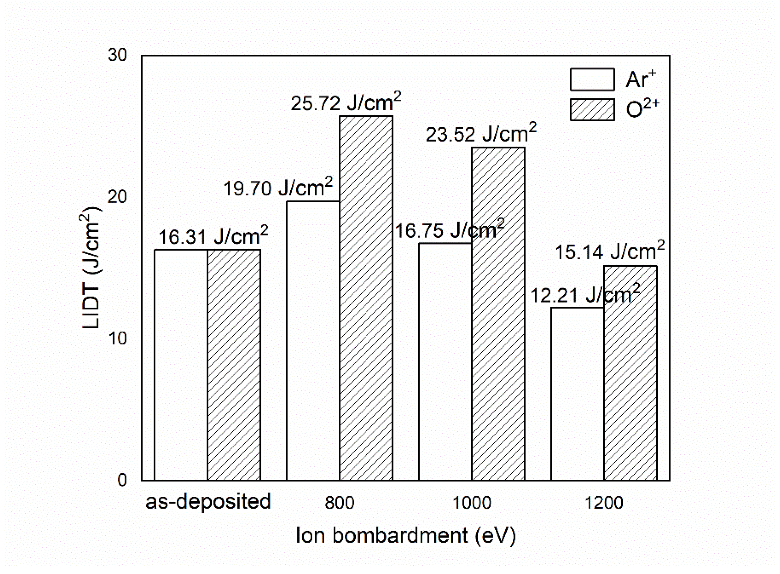
The effect of ion energy and species on LIDT of HfO2 thin films in the ion post-treatment are shown in Figure 6. The LIDT in the deposited HfO2 thin film was found by 16.31 J/cm2, but the maximum thresholds of 19.7 J/cm2 and 25.72 J/cm2 were observed in the irritated films at 800 eV under Ar and O2, respectively. This shows ion post-treatment had a significant contribution on the laser damage resistance. As shown in Figure 5a, the thin film became denser after post-treatment with Ar+ plasma. A plasma-treated ion beam can lessen the quantity of thermal defects for irradiated HfO2 thin film, whose thermal conductivity was higher than that of the as-deposited samples [16,17]. Furthermore, ion treatment is also likely to be a feasible method of making the defects in the coatings stabilized to avoid laser damage. However, the LITD decreased gradually with the further increase in the ion energy, and the LIDT of HfO2 irradiated at 1200 eV was even lower than that of the as-deposited, whose values were only 12.21 J/cm2 for Ar+ plasma and 15.14 J/cm2 for O2+ plasma. A conceivable explanation for the decline in the LIDT value of the irradiated samples is that irradiation by high energy ions results in an increase in internal defects to reduce the laser damage resistance.
Although changes in the LIDT with the increase in the energy were similar for ion post-treatment with Ar+ and O2+ plasma, the films irritated by O2+ plasma had higher LITD at the same ion energy. The reduction in the substoichiometric ratio oxygen to hafnium on irradiation might have a bearing on this difference [18,19], because the decrease in the sub-oxide component of HfO2 films during irradiation was correlated with the O2 ion bombardment. In fact, the two most essential factors that bear on the LIDT of thin film were defect density and absorption. The theory of electron-avalanche-ionization can be used to expound and explicate the effect of substoichiometer compositions on LIDT of HfO2 thin films. Ion beam bombardment of oxygen plasma could repair oxygen vacancies and reduce the absorption of the as-deposited films. The oxygen post-treatment on the anti-reflective film reported by Yuan [20] is in good agreement with these results. However, they performed a direct contrast with those reported for HfO2 films irradiated using end-hall ion source, which were found to be little changed in the LITD on low energy oxygen ions.
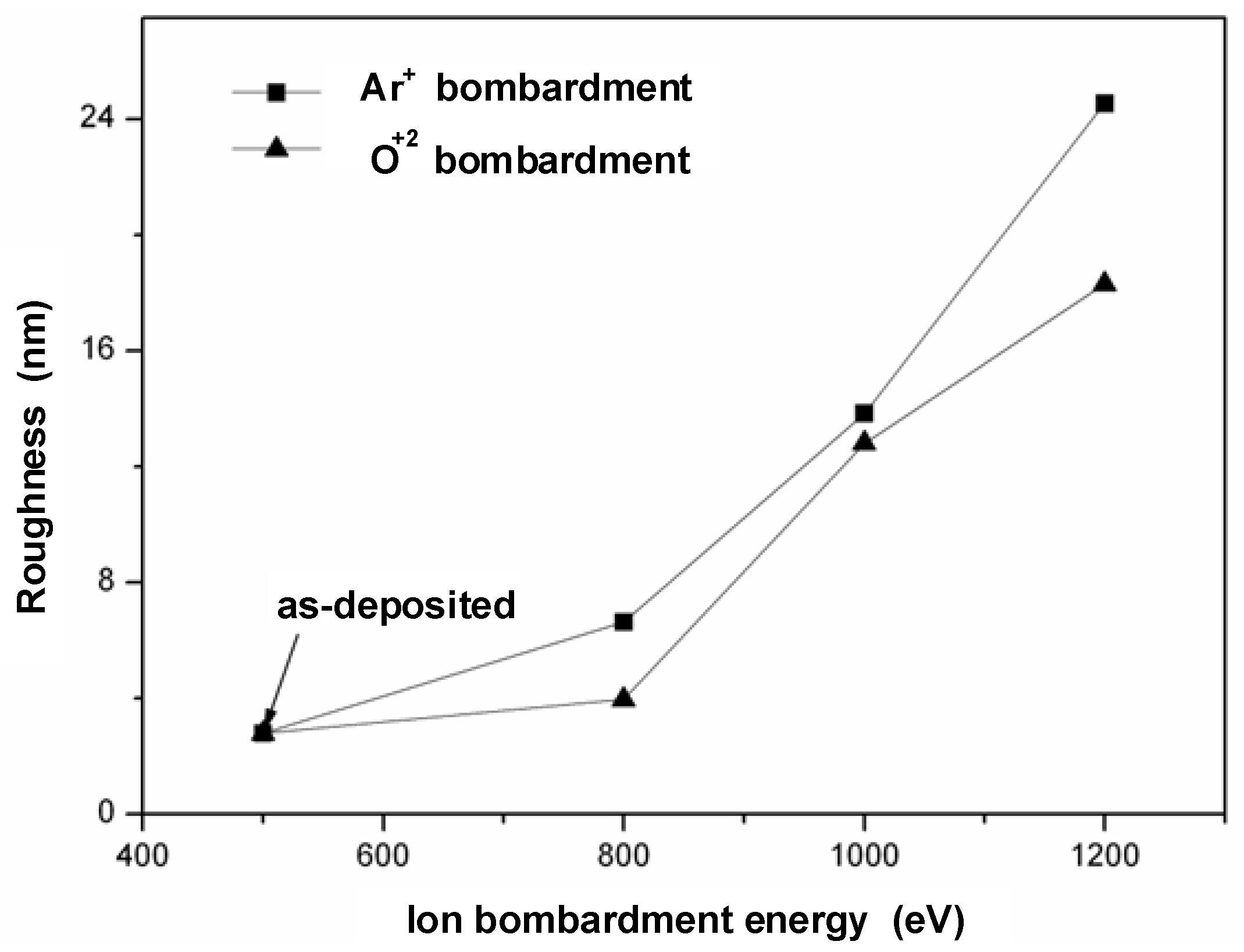
Figure 7 shows the graph of the RMS roughness values of irradiated films for different ion bombardment energies. A similar increasing trend of RMS value with an increase in ion bombardment energy for each film irradiated with Ar and O2 plasma was observed, but the increase in each change is not alike. As for newly deposited films, the minimum roughness value was 2.8 nm, which indicated an atomically smooth surface. With an increase from 800 to 1200 eV in the ion energy, the surface roughness for post-treated film in O2 increased gradually from 3.9 to 18.3 nm, but the surface films post-treated in Ar were rougher, increasing from 6.6 to 24.5 nm. This may be due to the larger mass of Ar than O2 [21,22]. The thermal spike phenomenon caused by ion-bombardment is the main reason leading to an increase in the surface roughness of irradiated film.
For irradiated film, the surface roughness increased proportionately with ion energy but inversely with the LIDT value. It seems that the LIDT value might be related to the change in the surface roughness induced by ion bombardment [23], especially for films irradiated with high energetic plasma ions. Deeper and further analysis is still required for more fundamental comprehension.
4. Conclusions
HfO2 thin films were processed in advance by the EBE technique at 200 °C and then subjected to ion post-treatment in Ar and O2 plasma at various ion energies from 800 to 1200 eV, respectively. The influence of the ion energy and species on the optical properties, laser damage resistance, and surface morphology were systematically studied. The refractive index of the film increased with ion energy in the range of 800~1200 eV after argon ion post-treatment; however, the refractive index of thin films irradiated in oxygen plasma decreased with the increase in the ion energy except 1200 eV, and it was inclined to show a very minor shift towards lower values that rested on ion energy. The laser damage resistance of HfO2 was strongly dependent on ion irradiation energy and ion species. The LIDT of HfO2 films irradiated at certain ion energies were improved but decreased with the increase in ion energy up to 1000 eV. Moreover, O2 ion irradiation was better than argon ion as a means of improving LITD in thin film. The LITD in the films after ion post-treatment was inversely proportional to the surface roughness. A conclusion can be drawn that the laser damage properties of irradiated HfO2 thin film may be related to the change in the surface roughness induced by ion bombardment.
Author Contributions
Conceptualization, Y.X. and J.Z. (Jiwu Zhao); Data curation, J.Z. (Jiwu Zhao) and C.Z.; Formal analysis, J.Z. (Jiwu Zhao); Funding acquisition, Y.X.; Investigation, Y.X. and J.Z. (Jiwu Zhao); Methodology, J.Z. (Jin Zhang); Project administration, Y.X. and J.Z. (Jiwu Zhao); Resources, Y.X. and J.Z. (Jiwu Zhao); Supervision, J.Z. (Jiwu Zhao); Validation, J.Z. (Jin Zhang); Visualization, Q.W.; Writing—original draft, Y.X. All authors have read and agreed to the published version of the manuscript.
Funding
This study was financially supported by the intergovernmental international scientific and technological innova-tion cooperation of science and technology ministry of China (No.2018YFE0199200), This paper also been supported by the Science and Technology on Applied Physical Chemistry Laboratory.
Institutional Review Board Statement
Not applicable.
Informed Consent Statement
Not applicable.
Data Availability Statement
Not applicable.
Conflicts of Interest
The authors declare no conflict of interest.
References
- Zhao, Z.; Sun, J.; Zhu, M. Research to improve the optical performance and laser-induced damage threshold of hafnium oxide/silica dichroic coatings. Opt. Mater. 2021, 113, 110890. [Google Scholar] [CrossRef]
- Field, E.S.; Galloway, B.R.; Kletecka, D.E. Dual-wavelength laser-induced damage threshold of a HfO2/SiO2 dichroic coating developed for high transmission at 527 nm and high reflection at 1054 nm. Proc. SPIE Laser Damage. 2019, 11173, 1117314. [Google Scholar]
- Papernov, S.; Kozlov, A.A.; Oliver, J.B. Near-ultraviolet absorption annealing in hafnium oxide thin films subjected to continuous-wave laser radiation. Opt. Engineering. 2014, 53, 122504. [Google Scholar] [CrossRef]
- Fang, M.H.; Tian, P.Y.; Zhu, M.D. Laser-induced damage threshold in HfO2/SiO2 multilayer films irradiated by β-ray. Chin. Phys. B 2019, 28, 024215. [Google Scholar] [CrossRef]
- Dong, J.; Fan, J.; Mao, S. Effect of annealing on the damage threshold and optical properties of HfO2/Ta2O5/SiO2 high-reflection film. Chin. Opt. Lett. 2019, 17, 113101. [Google Scholar] [CrossRef]
- Pan, F.; Wang, J.; Liu, M.; Wei, Y.; Liu, Z.; Zhang, F.; Wang, Z.; Luo, J.; Wu, Q.; Li, S. Influence of ion assistance on optical properties, residual stress and laser induced damage threshold of HfO2 thin film by use of different ion sources. Proc. SPIE Optifab. 2019, 11175, 1117510. [Google Scholar]
- Balogh-Michels, Z.; Stevanovic, I.; Borzi, A. Crystallization behavior of ion beam sputtered HfO2 thin films and its effect on the laser-induced damage threshold. J. Eur. Opt. Soc. Rapid Publ. 2021, 17, 1–8. [Google Scholar] [CrossRef]
- Pan, F.; Wei, Y.; Zhang, F. Correlation between the structure and laser damage properties of ion assisted HfO2 thin films. Int. Soc. Opt. Photonics. 2019, 11064, 110640L. [Google Scholar]
- Mao, S.; Fan, J.; Zou, Y. Effect of two-step post-treatment on optical properties, microstructure, and nanosecond laser damage threshold of HfO2/TiO2/SiO2 multilayer high reflection films. J. Vac. Sci. Technol. A Vac. Surf. Film. 2019, 37, 061503. [Google Scholar] [CrossRef]
- Wang, Y.; Ma, Y.; Wang, D. Theoretical simulation analysis of long-pulse laser induced damage in a BK7: SiO2/HfO2 optical anti-reflective films. Optik 2018, 156, 530–535. [Google Scholar] [CrossRef]
- Liu, J.; Ling, X.; Liu, X. Mechanism of annealing effect on damage threshold enhancement of HfO2 films in vacuum. Vac. 2021, 189, 110266. [Google Scholar] [CrossRef]
- Negres, R.A.; Carr, C.W.; Laurence, T.A. Laser-induced damage of intrinsic and extrinsic defects by picosecond pulses on multilayer dielectric coatings for petawatt-class lasers. Opt. Engineering. 2016, 56, 011008. [Google Scholar] [CrossRef]
- Liu, J.; Li, X.; Yu, Z. Effect of laser conditioning on the LIDT of 532 nm HfO2/SiO2 thin films reflectors. Proc. SPIE/SIOM Pac. Rim Laser Damage. 2013, 8786, 87860Z. [Google Scholar]
- Alvisi, M.; De Tomasi, F.; Perrone, M.R. Laser damage dependence on structural and optical properties of ion-assisted HfO2 thin films. Thin Solid Film. 2001, 396, 44–52. [Google Scholar] [CrossRef]
- Zhang, D.; Zhu, M.; Li, Y. Laser-induced damage of 355 nm high-reflective mirror caused by nanoscale defect. J. Wuhan Univ. Technol. Mater. Sci. Ed. 2017, 32, 1057–1060. [Google Scholar] [CrossRef]
- Li, C.; Zhao, Y.; Cui, Y. Investigation on picosecond laser-induced damage in HfO2/SiO2 high-reflective coatings. Opt. Laser Technol. 2018, 106, 372–377. [Google Scholar] [CrossRef]
- Jena, S.; Tokas, R.B.; Rao, K.D. Influence of oxygen partial pressure on microstructure, optical properties, residual stress and laser induced damage threshold of amorphous HfO2 thin films. J. Alloys Compd. 2019, 771, 373–381. [Google Scholar] [CrossRef]
- Xu, Y.; Dunlap, D.H.; Emmert, L.A. Laser-driven detonation wave in hafnium oxide film: Defect controlled laser damage and ablation. J. Appl. Phys. 2020, 128, 123101. [Google Scholar] [CrossRef]
- Wei, Y.; Xu, Q.; Wang, Z. Growth properties and optical properties for HfO2 thin films deposited by atomic layer deposition. J. Alloys Compd. 2018, 735, 1422–1426. [Google Scholar] [CrossRef]
- Yuan, H.; Zhang, G.; Kan, S. Influence of the deposition method on the laser induced damage threshold of HfO2 thin films. J. Huazhong Univ. Sci. Technol. (Nat. Sci. Ed.) 2007, 35, 108–111. [Google Scholar]
- Wang, C.; Jin, Y.; Zhang, D. A comparative study of the influence of different post-treatment methods on the properties of HfO2 single layers. Opt. Laser Technol. 2009, 41, 570–5736. [Google Scholar] [CrossRef]
- Jena, S.; Tokas, R.B.; Rao, K.D. Annealing effects on microstructure and laser-induced damage threshold of HfO2/SiO2 multilayer mirrors. Appl. Opt. 2016, 55, 6108–6114. [Google Scholar] [CrossRef] [PubMed]
- Kozlov, A.A.; Papernov, S.; Oliver, J.B. Study of the picosecond laser damage in HfO2/SiO2 based thin-film coatings in vacuum. Proc. SPIE Laser Damage. 2017, 10014, 100141Y. [Google Scholar]
Figure 1.
Typical HfO2 thin film measured data and fitting curves with fits to the Cauchy formula. The optical model is shown in the inserts at the top right corner.
Figure 1.
Typical HfO2 thin film measured data and fitting curves with fits to the Cauchy formula. The optical model is shown in the inserts at the top right corner.

Figure 2.
Schematic diagram of the apparatus used for in situ image laser damage testing system.

Figure 3.
Comparison of XPS patterns of HfO2 films before and after O ions treatment at an ion energy of 1000 eV; (a) O1s spectra of as-deposited films; (b) Hf4f spectra of as-deposited films; (c) O1s spectra of irradiated films; and (d) Hf4f s spectra of irradiated films.
Figure 3.
Comparison of XPS patterns of HfO2 films before and after O ions treatment at an ion energy of 1000 eV; (a) O1s spectra of as-deposited films; (b) Hf4f spectra of as-deposited films; (c) O1s spectra of irradiated films; and (d) Hf4f s spectra of irradiated films.

Figure 4.
XRD phase diagram of HfO2 film; * indicates the SiO2 (217) peak.

Figure 5.
Refractive index of HfO2 film irradiated at various ion energies under Ar (a) and O2 (b) atmosphere.
Figure 5.
Refractive index of HfO2 film irradiated at various ion energies under Ar (a) and O2 (b) atmosphere.

Figure 6.
Laser damage threshold of HfO2 film irradiated at various ion energies under Ar and O2 atmosphere.
Figure 6.
Laser damage threshold of HfO2 film irradiated at various ion energies under Ar and O2 atmosphere.

Figure 7.
Variation of roughness of HfO2 films with Ar and O2 ion energy.

Publisher’s Note: MDPI stays neutral with regard to jurisdictional claims in published maps and institutional affiliations. |
© 2022 by the authors. Licensee MDPI, Basel, Switzerland. This article is an open access article distributed under the terms and conditions of the Creative Commons Attribution (CC BY) license (https://creativecommons.org/licenses/by/4.0/).
Share and Cite
MDPI and ACS Style
Xi, Y.; Zhao, J.; Zhang, J.; Zhang, C.; Wu, Q. The Influence of Ion Beam Bombardment on the Properties of High Laser-Induced Damage Threshold HfO2 Thin Films. Crystals 2022, 12, 117. https://0-doi-org.brum.beds.ac.uk/10.3390/cryst12010117
AMA Style
Xi Y, Zhao J, Zhang J, Zhang C, Wu Q. The Influence of Ion Beam Bombardment on the Properties of High Laser-Induced Damage Threshold HfO2 Thin Films. Crystals. 2022; 12(1):117. https://0-doi-org.brum.beds.ac.uk/10.3390/cryst12010117
Chicago/Turabian StyleXi, Yingxue, Jiwu Zhao, Jin Zhang, Changming Zhang, and Qi Wu. 2022. "The Influence of Ion Beam Bombardment on the Properties of High Laser-Induced Damage Threshold HfO2 Thin Films" Crystals 12, no. 1: 117. https://0-doi-org.brum.beds.ac.uk/10.3390/cryst12010117
Note that from the first issue of 2016, this journal uses article numbers instead of page numbers. See further details here.