Communication

Identification, Characterization, and Preliminary X-ray Diffraction Analysis of a Single Stranded DNA Binding Protein (LjSSB) from Psychrophilic Lacinutrix jangbogonensis PAMC 27137

1
Research Unit of Cryogenic Novel Material, Korea Polar Research Institute, Incheon 21990, Korea
2
New Drug Development Center, Daegu-Gyeongbuk Medical Innovation Foundation, Daegu 41061, Korea
3
Division of Emerging Infectious Diseases Bureau of Infectious Disease Diagnosis Control, Korea Disease Control and Prevention Agency, Cheongju 28159, Korea
4
Division of Life Sciences, Korea Polar Research Institute, Incheon 21990, Korea
5
Department of Polar Sciences, University of Science and Technology, Incheon 21990, Korea
6
Department of Chemistry, Sookmyung Women’s University, Seoul 04310, Korea
*
Author to whom correspondence should be addressed.
These authors contributed equally to this work.
Submission received: 23 March 2022 / Revised: 5 April 2022 / Accepted: 8 April 2022 / Published: 11 April 2022
(This article belongs to the Special Issue Crystallographic Studies of Enzymes (Volume II))

Abstract

:
Single-stranded DNA-binding proteins (SSBs) are essential for DNA metabolism, including repair and replication, in all organisms. SSBs have potential applications in molecular biology and in analytical methods. In this study, for the first time, we purified, structurally characterized, and analyzed psychrophilic SSB (LjSSB) from Lacinutrix jangbogonensis PAMC 27137 isolated from the Antarctic region. LjSSB has a relatively short amino acid sequence, consisting of 111 residues, with a molecular mass of 12.6 kDa. LjSSB protein was overexpressed in Escherichia coli BL21 (DE3) and analyzed for binding affinity using 20- and 35-mer deoxythymidine oligonucleotides (dT). In addition, the crystal structure of LjSSB at a resolution 2.6 Å was obtained. The LjSSB protein crystal belongs to the space group C222 with the unit cell parameters of a = 106.58 Å, b = 234.14 Å, c = 66.14 Å. The crystal structure was solved using molecular replacement, and subsequent iterative structure refinements and model building are currently under progress. Further, the complete structural information of LjSSB will provide a novel strategy for protein engineering and for the application on molecular biological techniques.

1. Introduction

For important cellular processes, chromosomal DNA must be in a single-stranded form. Therefore, single-stranded DNA (ssDNA) intermediates are created by DNA unwinding and serve as templates for myriad cellular functions [1]. However, ssDNA is less stable and more sensitive to chemicals than double-stranded DNA (dsDNA); thus, ssDNA is more prone to damage than dsDNA. DNA lesions can interfere with essential cellular processes, ultimately affecting cell viability. These problems can be solved by encoding specialized ssDNA-binding proteins (SSBs) [2]. SSBs bind to ssDNA with high affinity in a sequence-independent manner to protect it from chemical and enzymatic damage [3]. SSBs are present in all living organisms and play essential roles in many processes related to DNA metabolism, such as DNA replication, repair, and homologous genetic recombination [4]. Most bacterial SSBs comprise two domains: an N-terminal domain called oligosaccharide/oligonucleotide binding (OB) domain, composed of a minimum of five β-sheets arranged as a β barrel capped by a single α helix and responsible for both ssDNA binding and oligomerization, and a C-terminal domain, that involves protein-protein interaction [5,6,7]. The OB domain is separated from the highly conserved last nine amino acids of the C-terminal domain by a proline-or glycine-rich linker, called intrinsically disordered linker (IDL) [8]. The C-terminal domain of SSB consists of IDL and last nine highly conserved residues, called acidic tip, and interacts with various other proteins involved in cell survival [9,10]. Although SSBs are found in every organism, they have little sequence similarity and differ in their subunit composition and oligomerization state [11]. While most bacterial SSBs form a homotetramer and have a single OB fold per polypeptide, eukaryotic SSBs, commonly called replication protein A (RPA), generally function as heterotrimeric complexes that contain six OB folds distributed among three subunits [12,13,14].
Due to these properties, there has been growing interest in SSBs owing to their potential applications in molecular biology and analytical methods. An increasing number of studies have found that SSBs increase amplification efficiency. Previous reports have shown that PCR application of an ssDNA-binding protein isolated from bacteriophage T4-infected Escherichia coli, named gene 32 protein, results in an increased amplification efficiency [15,16]. In particular, thermostable SSBs can be used in applications that require extremely high temperatures, such as nucleic acid amplification and sequencing [17]. Furthermore, the affinity of SSB towards ssDNA has been successfully utilized for rolling circle amplification (RCA) [18]. The addition of SSBs during the RCA reaction prevents the generation of double-stranded DNA, and consistently produces single-stranded products without termination. Although mesophilic and thermophilic SSBs have been identified and extensively studied, there is little information about psychrophilic SSBs. Cold-adapted enzyme proteins are useful for molecular biosciences due to the need for enzymes to be used in sequential reactions, and to be inactivated after performing their individual function. Heat-labile enzymes enable heat inactivation at temperatures that do not cause double-stranded DNA to melt, and their use obviates the need to use chemical extraction processes [19]. Particularly, molecular biological techniques, such as isothermal amplification or whole genome amplification, require enzymes that have activity at a relatively low temperature because these techniques have to be performed within room temperature range. Therefore, the characterization of cold-active SSBs offers an attractive alternative to other thermostable SSBs in molecular biology applications.
In this study, we identified and purified a psychrophilic SSB protein, called LjSSB, from a Lacinutrix jangbogonensis PAMC 27137 Gram-negative strain isolated from an Antarctic region [20]. We investigated whether LjSSB has a different binding affinity pattern depending on ssDNA length. Furthermore, LjSSB was crystallized for X-ray diffraction experiments to obtain structural information. We successfully determined LjSSB structure, and further iterative refinement and model building are currently in progress. We believe that our biochemical and structural analyses will help prospective enzyme engineering for applications in molecular biology.

2. Materials and Methods

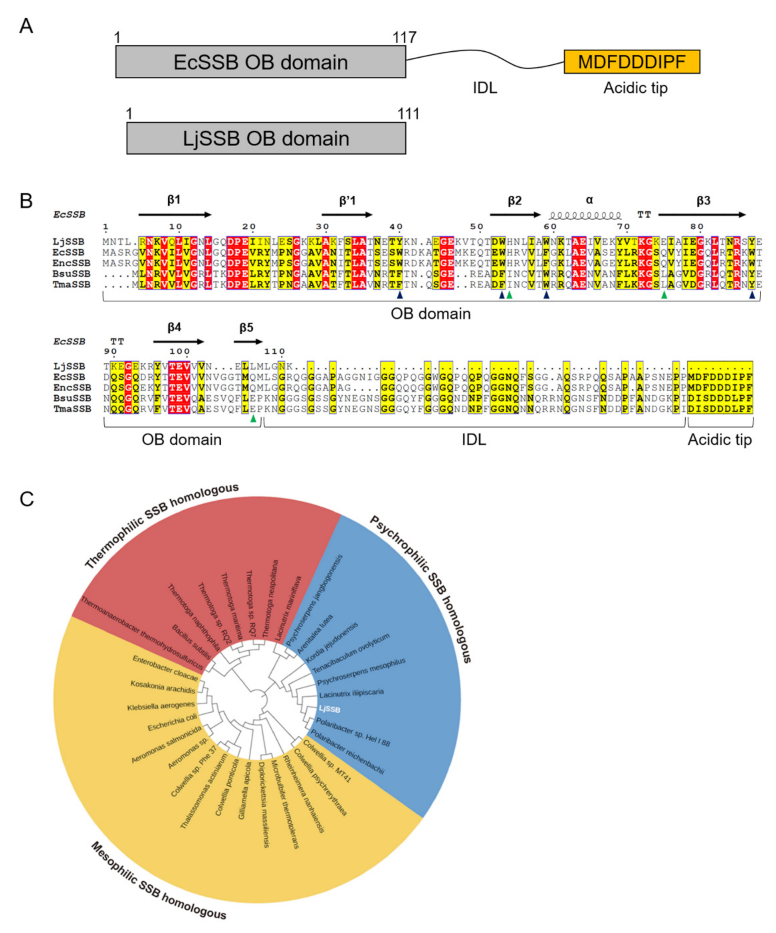
2.1. Sequence Analysis for LjSSB

To investigate the properties of the amino acid sequence of LjSSB, the sequence was compared with those of several previously classified SSBs. A total of 32 amino acid sequences containing LjSSB were used for multiple sequence alignment, and a phylogenetic tree was generated based on the neighbor-joining method using ClustalW [21] and MEGA-X [22].

2.2. Cloning, Expression, and Purification of the LjSSB from L. jangbogonensis

Genomic DNA of PAMC 27,137 was isolated using a DNA purification kit according to the manufacturer’s instructions (Promega, Madison, WI, USA). For cloning into the plasmid, the LjSSB gene was amplified from genomic DNA by PCR. The PCR product was purified and cloned into the pET22b(+) vector, which has a C-terminal hexa-histidine tag between the NdeI and XhoI restriction sites. Subsequently, the cloned sequence was estimated by Sanger sequencing using a T7 promoter and terminator primer pair. For protein expression, plasmids harboring LjSSB were transformed into E. coli BL21 (DE3), and transformed cells were cultured in Luria–Bertani (LB) broth. When OD600 reached 0.6–0.8, cells were induced by 0.3 mM IPTG and incubated at 25 °C for 16 h. Subsequently, cells were harvested by centrifugation at 8000× g for 20 min at 4 °C. For purification of SSB proteins, pelleted cells were resuspended in lysis buffer containing 20 mM Tris-HCl pH 8.0, and 200 mM NaCl and lysed by sonication. The supernatant was subjected to a column charged with a Ni2+-chelated resin. The column was washed with washing buffer (20 mM Tris-HCl, pH 8.0, 200 mM NaCl, and 30 mM imidazole) and the protein was eluted by elution buffer (20 mM Tris-HCl pH 8.0, 200 mM NaCl, and 300 mM imidazole). This was followed by further purification via gel filtration chromatography using a HiLoad 16/60 Superdex 200 column (GE Healthcare, Chicago, IL, USA) pre-equilibrated by buffer with same composition of lysis buffer.

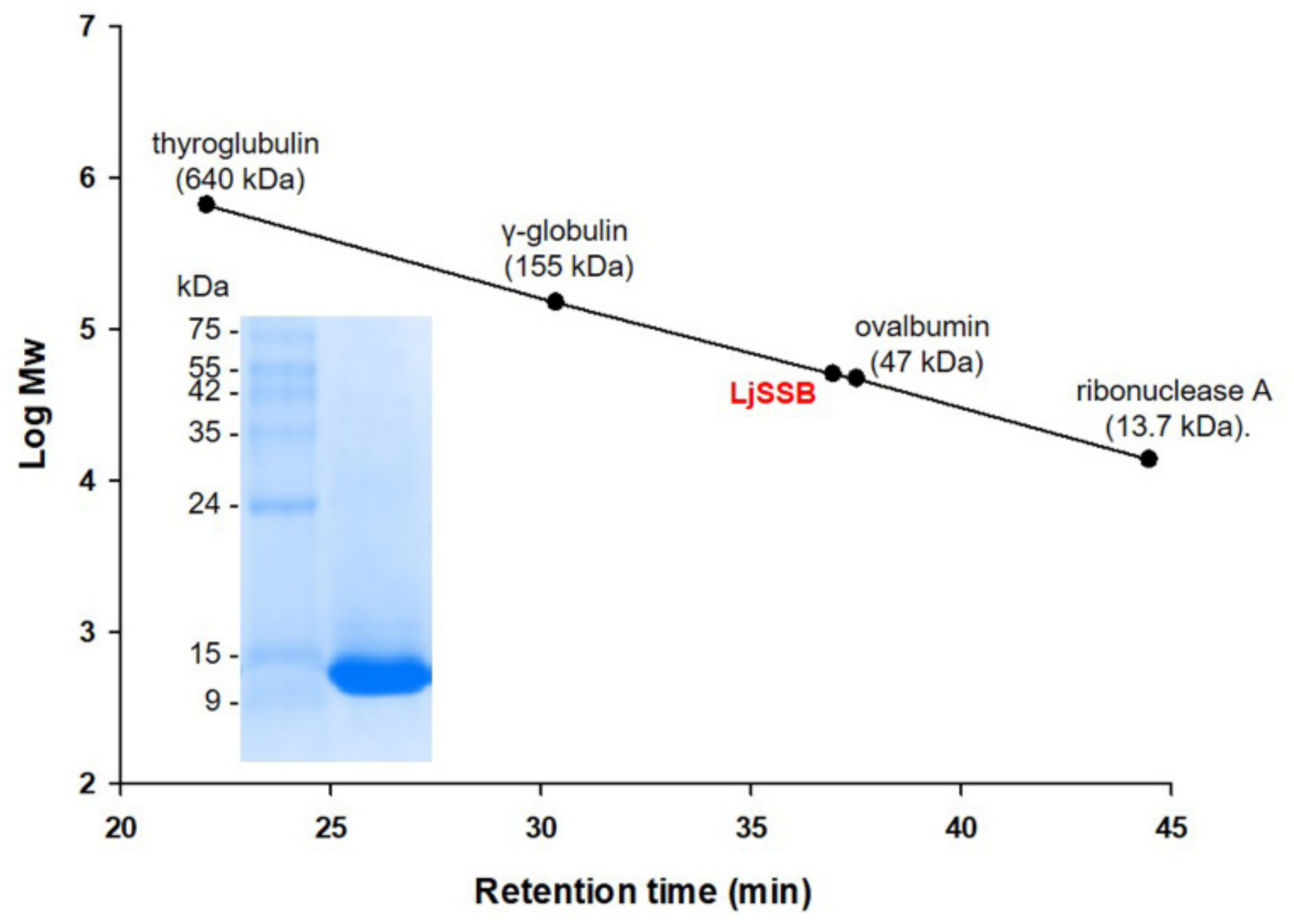
2.3. Estimation of Oligomerization State

The oligomerization state of LjSSB was determined using analytical size-exclusion chromatography. The molecular mass of recombinant SSB was measured by elution analysis of standard proteins from a Superdex 200 10/300 GL column (GE Healthcare, IL, USA). The column was calibrated using molecular mass standards: thyroglobulin (640 kDa), γ-globulin (155 kDa), ovalbumin (47 kDa), and ribonuclease A (13.7 kDa).

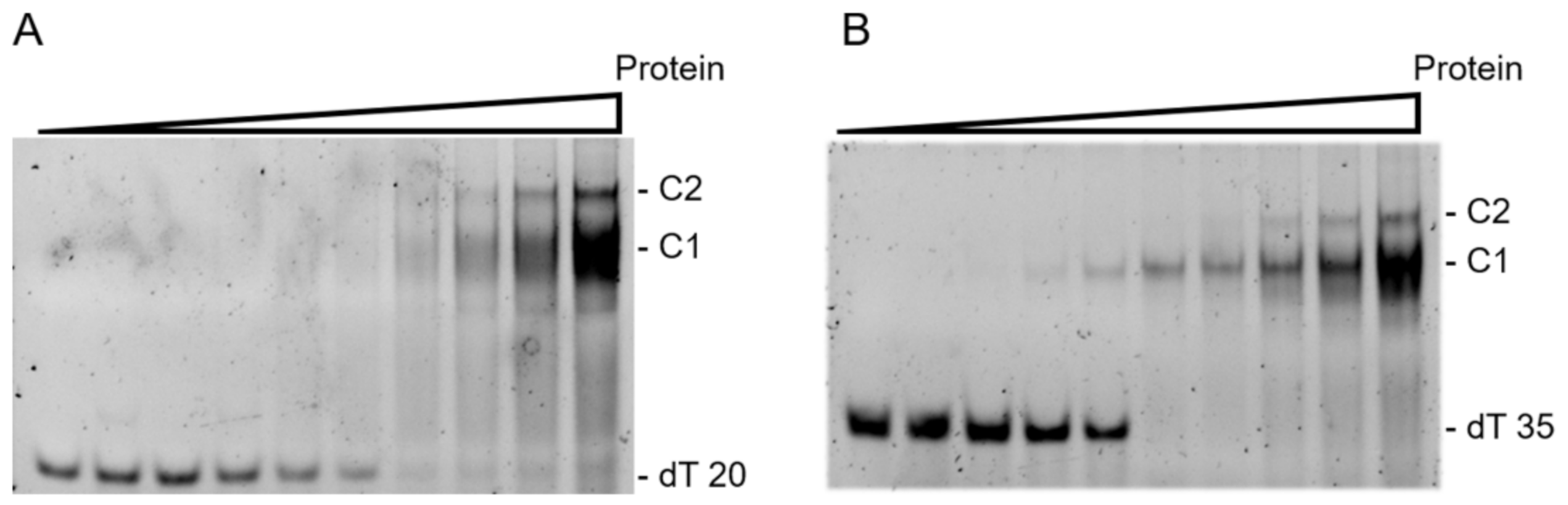
2.4. Enzymatic Analysis Using Gel Electrophoresis Mobility Shift Assay (EMSA)

Deoxythymidine (dT) oligonucleotides (20- and 35-mer) were synthesized by Macrogen (Seoul, Korea). LjSSB and ssDNA binding reaction was performed in 20 μL volumes containing each various concentrations of LjSSB (0.125, 0.25, 0.5, 1, 2, 4, 8, 16, and 32 μM) with 500 nM of synthesized dT20 and dT35 in a binding buffer (20 mM Tris—HCl pH 8.0, and 200 mM NaCl). The reaction mixtures were incubated for 15 min at room temperature (25 °C). Subsequently, the reaction products with oligonucleotides were loaded onto 8% acrylamide gel and separated by electrophoresis in a 1X TBE buffer (89 mM Tris borate, pH 8.0, and 2 mM EDTA). The gel was stained with SYBR Gold (Thermo Fisher Scientific, Waltham, MA, USA) for 15 min at room temperature (RT, at 25 °C). Bands corresponding to unbound ssDNA and SSB-ssDNA complex were visualized under UV light and photographed.

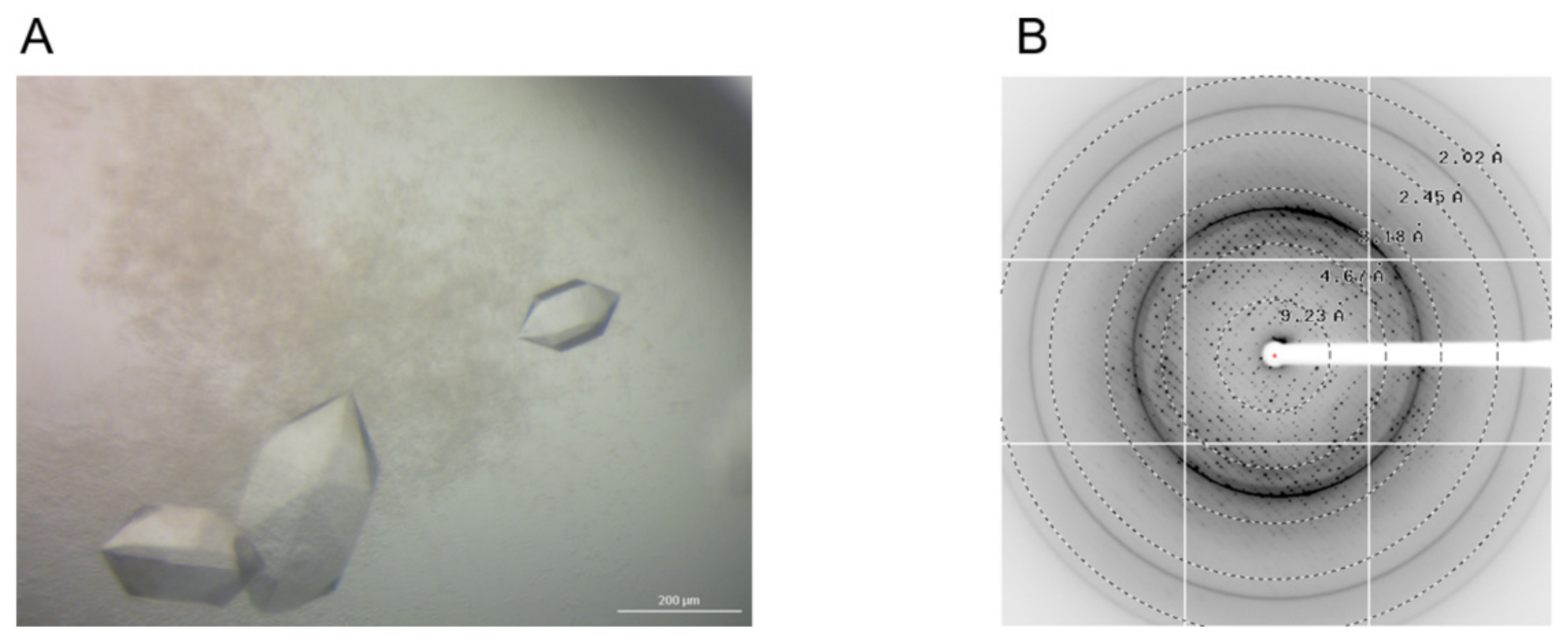
2.5. Crystallization, Data Collection, and Refinement for LjSSB

The crystallization of LjSSB was performed using the following procedure: the LjSSB was concentrated to 20 mg/mL using a 3 kDa molecular weight cutoff spin concentrator (Millipore, Burlington, MA, USA). Initial crystallization screening of LjSSB was performed by the sitting-drop vapor-diffusion method, using commercially available screening solution kits: MCSG I to IV (Anatrace Inc., Maumee, OH, USA), SaltRx HT™ (Hampton Research Corp., Aliso Vieojo, CA, USA), and JCSG™ (Molecular Dimensions Inc., Altamonte Springs, FL, USA). Each crystallization drop was prepared by equilibrating the mixture of 0.5 μL reservoir solution and 0.5 μL protein solution against 80 μL reservoir solution using a mosquito® liquid-handling robot (TTP Labtech Ltd., Melbourn, Hertfordshire, UK). Diffraction data for LjSSB were obtained under conditions comprising 0.1 M of phosphate-citrate pH 4.2 and 1.6 M of NaH2PO4/0.4M K2HPO4. Subsequently, crystals were transferred to perfluoropolyether oil PFO-X175/08 (Hampton Research Corp., CA, USA) to protect cryodamage and flash-frozen in a liquid nitrogen stream. A single crystal was mounted on the goniometer sample holder. A total of 360 diffraction image data of LjSSB were collected using an ADSC Quantum 315 CCD detector on beamline 5C at the Pohang Accelerator Laboratory (PAL, Pohang, Korea) by rotating with 1° of oscillation per frame. X-ray diffraction data were collected at a resolution of 2.6 Å. The datasets were indexed, processed, and scaled using the XDS program [23]. The initial crystal structure of LjSSB was determined using the molecular replacement (MR) method with a single-stranded DNA binding protein from Escherichia coli (EcSSB, PDB code: 4MZ9) as a search model. The collection data are summarized in Table 1.

3. Results and Discussion

3.1. Sequence Analysis for LjSSB

SSB consists of two domains: an N-terminal OB-domain and a C-terminal domain. The N-terminal domain of SSB is involved in ssDNA binding, and the C-terminal domain is composed of IDL and an acidic tip and is involved in protein-protein interactions, as with DNA repair or recombination proteins [24]. The results of genomic analysis of L. jangbogonensis PAMC 27137 (GenBank accession No. JSWF00000000) [20] indicated that they have three putative SSB genes. We attempted to clone and express all three; however, two SSBs were expressed in insoluble form, and only one SSB was soluble and expressed as recombinant proteins in E. coli. The soluble SSB, called LjSSB, and the LjSSB predicted protein contained 111 amino acid residues. Among the known bacterial SSB proteins, LjSSB has the smallest molecular size.
Comparative protein sequence analysis revealed that LjSSB has an N-terminal OB fold (Figure 1A) but not a C-terminal domain. Although the results of multiple sequence alignment displayed overall sequence identities of 45% to E. coli (EcSSB, GenBank ID: EEV5779109.1), 45% to Enterobacter cloacae (EncSSB, GenBank ID: WP_094085120.1), 36% to Bacillus subtilis (BsuSSB, GenBank ID: WP_153257179.1), and 33% to Thermotoga maritima (TmaSSB, GenBank ID: WP_004081225.1), they also showed that specific residues involved in binding ssDNA were highly conserved (Figure 1B). However, oligomerization-related residues were less conserved than ssDNA binding sites.
Multiple sequence alignments for evolutionary analysis are classified into three groups based on their origin: psychrophilic, mesophilic, and thermophilic SSB. The phylogenetic tree showed that LjSSB was part of the psychrophilic SSB group (Figure 1C).
Figure 1. Sequence analysis for LjSSB. (A) The comparison of domain architecture of LjSSB and EcSSB. In contrast with EcSSB, LjSSB does not have a C-terminal domain. (B) Multiple sequence alignment for SSBs. LjSSB from Lacinutrix jangbogonensis PAMC 27,137 in this study; EcSSB from Escherichia coli (GenBank ID: EEV5779109.1); EncSSB from Enterobacter cloacae (GenBank ID: WP_094085120.1); BsuSSB from Bacillus subtilis (GenBank ID: WP_153257179.1); and TmaSSB from Thermotoga maritima (GenBank ID: WP_004081225.1). The secondary structure deduced from EcSSB (PDB code: 1KAW) is showed on the top of aligned sequences. The strictly conserved regions are highlighted in red, and homologous regions are highlighted in yellow. The ssDNA binding site and oligomerization-involved residues from EcSSB are labeled using blue and green triangles, respectively. (C) The amino acid sequences for psychrophilic, mesophilic, and thermophilic SSBs were aligned using ClustalW, followed by construction of neighbor-joining (NJ) phylogenetic trees with 1000 bootstrap replicates using MEGA-X software. The tree display was obtained with online iTOL [25].
Crystals 12 00538 g001

3.2. Oligomerization Status for LjSSB

SSB proteins have a variety of oligomeric states, ranging from monomers to pentamers [26,27,28,29]. In the case of bacterial SSBs, mesophilic SSB function as homotetramers with a single OB-domain subunit. Thermophilic SSBs from bacteria, such as Deinococcus radiodurans and Thermus aquaticus, exist as homodimers. Analytical gel filtration was performed to determine the oligomeric status of the proteins in the solution. The elution profile relative to standard proteins indicated that the native molecular mass of LjSSB was approximately 50 kDa, which is nearly four times the molecular mass of the LjSSB monomer (12.6 kDa) (Figure 2). Since the molecular mass of LjSSB is 50.4 kDa as calculated from its amino acid composition in the case of homotetramers, the result implied that LjSSB existed as a stable homotetramer in the solution.

3.3. Binding Properties for LjSSB with ssDNA

Most bacterial SSBs have different binding modes for ssDNA of various lengths [7]. To investigate the binding mode and affinity of LjSSB for ssDNA, we performed gel shift assay (EMSA) using different concentrations of purified LjSSB (0–32 μM) and 20- and 35-mer oligonucleotides consisting of deoxythymidine (dT20 and dT35, respectively). As shown in Figure 3A, when LjSSB was incubated with dT20, intensity of the unbound DNA band decreased, but no significant band shift was observed at a low protein concentration (lanes 3 to 5 in Figure 3A). This result suggested that a low concentration of LjSSB may not form a stable complex with dT20. Nevertheless, at an LjSSB concentration of 1 μM (lane 7 in Figure 3A), a significant band shift was observed, indicating that LjSSB formed a single complex with dT20. Furthermore, when the LjSSB concentration was increased (lanes 8–10 in Figure 3A), another slow-migrating band appeared. This slow migrated second band indicated that LjSSB formed a second complex with dT20 under a high concentration of protein. At least two LjSSB tetramers may be involved per oligonucleotide, despite the short length of dT20. This phenomenon was also observed in LjSSB incubated with dT35 (Figure 3B). At low concentrations of LjSSB, 0.5 to 2 μM (lanes 4 to 6 in Figure 3B), a single complex was observed, whereas a second complex was detected under high concentrations of protein (lanes 8 to 10 in Figure 3B). The EMSA results for dT20 and dT35 with LjSSB indicated that LjSSB forms two types of complexes with dT20 and dT35 according to protein concentration, which was a distinctive feature from other SSBs, that had C-termini and formed only a single complex with short ssDNA [30,31]. When other SSBs with C-terminal domains were incubated with short ssDNA (15–40 nt), only a single complex was detected, contrary to the observed results for LjSSB. These different features of LjSSB are considered to derive from the absence of a C-terminal domain because the IDL and acidic tip of the C-terminal domain are involved in mediating ssDNA binding [32,33].

3.4. Crystallization and X-ray Structure Analysis of LjSSB

To obtain structural information on LjSSB, crystallization screening and X-ray diffraction experiments were performed. The crystallization conditions of LjSSB were screened using more than 1000 different crystallization buffers. The best single crystal was obtained under 0.1 M of phosphate-citrate pH 4.2 and 1.6 M of NaH2PO4/0.4 M K2HPO4 (Figure 4A). To prevent freezing damage, the single crystals were soaked in a cryoprotectant, perfluoropolyether oil PFO-X175/08. Subsequently, X-ray experiments were performed by mounting the samples under a liquid nitrogen stream. The 360 frames of the diffraction images were collected at a resolution of approximately 2.6 Å (Figure 4B). The acquired diffraction data were indexed, integrated, and scaled using the XDS program [23]. The crystal belongs to the orthorhombic space group C222, with the unit cell parameters a = 106.58 Å, b = 234.14 Å, c = 66.14 Å. Molecular replacement was performed for phase determination using MOLREP [34] and Phaser [35]. The initial structure of LjSSB was determined using EcSSB (PDB code: 1KAW) as a reference model. Refinement and model building are currently underway using the Phenix [36] and COOT software programs [37].

4. Conclusions

In this study, we identified the biochemical properties of a psychrophilic SSB, LjSSB, from L. jangbogonensis PAMC 27137. The LjSSB gene was identified, expressed in E. coli, and the resulting protein was purified using Ni+ affinity column and gel filtration chromatography. Sequence alignment and PAGE results indicated that LjSSB is the smallest known bacterial SSB protein because it has only an N-terminal domain. In addition, LjSSB has a different pattern of binding affinity according to ssDNA length. Structural refinement and model building are currently in progress. We believe that further structural analysis of LjSSB will provide new insights for engineering SSBs for application in molecular biology techniques.

Author Contributions

Conceptualization, W.C. and H.-W.K.; methodology, W.C., H.J. and A.P.; validation, W.C. and J.S.; formal analysis, W.C., H.J., S.C.S. and A.P.; writing—original draft preparation, W.C. and H.-W.K.; writing—review and editing, W.C., S.C.S., J.H.L., T.D.K. and H.-W.K., visualization, W.C. and J.S.; supervision, H.-W.K.; funding acquisition, H.-W.K. All authors have read and agreed to the published version of the manuscript.

Funding

This research was supported by a National Research Foundation of Korea Grant from the Korean Government (MSIT; Ministry of Science and ICT) (NRF-2021M1A5A1075524) (KOPRI-PN22014). This research was also supported by the Korea Polar Research Institute (KOPRI-PE22160).

Institutional Review Board Statement

Not applicable.

Informed Consent Statement

Not applicable.

Data Availability Statement

Not applicable.

Acknowledgments

We would like to thank the staff at BL-5C of the Pohang Accelerator Laboratory (Pohang, Korea) for their kind help with the X-ray diffraction data collection.

Conflicts of Interest

The authors declare no conflict of interest.

References

  1. Marceau, A.H. Functions of single-strand DNA-binding proteins in DNA replication, recombination, and repair. In Methods in Molecular Biology; Walker, J.M., Ed.; Humana Press: Totowa, NJ, USA, 2012; Volume 922, pp. 1–21. [Google Scholar]
  2. Lohman, T.M.; Ferrari, M.E. Escherichia coli single-stranded DNA-binding protein: Multiple DNA-binding modes and cooperativities. Annu. Rev. Biochem. 1994, 63, 527–570. [Google Scholar] [CrossRef]
  3. Mishra, G.; Levy, Y. Molecular determinants of the interactions between proteins and ssDNA. Proc. Natl. Acad. Sci. USA 2015, 112, 5033–5038. [Google Scholar] [CrossRef] [PubMed] [Green Version]
  4. Pestryakov, P.E.; Lavrik, O.I. Mechanisms of single-stranded DNA-binding protein functioning in cellular DNA metabolism. Biochemistry (Moscow) 2008, 73, 1388–1404. [Google Scholar] [CrossRef] [PubMed]
  5. Theobald, D.L.; Mitton-Fry, R.M.; Wuttke, D.S. Nucleic acid recognition by OB-fold proteins. Annu. Rev. Biophys. Biomol. Struct. 2003, 32, 115–133. [Google Scholar] [CrossRef] [PubMed] [Green Version]
  6. Flynn, R.L.; Zou, L. Oligonucleotide/oligosaccharide-binding fold proteins: A growing family of genome guardians. Crit. Rev. Biochem. Mol. Biol. 2010, 45, 266–275. [Google Scholar] [CrossRef] [PubMed] [Green Version]
  7. Antony, E.; Lohman, T.M. Dynamics of E. coli single stranded DNA binding (SSB) protein-DNA complexes. Semin. Cell Dev. Biol. 2019, 86, 102–111. [Google Scholar] [CrossRef] [PubMed] [Green Version]
  8. Shinn, M.K.; Kozlov, A.G.; Nguyen, B.; Bujalowski, W.M.; Lohman, T.M. Are the intrinsically disordered linkers involved in SSB binding to accessory proteins? Nucleic Acids Res. 2019, 47, 8581–8594. [Google Scholar] [CrossRef]
  9. Costes, A.; Lecointe, F.; McGovern, S.; Quevillon-Cheruel, S.; Polard, P. The C-terminal domain of the bacterial SSB protein acts as a DNA maintenance hub at active chromosome replication forks. PLoS Genet. 2010, 6, e1001238. [Google Scholar] [CrossRef] [Green Version]
  10. Bianco, P.R.; Pottinger, S.; Tan, H.Y.; Nguyenduc, T.; Rex, K.; Varshney, U. The IDL of E. coli SSB links ssDNA and protein binding by mediating protein–protein interactions. Protein Sci. 2017, 26, 227–241. [Google Scholar] [CrossRef] [Green Version]
  11. Liu, T.; Huang, J. Replication protein A and more: Single-stranded DNA-binding proteins in eukaryotic cells. Acta Biochim. Biophys. Sin. 2016, 48, 665–670. [Google Scholar] [CrossRef] [Green Version]
  12. Raghunathan, S.; Kozlov, A.G.; Lohman, T.M.; Waksman, G. Structure of the DNA binding domain of E. coli SSB bound to ssDNA. Nat. Struct. Biol. 2000, 7, 648–652. [Google Scholar] [CrossRef] [PubMed]
  13. Fan, J.; Pavletich, N.P. Structure and conformational change of a replication protein A heterotrimer bound to ssDNA. Genes Dev. 2012, 26, 2337–2347. [Google Scholar] [CrossRef] [PubMed] [Green Version]
  14. Ashton, N.W.; Bolderson, E.; Cubeddu, L.; O’Byrne, K.J.; Richard, D.J. Human single-stranded DNA binding proteins are essential for maintaining genomic stability. BMC Mol. Biol. 2013, 14, 9. [Google Scholar] [CrossRef] [PubMed] [Green Version]
  15. Rapley, R. Enhancing PCR amplification and sequencing using DNA-binding proteins. Mol. Biotechnol. 1994, 2, 295–298. [Google Scholar] [CrossRef]
  16. Dąbrowski, S.; Kur, J. Cloning, overexpression, and purification of the recombinant his-tagged SSB protein of Escherichia coli and use in polymerase chain reaction amplification. Protein Expr. Purif. 1999, 16, 96–102. [Google Scholar] [CrossRef]
  17. Perales, C.; Cava, F.; Meijer, W.J.J.; Berenguer, J. Enhancement of DNA, cDNA synthesis and fidelity at high temperatures by a dimeric single-stranded DNA-binding protein. Nucleic Acids Res. 2003, 31, 6473–6480. [Google Scholar] [CrossRef] [Green Version]
  18. Ducani, C.; Bernardinelli, G.; Högberg, B. Rolling circle replication requires single-stranded DNA binding protein to avoid termination and production of double-stranded DNA. Nucleic Acids Res. 2014, 42, 10596–10604. [Google Scholar] [CrossRef] [Green Version]
  19. Cavicchioli, R.; Charlton, T.; Ertan, H.; Omar, S.M.; Siddiqui, K.S.; Williams, T.J. Biotechnological uses of enzymes from psychrophiles. Microb. Biotechnol. 2011, 4, 449–460. [Google Scholar] [CrossRef] [Green Version]
  20. Lee, Y.M.; Shin, S.C.; Baek, K.; Hwang, C.Y.; Hong, S.G.; Chun, J.; Lee, H.K. Draft genome sequence of the psychrophilic bacterium Lacinutrix jangbogonensis PAMC 27137T. Mar. Genom. 2015, 23, 31–32. [Google Scholar] [CrossRef]
  21. Thompson, J.D.; Gibson, T.J.; Higgins, D.G. Multiple sequence alignment using clustalW and clustalX. In Current Protocol in Bioinformatics; Baxevanis, A.D., Pearson, W.R., Stein, L.D., Storomo, G.D., Yates, J.R., III, Eds.; John Wiley & Sons: Hoboken, NJ, USA, 2002; pp. 2–3. [Google Scholar] [CrossRef]
  22. Kumar, S.; Stecher, G.; Li, M.; Knyaz, C.; Tamura, K. MEGA X: Molecular evolutionary genetics analysis across computing platforms. Mol. Biol. Evol. 2018, 35, 1547–1549. [Google Scholar] [CrossRef]
  23. Kabsch, W. XDS. Acta Crystallogr. D Biol. Crystallogr. 2010, 66, 125–132. [Google Scholar] [CrossRef] [PubMed] [Green Version]
  24. Shereda, R.D.; Kozlov, A.G.; Lohman, T.M.; Cox, M.M.; Keck, J.L. SSB as an organizer/mobilizer of genome maintenance complexes. Crit. Rev. Biochem. Mol. Biol. 2008, 43, 289–318. [Google Scholar] [CrossRef] [PubMed] [Green Version]
  25. Letunic, I.; Bork, P. Interactive Tree of Life (iTOL) v5: An online tool for phylogenetic tree display and annotation. Nucleic Acids Res. 2021, 49, W293–W296. [Google Scholar] [CrossRef] [PubMed]
  26. Shamoo, Y.; Friedman, A.M.; Parsons, M.R.; Konigsberg, W.H.; Steitz, T.A. Crystal structure of a replication fork single-stranded DNA binding protein (T4 gp32) complexed to DNA. Nature 1995, 376, 362–366. [Google Scholar] [CrossRef] [PubMed]
  27. Bernstein, D.A.; Eggington, J.M.; Killoran, M.P.; Misic, A.M.; Cox, M.M.; Keck, J.L. Crystal structure of the Deinococcus radiodurans single-stranded DNA-binding protein suggests a mechanism for coping with DNA damage. Proc. Natl. Acad. Sci. USA 2004, 101, 8575. [Google Scholar] [CrossRef] [Green Version]
  28. Wold, M.S. Replication protein A: A heterotrimeric, single-stranded DNA-binding protein required for eukaryotic DNA metabolism. Annu. Rev. Biochem. 1997, 66, 61–92. [Google Scholar] [CrossRef]
  29. Norais, C.A.; Chitteni-Pattu, S.; Wood, E.A.; Inman, R.B.; Cox, M.M. DdrB protein, an alternative Deinococcus radiodurans SSB induced by ionizing radiation. J. Biol. Chem. 2009, 284, 21402–21411. [Google Scholar] [CrossRef] [Green Version]
  30. Huang, Y.H.; Lee, Y.L.; Huang, C.Y. Characterization of a single-stranded DNA binding protein from Salmonella enterica serovar Typhimurium LT2. Protein J. 2011, 30, 102–108. [Google Scholar] [CrossRef]
  31. Huang, Y.-H.; Chen, I.C.; Huang, C.-Y. Characterization of an SSB–dT25 complex: Structural insights into the S-shaped ssDNA binding conformation. RSC Adv. 2019, 9, 40388–40396. [Google Scholar] [CrossRef] [Green Version]
  32. Kozlov, A.G.; Cox, M.M.; Lohman, T.M. Regulation of single-stranded DNA binding by the C termini of Escherichia coli single-stranded DNA-binding (SSB) protein. J. Biol. Chem. 2010, 285, 17246–17252. [Google Scholar] [CrossRef] [Green Version]
  33. Su, X.-C.; Wang, Y.; Yagi, H.; Shishmarev, D.; Mason, C.E.; Smith, P.J.; Vandevenne, M.; Dixon, N.E.; Otting, G. Bound or free: Interaction of the C-terminal domain of Escherichia coli single-stranded DNA-binding protein (SSB) with the tetrameric core of SSB. Biochemistry 2014, 53, 1925–1934. [Google Scholar] [CrossRef] [PubMed] [Green Version]
  34. Vagin, A.; Teplyakov, A. Molecular replacement with MOLREP. Acta Crystallogr. D Biol. Crystallogr. 2010, 66, 22–25. [Google Scholar] [CrossRef] [PubMed]
  35. Vagin, A.A.; Steiner, R.A.; Lebedev, A.A.; Potterton, L.; McNicholas, S.; Long, F.; Murshudov, G.N. REFMAC5 dictionary: Organization of prior chemical knowledge and guidelines for its use. Acta Crystallogr. D Biol. Crystallogr. 2004, 60, 2184–2195. [Google Scholar] [CrossRef] [PubMed] [Green Version]
  36. Afonine, P.V.; Grosse-Kunstleve, R.W.; Echols, N.; Headd, J.J.; Moriarty, N.W.; Mustyakimov, M.; Terwilliger, T.C.; Urzhumtsev, A.; Zwart, P.H.; Adams, P.D. Towards automated crystallographic structure refinement with phenix. refine. Acta Cryst. D Biol. Cryst. 2012, 68, 352–367. [Google Scholar] [CrossRef] [Green Version]
  37. Emsley, P.; Lohkamp, B.; Scott, W.G.; Cowtan, K. Features and development of Coot. Acta Cryst. D Biol. Cryst. 2010, 66, 486–501. [Google Scholar] [CrossRef] [Green Version]
Figure 2. Oligomerization state for LjSSB. Purified LjSSB protein in buffer A (20 mM Tris-HCl pH 8, 200 mM NaCl) was applied to a Superdex 200 10/300 GL column equilibrated with the same buffer. The LjSSB was detected at 280 nm; 12% of Coomassie Blue-stained SDS-PAGE for the purified LjSSB and molecular mass standards are also shown. The molecular mass of purified LjSSB is 12.6 kDa.
Crystals 12 00538 g002
Figure 3. Gel shift assay for Binding of LjSSB with dT20 and dT35. LjSSB (0, 0.125, 0.25, 0.5, 1, 2, 4, 8, 16, and 32 μM) was incubated with 500 nM of dT oligomers. (A) dT20 and (B) dT35 incubated with LjSSBs. C1 and C2 refer to complex 1 and complex 2, respectively.
Crystals 12 00538 g003
Figure 4. Crystal and preliminary X-ray diffraction analysis of LjSSB. (A) The LjSSB crystal obtained under 0.1 M of phosphate-citrate pH 4.2 and 1.6 M of NaH2PO4/0.4 M K2HPO4. (B) The diffraction image of LjSSB crystal. The diffraction spots are indicated at a resolution of 2.6 Å.
Crystals 12 00538 g004
Table 1. Initial X-ray diffraction data.
Data Collection
Wavelength (Å)0.9796
X-ray sourcePAL 5C
Rotation range per image (°)1
Exposure Time (s)0.5
Space groupC 2 2 2
Unit-cell parameters (Å, °)a = 106.58, b = 231.14, c = 66.14
Resolution range (Å) a50.00–2.60 (2.70–2.60)
No. of observed reflections71,319
No. of unique reflections a37,560 (3753)
Completeness (%) a99.4 (98.7)
Redundancy a7.1 (7.6)
Rsym a,b0.138 (0.771)
Rmeas a,b0.144 (0.800)
I/σ a14.23 (4.67)
CC(1/2) c (%)99.9 (94.6)
a Values in parentheses correspond to the highest-resolution shells. b Rmerge = ΣhΣi |I(h)i − <I(h)>|/ΣhΣi I(h), where I is the intensity of reflection h, Σh is the sum over all reflections, and Σi is the sum over i measurements of reflection h. c Percentage correlation between intensities from random half datasets.
Publisher’s Note: MDPI stays neutral with regard to jurisdictional claims in published maps and institutional affiliations.

