Article

Luminescent and Scintillation Properties of CeAlO3 Crystals and Phase-Separated CeAlO3/CeAl11O18 Metamaterials

1
Institute for Scintillation Materials, National Academy of Sciences of Ukraine, 61072 Kharkiv, Ukraine
2
Institute of Physics, Kazimierz Wielki University in Bydgoszcz, 85090 Bydgoszcz, Poland
3
Institute for Single Crystals, National Academy of Sciences of Ukraine, Kharkiv 61072, Ukraine
4
SSI Institute for Single Crystals, National Academy of Sciences of Ukraine, 61072 Kharkiv, Ukraine
5
Institute of Nuclear Physics, Polish Academy of Sciences, 31342 Krakow, Poland
*
Author to whom correspondence should be addressed.
Submission received: 29 April 2019 / Revised: 29 May 2019 / Accepted: 4 June 2019 / Published: 6 June 2019
(This article belongs to the Special Issue Crystals, Films and Nanocomposite Scintillators)

Abstract

:
This work is dedicated to the growth process and investigation of luminescent and scintillation properties of CeAlO3 single crystals and CeAlO3/CeAl11O18 metamaterials under e-beam and α-particles excitation. It has been shown that cathodoluminescence and radioluminescence spectra of CeAlO3 crystals contain two bands, peaking at 440 and 500 nm, and caused by the Ce3+ 5d–4f transitions into CeAl11O18 phase, which is present in these crystals as an admixture. Under 270 nm ultraviolet (UV) light excitation, a CeAlO3 crystal possesses complicated non-exponential luminescence decay, with the average decay time of 16 ns. The light yield of CeAlO3 crystals under α-particle excitation is about 16% and 12%, in respect to the standard Bi4Ge3O12 (BGO) crystal and Y3Al5O12:Ce (YAG:Ce) single crystalline film samples, respectively. The CeAlO3 scintillation decay is quite fast, with the decay time value t1/e in the 54–56 ns range.

1. Introduction

An interest in CeAlO3 crystals has been prompted by their ferroelectric, optical, and luminescent properties and the possibility to apply them as solid electrolytes, gaseous gauges, and catalysts [1]. Because of the complex obtaining procedure, before 2015, CeAlO3 crystals could only be made in powder or microcrystalline form. Recently, the procedure of obtaining bulk crystals by the Czochralski and edge-defined film-fed grown (EFG) methods has been developed [2]. These have opened wider perspectives for CeAlO3 application. As this material contains trivalent cerium, one of the most efficient activators of fast luminescence in scintillators, information on CeAlO3 luminescence and scintillation properties should be updated and reconsidered. The luminescence response of both CeAlO3 ceramics and some colored bulk crystals has been recorded under UV-irradiation. However, no emission under X-ray and gamma-excitation was observed [2]. Meanwhile, the luminescence properties of CeAlO3 crystals under excitation with higher ionization density, such as cathodoluminescence, α- and β-particles, have not been studied yet.
The Ce2O3-Al2O3 system also enables the preparation of metaphase columnar compositions. Such structures are known as eutectic metamaterials or phase-separated crystals [3]. As the examples, the binary GdAlO3:Ce/Al2O3 [4], CsI/NaCl and related systems [5], and LiF/CaF2/LiBaF3 ternary system [6] were introduced. Such metamaterials can be crystallized from eutectic compositions, and their grain size and quantity can be controlled by the growth rate and the melt composition [5]. The phase separation therein includes columns of a first, emitting crystal phase incorporated into the host of a second crystal phase having a higher refractive index than that of the first crystal phase to provide a light guide function. A unidirectional phase-separated structure provides a light guide function for crosstalk prevention without using partitions. It can be useful, for example, in computer tomography (CT) scanners with high spatial resolution.
This work overviews an obtaining procedure of the ordered CeAlO3/CeAl11O18 metaphase structures, as well as the study of the luminescence properties of CeAlO3 crystals and CeAlO3/CeAl11O18 structures under excitation by selective UV light, e-beam, α- and β-particles, and high-energy X-ray quanta.

2. Materials and Methods

2.1. Fabrication of CeAlO3 Samples

CeAlO3 bulk crystals were grown on a CeAlO3 seed by the EFG method from tungsten (W) crucibles in an Ar + CO reducing atmosphere. A raw material with the stoichiometric CeAlO3 composition was synthesized from 99.99% purity (4N-grade) CeO2 and Al2O3 powders under the same reducing atmosphere. Crystals were pulled from the melt at a rate of 3–7 mm/hour. The length of ingots was up to 100 mm and the cross section was up to 2 × 20 mm2. The crystal growth procedure was described in detail in [2]. The melting point of CeAlO3 is around 2050 °C [2]. The samples were extracted from the grown ingots and polished for optical and scintillation measurements. Then some samples were annealed at 1300 °C under an Ar and CO reducing atmosphere, or in a vacuum.

2.2. Phase Analysis

Structure and phase composition were determined with a Siemens D500 diffractometer. In prior study [2] X-ray diffraction (XRD) analysis of single crystalline samples did not show any admixture phases, except the CeAlO3 tetragonal phase, space group I4/mcm, though reflections similar to the isostructural LaAl11O18 phase were obtained in the sintered raw material.

2.3. Element Analysis

The surface composition of the studied samples, with the relative error ±1%, was controlled using a JSM 6390 LVX (Peabody, MA, USA) scanning electron microscope (SEM) with the MAXN X-ray microanalysis system. Structure and phase composition of samples were determined using a Siemens D500 diffractometer (Berlin, Germany). The phases were identified using EVA and SEARCH software and the PDF-1 database.

2.4. Luminescent and Scintillation Measurements

All luminescent and scintillation measurements were carried out at room temperature (RT). The cathodoluminescence (CL) spectra were measured using a SEM JEOL JSM-820 electron microscope (Peabody, MA, USA) equipped with a Stellar Net spectrometer and TE-cooled CCD detector working in the 200–925 nm range. The scintillation light yield (LY), and luminescence decay measurements were performed with a shaping time of 12 μs using the setup based on a Hamamatsu H6521 PMP, multi-channel analyzer and digital TDS3052 oscilloscope under excitation by α-particles of a Pu239 (5.15 MeV) source. The photoluminescence (PL) emission and excitation spectra of the crystals were measured using an Edinburgh Instruments FS5 spectrofluorometer. For thermal stimulated luminescence (TSL) of the samples under study, we used an automatic Risø TL/OSL-DA20 reader (Roskilde, Denmark) and excitations by α-particles (500 s; 49.976 Gy, 241Am source) and β-particles (10 s; 0.97 Gy, 90Sr/90Y source).

3. Results and Discussion

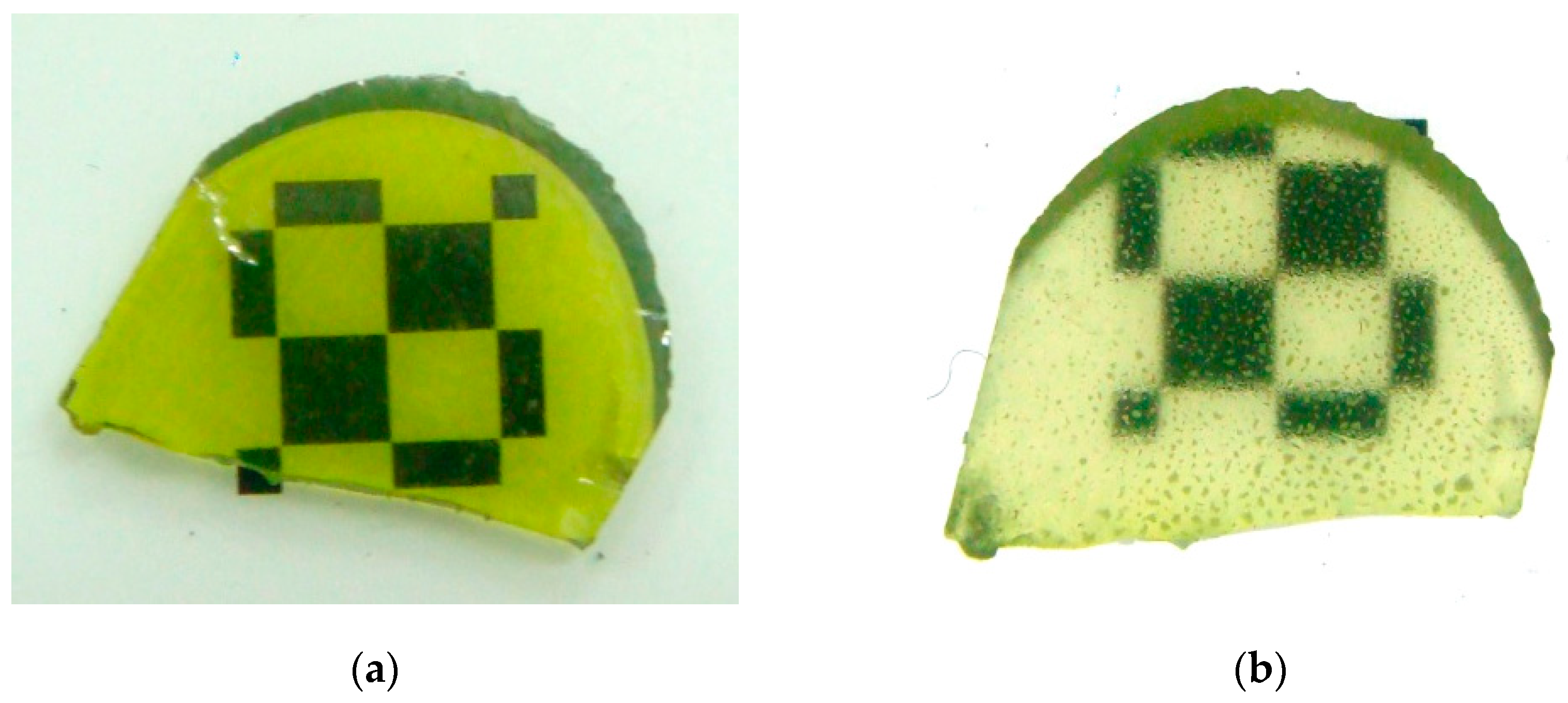
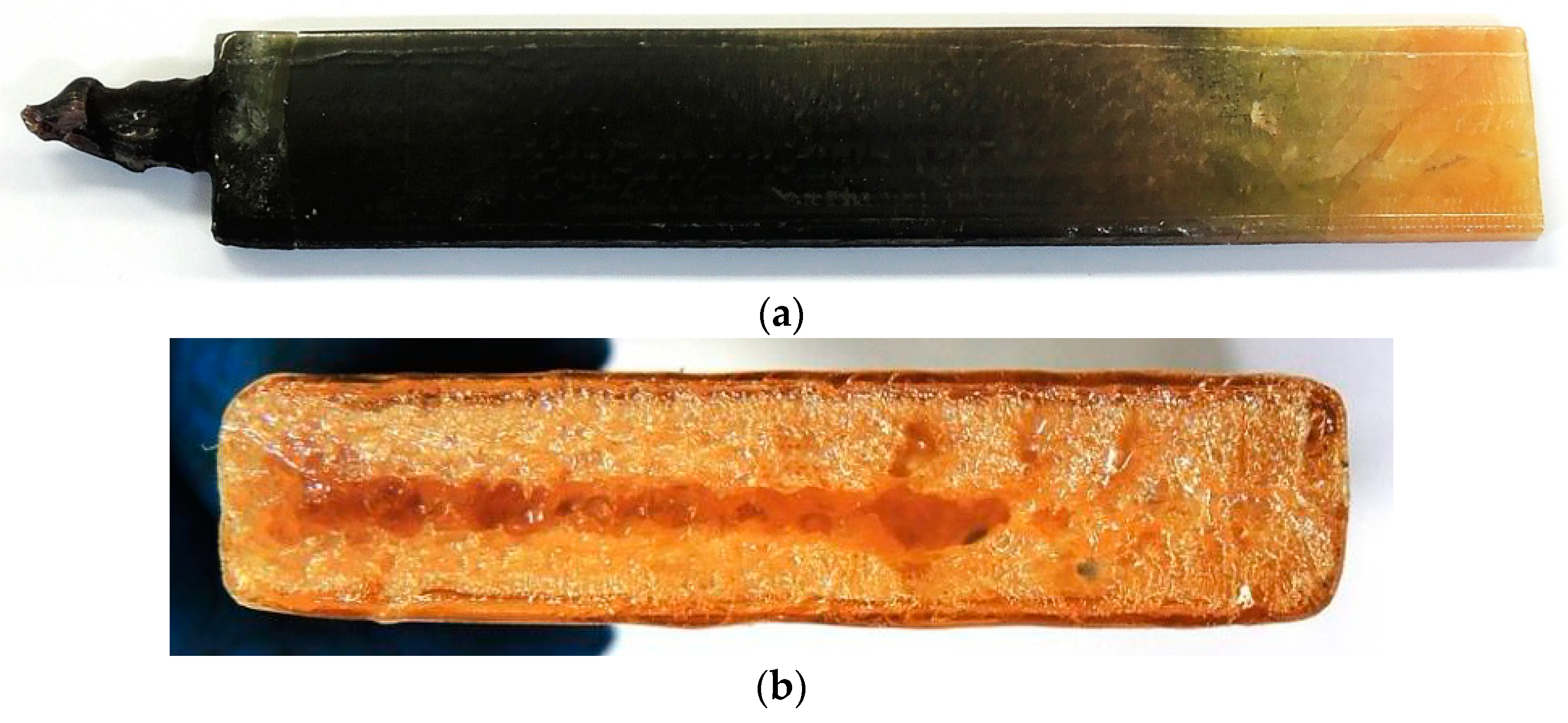
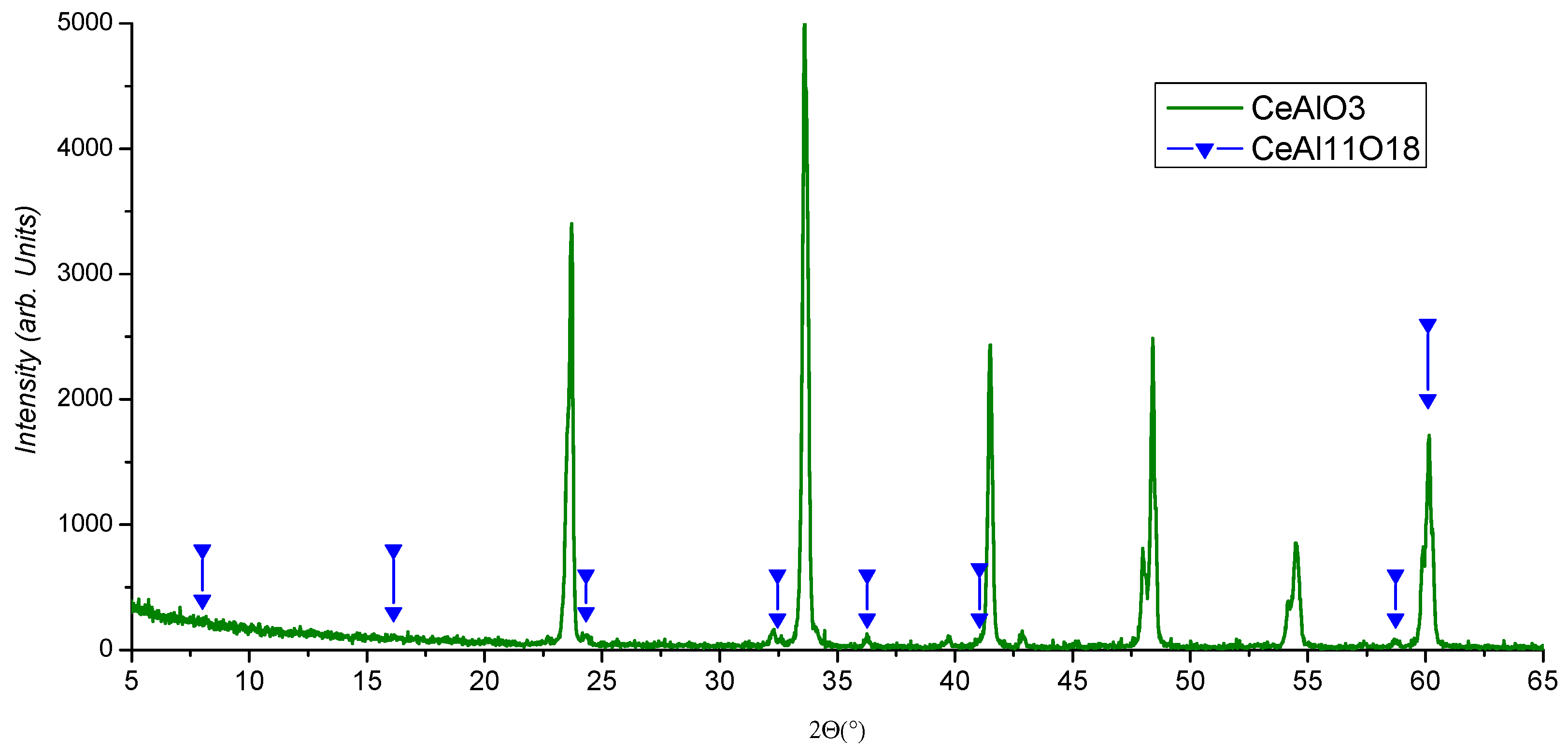
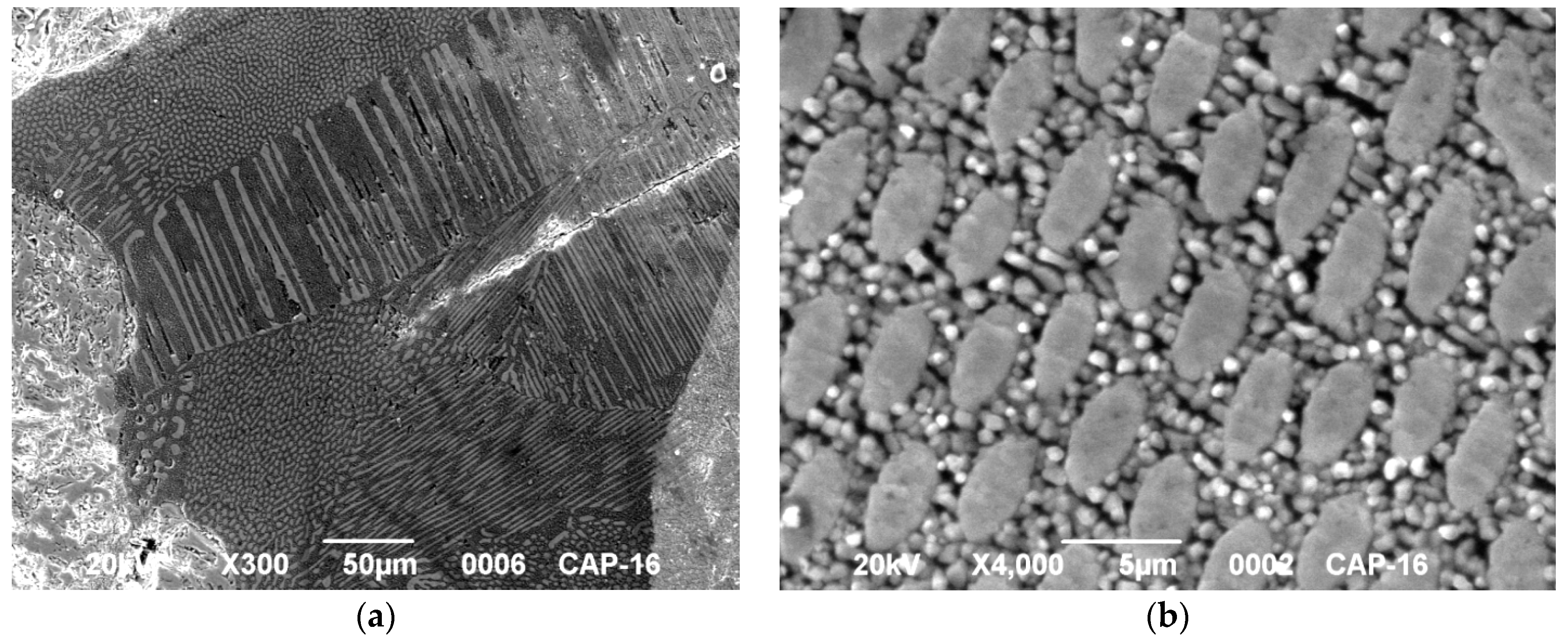
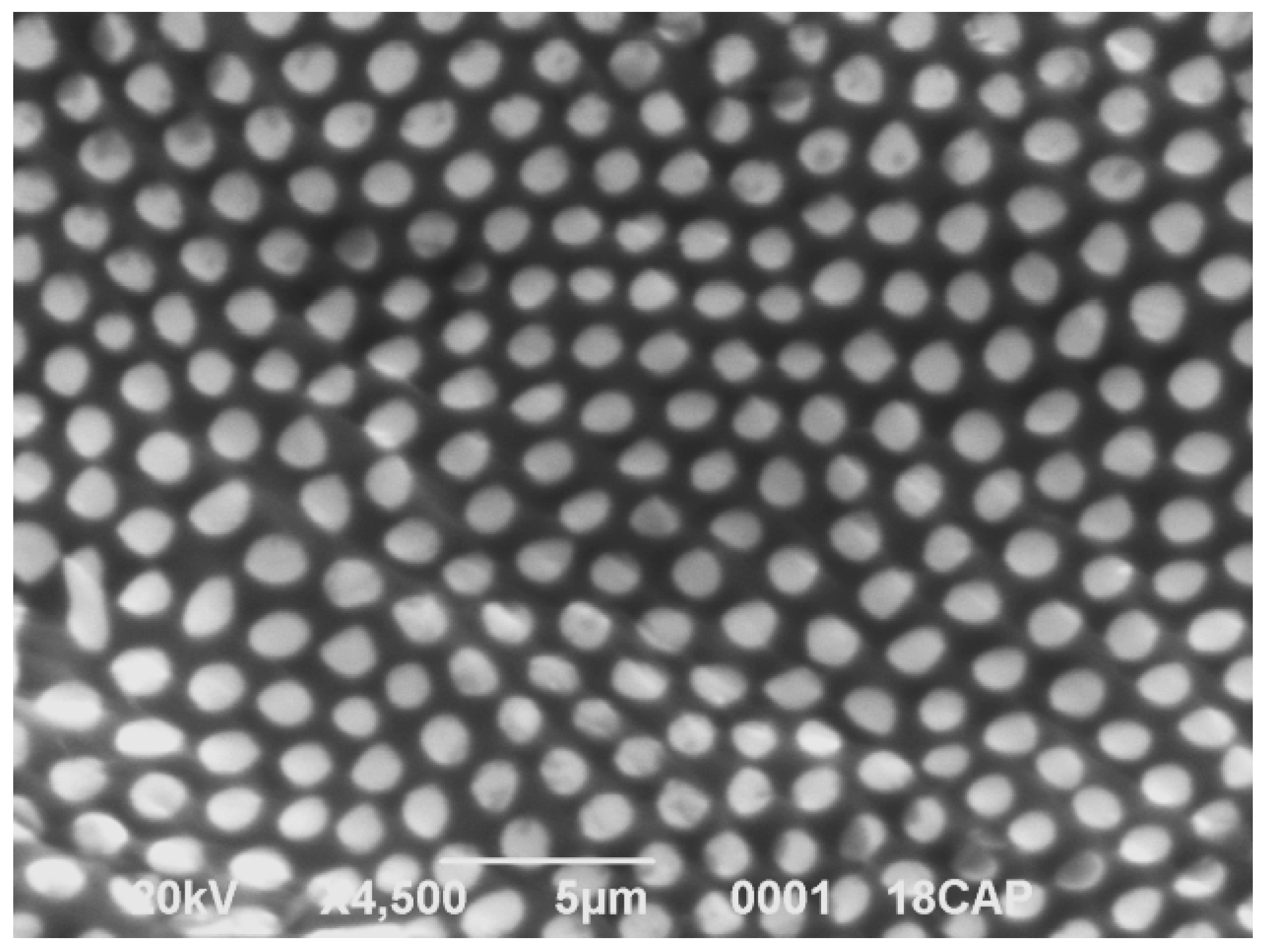
3.1. Structure and Composition of CeAlO3 Crystals and CeAlO3/CeAl11O18 Metaphase Systems

As-grown CeAlO3 single crystals are colored as shown in Figure 1a. The coloration disappears after 2–4 h of post-growth annealing at 1300 °C in the Ar and CO reducing atmosphere. Meanwhile, we noticed that the bleaching process is not uniform in the crystal bulk, and some colored spots remain at the surface at intermediate stage (Figure 1b). The photos of the studied colored and transparent samples are presented in Figure 1.
Some ingots were polycrystalline (Figure 2a,b) and show a visible emission under UV light irradiation, as seen in Figure 2c. Microanalysis showed that while the light-emitting areas have composition around Ce0,2Al1,8O3, the composition of other grains was between Ce1,1Al0,9O3 and Ce1,2Al0,8O3. This almost corresponds to CeAl11O18 in the light-emitting grains and CeAlO3 in the rest of the grains.
The XRD data (Figure 3) show the presence of a small amount of CeAl11O18 phase in addition to the main CeAlO3 phase. No evidence of other phases have been obtained. From SEM images (Figure 4a), it can clearly be seen that the cut of EGF-grown crystal contains CeAl11O18 phase inclusions (light areas) arranged into columns oriented along the different directions and embedded into the CeAlO3 host phase (dark areas). Under higher resolution (Figure 4b), one can see the CeAl11O18 columns of oval shape with the 3–5 µm size are surrounded by smaller grains with the size less than 1 µm and composition around CeAlO3.
As the eutectic composition between CeAlO3 and CeAl11O18 is Ce0,46Al1,54O3 [7], the Al2O3 content has to be increased for directional synthesis of the phase-separated columnar structure. While in the shown crystals, phase-separated structures were occasionally formed, such structures can be formed intentionally by a shift of melt composition, or by solid phase synthesis. The latter was implemented in this work by the annealing of visually homogeneous CeAlO3 crystals in contact with a single Al2O3 crystal at 1700 °C. While the composition of the crystal surface before the annealing was precisely CeAlO3, after annealing, the surface integral composition shifted to Ce0,38Al1,62O3. Herein, the measured compositions of the light and dark areas in Figure 5 are Ce0,54Al1,46O3, and Ce0,26Al1,74O3, correspondingly, with larger and smaller Ce/Al ratios relative to the Ce0,46Al1,54O3 eutectic composition. The compositions of light and dark areas are close to CeAlO3 and CeAl11O18, because the spatial resolution of the method is limited by 1 µm and it is barely possible to determine the composition of a single ~1 µm size area. This picture is very similar to the microstructure of the Tb3Sc2Al3O12-TbScO3 binary eutectic grown by the micro-pulling down method [3]. As no data on CeAlO3 and CeAl11O18 refractive indices are known at the moment, it is not possible to evaluate precisely the wave guiding properties of such columnar structure. However, the bright emission from the CeAl11O18-designated areas and the high refraction index of 1.98 in LaAlO3 homologue with perovskite structure [8] provides evidence that the CeAl11O18 refractive index is lower.

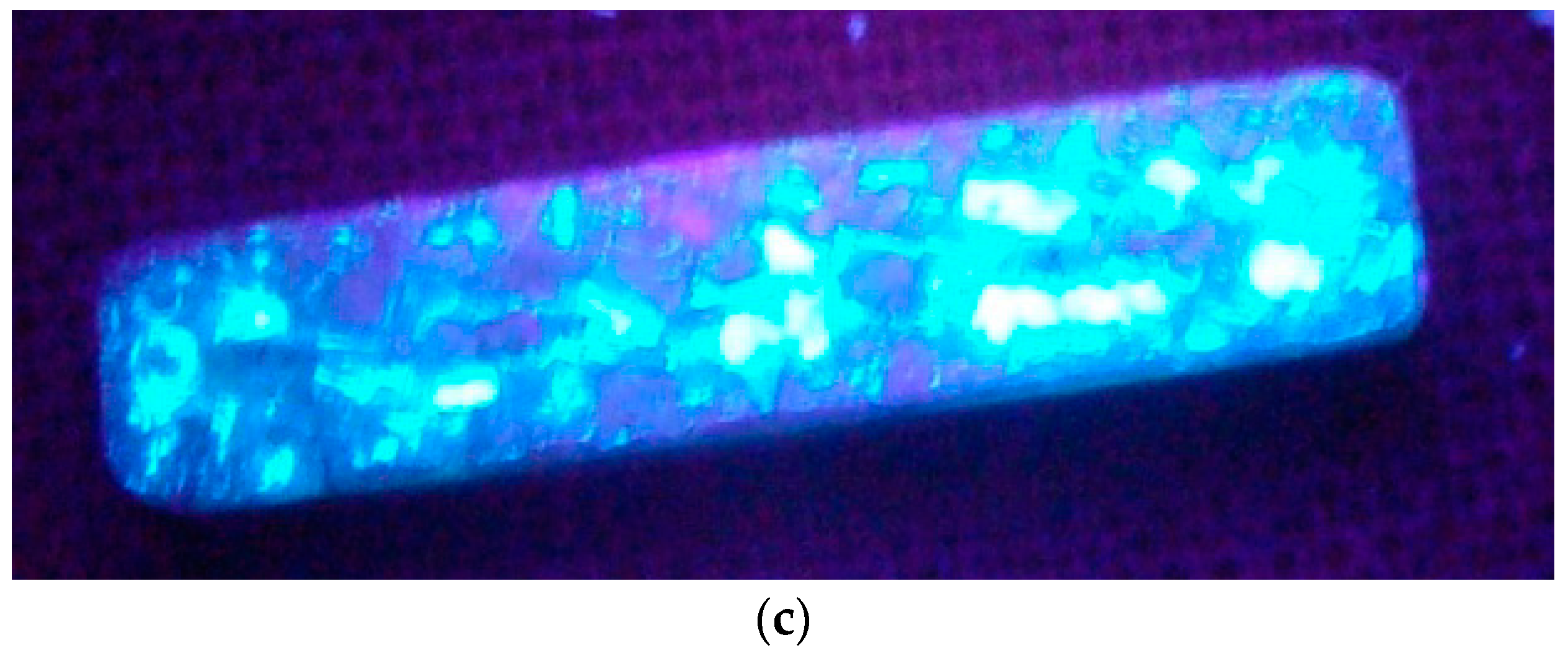
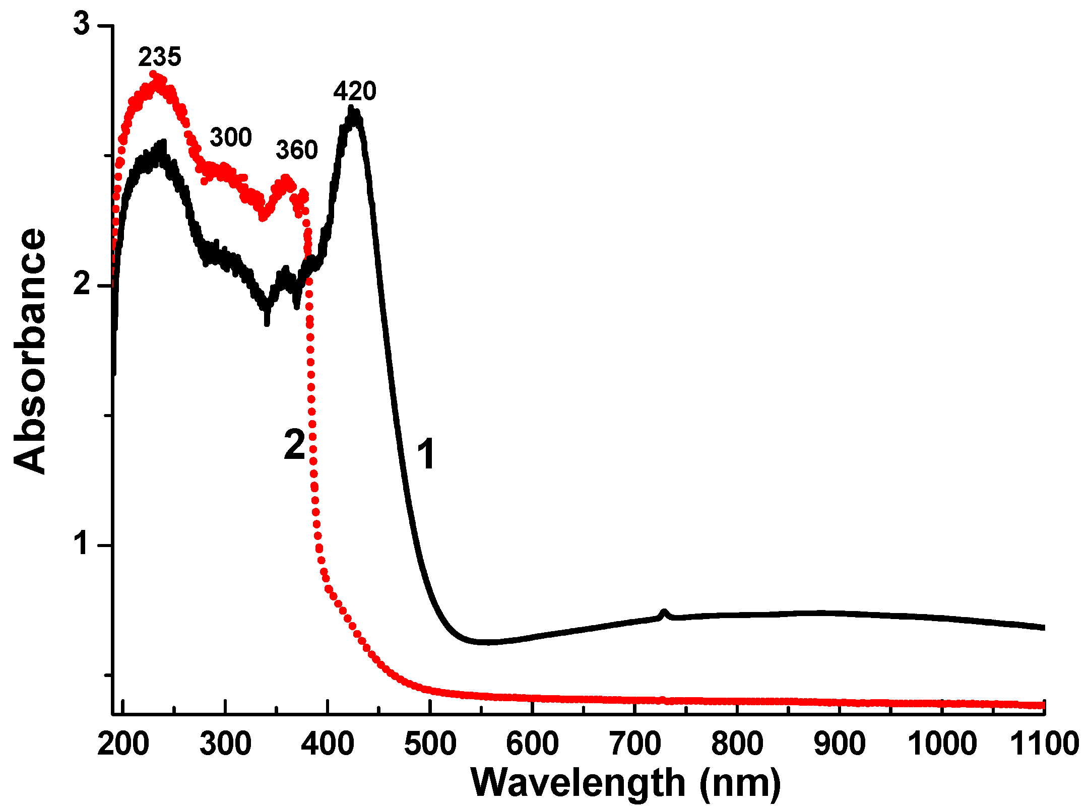
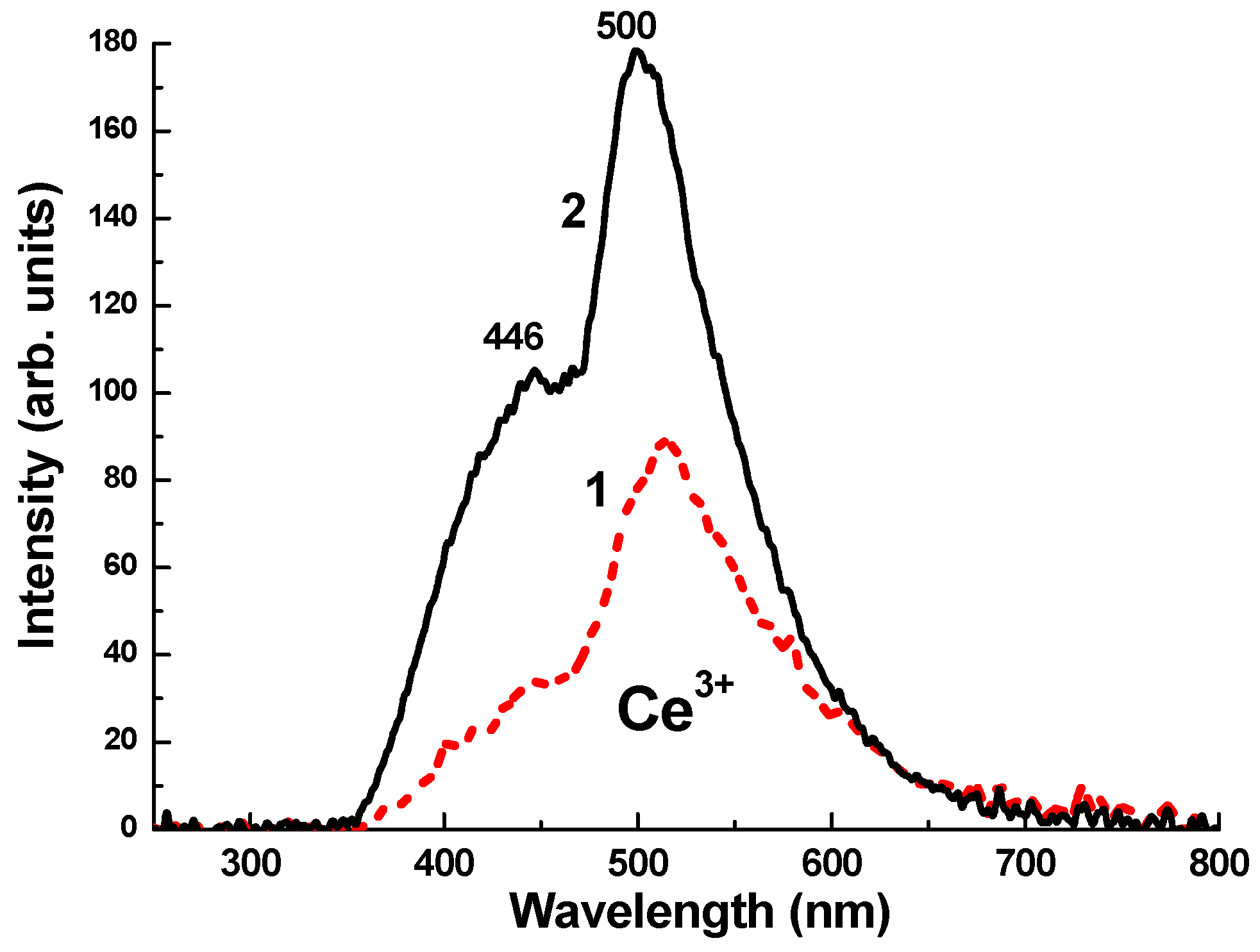
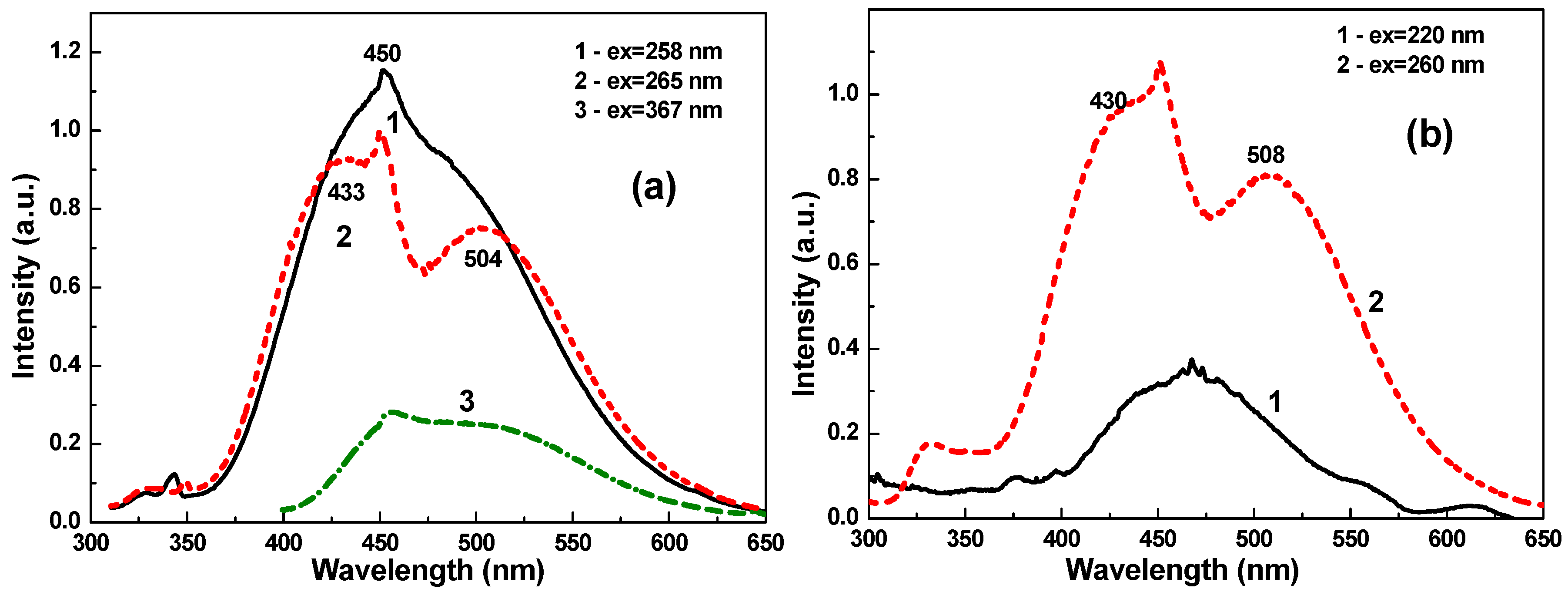
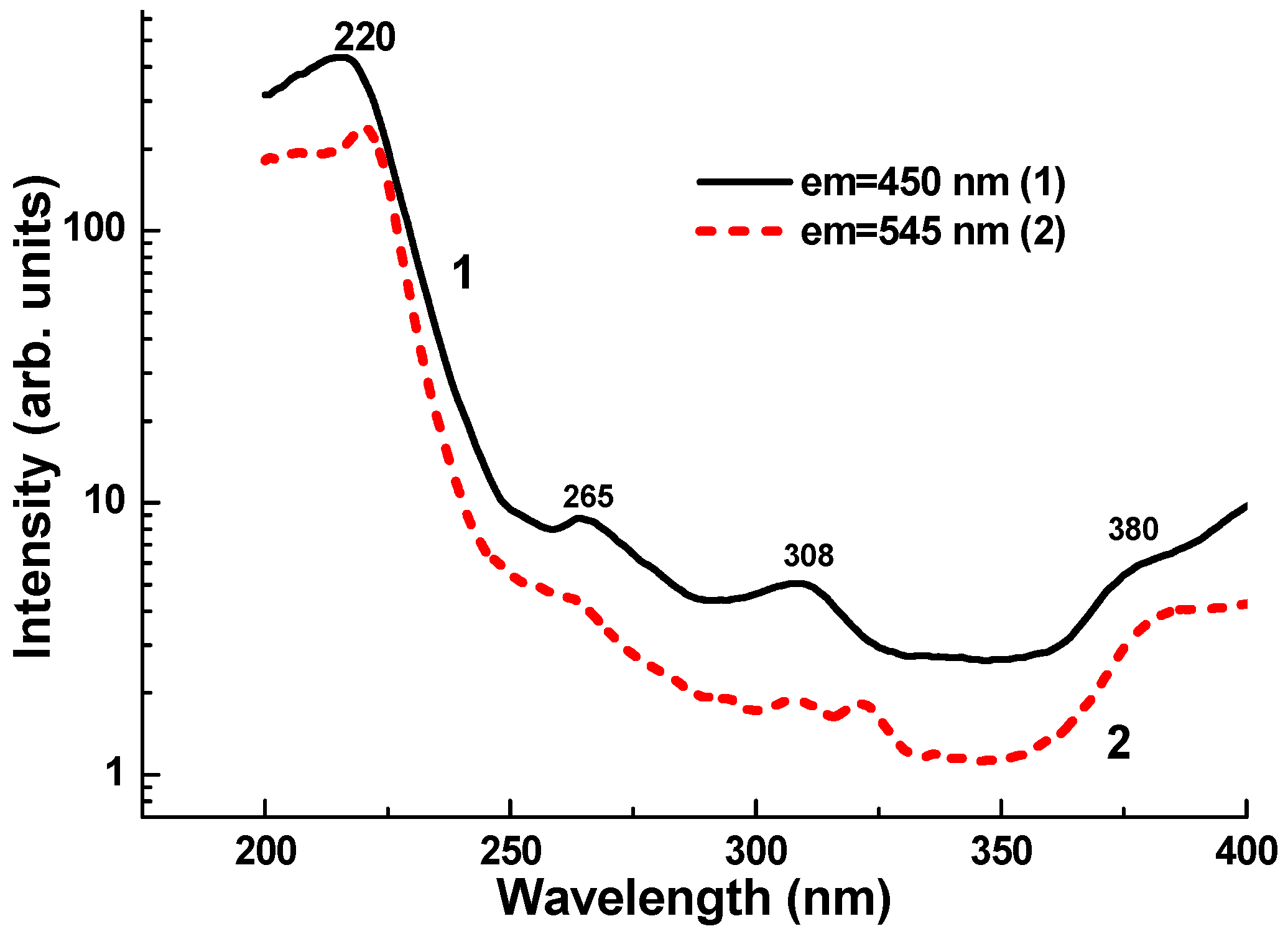
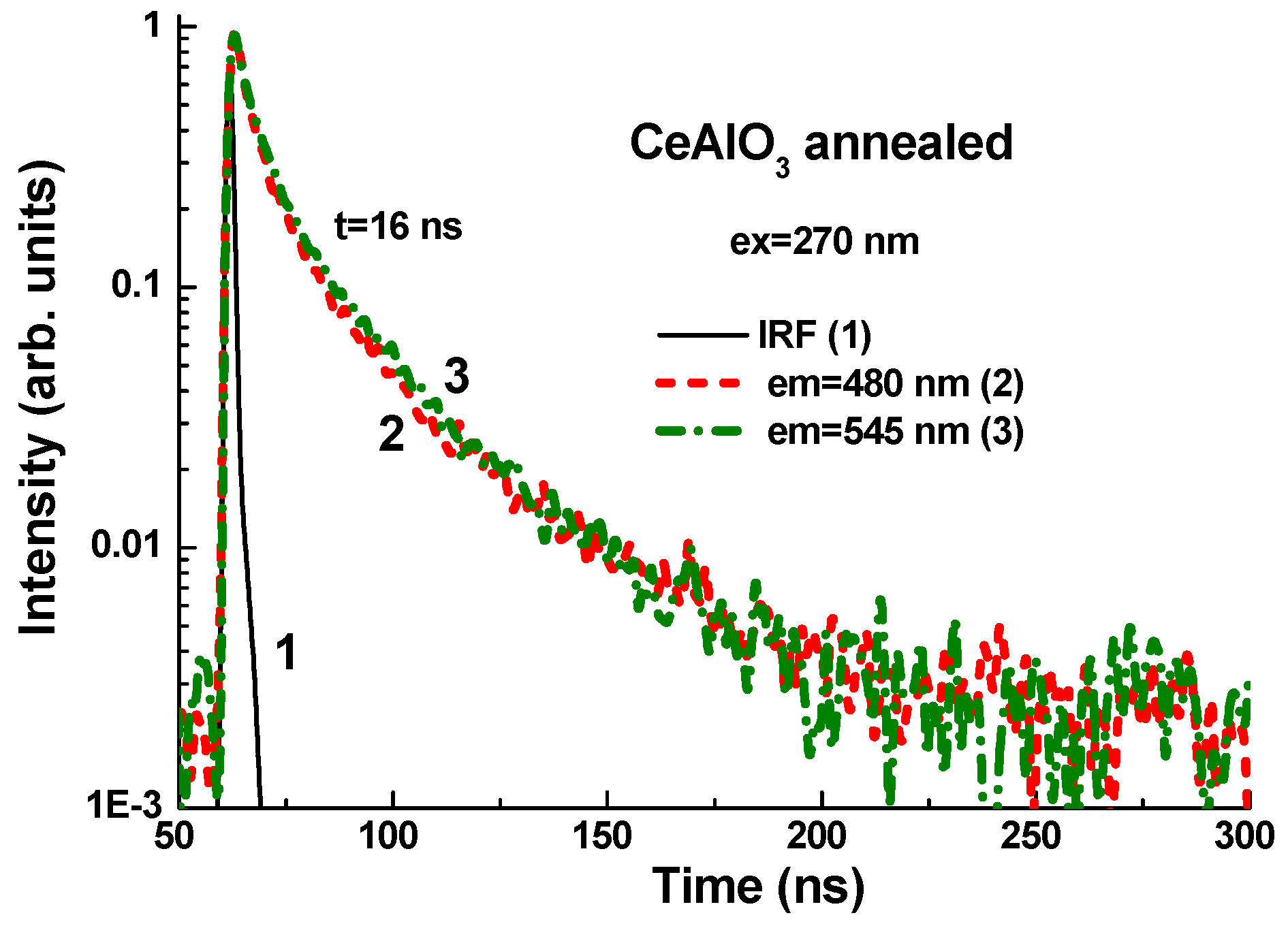
3.2. Optical and Luminescent Properties

The absorption spectra of as-grown and annealed samples are presented in Figure 6. Absorption of the as-grown CeAlO3 crystals is characterized by the main band peaking at 420 nm. As this band completely disappears after annealing in an Ar + CO reducing atmosphere (Figure 6, curve 2), eventually promoting the Ce4+→Ce3+ transfer, it is likely that this band is related to O2−→Ce4+ charge transfer transitions in the CeAlO3 host, similar to other Ce-containing materials [9].
The luminescence of CeAlO3 crystals under excitation by e-beam and α- and β-particles was registered for the first time (Figure 7). Under e-beam excitation, the observed double luminescence band in CeAlO3 crystals, peaking at 446 and 500 nm, is probably related to the Ce3+ 5d-4f transition into the CeAl11O18 admixture phase. The CL intensity is significantly larger in the annealed sample due to the increase of Ce3+-emitting centers concentration and the decrease of luminescence reabsorption by the CT-related absorption band, which peaked at 420 nm (see Figure 7).
However, we noticed that in annealed CeAlO3 crystals, the UV-excited blue photoluminescence is emitted not from the overall volume of the CeAlO3 bulk crystal, but mainly from colored spots (see Figure 1b). The shapes of the photoluminescence spectra of the spots at the crystal surface and sintered raw material powders are similar (Figure 8), which points to the emission of CeAl11O18 phase embedded in the CeAlO3 crystals, as suggested in [2].
Indeed, the comparison of UV-excited photoluminescence spectra of the metaphase structure and colored spots at the transparent CeAlO3 crystal surface (Figure 8) confirms the similar nature of the luminescence response. At 260–265 nm excitation, a wide band with the main peaks near 430–450 and 504–508 nm is observed. Therefore, we attribute the UV-excited luminescence in CeAlO3 crystal to the CeAl11O18 phase admixture in the raw material powders, as well as in the single- and polycrystalline samples.
Excitation spectra of these main luminescence bands are of quite similar shape (Figure 9). Several distinguished peaks at 220, 265, and 308 nm, as well as the complex peaks at 380 nm, show that the excitation spectra are related to 4f–5d transitions in Ce3+ ions. Most probably, these two group of excitation bands are connected to the transition from the 2F5/2 level of ground state to the 2E and T2g excited levels of Ce3+ ions in the CeAl11O18 host. However, such conclusions need more careful experimental confirmation.
The luminescence decay curves of annealed CeAlO3 crystal sample are shown in Figure 10. Generally, the decay curves monitored at 480 and 540 nm under UV excitation and at 270 nm are quite similar to each other and strongly not exponential. Such shape of the decay curves points at Ce3+ luminescence quenching due to some non-radiative process. For this reason we have calculated the average time t1/e of the photoluminescence intensity decay to 1/e level. This value is equal to 16 ns (Figure 10) and is typical for the Ce3+ decay time in perovskite hosts [10].

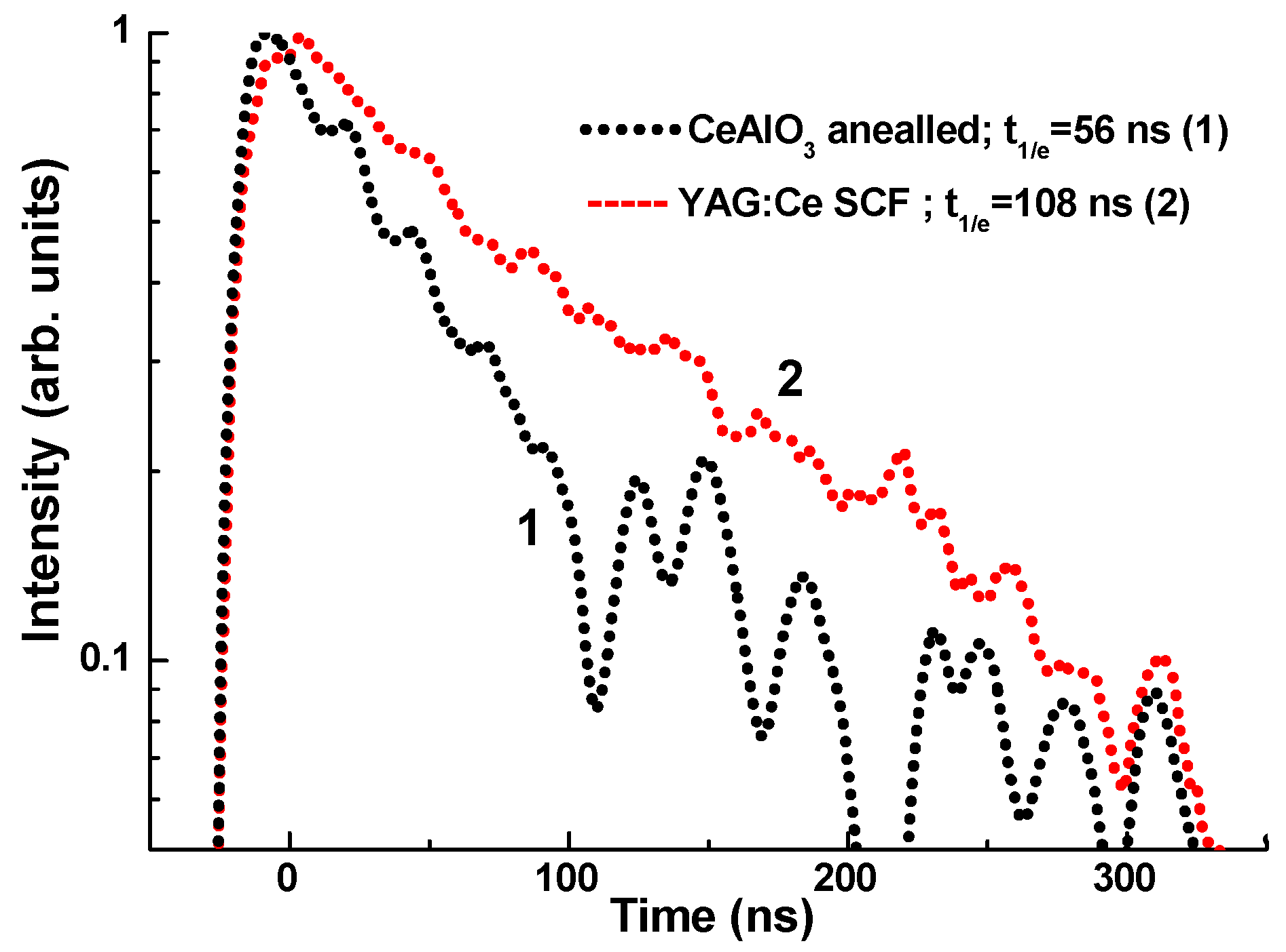
3.3. Scintillation Properties of CeAlO3 Single Crystals

Apart from the fact that the CeAlO3 single and polycrystals possess very weak luminescence at room temperature under soft X-rays and γ-radiation [2], the scintillation light yield and scintillation decay of CeAlO3 crystals under α-particle excitation can be seen. Namely, under α- particle excitation by 239Pu sources (5.15 MeV), the light yield of annealed CeAlO3 crystals is equal to about 16% and 12% in respect to the standard BGO crystal and YAG:Ce SCF sample with the light yields of 1950 and 2600 photon/MeV, respectively. The scintillation response of the annealed CeAlO3 crystal is quite fast, and the respective scintillation decay time is equal to 56 ns (Figure 11).

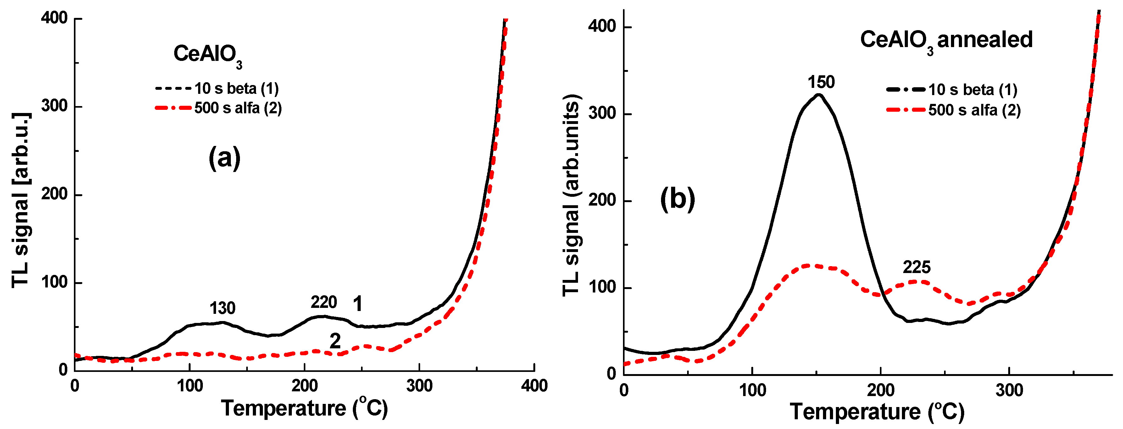
3.4. Thermoluminescence

CeAlO3 single crystals after irradiation by high-energy X-rays and α-particles show weak thermoluminescence (TL, Figure 12a). Indeed, the TL peaks in the 120–150 °C and 220–225 °C ranges were resolved mainly after β-particle irradiation (Figure 12a). Meanwhile, after annealing in the reduced atmosphere, the 150 °C peak intensity increased by 6 times after irradiation with β-particles, while the 220 °C peak intensity remained the same (Figure 12b). It is worth noting that after α-particle irradiation, the 150 and 225 K peaks’ intensities also increased non-proportionally by 6.26 and 3.8 times, respectively.
Taking into account that Ce3+ ions typically serve as the hole trapping centers, the observed TL peaks in the 130–150 °C and 220–225 °C ranges correspond to electron trapping centers. The defects responsible for such deep centers in CeAlO3 single crystals could be oxygen vacancies trapping one or two electrons (F+ and F centers, respectively). We can suppose that the concentration of oxygen vacancies is low in as-grown CeAlO3 crystals, and this fact caused the very weak TL signal in this sample (Figure 12a). Meanwhile, after annealing under the reduction atmosphere, the concentration of oxygen vacancies and related F+ and F centers could significantly increase. That may lead to the observed TL intensity increase (Figure 12b).

4. Conclusions

The growth process and luminescent and scintillation properties of CeAlO3 single crystals have been considered in this work. We have shown the possibility of creating CeAlO3-CeAl11O18-based scintillating metamaterials using the combination of the EFG growth method and post-growth high-temperature annealing of CeAlO3 crystals in a reducing atmosphere or in vacuum.
Cathodoluminescence and radioluminescence in CeAlO3 single crystals under e-beam excitation and α-particles excitation were registered for the first time. Under such types of excitation, CeAlO3 single crystals possess double pealed luminescence in the visible range at 440 and 500 nm. This is related to Ce3+ 5d–4f transition in the CeAl11O18 phase, which is present in CeAlO3 crystals as an admixture. The CL and RL intensity significantly increased in CeAlO3 crystals after annealing at 1700 °C in an Ar and CO reducing atmosphere. Such annealed CeAlO3 crystal also showed more intense thermoluminescence peaks in the 130–150 °C and 220–225 °C ranges, due to the larger concentration of oxygen vacancies and related traps compared to the as-grown counterpart.
We have also found that CeAlO3 crystals show a quite fast scintillation response under α- particle excitation, with a decay time about of 56 ns. However, the scintillation light yield of annealed CeAlO3 crystals is not high and equal to 310–315 photon/MeV under α-particle excitation by a 239Pu (5.15 MeV) source. At the same time, after the optimization of growth and thermal treatment conditions, the heavy CeAlO3 single crystal scintillators are promising for selective registration of high-energy particles, namely in the form of thin (up to 1 mm) plates, or could be used as the substrates in composite film-substrate scintillators based on the liquied phase epitaxy (LPE) grown structures of perovskite compounds [11,12].

Author Contributions

O.S. and Y.Z. analyzed experimental materials and wrote the test of the text the paper. P.A., S.T., I.G., and G.T. performed the experiments on growth of single crystals and metaphase materials, as well as co-wrote the growth part of the paper. T.Z. performed the luminescence and scintillation measurements. W.G. and P.B. performed the TSL measurements. P.M. performed SEM study and element analysis. A.P. performed XRD analysis.

Funding

The work was supported by the Polish NCN 2016/21/B/ST8/03200 and Ukrainian MES SL-76 F projects.

Conflicts of Interest

The authors declare no conflict of interest.

References

  1. Irvine, J.T.S.; Connor, P. Solid Oxide Fuels Cells: Facts and Figures, Green Energy and Technology; Springer-Verlag: London, UK, 2013. [Google Scholar]
  2. Arhipov, P.; Tkachenko, S.; Gerasymov, I.; Sidletskiy, O.; Hubenko, K.; Vasyukov, S.; Shiran, N.; Baumer, V.; Mateychenko, P.; Fedorchenko, A.; et al. Growth and characterization of large CeAlO3 perovskite crystals. J. Cryst. Growth 2015, 430, 116–121. [Google Scholar] [CrossRef]
  3. Pawlak, D.A. Metamaterials and photonic crystals—Potential applications for self-organized eutectic micro-and nanostructures. Sci. Plena 2018, 4, 014801. [Google Scholar]
  4. Ohashi, Y.; Yasui, N.; Yokota, Y.; Yoshikawa, A.; Den, T. Submicron-diameter phase-separated scintillator fibers for high-resolution X-ray imaging. Appl. Phys. Lett. 2013, 102, 051907. [Google Scholar] [CrossRef]
  5. Yasui, N.; Ohashi, Y.; Den, T.; Horie, R. Scintillator body, method for manufacturing the same, and radiation detector. Patent WO 2011/093176 A2, 4 August 2011. [Google Scholar]
  6. Hishinuma, K.; Kamada, K.; Kurosawa, S.; Yamaji, A.; Pejchal, J.; Yokota, Y.; Ohashi, Y.; Yoshikawa, A. LiF/CaF2/LiBaF3 ternary fluoride eutectic scintillator. Jpn. J. Appl. Phys. 2015, 54, 04DH04. [Google Scholar] [CrossRef]
  7. Cuneyt, T.A.; Akinc, M. Phase relations in the system Ce2O3-Al2O3 in inert and reducing atmospheres. J. Am. Chem. Soc. 1994, 77, 2961–2967. [Google Scholar]
  8. Merker, L.; Herrington, K.D. Transmission spectra of rare earth titanates and aluminates. Appl. Opt. 1964, 3, 1311–1313. [Google Scholar] [CrossRef]
  9. Blasse, G.; Grabmaier, B.C. Luminescent Materials; Springer: Berlin/Heidelberg, Germany, 1994. [Google Scholar]
  10. Zorenko, Y.; Gorbenko, V.; Zorenko, T.; Voznyak, T.; Riva, F.; Douissard, P.A.; Martin, T.; Fedorov, A.; Suchocki, A.; Zhydachevski, Y. Growth and luminescent properties of single crystalline films of Ce3+ doped Pr1-xLuxAlO3 and Gd1-xLuxAlO3 perovskites. J. Cryst. Growth 2017, 457, 220–226. [Google Scholar] [CrossRef]
  11. Witkiewicz-Lukaszek, S.; Gorbenko, V.; Zorenko, T.; Sidletskiy, O.; Gerasymov, I.; Fedorov, A.; Yoshikawa, A.; Mares, J.A.; Nikl, M.; Zorenko, Y. Development of composite scintillators based on single crystalline films and crystals of ce3+-doped (Lu,Gd)3(Al,Ga)5O12 mixed garnet compounds. Cryst. Growth Des. 2018, 18, 1834–1842. [Google Scholar] [CrossRef]
  12. Witkiewicz-Lukaszek, S.; Gorbenko, V.; Zorenko, T.; Paprocki, K.; Sidletskiy, O.; Gerasymov, I.; Mares, J.A.; Kucerkova, R.; Nikl, M.; Zorenko, Y. Novel all-solid-state composite scintillators based on the epitaxial structures of LuAG garnet doped with Pr, Sc, and Ce Ions. IEEE Trans. Nucl. Sci. 2018, 65, 2114–2119. [Google Scholar] [CrossRef]
Figure 1. CeAlO3 single crystalline samples before (a) and after (b) annealing at 1700 °C in the Ar and CO reducing atmosphere.
Crystals 09 00296 g001
Figure 2. Photos of as-grown crystal (a), its transversal cut (b), and the same transversal cut under illumination with UV light (c).
Crystals 09 00296 g002aCrystals 09 00296 g002b
Figure 3. XRD of the transverse cut of polycrystalline sample grown by the edge-defined film-fed (EFG) technique.
Crystals 09 00296 g003
Figure 4. SEM images of crystallites at the transverse cut: (a) ×300 magnification; (b) ×4000 magnification.
Crystals 09 00296 g004
Figure 5. SEM image (magnification ×4500) of the eutectic structure at the surface of CeAlO3 crystal annealed at 1700 °C under vacuum in contact with Al2O3 crystal.
Crystals 09 00296 g005
Figure 6. Absorption spectra of as-grown (1) and annealed (2) CeAlO3 single crystals with a thickness of 1 mm.
Crystals 09 00296 g006
Figure 7. Cathodoluminescence spectra of CeAlO3 single crystals: 1, as-grown; 2, annealed.
Crystals 09 00296 g007
Figure 8. Luminescence spectra of CeAlO3 eutectic structure (a) and colored spots at the surface of transparent CeAlO3 single crystal and (b) under excitation with the different wavelengths in the UV range.
Crystals 09 00296 g008
Figure 9. Photoluminescence excitation spectra of a CeAlO3 crystal. Annealed sample monitored at 450 nm (1) and annealed CeAlO3 crystal monitored at 545 nm (2).
Crystals 09 00296 g009
Figure 10. Normalized PL decay of annealed CeAlO3 crystals under excitation at 270 nm and registration of emission at 480 nm (2) and 540 nm (3). (1) Instrumental response function (IRF) of laser pulse.
Crystals 09 00296 g010
Figure 11. Normalized scintillation decay curves of annealed CeAlO3 crystals under α-particle excitation of 239Pu sources in comparison with a YAG:Ce SCF standard sample (2).
Crystals 09 00296 g011
Figure 12. Thermoluminescence (TL) glow curves of as-grown (a) and annealed (b) CeAlO3 crystals after α- and β- particle excitation with 239Pu (1) and 90Sr/90Y (2) sources.
Crystals 09 00296 g012

