Communication

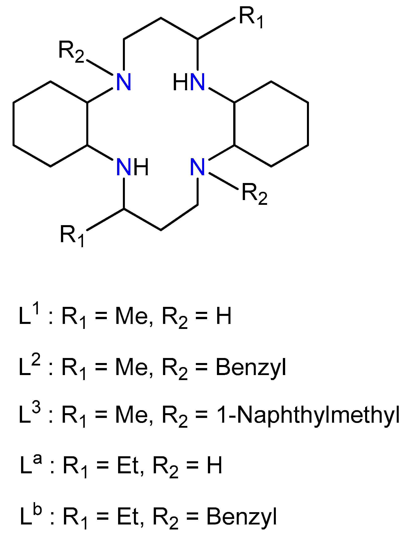
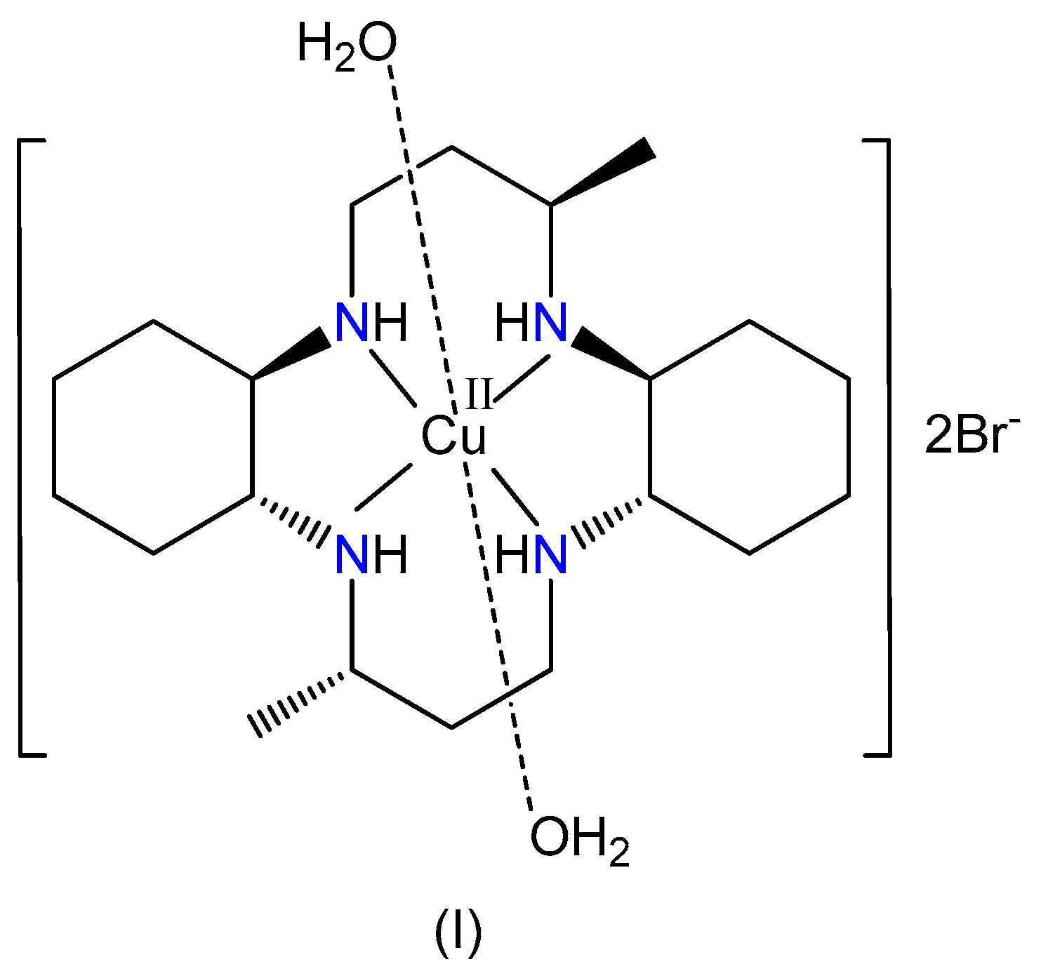
Synthesis, Crystal Structure, Spectroscopic Properties, and Hirshfeld Surface Analysis of Diaqua [3,14-dimethyl-2,6,13,17 tetraazatricyclo(16.4.0.07,12)docosane]copper(II) Dibromide

1
Department of Chemistry, Andong National University, Andong 36729, Korea
2
Department of Inorganic Chemistry, Faculty of Chemical and Food Technology, Slovak University of Technology, 81237 Bratislava, Slovakia
3
Department of Physical Chemistry, Faculty of Chemical and Food Technology, Slovak University of Technology, 81237 Bratislava, Slovakia
*
Authors to whom correspondence should be addressed.
Submission received: 5 June 2019 / Revised: 17 June 2019 / Accepted: 26 June 2019 / Published: 28 June 2019

Abstract

:
A newly prepared Cu(II) complex salt, Cu(L1)(H2O)2Br2, where L1 is 3,14-dimethyl-2,6,13,17-tetraazatricyclo(16.4.0.07,12) docosane, is characterized by elemental and crystallographic analyses. The Cu(II) center is coordinated by four nitrogen atoms of macrocyclic ligand and the axial position by two water molecules. The macrocyclic ligand adopts an optimally stable trans-III conformation with normal Cu–N bond lengths of 2.018 (3) and 2.049 (3) Å and long axial Cu1–O1W length of 2.632 (3) Å due to the Jahn–Teller effect. The complex is stabilized by hydrogen bonds formed between the O atoms of water molecules and bromide anions. The bromide anion is connected to the neighboring complex cations and water molecules through N–H···Br and O–H···Br hydrogen bonds, respectively. The g-factors obtained from the electron spin resonance spectrum show the typical trend of g > g > 2.0023, which is in a good accordance to the dx2-y2 ground state. It reveals a coordination sphere of tetragonal symmetry for the Cu(II) ion. The infrared and electronic absorption spectral properties of the complex are also discussed. Hirshfeld surface analysis represents that the H···H, H···Br/Br···H and H···O/O···H contacts are the major molecular interactions in the prepared complex.

Graphical Abstract

1. Introduction

Recently, the mobilization of stem cell in bone marrow and anti-HIV effect by the cyclam derivatives and their transition metal complexes have been found [1,2,3,4]. The metal complex with the cyclam skeleton can take either trans or cis geometrical isomers because of its structural flexibility. A total of five conformational trans isomers caused by the chiral secondary amine nitrogen atoms. Furthermore, three cis-conformers are possible [5,6]. The macrocycle conformation in the complex is important factor for CXCR4 chemokine receptor recognition [1,2,3]. The knowledge on the coordination behavior and conformation of the cyclam derivative plays important role for development of novel and highly effective anti-HIV drugs. In this context, investigations on various constrained cyclam ligands (Scheme 1) containing cyclohexane subunits and methyl or ethyl groups have proved instructive.
The crystallographic study of [Cu(L1)(H2O)2]Cl2, [Cu(L1)(H2O)2](BF4)2·2H2O, [Cu(L1)(ClO4)2], [Cu(L1)(NO3)2], [Cu(L1)(NO3)2]·3H2O, [Cu(L1)(NCS)2]·2H2O, [Cu(L1)(NCS)]SCN, and [Cu(L1)(N3)]ClO4·H2O have been performed previously [7,8,9,10,11,12,13,14]. In these Cu(II) complexes, the macrocycles adopt the most stable trans–III conformation. The ligand containing cyclohexane ring and methyl group has sometimes showed different coordination behaviors comparing to those of the complexes with cyclam ligand. For example, crystallographic analysis of [Cu(L2)](ClO4)2 revealed that the copper(II) ion was tetracoordinated in a square planar configuration somewhat distorted towards tetrahedral geometry [15] whereas the [Cu(L3)](ClO4)2·2CH3CN had a square planar geometry [16]. The structural and physical properties of the Cu(II) and Ni(II) complexes with macrocycles containing cyclohexane ring and ethyl group were also reported [17,18,19,20,21,22].
The structural characteristics, like chelate ring size and substituent influence the chemical properties and geometries of Cu(II) complexes. Anionic species is also an important factor in coordination chemistry, molecular assembly and medicine [23,24].
To further investigate on the effect of counter anion and water molecule to the crystal packing behavior, we report here the preparation, structural characterization, spectral properties and Hirshfeld surface analysis of a new bromide salt, [Cu(L1)(H2O)2]Br2, (I) (Scheme 2).

2. Experimental Section

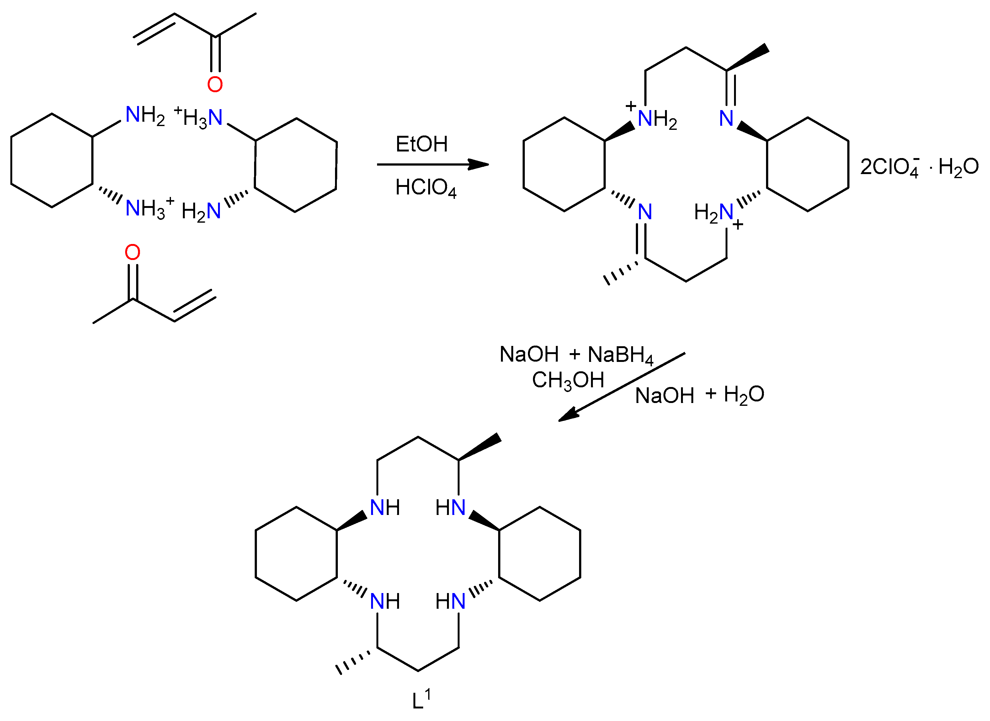
2.1. Synthesis of L1

The macrocycle L1 was synthesized according to a previously published procedure [25]. The synthetic process of ligand L1 is summarized in Scheme 3.
The yield was 88.7%. Anal. Calcd (%). for C20H40N4: C, 71.37; H, 11.98; N, 16.65. Found (%): C, 71.63; H, 12.12; N, 16.50.

2.2. Synthesis of [Cu(L1)(H2O)2]Br2, (I)

A 50 mL aqueous solution of 0.5 mmol CuBr2 was prepared and a 20 mL aliquot was removed, and the macrocycle L1 (0.091 g, 0.25 mmol) was added. The resulting solution was refluxed for 60 min at 373 K. After cooling to 298 K, the pH was adjusted to 3.0 by the addition of 1.0 M HCl. Colorless and violet mixed crystals were formed from the solution over the next few days. The product mixture was added to 30 mL MeOH-acetone (1:2) solution under stirring, and the stirring was continued for 30 min at 298 K. The colorless [H4L1]Cl4·4H2O crystals was removed by filtration [26]. The filtrate was left at 298 K. After few days, violet crystals of [Cu(L1)(H2O)2]Br2 were given. The yield was 75.2%. The complex (I) is stable in air, and soluble in MeOH, EtOH, DMF or DMSO whereas it is insoluble in H2O, i-PrOH, CH3CN, THF and acetone. Anal. Calcd (%). for C20H44CuN4O2Br2: C, 40.31; H, 7.44; N, 9.40. Found (%): C, 40.76; H, 7.40; N, 9.45.

2.3. Physical Measurements

The electronic spectrum (190–1100 nm) was recorded in a Nujol suspension using a Specord 250 Plus spectrometer (Carl Zeiss Spectroscopy GMBH, Jena, Germany). The FT-infrared spectrum was measured with a JASCO 460 FT-IR spectrometer (JASCO, Tokyo, Japan). Elemental analyses for C, H and N was carry out on a Vario MICRO cube Elemental analyzer (Elementar Analysensysteme GmbH, Langenselbold, Germany). The first derivative X-band EPR spectrum of powdered [Cu(L1)(H2O)2]Br2 sample was measured on an EMX EPR spectrometer (Bruker, Karlsruhe, Germany) at 298 K. The sample-containing quartz EPR tube (Bruker) was inserted inside the EPR cavity by the previously described procedure [27]. From the experimental EPR spectrum, the spin Hamiltonian parameters were found by WinEPR [28], and then refined by spectral line simulation using Bruker program SimFonia (Shareware, Bruker, Karlsruhe, Germany) [29].

2.4. Crystal Structure Analysis

Data collection and cell refinement of complex (I) were performed using a four-circle diffractometer StadiVari at 100 K. The diffractometer was equipped by HPAD detector Pilatus3R 300 K and microfocused X-ray source Genix3D Cu HF. The diffraction intensities were corrected for Lorentz and polarization factors. The structures were solved by a dual-space method using the SHELXT-2018 [30] and refined by the full matrix least-squares procedure in SHELXL-2018 [31]. Multi-scan absorption corrections were applied using Stoe LANA software (STOE & Cie GmbH, Darmstadt, Germany) [32]. Molecular graphics were drawn using DIAMOND-3 (Crystal Impact GbR, Bonn, Germany) [33]. The crystal data, data collection conditions and refinements are provided in Table 1. The crystallographic data for the complex (I) was deposited in CCDC 1912362.

2.5. Hirshfeld Surface Analysis

CrystalExplorer program was used for the Hirshfeld surface analysis [34], and the normalized contact distance, dnorm, was mapped onto the Hirshfeld surface [35,36].

3. Results and Discussion

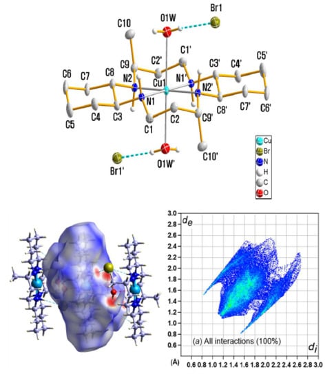
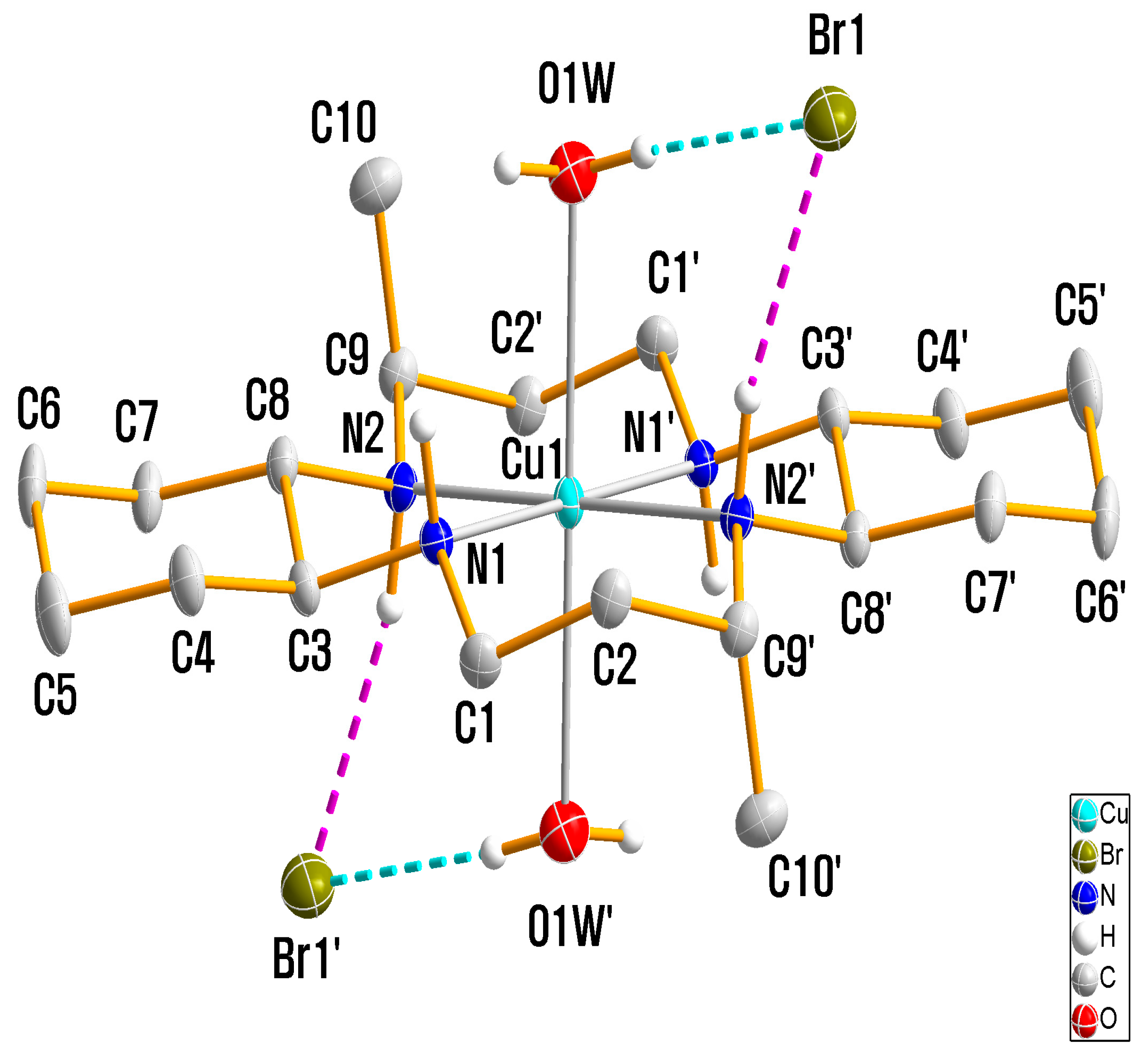
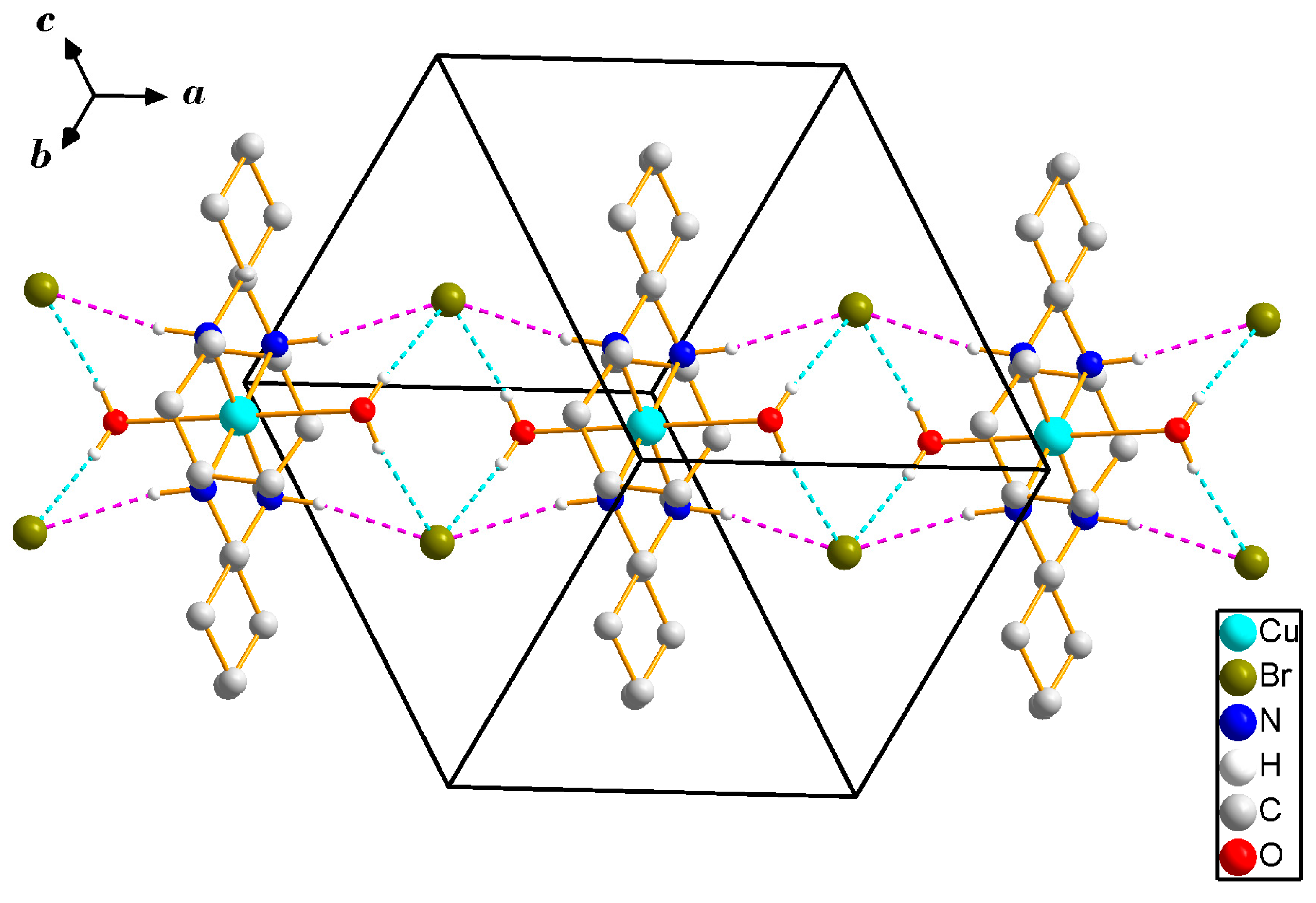
3.1. X-ray Crystallography

The complex (I) crystallized the triclinic space group Pī with dimensions of a = 9.2244 (7), b = 11.5540 (8), c = 12.8659 (9) Å, and β = 97.722 (6)°. The space group of new bromide salt differed from the monoclinic space group P21/c of [Cu(L1)(H2O)2]Cl2 or P21/n of [Cu(L1)(H2O)2](BF4)2·2H2O with two mononuclear formula units per unit cell [7,8]. The asymmetric unit contains one half of [Cu(L1)(H2O)2]2+ cation and one bromide anion. The important bond lengths and angles are listed in Table 2.
Figure 1 shows an ellipsoid plot (50% probability level) of the complex (I) together with the atomic numbering scheme.
The stereochemistry of the prepared Cu(II) complex can be considered square planar or tetragonal geometry. A semi-coordinated bond’s concept was introduced to explain a situation where the axial ligands occupies the tetragonal position in a square planar Cu(II) complex with an atom in the bond length range of 2.5–3.0 Å [36]. The Cu(II) geometry in complex (I) can be described as tetragonally distorted geometry with four N atoms from the macrocycle and two O atoms from water molecules. The macrocyclic skeleton adopted the most stable trans-III (RRSS) conformation. The two methyl groups on the six-membered chelate rings and the two-(CH2)4-parts of the cyclohexane backbones adopted anti configurations with respect to the macrocyclic plane. The Cu–N distances of 2.018 (3) and 2.049 (3) Å can be compared to related complexes in Table 3.
The axial Cu1–O1W bond length [2.632(3) Å] was slightly shorter than the corresponding lengths in [Cu(L1)(H2O)2]Cl2 [2.649(2) Å] [7] and [Cu(L1)(H2O)2](BF4)2·2H2O [2.693(3) Å], but longer than that of [Cu(La)(H2O)2](SCN)2 [2.569(2) Å] [19]. The Cu1–OW bond was not perpendicular to the CuN4 plane, with actual N1–Cu1–O1W and N2–Cu1–O1W angles of 88.22(11) and 95.89(11)°, respectively. The ratio of the average equatorial bond length RS and the average axial bond length RL was used as a measure of tetragonal distortion [36]. The ca. tetragonality factor (T = RS/RL) was 0.773, similar to the calculated mean of ca. 0.84 for those of previously reported many Cu(II) complexes [37].
The ligand L1 contains cyclam in its backbone with two cyclohexane subunits. As it is typically observed, the five-membered chelate rings adopt a gauche conformation, whereas the six-membered rings adopt slightly distorted chair conformations due to the attached methyl group. The bond angles of the five- and six-membered chelate rings around Cu(II) were 84.60 (11) and 95.40 (11)°, respectively. The cyclohexane rings also adopted chair conformation, with the N atoms in equatorial positions. The average C–N and C–C distances, as well as the C–N–C and C–C–N angles in the macrocyclic ligands, were similar to those for a number of macrocyclic tetraamine complexes [15,16].
The supramolecular architecture involves hydrogen-bonding interactions of the N–H groups of the macrocycle ligands and O–H groups of the water molecules as donors, and the bromide anions as acceptors, resulting in a three-dimensional network structure (Figure 2 and Figure 3). The Br anions remain outside the coordination sphere [Cu–Br (4.768 Å)] and are connected to the semi-coordinated water molecules through O–H···Br (cyan) hydrogen bonds. Both hydrogens on N1 and N2 of the macrocyclic ligand were involved in very weak N–H···Br (pink) interactions. These hydrogen-bonded networks stabilized the crystal structure.

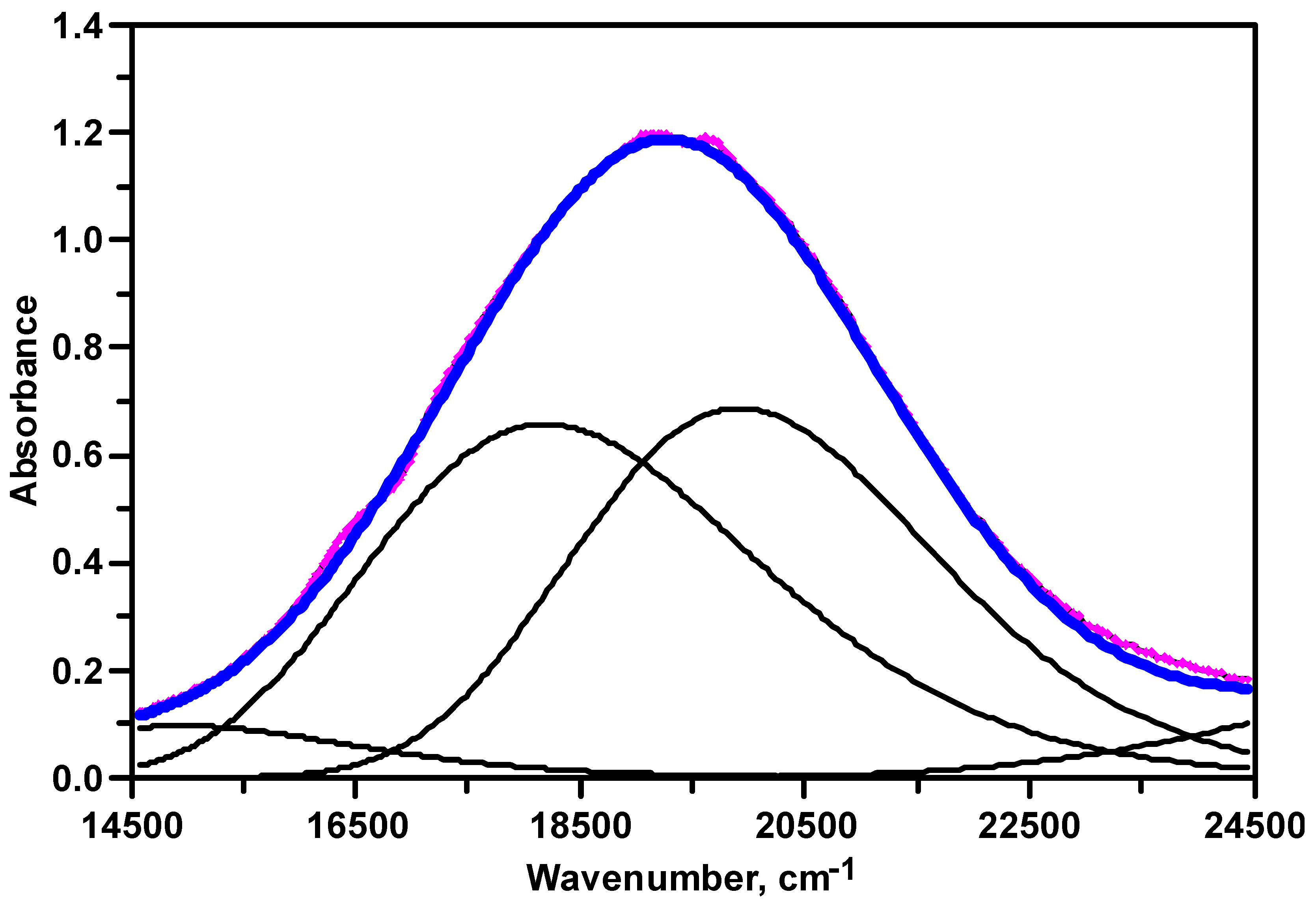
3.2. UV–Visible Spectroscopy

The electronic spectrum of complex (I) exhibited one broad d-d band at 19,920 cm−1 in the Nujol suspension. The absorption maximum, νmax, is comparable to those of [Cu(L1)(H2O)2](BF4)2·2H2O, [Cu(L1)(ClO4)2], and [Cu(La)(H2O)2](SCN)2 with maxima at 19,570, 19,530, and 19,635 cm−1, respectively [8,19]. The similar position and shape of the absorption bands reveal the presence of a CuN4O2 chromophore in a tetragonally elongated geometry. In complex (I), two water molecules were weakly semi-coordinated to the Cu(II) ion at the axial position. For the tetragonal geometry, the order of one-electron energy levels is: eg (xz, yz) < b2g (xy) < a1g (z2) < b1g (x2y2). The 2D term of the free Cu(II) ion (3d9) in an octahedral field splits into a higher energy 2T2g level and lower 2Eg level. The 2Eg and 2T2g levels for a tetragonally distorted Cu(II) complex can be further split into 2B1g, 2A1g, 2B2g, and 2Eg states. By the way, the shape of the observed d-d absorption band is slightly asymmetric. In order to suggest a standard model for the absorption band splitting of Cu (II) compounds of this type, the Gaussian curve (dotted line) was used to resolve into two main components as shown in Figure 3. Two maxima of the resolved bands were observed at 18,180 and 19,920 cm−1, respectively. The two bands were assigned in order of increasing energy: 2B1g2B2g and 2B1g2Eg [38]. Much weaker intensity and lower energy absorptions at 12,035 cm−1 arose from the 2B1g2A1g transition. The energy level sequence depends on the degree of tetragonal distortion due to the ligand-field effect.

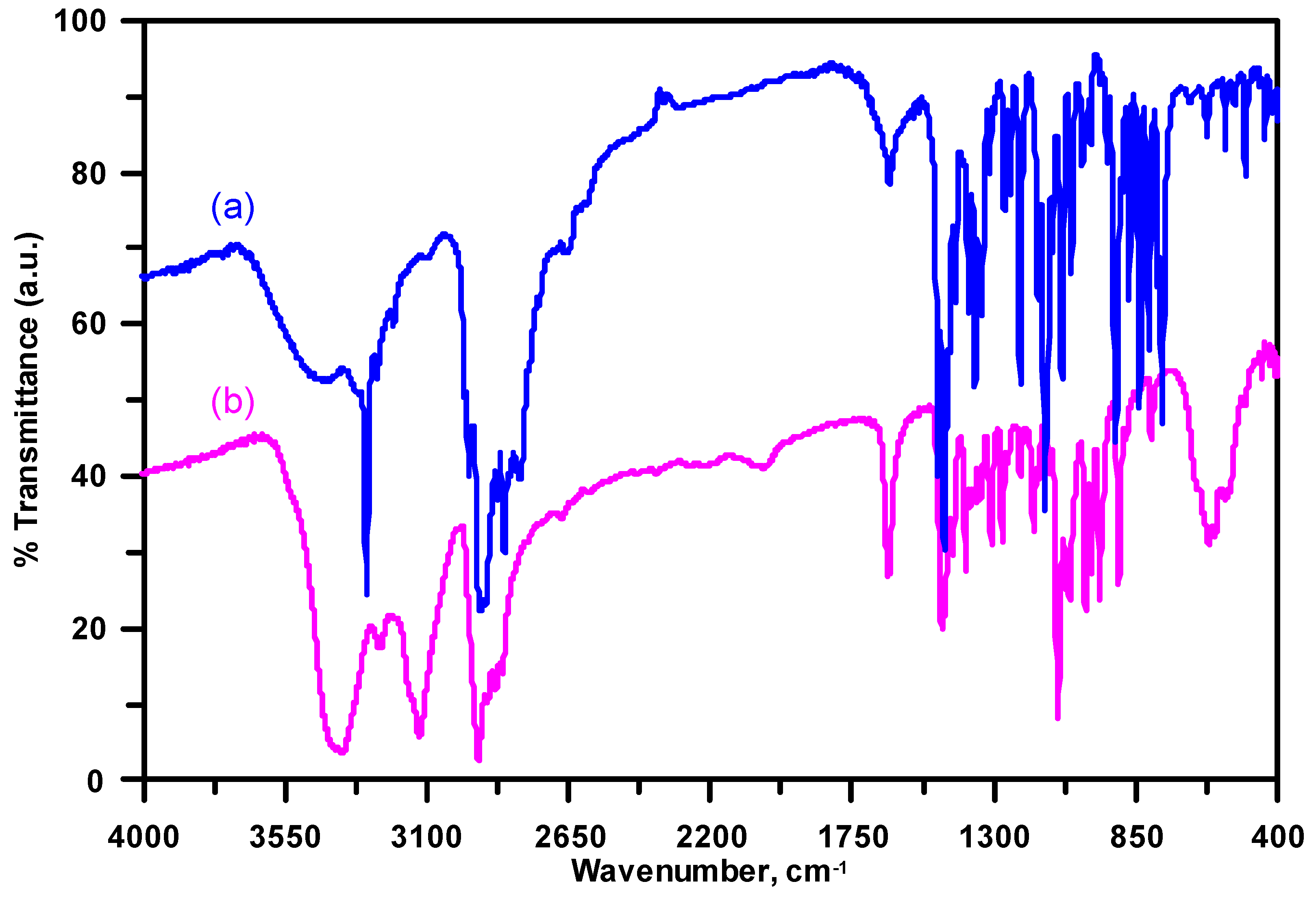
3.3. Infrared Spectroscopy

The FT-IR (Fourier transform infrared) spectra of (a) the ligand L1 and (b) its complex (I) are shown in Figure 4. Absorption band at 3494 cm−1 of ligand, and two peaks at 3389 and 3360 cm−1 of complex (I) are attributed to ν(O-H) stretching vibration of the water molecules. The band at 1634 cm−1 can be assigned to δ(H-O-H) bending mode. The intense bands observed in the frequency region 3340–3050 and 3000–2800 cm−1 easily are assigned to the ν(N—H) and ν(C—H) stretching modes, respectively [39]. The decreased asymmetric and symmetric stretching ν(N—H) frequencies compared to those of the free ligands may be due to the coordination and hydrogen bonding interaction of the secondary amine groups.
The bands observed in the region 1500–1000 cm−1 are assigned to ν(C—C) and ν(C—N) stretching modes. Two absorptions at 1465 and 1429 cm−1 for ligand L1, and 1476 and 1421 cm−1 for complex (I) are attributed to the δ(CH2) stretching modes. IR spectroscopy is useful to assign the cis or trans geometry of transition metal complexes with cyclam derivatives [5,6]. Generally, the trans isomer reveals two groups of bands, overlapping peaks near 890 cm−1 due to γ(N—H) wagging mode and one band near 800 cm−1 arising from the ρ(CH2) rocking vibration. However, the cis isomer reveals at least three bands at 890–830 cm−1 arising from the γ(N—H) wagging vibration, while the ρ(CH2) rocking vibration splits into two peaks at 830–790 cm−1 [40,41,42,43]. In complex (I), two bands at 879 and 845 cm−1 and a single absorption band at 801 cm−1 are assigned to the γ(N—H) wagging and ρ(CH2) rocking frequencies, respectively. The three absorptions peaks in the vibrational regions are consistent with the trans-configuration of the constrained cyclam ligand in complex (I). The wagging and rocking deformation bands are not significantly affected by the counter anion or transition metal ion [5,6,40,41,42,43].

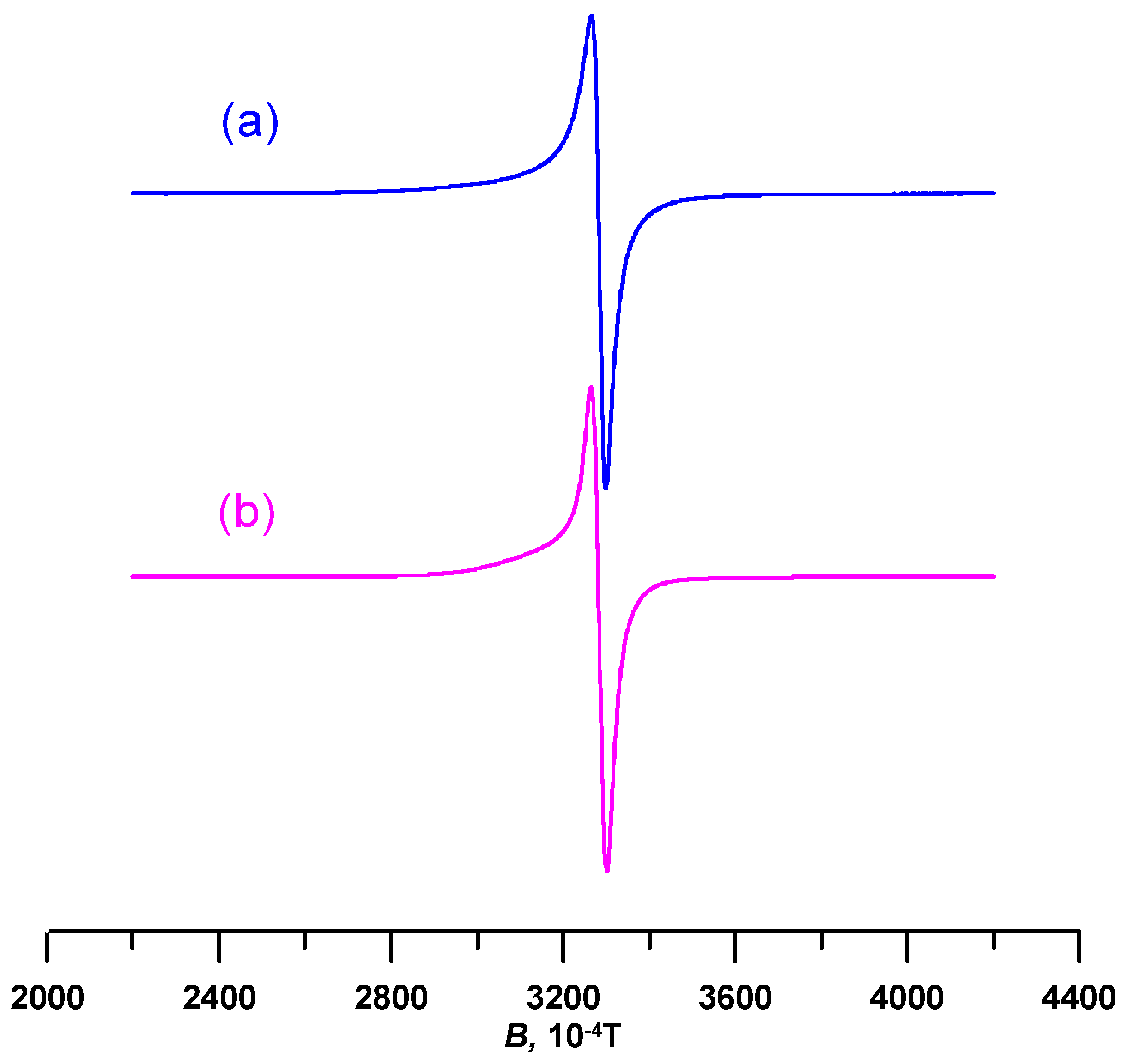
3.4. Electron Paramagnetic Resonace (EPR) Spectroscopy

The experimental and calculated Cu(II) EPR spectra of the powdered complex (I) recorded at RT are exhibited in Figure 5a,b, respectively. The Cu(II) EPR spectrum with axially symmetric spectral lines and unresolved hyperfine splitting was obtained. The spin Hamiltonian parameters refined by computer simulation of the experimental Cu(II) EPR spectrum is as follows: g = 2.153 ± 0.002, g = 2.048 ± 0.002. The g-factors showed a typical trend of g > g > 2.0023, consisting of the dx2y2 ground electronic state, indicating coordination spheres of distorted tetragonal symmetry for central Cu (II) ion. The geometric parameter G = (g − 2)/(g − 2) = 3.188 was calculated, with G < 4 indicating the presence some exchange interactions between the Cu(II) ions [37,44]. The EPR spectrum of the Cu(II) complex, which was recorded at a wide magnetic field of 800 mT, confirmed that the typical EPR resonances of Cu(II) dimeric structure are not present. The found spin Hamiltonian parameters are in good accordance with previously presented g-values of similar monomeric Cu(II) complexes [37,44,45].

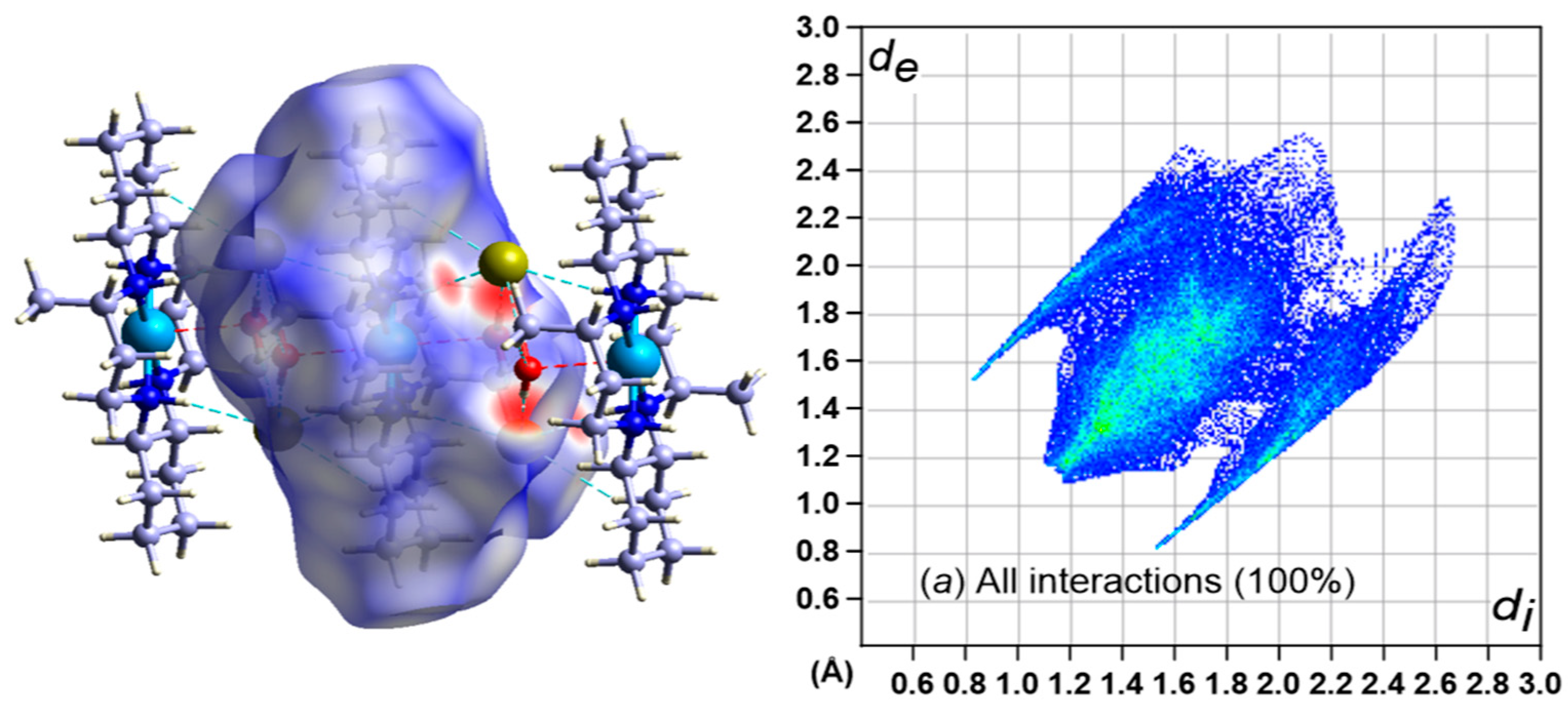
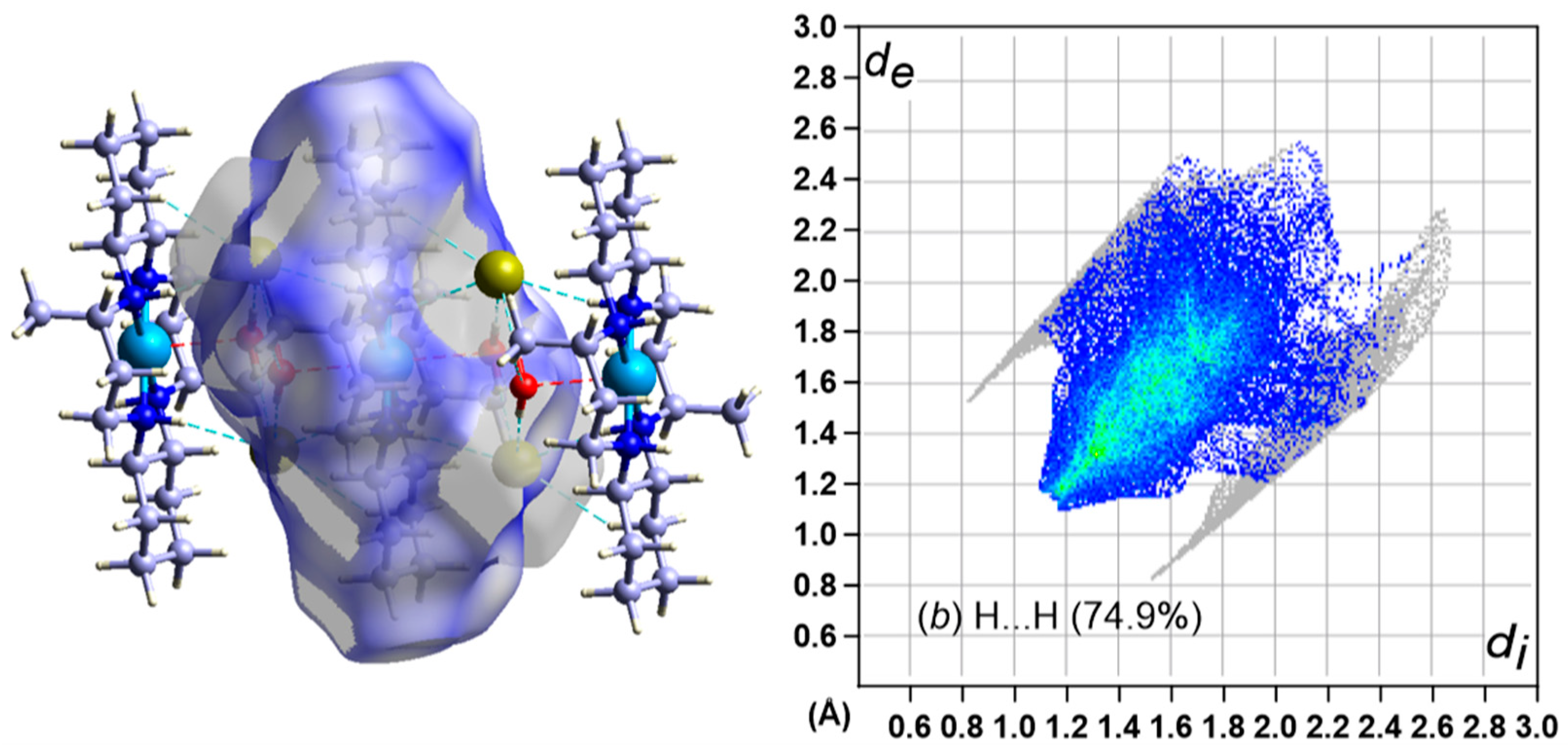
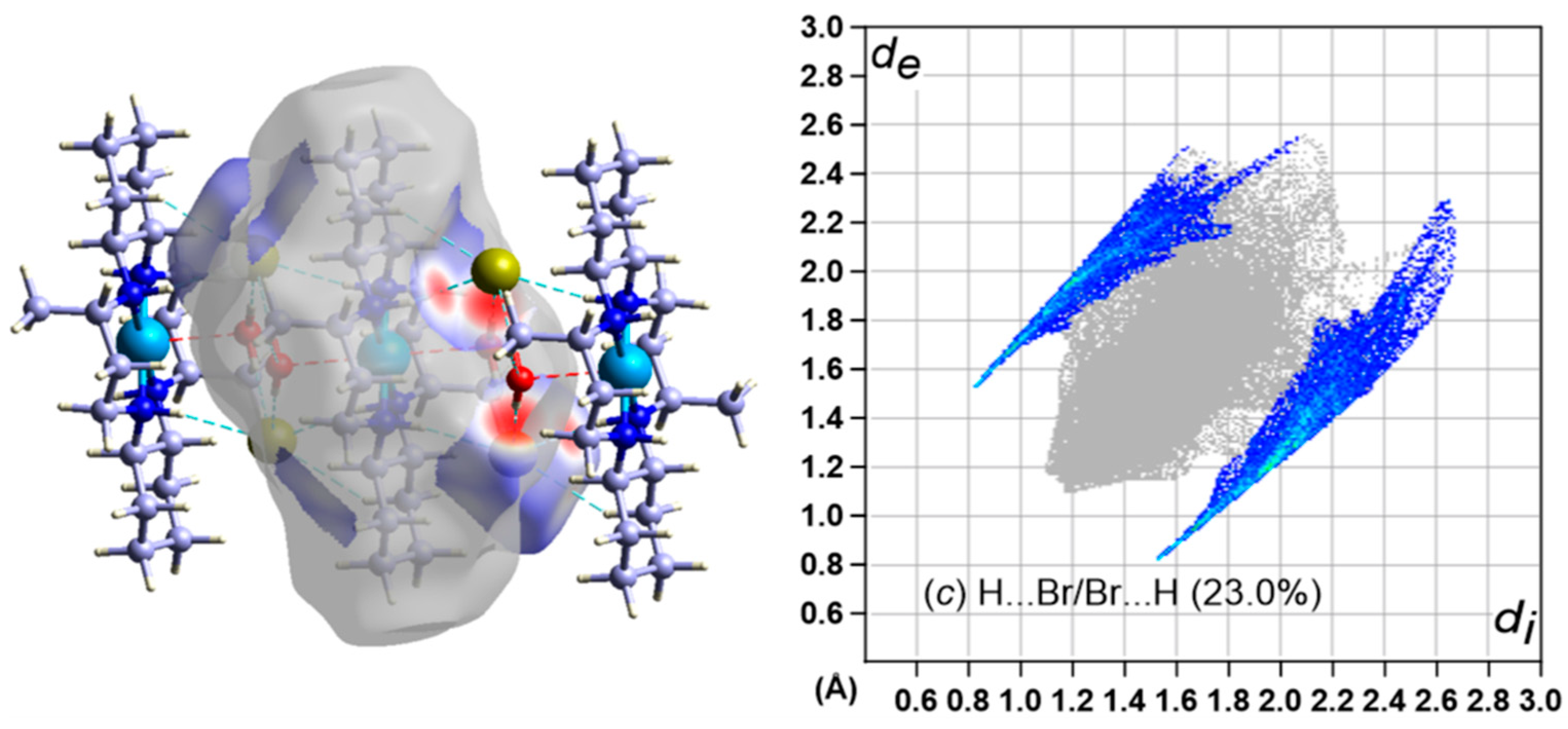
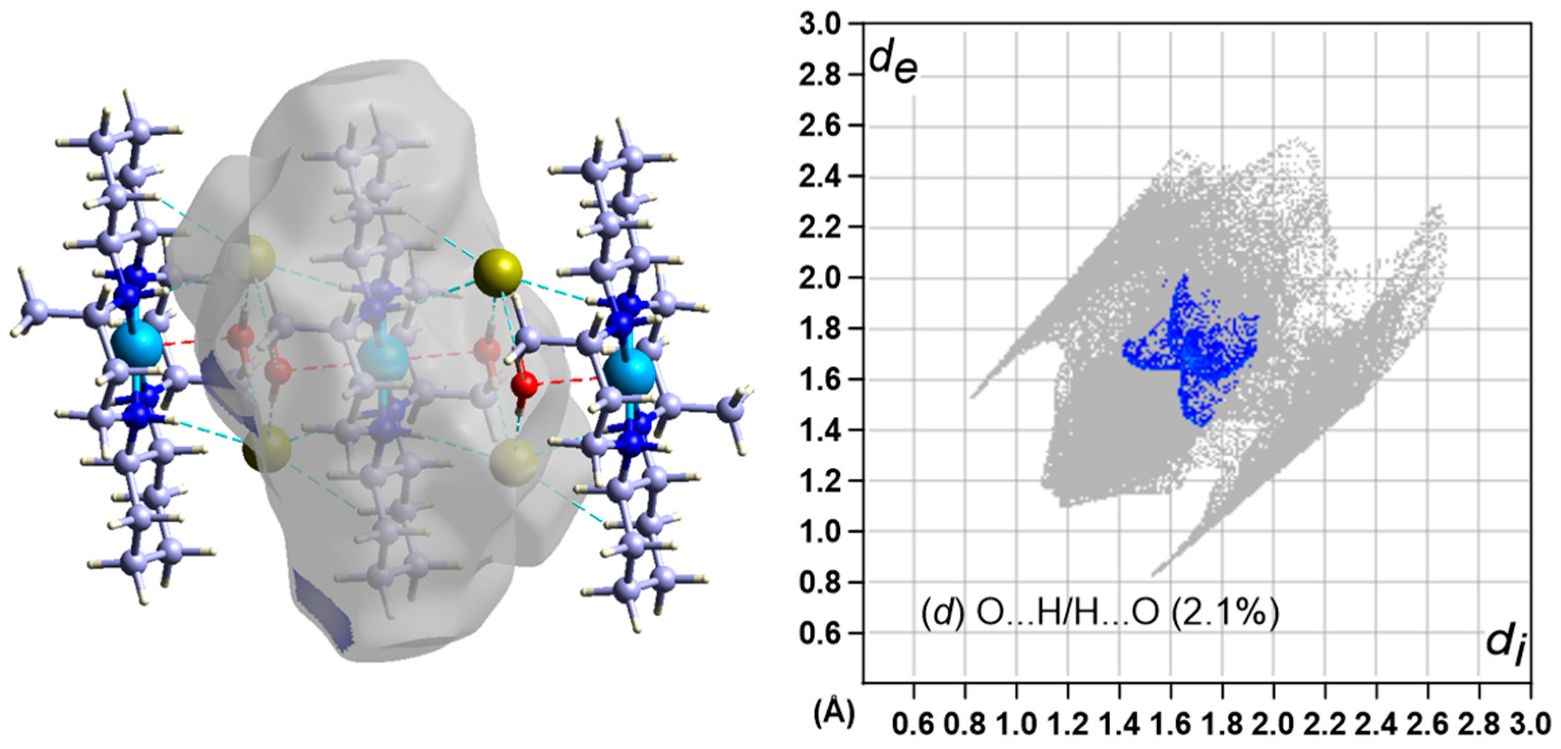
3.5. Hirshfeld Surface Analysis

Various interactions in the crystal were analyzed by Hirshfeld surfaces (HS) analysis [46] and corresponding the two-dimensional (2D) fingerprint plots [47]. The HS is mapped with dnorm and the 2D fingerprint plot of the complex (I) crystal is shown in Figure 6, Figure 7, Figure 8 and Figure 9.
In the HS with dnorm (Figure 7, left), negative values of dnorm are denoted by the red color denoting contacts shorter than the sum of the van der Waals (vdW) radii of the bonding atoms. The white surface indicates the intermolecular distances that are similar to the vdW contacts with dnorm equal to zero [48]. In turn, contacts longer than the sum of the associated vdW radii with positive dnorm values are denoted by the blue color.
Most intermolecular interactions (Figure 7, Figure 8 and Figure 9, right) are H···H (74.9%) and H···Br/Br···H (23.0%), with some contribution from H···O/O···H (2.1%). A large number of H···H and H···/Br···H interactions suggest that vdW interactions and hydrogen bonding play major roles in the crystal packing [49,50].

4. Conclusions

Electronic absorption and IR spectral properties of complex (I) are in agreement with the crystallographic results, showing that the complex adopted an elongated tetragonal octahedral geometry with a square planar CuN4 arrangement with two waters occupying axial positions. The uncoordinated bromide anions remain outside the coordination sphere are connected to the water molecules and complex cations through hydrogen bonds. The crystals are stabilized by the hydrogen bonds between hydrogen atoms of secondary N atoms, O atoms of the water molecules, and the Br anions. The EPR spectrum is acquired at RT and the g-factors, which are refined by computer simulation, exhibit a typical trend: g > g > 2.0023. Furthermore, this is in good accord with the dx2-y2 ground state, indicating a coordination sphere of distorted tetragonal geometry for the Cu(II) ion.

Author Contributions

S.J. prepared the complex and measured IR, UV–Visible spectra. J.M. collected the single-crystal X-ray diffraction data and solved the structure; M.M. measured, analyzed and interpreted the EPR spectrum; M.V. conceived the work, contributed analysis equipment and acted as scientific coordinator; J.-H.C. interpreted the IR and UV-Visible spectra, performed the HS analysis and wrote the manuscript.

Funding

This research was funded by the bilateral collaboration program (NRF 2018K1A3A1A39065843) between South Korea and Slovakia. The APC was funded by Slovak University of Technology.

Acknowledgments

This work was supported by the Slovak Research and Development Agency under the contact No. APVV SK-KR18-0010 and by the Slovak Grant Agency, Projects VEGA 1/0686/17 and 1/0639/18. M.M. and M.V. thank Ministry of Education, Science, Research and Sport of the Slovak Republic for funding within the scheme “Excellent research teams”.

Conflicts of Interest

The authors declare no conflict of interest.

References

  1. Valks, G.C.; McRobbie, G.; Lewis, E.A.; Hubin, T.J.; Hunter, T.M.; Sadler, P.J.; Pannecouque, C.; De Clercq, E.; Archibald, S.J. Configurationally restricted bismacrocyclic CXCR4 receptor antagonists. J. Med. Chem. 2006, 49, 6162–6165. [Google Scholar] [CrossRef] [PubMed]
  2. Ronconi, L.; Sadler, P. Using coordination chemistry to design new medicines. Coord. Chem. Rev. 2007, 251, 1633–1648. [Google Scholar] [CrossRef]
  3. De Clercq, E. Highlights in the discovery of antiviral drugs: A personal retrospective. J. Med. Chem. 2010, 53, 1438–1450. [Google Scholar] [CrossRef] [PubMed]
  4. Ross, A.; Choi, J.-H.; Hunter, T.M.; Pannecouque, C.; Moggach, S.A.; Parsons, S.; De Clercq, E.; Sadler, P. Zinc(II) Complexes of Constrained Antiviral Macrocycles. Dalton Trans. 2012, 41, 6408–6418. [Google Scholar] [CrossRef] [PubMed]
  5. Choi, J.-H. Crystal structure and spectroscopic properties of trans-bis(nicotinato)(1,4,8,11-tetraazacyclotetradecane)chromium(III) perchlorate. Inorg. Chim. Acta 2009, 362, 4231–4236. [Google Scholar] [CrossRef]
  6. Subhan, M.A.; Choi, J.-H.; Ng, S.W. Synthesis, crystal structure and spectroscopic properties of (acetylacetonato)(1,4,8,11-tetraazacyclotetradecane)chromium(III) diperchlorate. Z. Anorg. Allg. Chem. 2011, 637, 2193–2197. [Google Scholar] [CrossRef]
  7. Choi, J.-H.; Tanmaya, T.; Spiccia, L. Syntheses, structural and spectroscopic properties of the copper(II) complexes of constrained macrocyclic ligands. Z. Anorg. Allg. Chem. 2012, 638, 146–151. [Google Scholar] [CrossRef]
  8. Choi, K.-Y.; Kim, J.C.; Jensen, W.P.; Suh, I.H.; Choi, S.S. Copper(II) and nickel(II) complexes with 3,14-dimethyl-2,6,13,17-tetraazatricyclo[16.4.0.07,12]docosane, [M(C20H40N4)]Cl2·2H2O (M = Cu II and Ni II). Acta Crystallogr. 1996, C52, 2166–2168. [Google Scholar]
  9. Choi, J.-H.; Ryoo, K.S.; Park, K.M. [3,14-Dimethyl-2,6,13,17-tetraazatricyclo(16.4.0.07,12)docosane-κ4N]bis(perchlorato-κO)copper(II). Acta Crystallogr. 2007, E63, m2674–m2675. [Google Scholar]
  10. Choi, J.-H.; Subhan, M.A.; Ng, S.W. [3,14-Dimethyl-2,6,13,17-tetraazatricyclo(16.4.0.07,12)docosane-κ4N]bis(nitrato-κO)copper(II). Acta Crystallogr. 2012, E68, m190. [Google Scholar]
  11. Choi, J.-H.; Suzuki, T.; Kaizaki, S. [3,14-Dimethyl-2,6,13,17-tetraazatricyclo(16.4.0.07,12)docosane-κ4N]dinitratocopper(II) trihydrate. Acta Crystallogr. 2006, E62, m2383–m2385. [Google Scholar]
  12. Subhan, M.A.; Ryoo, K.S.; Choi, J.-H. Crystal structure and spectroscopic properties of [3,14-dimethyl-2,6,13,17-tetraazatricyclo(16.4.0.07,12)docosane]copper(II) dithiocyanate dihydrate. Int. J. Chem. Sci. 2015, 13, 593–604. [Google Scholar]
  13. Subhan, M.A.; Ng, S.W.; Lee, C.S.; Choi, J.-H. Synthesis, crystal structure, and spectroscopic properties of isothiocyanato[3,14-dimethyl-2,6,13,17-tetraazatricyclo(16.4.0.07,12)docosane]copper(II) thiocyanate. J. Struct. Chem. 2017, 58, 742–749. [Google Scholar] [CrossRef]
  14. Choi, K.-Y. Synthesis and crystal structure of a macrocyclic azidotetraamine copper(II) complex. J. Chem. Crystallogr. 1998, 28, 875–878. [Google Scholar] [CrossRef]
  15. Choi, J.-H.; Clegg, W.; Harrington, R.W. Crystal structure of [2,13-dibenzyl-5,16-dimethyl-2,6,13,17-tetraazatricyclo(14,4,0,07.12)docosane]copper(II) diperchlorate. J. Chem. Crystallogr. 2010, 40, 80–84. [Google Scholar] [CrossRef]
  16. Choi, J.-H.; Clegg, W.; Gary, G.S. Synthesis, crystal structure and spectroscopic properties of [2,13-bis(1-naphthalenylmethyl)-5,16-dimethyl-2,6,13,17-tetraazatricyclo(14,4,0,07.12)docosane]copper(II) diperchlorate acetonitrile disolvate. Z. Anorg. Allg. Chem. 2010, 636, 1612–1616. [Google Scholar] [CrossRef]
  17. Lim, J.H.; Kang, J.S.; Kim, H.C.; Koh, E.K.; Hong, C.S. Synthesis, crystal structures, and magnetic properties of cyano-bridged honeycomb like layers MV-CuII (M=Mo, W) chelated by a macrocyclic ligand. Inorg. Chem. 2006, 45, 7821–7827. [Google Scholar] [CrossRef] [PubMed]
  18. Moncol, J.; Mazúr, M.; Valko, M.; Choi, J.-H. Synthesis, structural characterization, EPR spectroscopy and Hirshfeld surface analysis of novel Cu2+ doped 3,14-diethyl-2,13-diaza-6,17-diazoniatricyclo(16.4.0.07,12)docosane diperchlorate. Acta Crystallogr. 2019, C75, 612–622. [Google Scholar] [CrossRef] [PubMed]
  19. Choi, J.-H.; Subhan, M.A.; Ng, S.W. Syntheses, crystal structures and spectroscopic properties of [Cu(L)(NO3)2] and [Cu(L)(H2O)2](SCN)2 with 3,14-diethyl-2,6,13,17-tetraazatricyclo(16.4.0.07,12)docosane (L). J. Coord. Chem. 2012, 65, 3481–3491. [Google Scholar] [CrossRef]
  20. Subhan, M.A.; Choi, J.-H. X-ray structure and spectroscopy of novel trans-[Ni(L)(NO3)2] and [Ni(L)](ClO4)2·2H2O complexes. Spectrochim. Acta Part A 2014, 123, 410–415. [Google Scholar] [CrossRef] [PubMed]
  21. Lim, I.-T.; Kim, C.-H.; Choi, K.-Y. Synthesis and structural characterization of nickel(II) complexes of 3,14-diethyl-2,6,13,17-tetraazatricyclo(14.4.0.07,12)docosane with inorganic salts. Polyhedron 2015, 100, 43–48. [Google Scholar]
  22. Subhan, M.A.; Moon, D.; Choi, J.-H. Synthesis, crystal structure determination, and spectroscopic characterization of [2,13-dibenzyl-5,16-diethyl-2,6,13,17-tetraazatricyclo(16.4.0.07,12)docosane]copper(II) dinitrate. Main Group Chem. 2017, 16, 27–36. [Google Scholar] [CrossRef]
  23. Jimenez, D.; Martinez-Manez, R.; Sancenon, F.; Ros-Lis, J.V.; Soto, J. A new chromo-chemodosimeter selective for sulfide anion. J. Am. Chem. Soc. 2003, 125, 9000–9001. [Google Scholar] [CrossRef] [PubMed]
  24. Martinez-Manez, R.; Sancenon, F. Fluorogenic and chromogenic chemosensors and reagents for anions. Chem. Rev. 2003, 103, 4419–4476. [Google Scholar]
  25. Kang, S.G.; Kweon, J.K.; Jung, S.K. Synthesis of new tetraaza macrocyclic ligands with cyclohexane ring and their Ni(II) and Cu(II) complexes. Bull. Korean Chem. Soc. 1991, 12, 483–487. [Google Scholar]
  26. Moon, D.; Choi, J.-H. Crystal structure of 3,14-dimethyl-2,6,13,17-tetraazoniatricyclo(16.4.0.07,12)docosane tetrachloride tetrahydrate from synchrotron X-ray data. Acta Crystallogr. 2018, E74, 1039–1041. [Google Scholar]
  27. Mazur, M.; Valko, M.; Klement, R.; Morris, H. Quantitative EPR spectroscopy with a TE104 double rectangular cavity. Part 1. A simple alignment procedure for the precision positioning of the sample. Anal. Chim. Acta 1996, 333, 249–252. [Google Scholar] [CrossRef]
  28. Thiele, H.; Etstling, J.; Such, P.; Hoefer, P. WINEPR; Bruker Analytic Gmb: Rheinstetten, Germany, 1992. [Google Scholar]
  29. Weber, R.T. WINEPR SimFonia; EPR Division, Bruker Instr. Inc.: Billerica, MA, USA, 1995. [Google Scholar]
  30. Sheldrick, G.M. SHELXT-Integrated space-group and crystal-structure determination. Acta Crystallogr. 2015, A71, 3–8. [Google Scholar] [CrossRef] [PubMed]
  31. Sheldrick, G.M. Crystal structure refinement with SHELXL. Acta Crystallogr. 2015, C71, 3–8. [Google Scholar]
  32. Koziskova, J.; Hahn, F.; Richter, J.; Kozisek, J. Comparison of different absorption corrections on the model structure of tetrakis(μ2-acetato)diaquadicopper(II). Acta Chim. Slovaca 2016, 9, 136–140. [Google Scholar] [CrossRef]
  33. Putz, H.; Brandenburg, K. DIAMOND-3; University of Bonn: Bonn, Germany, 2014. [Google Scholar]
  34. Turner, M.J.; McKinnon, J.J.; Wolff, S.K.; Grimwood, D.J.; Spackman, P.R.; Jayatilaka, D.; Spackman, M.A. CrystalExplorer17.5. The University of Western Australia. 2017. Available online: http://crystalexplorer.scb.uwa.edu.au/ (accessed on 27 June 2019).
  35. McKinnon, J.J.; Spackman, M.A.; Mitchell, A.S. Novel tools for visualizing and exploring intermolecular interactions in molecular crystals. Acta Crystallogr. 2004, B60, 627–668. [Google Scholar] [CrossRef]
  36. McKinnon, J.J.; Jayatilaka, D.; Spackman, M.A. Towards quantitative analysis of intermolecular interactions with Hirshfeld surfaces. Chem. Commun. 2007, 3814–3816. [Google Scholar] [CrossRef]
  37. Hathaway, B.J.; Billing, D.E. The electronic properties and stereochemistry of mononuclear complexes of copper(II) ion. Coord. Chem. Rev. 1970, 5, 43–207. [Google Scholar] [CrossRef]
  38. Hathaway, B.J.; Hodgson, P.G. Copper-ligand bond-lengths in axial complexes of the copper(II) ion. J. Inorg. Nucl. Chem. 1973, 35, 4071–4081. [Google Scholar] [CrossRef]
  39. Lever, A.B.P. Inorganic Electronic Spectroscopy, 2nd ed.; Elsevier: Amsterdam, The Netherlands, 1984. [Google Scholar]
  40. Nakamoto, K. Infrared and Raman Spectra of Inorganic and Coordination Compounds, Part A: Theory and Applications in Inorganic Chemistry; Part B: Applications in Coordination, Organometallic, and Bioinorganic Chemistry, 6th ed.; John Wiley & Sons: New York, NY, USA, 2009. [Google Scholar]
  41. Choi, J.-H.; Oh, I.-G.; Suzuki, T.; Kaizaki, S. Crystal structure and spectroscopic properties of oxalato(1,4,8,11-tetraazacyclotetradecane)chromium(III) perchlorate. J. Mol. Struct. 2004, 694, 39–44. [Google Scholar] [CrossRef]
  42. Choi, J.-H. Spectral properties and ligand field analysis of cis-dinitrito(1,4,8,11-tetraazacyclotetradecane)chromium(III) nitrate. Chem. Phys. 2000, 256, 29–35. [Google Scholar] [CrossRef]
  43. Choi, J.-H. Spectroscopic properties and ligand field analysis of cis-diazido(1,4,8,11-tetraazacyclotetradecane)chromium(III) azide. Spectrochim. Acta Part A 2000, 56, 1653–1660. [Google Scholar] [CrossRef]
  44. Choi, J.-H.; Oh, I.-G.; Linder, R.; Schönherr, T. Electronic spectroscopy and ligand field analysis of cis-carbonato(rac-5,5,7,12,12,14-hexamethyl-1,4,8,11-tetraazacyclotetradecane)chromium(III) chloride. Chem. Phys. 2004, 297, 7–12. [Google Scholar] [CrossRef]
  45. Hathaway, B.J.; Tomlinson, A.A.G. Copper ammonia complexes. Coord. Chem. Rev. 1970, 5, 1–43. [Google Scholar] [CrossRef]
  46. Goodman, B.A.; Raynor, J.B. Electron spin resonance of transition metal complexes. Adv. Inorg. Chem. Radiochem. 1970, 13, 135–362. [Google Scholar]
  47. Spackman, M.A.; Jayatilaka, D. Hirshfeld surface analysis. CrystEngComm 2009, 11, 19–32. [Google Scholar] [CrossRef]
  48. Moon, D.; Tanaka, S.; Akitsu, T.; Choi, J.-H. Molecular structure, spectroscopic properties, and Hirshfeld surface analysis of chlorobis(N-methyl-1,3-propanediamine)copper(II) tetrafluoroborate and azidobis(2,2-dimethyl-1,3-propanediamine)copper(II) azide. J. Mol. Struct. 2018, 1154, 338–347, Erratum in 2018, 1168, 329. [Google Scholar]
  49. Hathwar, V.R.; Sist, M.; Jørgensen, M.R.V.; Mamakhel, A.H.; Wang, X.; Hoffmann, C.M.; Sugimoto, K.; Overgaard, J.; Iversen, B.B. Quantitative analysis of intermolecular interactions in orthorhombic rubrene. IUCrJ 2015, 2, 563–574. [Google Scholar] [CrossRef] [PubMed]
  50. Spackman, M.A.; McKinnon, J.J.; Jayatilaka, D. Electrostatic potentials mapped on Hirshfeld surfaces provide direct insight into intermolecular interactions in crystals. CrystEngComm 2008, 10, 377–388. [Google Scholar] [CrossRef]
Scheme 1. Structures of various macrocyclic ligands.
Crystals 09 00336 sch001
Scheme 2. Chemical structure of [Cu(L1)(H2O)2]Br2, (I).
Crystals 09 00336 sch002
Scheme 3. Synthetic procedure of the ligand L1.
Crystals 09 00336 sch003
Figure 1. Molecular structure of complex (I).
Crystals 09 00336 g001
Figure 2. Crystal packing diagram of complex (I). Dashed lines represent hydrogen bonds.
Crystals 09 00336 g002
Figure 3. Visible spectrum (pink line) and resolved Gaussian curves (black line) of complex (I).
Crystals 09 00336 g003
Figure 4. FT-IR spectra of (a) free ligand L1 (blue line) and (b) complex (I) (pink line), respectively.
Crystals 09 00336 g004
Figure 5. (a) Experimental EPR spectrum (blue line) recorded at RT and (b) calculated EPR spectrum (pink line) of powdered complex (I).
Crystals 09 00336 g005
Figure 6. Hirshfeld surface mapped with (left) dnorm, and 2D fingerprint map (right) for all interactions.
Crystals 09 00336 g006
Figure 7. Hirshfeld surface mapped with (left) dnorm, and 2D fingerprint plot (right) for H···H interaction.
Crystals 09 00336 g007
Figure 8. Hirshfeld surface mapped with (left) dnorm, and 2D fingerprint plot (right) for H···Br/Br···H interactions.
Crystals 09 00336 g008
Figure 9. Hirshfeld surface mapped with (left) dnorm, and 2D fingerprint plot (right) for H···O/O···H interactions.
Crystals 09 00336 g009
Table 1. Crystal data, data collection conditions and refinements for complex (I).
Empirical formulaC20H44CuN4O2Br2
Formula weight595.95 g mol−1
Temperature100(1) K
Wavelength1.54186 Å
Crystal system, space groupTriclinic, Pī
Unit cell dimensionsa = 8.0777(4) Å, α = 98.879(4)°
b = 8.8129(4) Å, β = 110.204(3)°
c = 10.0932(5) Å, γ = 109.391(3)°
Volume650.97(5) Å3
Z1
Radiation typeCu
Density (calculated)1.633 Mg m−3
Absorption coefficient5.31 mm−1
Crystal size0.32 × 0.22 × 0.21 mm3
Theta range for data collection4.9 to 70.5°
Reflections collected19,409
Independent reflections9304 [Rint = 0.013]
Absorption correctionMulti-scan
Max. and min. transmission0.012 and 0.263
Refinement methodFull-matrix least-squares on F2
Data/restraints/parameters2241/0/134
Goodness-of-fit on F21.11
Final R indices (F2 > 2)R1 = 0.034, wR2
Extinction coefficient0.0050(3)
Largest diff. peak and hole (e Å−3)0.68 and −0.87
Table 2. Selected bond distances (Å) and angles (°) of complex (I).
Cu1–N12.018 (3)C2–C9i1.527 (5)
Cu1–N22.049 (3)C3–C41.531 (4)
Cu1–O1W2.632 (3)C3–C81.526 (5)
N1–C11.489 (4)C4–C51.528 (5)
N1–C31.488 (4)C5–C61.516 (6)
N2–C81.497 (4)C6–C71.528 (5)
N2–C91.498 (4)C7–C81.525 (4)
C1–C21.522 (5)C9–C101.521 (5)
N1–Cu1–N1i180.0N1–C1–C2110.8 (3)
N1i–Cu1–N295.40 (11)C1–C2–C9i116.1 (3)
N1–Cu1–N284.60 (11)N1–C3–C4113.4 (3)
N1–Cu1–O1Wi91.78 (10)N1–C3–C8106.4 (3)
N1–Cu1–O1W88.22 (10)C8–C3–C4111.3 (3)
N2–Cu1–N2i180.0C5–C4–C3110.6 (3)
N2–Cu1–O1W95.89 (10)C6–C5–C4110.8 (3)
N2–Cu1–O1Wi84.11 (10)C5–C6–C7110.7 (3)
O1Wi–Cu1—O1W180.0C8–C7–C6110.4 (3)
C1–N1–Cu1116.0 (2)N2–C8–C3106.3 (3)
C3–N1–Cu1108.07 (19)N2–C8–C7113.4 (3)
C3–N1–C1113.4 (3)C7–C8–C3111.7 (3)
C8–N2–Cu1107.20 (19)N2–C9–C2i109.1 (3)
C8–N2–C9114.6 (3)N2—C9–C10111.8 (3)
C9–N2–Cu1121.0 (2)C10–C9–C2i113.3 (3)
Symmetry code: (i) −x + 1, −y + 1, −z + 1.
Table 3. Selected bond distances (Å) and conformations of metal complexes with macrocyclic ligands.
CompoundM–NM–XConformationRef.
[Cu(L1)(H2O)2]Br22.018(3)–2.049(3)2.632(3)trans-IIIThis work
[Cu(L1)(H2O)2](BF4)2·2H2O2.028(2)–2.028(2)2.693(3)trans-III[7]
[Cu(L1)(H2O)2]Cl22.017(2)–2.038(2)2.649(2)trans-III[8]
[Cu(L1)(ClO4)2] 2.005(2)–2.048(2)2.623(2)trans-III[9]
[Cu(L1)(NO3)2]2.007(2)–2.044(2)2.463(2)trans-III[10]
[Cu(L1)(NO3)2]·3H2O2.021(2)–2.029(2)2.746(2)trans-III[11]
[Cu(L1)](NCS)2·2H2O2.020(7)–2.056(7)3.037(7)trans-III[12]
[Cu(L1)(NCS)]SCN2.013(2)–2.052(2)2.322(3)trans-III[13]
[Cu(L1)(N3)]ClO4·H2O2.030(3)–2.054(3)2.254(4)trans-III[14]
[Cu(L2)](ClO4)21.990(4)–2.050(4)3.496(3)trans-I[15]
[Cu(L2)](NO3)21.999(7) –2.095(7)2.964(7)trans-III[8]
[Cu(L3)](ClO4)2·2CH3CN2.030(3)–2.081(2)3.264(3)trans-III[16]
[Cu(La)(ClO4)2]2.016(2) –2.040(2) 2.762(2)trans-III[17]
[Cux(H2(1-x)La)(ClO4)2] (x = 0.69)2.015(3) –2.047(3) 2.795(3)trans-III[18]
[Cu(La)(NO3)2]2.014(2) –2.047(2) 2.506(2)trans-III[19]
[Cu(La)(H2O)2](SCN)22.021(2) –2.046(2)2.569(2)trans-III[19]
[Ni(La)(NO3)2]2.051(4) –2.094(4)2.198(3)trans-III[20]
[Ni(La)](ClO4)2·2H2O1.996(7) –2.042(7)2.973(7)trans-III[20]
[Ni(La)(N3)2]2.070(1) –2.105(1)2.176(1)trans-III[21]
[Cu(Lb)](NO3)22.023(2) –2.086(2) 2.936(2)trans-III[22]

