Hydrogel Based Sensors for Biomedical Applications: An Updated Review


1
Medical Device Research Institute, College of Science and Engineering, Flinders University, Adelaide 5042, SA, Australia
2
Institute for Nano Scale Science & Technology, College of Science and Engineering, Flinders University, Adelaide 5042, SA, Australia
*
Authors to whom correspondence should be addressed.
Polymers 2017, 9(8), 364; https://0-doi-org.brum.beds.ac.uk/10.3390/polym9080364
Submission received: 20 July 2017
/
Revised: 10 August 2017
/
Accepted: 12 August 2017
/
Published: 16 August 2017
(This article belongs to the Special Issue Hydrogels in Tissue Engineering and Regenerative Medicine)
Abstract
:Biosensors that detect and convert biological reactions to a measurable signal have gained much attention in recent years. Between 1950 and 2017, more than 150,000 papers have been published addressing the applications of biosensors in different industries, but to the best of our knowledge and through careful screening, critical reviews that describe hydrogel based biosensors for biomedical applications are rare. This review discusses the biomedical application of hydrogel based biosensors, based on a search performed through Web of Science Core, PubMed (NLM), and Science Direct online databases for the years 2000–2017. In this review, we consider bioreceptors to be immobilized on hydrogel based biosensors, their advantages and disadvantages, and immobilization techniques. We identify the hydrogels that are most favored for this type of biosensor, as well as the predominant transduction strategies. We explain biomedical applications of hydrogel based biosensors including cell metabolite and pathogen detection, tissue engineering, wound healing, and cancer monitoring, and strategies for small biomolecules such as glucose, lactate, urea, and cholesterol detection are identified.
1. Introduction
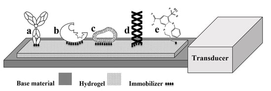
Biosensors that detect and convert biological reactions to a measurable signal have gained much attention in recent years. By integration of a biologically active component with an appropriate transducer, biosensors produce a measurable signal generated by chemical reactions. To achieve this purpose, a biosensor is made up of four main components, as shown in Figure 1.
A bioreceptor, as a molecular species—such as nucleic acid, enzyme, antibody, gene, or a biological system, such as a cell or an organ—employs a biochemical mechanism to interact with the analyte. While a transducer produces a measurable signal proportional to the bioreceptor–analyte interaction [1]. Using different immobilization techniques, bioreceptors are coupled with base materials [2]. Base materials can be made up of metals, polymers, glass, or composites. Moreover, hydrogels as a swell able polymer with enviro-sensitive properties are having a profound impact in a broad range of applications due to their exceptional physicochemical, mechanical, electrical, and optical properties [3,4,5]. Despite the period since their initial discovery in 1968, hydrogels have emerged as a promising platform with surprising and enormous potential for biomedical use. Their applications in this field are still in a developing phase [6,7,8,9,10].
In fact, searches for the word “biosensor” in the ISI Web of Science, PubMed, and Science Direct databases obtained 46,791, 47,017, and 72,477 hits respectively from the period 1950 to 1 February 2017. An advanced searches that combined “biosensors” and “hydrogel based” resulted in 702, 497, and 4047 findings in the ISI Web of Science, PubMed, and Science Direct databases. Totals of 28, 66 and 1577 papers were found for “biosensor + hydrogel based + biomedical application” searches in the ISI Web of Science, PubMed, and Science Direct databases, respectively. Through careful screening, the most published papers were revealed to be those referring to general specifications of bioreceptors and transducers. To our knowledge, however, critical reviews that describe hydrogel based biosensors for biomedical applications were rare.
The objectives of this review are to summarize the biomedical applications of hydrogel based biosensors, including bioreceptors that have been specifically recommended for hydrogels, the methods of immobilization, sensor design, and structural modifications, to identify potential roles of hydrogels for biomedical applications, and to suggest areas for future investigation.
2. Bioreceptors
Bioreceptors are biomolecular recognition components that are responsible for binding a specific particle of interest within a biosystem environment. Although many forms of bioreceptors are available to monitor numerous different particles that have been triggered for sensing, they can be categorized in five different major groups, as shown in Figure 2 [11,12].
Antibodies are Y-shaped, complex proteins used to identify foreign antigens, viruses, and bacteria. With specific binding capabilities, antibodies have been used in biosensors. The “lock and key fit”—as a unique property of a specific geometrical configuration—is the capability that an antigen-specific antibody exhibits when employed as a biosensor [13]. Exploitation of an animal immune system is a usual method for production of polyclonal, monoclonal, and recombinant antibodies, among which, polyclonal antibodies are popular for their frequent use as immune sensors [14]. Recombinant antibodies have been selected to detect structurally diverse antigens including haptens, proteins, and carbohydrate moieties.
Enzymes are another type of bioreceptor attracting attention for their specific binding capabilities and catalytic activities that amplify detection [15]. The activities are bestowed by the enzyme amino acid residues. For some of these capabilities, however, some enzymes require a cofactor, such as inorganic ions or coenzymes, e.g., complex metalorganic/organic molecules [16]. The regulatory nature of enzymes has made them attractive for use as biosensors to quantify catalytic reactions, including heat, light, and charges, i.e., protons and electrons [17]. Regulation changes of the enzymes’ selective conformation serve as a biorecognition method in biosensors [18].
Nucleic acids use the pairs of C:G (cytosine:guanosin) and A:T (adenine:thymine) in DNA for specificity of biorecognition. Hybridization of a certain sequence of a DNA molecule to a specific labeled molecule can be utilized in biosensors [19]. In fact, the DNA portion with the specific sequence is immobilized to the base material of the biosensor and facilitates biosensing upon interaction with the complementary sequence that exists on the targeted molecule [20]. Nucleic acid ligands known as aptamers are included in a nucleic acid bioreceptor’s family that is isolated from libraries of oligonucleotides through an in vitro “systematic evolution of ligands” process [21,22]. In contrast to nucleic acids, these short, single-strand oligonucleotides are believed to distinguish their targets by shape rather than by sequence. Therefore, a wide range of small molecules, cells, and proteins can be detected by using aptamer bioreceptors [23]. Within these families, catalytic aptamers —“aptazymes”—have been introduced, that have high capability of recognizing metabolic intermediates at a very low concentration [24]. Aptazymes, like enzyme bioreceptors, can be denatured frequently without loss of their catalytic or binding capabilities, and their relatively high signal-to-noise ratios make them more attractive than enzymes [25].
For biorecognition by cells or cellular structures (microorganisms), bioreceptors can be performed by either a specific cellular structure or an entire cell binding to certain species. As certain chemicals are taken up by cellular structures for digestion, a kind of chemical-specific biosensor is fashioned [26,27]. Bacteria and fungi microorganisms are categorized in the cell bioreceptor’s group and have been used for toxicity detection [28]. In the bioreceptors of a cellular structure, abundant carrier proteins that provide facilities to transport a chemical compound from one place to another (surfaces to internal parts or between cells) can be used for molecular recognition [29,30,31].
Biomimetic receptors are artificial fabrications employed to mimic a bioreceptor [32,33]. Methods used for this purpose include artificial membrane production [34,35,36], using genetically engineered molecules such as lectin based or peptide nucleic acid based molecules [37,38,39], and molecular imprinting [40,41]. Among them, the molecular imprinting technique, which consists of mixing biomolecules with monomers and a crosslinking agent, has attracted much attention [42,43,44]. With this method, selective binding sites based on molecular templates are introduced to synthetic polymers. Figure 3 gives a schematic representation of the fabrication of a molecular imprint biosensor. Advantages and limitations of different bioreceptors are listed in Table 1.
3. Hydrogels for Biosensing
As water swellable three-dimensional structures, hydrogels are formed by chemical (covalent bonds) or physical (non-covalent interactions) crosslinking. These smart materials with excellent biocompatibility are considered to interface progressively with biosystems, but suffer from side effects [51,52,53]. Many features have made them popular for biosensing applications: interaction with biological components at the molecular level; their regulating viscoelastic properties; being reactive to external stimuli; possessing antifouling characteristics; and the existence of a wide range of well-known synthesis methods for incorporating bioreceptors into their highly wet structure.
Hydrogel-based biosensors can detect biological events in two ways. The first includes hydrogels without bioreceptors, whose swelling properties change in response to selected biological interactions [54]. Ionic hydrogels with environmental sensitivity—for example to pH, temperature, and electrical field—have been widely used in this group [55]. pH-sensitive hydrogels either admit or release protons in response to appropriate ionic strength alterations in the surrounding aqueous biosystem. The more ionized ionic hydrogels are, the more electrostatic repulsion between polymeric chains will be created, leading to a negative or positive swelling ratio [56]. The same scenario has been observed for polyelectrolyte gels in response to an electric field, the intensity of which was responsible for swelling and de-swelling processes. The higher the electric field intensity, the more fixed charges will exist that affect the degree of swelling [57]. A copolymer of hydrophobic and hydrophilic monomers can introduce a thermo-responsive hydrogel, where the ratio of the monomers is responsible for phase transition in response to changes in biosystem temperature [58]. Apart from the reasons mentioned for phase transition, the biological interactions in a biosystem cause swelling alteration and may translate to a macroscopic response. Hence, the macroscopic response needs to be used as optical, conductometric, amperometric, or mechanical readouts for biosensing.
With respect to hydrogel porous structures and their unique large internal surfaces, the second way of detecting biological events involves hydrogel-based biosensors that can accommodate bioreceptors for detection of biochemical or biological interactions. With this method, hydrogel immobilized bioreceptors are employed for hosting biomolecular recognition components to identify a definite event of a biosystem. Of major importance for designing this type of biosensor are stable immobilization of bioreceptors, surface bonding strategies, prevention of nonspecific protein adsorption to the hydrogel surface, probe density, flexibility, and swelling kinetics.
3.1. Polyvinyl Alcohol
Polyvinyl alcohol (PVA) hydrogels have been widely used in various biomedical applications due to their biocompatibility, hydrophilic properties, and biomechanical characteristics. Relying on their physical crosslinking, the performance PVA and its composites with glucose [59,60,61,62,63,64,65], triglyceride [66,67], ethanol [68], urea [69,70,71,72], hydrogen peroxide [73,74], toxicity [75,76,77], and genetic [78] sensors have been investigated. The flexibility of PVA hydrogels as well as their stability under a wide range of environmental conditions allows them to mimic soft tissue and to minimize inflammation and fibrosis, an ability that is necessary for implantable sensors.
3.2. Polyethylene Glycol
Polyethylene glycol (PEG), a hydrophilic biomaterial, has excellent antifouling properties conferred by its low interfacial energy, resisting protein and cell surface adhesion [79,80,81]. With its biocompatibility, PEG is widely used as a biosensor with antifouling characteristics. In recent studies, PEG and its hybrids have been employed for the fabrication of electrochemical [82,83,84,85,86,87,88,89,90], optical [91,92,93,94,95,96,97,98], and mass based [99,100,101,102] biosensors.
3.3. Polyacrylate Families
Polyacrylic acid, polyhydroxyethyl methacrylate, polyacrylamide, and poly(N-isopropylacrylamide) are some examples of stimulus-responsive hydrogels used mainly for pH and temperature sensing within a biosystem. As their hydrophilicity hinges on charged group density, ionic hydrogels can reversibly swell and de-swell relative to changes in surrounding conditions. Recently, fabrication patterning techniques have been noted as an essential development phase for greater utilization of the sensing properties of ionic hydrogels, especially when they are used without utilizing immobilized bioreceptors. Micromolding, microlithography (photomask, ion beam, and optical maskless) [103], wet-etching, microcontact printing, and evaporation-induced self-assembly are well-known rapid prototyping methods reported in some studies. Development sensing systems for taste [104], ammonia [105,106], glucose [107,108,109], pH [103,110,111,112,113,114,115,116,117,118,119,120,121,122,123,124,125,126,127,128,129], humidity [130,131,132], chemical and biochemical molecules [133,134,135,136,137,138,139,140,141,142,143], and temperature [144] have been reported using polyacrylate family hydrogels.
3.4. Hydrogels with Biological Origin
Polysaccharides and polypeptides including alginate, chitin and chitosan, agarose, cellulose, dextran, and hyaluronic acid are biomaterials with gel-forming properties. The unique properties of these biological origin hydrogels—including biocompatibility, hydrophilicity, heavy metal ion chelation, high protein affinity, ease of surface chemical modification granted by the availability of reactive functional groups, low cost of preparation, acceptable mechanical properties, and facile fabrication method even in small geometries—make them attractive for biosensor applications [145]. The numerous applications of biological origin hydrogel with immobilized bioreceptors reveal that they are widely utilized in the fabrication of biosensors [46,47,146,147,148,149,150,151,152,153,154,155,156].
3.5. Electroconductive Hydrogels
Electroconductive hydrogels, combinations of hydrated structures with electronic functionality, have attracted attention in the biomaterials field due to their unique properties. In such hydrogels, conductive polymers facilitate electron transport across the interface, while a large surface area with greater diffusivity is provided by porous hydrogels. The flexibility and processability of these hydrogels when conducting electrons, as well as functionalities conferred by chemical modifications, make them prominent in biosensors’ technology. Polypyrrole, polyaniline, and poly(ethylenedioxy thiophene) are the most common conducting polymers used for conductive hydrogel fabrication. Electrochemical enzyme-immobilized biosensors are redox active, hence their reaction with the biosystem environment results in electron transfer across the electroconductive hydrogel that produces a current or alters the potential to generate voltage. On the other hand, the doping/de-doping mechanism of conductive hydrogels leads to changes in surface resistance, current, or voltage that can be monitored to gauge response to concentrations. Also, the impedance between electrode and environment decreases due to the ionic conductivity of electroconductive hydrogels. Electroconductive hydrogels have been used for the detection of vitamins [157], glucose [158,159,160,161,162,163,164], human metabolites [165], cell viability and function [166], lactate [167,168], DNA [169,170,171], dopamine [172], peptide [173], tumors [174,175], and hydrogen peroxide [176].
4. Immobilization Techniques
Utilizing a reliable strategy for the immobilization of bioreceptors on a hydrogel surface is one of the most important and critical steps in biosensor design and fabrication. Selection of an appropriate immobilization strategy, monitoring of the bioreceptor’s degradation and viability, availability of reactive groups, and binding type (covalent or non-covalent) all need to be taken into account [177]. In general, bioreceptors can be immobilized on the hydrogel surface through physical adsorption and entrapment, covalent binding, crosslinking methods, or a combination of some of these techniques. Based on the technique selected, the immobilization method can be reversible or irreversible [173]. In the case of irreversible immobilization, the attached bioreceptor cannot be detached without destroying either the hydrogel microstructure or the activity of the bioreceptor.
Hydrogels that are prepared by the solution polymerization are appropriate for bioreceptor immobilization by entrapping; however, bioreceptor leakage is undesirable [178]. The entrapment method is preferable if the average intermolecular distance of the hydrogel (hydrogel porosity) is less than the size of the bioreceptor. Due to the stability of covalent bonds shaped between hydrogel and bioreceptor, covalent binding immobilization is the most widely used of the different immobilization methods [179]. However, because release of the bioreceptor upon use is minimized in this method, special attention is required to avoid blockage of the active site by the possible formation of covalent linkages. Crosslinking agents with functionalities greater than 2 are widely used in crosslinking-immobilization techniques. The method is mainly based on the ability of a crosslinker to react with a bioreceptor and the hydrogel surface.
Some costly irreversible methods are available. Affinity ligand binding and physical adsorption is a simple immobilization technique in which bioreceptors are attached to the hydrogel surface through van der Waals reactions, salt linkage, or hydrogen binding. The functionality of the bioreceptors is preserved using this technique; however, the immobilization process may be reversed by environmental conditions. The limitations of this technique are weak reproducibility, random orientation of bioreceptors attached on the hydrogel surface, and weak attachment. Chelation, which is known as metal binding, is an excellent choice for an immobilization technique when hydrogels of biological origin are employed in biosensor structures. Using a heating or a neutralization process, precipitation of a metal salt or hydroxide leads to their binding through coordination of nucleophilic groups. Depending on the mode of bond cleavage and formation and on the environment and conditions of the reaction site, unoccupied binding sites remain free for interaction with bioreceptors. Non-uniformity of the bioreceptors’ adsorption, as well as metal ion leakage, make the chelation method less reproducible; however, using chelator ligands has improved that immobilization technique. Establishing disulfide bonds between bioreceptors and hydrogels is a unique physical immobilization method. The formed stable covalent bonds tend to be broken by reacting with suitable agents or when modulation of reactivity under alteration of the solution’s ionic strength occurs.
Stabilization of bioreceptors is another important aspect of biosensor fabrication that is performed after immobilization, and it refers to shelf or operation stability. The capability of a bioreceptor to retain its activity or conformation is known as stabilization. The best-known process to provide stability to bioreceptors is the use of polyelectrolytes to increase the stability of certain enzymes and antibodies. The sugar (sugar-alcohol) component of a polyelectrolyte modifies the bioreceptor’s environment by replacing it with free water. The protecting hydration shell helps the protein to maintain both its structure and its activity. Some strategies that offer a combination of polymer and polyelectrolytes have synergic effects on the stabilization of bioreceptors [179]. Selected methods that have been proposed are lyophilization, vacuum drying, continuous culture, and encapsulation for microorganism stabilization in biosensors [177].
5. Transducing Strategies
As in other sensors, the transduction methods in biosensors can be classified into one of three main methods: optical, electrochemical, or mass based detection, as was indicated in Figure 1. The chemical reactions between immobilized bioreceptors and targeted biosystems that lead to electron (ion) consumption (production) form the fundamental principle for electrochemical biosensors. The rate and amount of electron consumption or production affects the measurable electrical properties (i.e., potential, current) of a biosystem. Optical transducers measure alterations in selected optical properties that occur as a consequence of changes in the biosystem’s environment. The mass measurement of minor changes is another form of transduction that has been used with biosensors. Mass change due to the binding of chemicals can be measured electrically using a mass based transducer.
Electrochemically based detection techniques—including amperometric, conductometric, potentiometric, and impedimetric—as shown in Figure 4a–d respectively, are more complementary than other techniques, such as optical and mass based methods, so that hydrogel based biosensors have restricted influence in this category. With the objective of miniaturization of biosensors, pH, or electroconductive hydrogels integrated to electrode conductimetric transducers have been used for sensing glucose, monosaccharaides, and nucleotides [178,180,181,182].
As the principle of transducing methods mentioned has been well described previously, a brief review is provided here. In optical techniques, as shown in Figure 4d, detection may be based on absorption, fluorescence, and light scattering. Recognition of a biological event using hydrogel based biosensors is mainly transduced by measuring the change of fluorescence arising from hydrogel-free swelling, with high sensitivity and selectivity to low bimolecular concentration. Three types of fluorescence biosensing are direct sensing of a molecule before and after reaction, indirect sensing by transducing a dye that shows a specific biomolecule, and generation of a fluorescence signal, which is known as fluorescence energy transfer. Detection of a refractive index is another optical transduction technique that is used in preference with thin layer hydrogels coated by a metal coating. The sensitivity of the measured refractive index at the hydrogel–metal layer interface relates to metallic coating’s surface nanostructure, periodicity, and efficiency of adhesion. Changes in the reflection of incident light at hydrogel-transducer and hydrogel-bio-system edges provide another method of optical biosensing. By the embedding of a crystalline colloidal array within the hydrogel of a biosensor and by its subsequent rearrangement due to swelling/ de-swelling processes, shifts in the wavelength of diffracted lights can be measured. This method, known as Bragg diffraction, is widely used in biomedical applications.
In hydrogel based biosensors whose mechanical work is measured for biosensing, the wide range of mechanical transduction methods available includes pressure sensors, capacitive sensors, cantilever based sensors, bending plates, and microgravimetric sensors, as shown in Figure 4e,f. Pressure and capacitive biosensors are less common than other transducing methods because the multiple processing steps involved in fabrication make them expensive, and they suffer from insensitivity. In contrast, changes in mass, temperature, or stress can be transduced in both dynamic and static modes by using a microcantilever detection method. Changes in hydrogel properties that lead to swelling variation result in alteration of surface stress and bend microcantilever transducers.
6. Biomedical Applications of Biosensors
Numerous investigations, research studies, and innovations relating to the physical, chemical, mechanical, and biocompatibility properties of stimulus-sensitive hydrogels have contributed to our understanding of their novel potentials for biological signal sensing in medical and biomedical activities. Using biosensors to monitor physiochemical changes in the body provides opportunities for early diagnosis, treatment, and management of disease. Despite some limitations concerning accuracy, significant progress has been made in producing advanced biomaterials that facilitate a new generation of biosensor design and construction, minimizing imprecision and slow responses to physiological conditions, for enhanced therapeutic effect. As well, the successful integration of small bioreceptors with sensing components, a vital step towards miniaturization, has made the biosensing process more developed and attractive [183].
6.1. Cell Metabolite Detection
Cultivation of cells and intermittent cell or media culture collection are outdated methods for measuring cellular metabolism. Not only do they require many cell and reagents, but they also yield only slight information about the dynamics of metabolism and cell multiplexing. To overcome these inefficiencies of traditional methods, new biosensing approaches have been developed to monitor biosystem changes at the site of the cell. That possibility has been revealed by different research groups [184,185,186,187]. The detection of hydrogen peroxide secreted by stimulated macrophages [188], extracellular lactate [189], insulin [178,190], pH and calcium ion [191], and nitric oxide [192] has been reported in cell metabolite analyses. Currently, the key concepts for measuring cell metabolism are microfabrication techniques including photolithography [193,194,195], hybrid processes of thin-film and laminate technology [48,196,197], microcontact printing [48,198,199], microfluidic patterning and micro-channels [48,197,200,201,202], laminar flow and stencil patterning [202].
These methods have achieved high sensitivity and specificity; however, they have not been adopted in the mainstream for long-term monitoring of cell metabolism due to concerns about their stability and efficient operation in complex biosystem media and also their biocompatibility. However, covalent bonding of bioreceptors has been used to address their increasing stability in long-term applications, when the amount of immobilized bioreceptors at the cellular level is limited [203].
6.2. Tissue Engineering
As immobilizing scaffolds for bioreceptors, hydrogels have been employed to detect and measure the concentration of specific biomolecular interaction and kinetics in tissue engineering. With its extracellular signaling activity, adenosine triphosphate (ATP) is a vital multifunctional molecule with a key role in cellular metabolism. Recently, hydrogel based biosensors have been used for measuring ATP in situ and in vivo [204], as the application of previous methods based on luciferase has been limited by low sensitivity, resolution, and accuracy. Tissue engineers can identify a wide range of cellular activities [205,206] including cell proliferation, migration and differentiation, apoptosis, cytokine release, and necrosis by measuring extracellular ATP [207,208,209], adenosine diphosphate (ADP) [210,211], and uridine triphosphate (UTP) using biosensors [212].
The detection of targeted nucleic acids in tissue engineered scaffold has been reported using different biosensing strategies and biomaterials; however, their pre-preparation and fabrication were reported to be difficult [49,213,214,215]. Therefore, work on the integration of hydrogels of biological origin with electrochemical biosensors has recently been strongly focused on making biosensing easier and relatively inexpensive [49,216].
The fundamentals of cell biology for tissue engineering applications can be quantified by measuring the activity of functional protein molecules. For example, as a response to tissue remodeling processes, matrix metalloproteinase (MMP) that is released by cells can be used as a biomarker [217,218]. Detecting interactions between scaffold microenvironment, stem cells, and surrounding tissue is important for monitoring changes in the mechanical and physical properties of scaffolds that may lead to alteration of properties or scaffold disassembly. Labeling methods are not suitable for non-invasively monitoring scaffolds and their suboptimal biomolecular changes over time. Therefore, label-free methods are employed for real time screening of irregularity of extracellular proteins [219,220,221].
6.3. Wound Healing Biosensors
Wound healing can be better managed by using biosensors for early identification of wound infection and remote screening of wound factors. It is well known that during wound healing phases, inflammation and proliferation are responsible for producing exudate that contains electrolytes, MMPs, and proteins, whose concentration changes can be associated with the risk of development of wound problems [222]. Biosensor research in wound monitoring has primarily been performed for physiological indications, such as temperature, pH, and moisture [223,224,225], while uric acid in wound exudate is highly correlated with wound bacterial infection [226,227,228]. Complete approaches towards using biosensors for wound exudates to detect bacteria, temperature, oxygen, and enzymes, as well as to monitor pH, have been presented in review papers [229,230,231,232].
6.4. Cancer Monitoring
The sensing of two inflammatory signals—H2O2 and cytokines (TNF-α)—is important for early cancer detection. As a small molecule, H2O2 can usually be detected by enzyme based biosensors, whereas antibody based biosensors (immune-sensors) are used to detect TNF-α [233]. The imprinting capability of acrylamide hydrogels integrated with quartz crystal microbalance sensors for loading of proteins via crystallization has been used for cancer detection [234]. Poly(ethylene glycol) diacrylate hydrogel matrix that contains ferrocene-coupled superoxide dismutase has been introduced as a novel sensitive hydrogel based sensor for detection of superoxide anions released by cancer cells [193]. Ultrasensitive label-free lectin based biosensors [67], chitosan based biosensors [156], and PEG based biosensors [87,95,235] have been reported as applicable for early diagnosis of cancer. The application of novel nanoparticles (graphene, gold, silver, etc.) in combination with hyper-branched hydrophilic polymers in the immune-sensing of carcinoembryonic antigen has been discussed in detail [175] and the performance improvement of nanostructured metal oxide that could be coupled with hydrogel-based biosensors via engineering of morphology, size, interface with biomolecules, functionality, and adsorption ability has been reported [236]. Substitution of the electric cell-substrate impedance sensing method performed by monitoring specifically labeled cells, to overcome the limitations of conventional cancer detection techniques, has been used to monitor the viability, morphology, and change in environment of the adhered cells [237,238,239].
The use of molecules with defined properties as biosensors for cancer detection on the basis of their ability to bind to different targets, their chemical and thermal stability, synthesis, and storage has been explained [50].
6.5. Pathogen Detection
Significant efforts have been focused on the development of hydrogel based biosensors for detection of viruses [235,240,241,242]. Viruses that have been detected using hydrogel based sensors include the influenza virus [243,244,245], hepatitis B virus [246], different pathogens [13,45], and West Nile virus protein domain III [247].
6.6. Detection of Small Molecules
Monitoring small molecules such as glucose and cholesterol with high sensitivity and specificity offers important opportunities in primary diagnosis, early treatment, and better management of chronic disease. Table 2 indicates some specifications of hydrogel based biosensors for the detection of small molecules.
7. Future Outlook
Biomedical application of hydrogel-based biosensors was discussed in this study. Different hydrogels with biosensing abilities were identified. The most frequently used hydrogels are polyvinyl alcohol, polyethylene glycol, polyacrylate families, and electroconductive hydrogels. Hydrogels with biological sources include alginate, chitin, chitosan, agarose, cellulose, dextran, and hyaluronic acids. Bioreceptors to be immobilized on hydrogel-based biosensors, their advantages and disadvantages, and immobilization techniques were discussed here. Our review study showed that hydrogel based biosensors have been used for different biomedical purposes including cell metabolite and pathogen detection, tissue engineering, wound healing, and cancer monitoring, and detection strategies for small biomolecules such as glucose, lactate, urea and cholesterol. In fact, 3D swellability of hydrogels conducting electric signals, their permeability to different molecules and negligible interaction to different swelling media as well as their ability to optimal immobilization of different biomolecules suggests their roles in the future of biosensor fabrication. Some limitations including life time, storage, and adaptation with transducers for rapid quantitative analysis reveal that there is still a long way for hydrogel based biosensors to go before they are used in commercialized health management systems.
Meanwhile, the signal enhancement produced by biosensors that leads to improved measurement is conferred by labeling targeted cells. However, this process is a potential source of complexity, experimental error, and uncertainties that can place biomolecules at risk of alteration of properties. Associated costs are also involved in biosensor fabrication. Therefore, substitution of label-free biosensing strategies is essential to rendering this platform more precise, more cost effective, quicker, and more sensitive. Further important progress is required in multiple-target detection ability of the new generation of biosensors to provide wide-ranging sensing of targets in one measurement platform.
Author Contributions
Javad Tavakoli and Youhong Tang conceived and designed the whole structure of the review; Javad Tavakoli imitated and wrote the first draft and Youhong Tang finalized the draft.
Conflicts of Interest
The authors declare no conflict of interest.
References
- Vo-Dinh, T. Nanobiosensors: Probing the sanctuary of individual living cells. J. Cell. Biochem. 2002, 87, 154–161. [Google Scholar] [CrossRef] [PubMed]
- Ertürk, G.; Mattiasson, B. Molecular imprinting techniques used for the preparation of biosensors. Sensors 2017, 17, 288. [Google Scholar] [CrossRef] [PubMed]
- Jabbari, E.; Tavakoli, J.; Sarvestani, A.S. Swelling characteristics of acrylic acid polyelectrolyte hydrogel in a dc electric field. Smart Mater. Struct. 2007, 16, 1614. [Google Scholar] [CrossRef]
- Cretich, M.; Pirri, G.; Damin, F.; Solinas, I.; Chiari, M. A new polymeric coating for protein microarrays. Anal. Biochem. 2004, 332, 67. [Google Scholar] [CrossRef] [PubMed]
- Dehbari, N.; Tavakoli, J.; Zhao, J.; Tang, Y. In situ formed internal water channels improving water swelling and mechanical properties of water swellable rubber composites. J. Appl. Polym. Sci. 2017, 134. [Google Scholar] [CrossRef]
- Tavakoli, J.; Tang, Y. Honey/PVA hybrid wound dressings with controlled release of antibiotics: Structural, physico-mechanical and in vitro biomedical studies. Mater. Sci. Eng 2017, 77, 318–325. [Google Scholar] [CrossRef] [PubMed]
- Han, L.; Lu, X.; Wang, M.; Gan, D.; Deng, W.; Wang, K.; Fang, L.; Liu, K.; Chan, C.W.; Tang, Y.; et al. A mussel-inspired conductive, self-adhesive, and self-healable tough hydrogel as cell stimulators and implantable bioelectronics. Small 2017, 13. [Google Scholar] [CrossRef] [PubMed]
- Chen, S.; Duan, J.; Tang, Y.; Qiao, S.Z. Hybrid hydrogels of porous graphene and nickel hydroxide as advanced supercapacitor materials. Chem. Eur. J. 2013, 19, 7118–7124. [Google Scholar] [CrossRef] [PubMed]
- Peng, R.; Yu, Y.; Chen, S.; Yang, Y.; Tang, Y. Conductive nanocomposite hydrogels with self-healing property. RSC Adv. 2014, 4, 35149–35155. [Google Scholar] [CrossRef]
- Salina, M.; Giavazzi, F.; Lanfranco, R.; Ceccarello, E.; Sola, L.; Chiari, M.; Chini, B.; Cerbino, R.; Bellini, T.; Buscaglia, M. Multi-spot, label-free immunoassay on reflectionless glass. Biosens. Bioelectron. 2015, 74, 539–545. [Google Scholar] [CrossRef] [PubMed]
- Nicu, L.; Leïchlé, T. Biosensors and tools for surface functionalization from the macro-to the nanoscale: The way forward. J. Appl. Phys. 2008, 104, 12. [Google Scholar] [CrossRef]
- Mittal, S.; Kaur, H.; Gautam, N.; Mantha, A.K. Biosensors for breast cancer diagnosis: A review of bioreceptors, biotransducers and signal amplification strategies. Biosens. Bioelectron. 2017, 88, 217–231. [Google Scholar] [CrossRef] [PubMed]
- Caygill, R.L.; Blair, G.E.; Millner, P.A. A review on viral biosensors to detect human pathogens. Anal. Chim. Acta 2010, 681, 8–15. [Google Scholar] [CrossRef] [PubMed]
- Conroy, P.J.; Hearty, S.; Leonard, P.; O’Kennedy, R.J. Antibody production, design and use for biosensor-based applications. Semin. Cell Dev. Biol. 2009, 20, 10–26. [Google Scholar] [CrossRef] [PubMed]
- Pita, M.; Minko, S.; Katz, E. Enzyme-based logic systems and their applications for novel multi-signal-responsive materials. J. Mater. Sci. 2008, 20, 457–462. [Google Scholar] [CrossRef] [PubMed]
- Castillo, J.; Gaspar, S.; Leth, S.; Niculescu, M.; Mortari, A.; Bontidean, I.; Soukharev, V.; Someanu, S.A.; Ryabov, A.D.; Csoregi, E. Biosensors for life quality: Design, development and applications. Sens. Actuator B 2004, 102, 179–194. [Google Scholar] [CrossRef]
- Ansari, S.A.; Husain, Q. Potential applications of enzymes immobilized on/in nano materials: A review. Biotechnol. Adv. 2012, 30, 512–523. [Google Scholar] [CrossRef] [PubMed]
- Ispas, C.R.; Crivat, G.; Andreescu, S. Review: Recent developments in enzyme-based biosensors for biomedical analysis. Anal. Lett. 2012, 45, 168–186. [Google Scholar] [CrossRef]
- Sassolas, A.; Leca-Bouvier, B.D.; Blum, L.J. DNA biosensors and microarrays. Chem. Rev. 2008, 108, 109–139. [Google Scholar] [CrossRef] [PubMed]
- Odenthal, K.J.; Gooding, J.J. An introduction to electrochemical DNA biosensors. Analyst 2007, 132, 603–610. [Google Scholar] [CrossRef] [PubMed]
- Stoltenburg, R.; Reinemann, C.; Strehlitz, B. FluMag-SELEX as an advantageous method for DNA aptamer selection. Anal. Bioanal. Chem. 2005, 383, 83–91. [Google Scholar] [CrossRef] [PubMed]
- Turner, A.P. Biosensors: Sense and sensibility. Chem. Soc. Rev. 2013, 42, 3184–3196. [Google Scholar] [CrossRef] [PubMed]
- Tombelli, S.; Minunni, M.; Mascini, M. Analytical applications of aptamers. Biosens. Bioelectron. 2005, 20, 2424–2434. [Google Scholar] [CrossRef] [PubMed]
- Famulok, M.; Hartig, J.S.; Mayer, G. Functional aptamers and aptazymes in biotechnology, diagnostics, and therapy. Chem. Rev. 2007, 107, 3715–3743. [Google Scholar] [CrossRef] [PubMed]
- Mariani, S.; Pino, L.; Strambini, L.M.; Tedeschi, L.; Barillaro, G. 10000-fold improvement in protein detection using nanostructured porous silicon interferometric aptasensors. ACS Sensors 2016, 1, 1471–1479. [Google Scholar] [CrossRef]
- Wang, P.; Wu, C.; Cai, H.; Hu, N.; Zhou, J.; Wang, P. Cell-based biosensors and its application in biomedicine. Sens. Actuator B 2005, 108, 576–584. [Google Scholar] [CrossRef]
- Seliktar, D. Designing cell-compatible hydrogels for biomedical applications. Science 2012, 336, 1124–1128. [Google Scholar] [CrossRef] [PubMed]
- Pandya, H.J.; Kanakasabapathy, M.K.; Verma, S.; Chug, M.K.; Memic, A.; Gadjeva, M.; Shafiee, H. Label-free electrical sensing of bacteria in eye wash samples: A step towards point-of-care detection of pathogens in patients with infectious keratitis. Biosens. Bioelectron. 2017, 91, 32–39. [Google Scholar] [CrossRef] [PubMed]
- Fang, Y. Label-free cell-based assays with optical biosensors in drug discovery. Assay Drug Dev. Technol. 2006, 4, 583–595. [Google Scholar] [CrossRef] [PubMed]
- Banerjee, P.; Lenz, D.; Robinson, J.P.; Rickus, J.L.; Bhunia, A.K. A novel and simple cell-based detection system with a collagen-encapsulated B-lymphocyte cell line as a biosensor for rapid detection of pathogens and toxins. Lab. Investig. 2008, 88, 196–206. [Google Scholar] [CrossRef] [PubMed]
- Banerjee, P.; Bhunia, A.K. Mammalian cell-based biosensors for pathogens and toxins. Trends Biotechnol. 2009, 27, 179–188. [Google Scholar] [CrossRef] [PubMed]
- Sansone, F.; Baldini, L.; Casnati, A.; Ungaro, R. Calixarenes: From biomimetic receptors to multivalent ligands for biomolecular recognition. New J. Chem. 2010, 34, 2715–2728. [Google Scholar] [CrossRef]
- Kowalczyk, S.W.; Blosser, T.R.; Dekker, C. Biomimetic nanopores: Learning from and about nature. Trends Biotechnol. 2011, 29, 607–614. [Google Scholar] [CrossRef] [PubMed]
- Mecke, A.; Dittrich, C.; Meier, W. Biomimetic membranes designed from amphiphilic block copolymers. Soft Matt. 2006, 2, 751–759. [Google Scholar] [CrossRef]
- Parodi, A.; Quattrocchi, N.; van de Ven, A.L.; Chiappini, C.; Evangelopoulos, M.; Martinez, J.O.; Brown, B.S.; Khaled, S.Z.; Yazdi, I.K.; Enzo, M.V.; et al. Synthetic nanoparticles functionalized with biomimetic leukocyte membranes possess cell-like functions. Nat. Nanotechnol. 2013, 8, 61–68. [Google Scholar] [CrossRef] [PubMed] [Green Version]
- Rossi, C.; Chopineau, J. Biomimetic tethered lipid membranes designed for membrane-protein interaction studies. Eur. Biophys. J. 2007, 36, 955–965. [Google Scholar] [CrossRef] [PubMed]
- Hussain, M.; Wackerlig, J.; Lieberzeit, P.A. Biomimetic strategies for sensing biological species. Biosensors 2013, 3, 89–107. [Google Scholar] [CrossRef] [PubMed]
- Zeng, X.; Andrade, C.A.; Oliveira, M.D.; Sun, X.L. Carbohydrate–protein interactions and their biosensing applications. Anal. Bioanal. Chem. 2012, 402, 3161–3176. [Google Scholar] [CrossRef] [PubMed]
- Arnaud, J.; Audfray, A.; Imberty, A. Binding sugars: From natural lectins to synthetic receptors and engineered neolectins. Chem. Soc. Rev. 2013, 42, 4798–4813. [Google Scholar] [CrossRef] [PubMed]
- Ye, L.; Mosbach, K. Molecular imprinting: Synthetic materials as substitutes for biological antibodies and receptors. Chem. Mater. 2008, 20, 859–868. [Google Scholar] [CrossRef]
- Sarikaya, M.; Tamerler, C.; Jen, A.K.Y.; Schulten, K.; Baneyx, F. Molecular biomimetics: Nanotechnology through biology. Nat. Mater. 2003, 2, 577–585. [Google Scholar] [CrossRef] [PubMed]
- Zaidi, S.A. Molecular imprinting polymers and their composites: A promising material for diverse applications. Biomater. Sci. 2017, 5, 388–402. [Google Scholar] [CrossRef] [PubMed]
- El-Sharif, H.; Stevenson, D.; Reddy, S. MIP-based protein profiling: A method for interspecies discrimination. Sens. Actuators B 2017, 241, 33–39. [Google Scholar] [CrossRef]
- Song, H.S.; Kwon, Q.S.; Kim, J.H.; Conde, J.; Artzi, N. 3D hydrogel scaffold doped with 2D graphene materials for biosensors and bioelectronics. Biosens. Bioelectron. 2017, 89, 187–200. [Google Scholar] [CrossRef] [PubMed]
- Skottrup, P.D.; Nicolaisen, M.; Justesen, A.F. Towards on-site pathogen detection using antibody-based sensors. Biosens. Bioelectron. 2008, 24, 339–348. [Google Scholar] [CrossRef] [PubMed]
- Krajewska, B. Application of chitin- and chitosan-based materials for enzyme immobilizations: A review. Enzyme Microb. Technol. 2004, 35, 126–139. [Google Scholar] [CrossRef]
- Kotanen, C.N.; Moussy, F.G.; Carrara, S.; Guiseppi-Elie, A. Implantable enzyme amperometric biosensors. Biosens. Bioelectron. 2012, 35, 14–26. [Google Scholar] [CrossRef] [PubMed]
- Weltin, A.; Kieninger, J.; Urban, G.A. Microfabricated, amperometric, enzyme-based biosensors for in vivo applications. Anal. Bioanal. Chem. 2016, 408, 4503–4521. [Google Scholar] [CrossRef] [PubMed]
- Liu, J. Oligonucleotide-functionalized hydrogels as stimuli responsive materials and biosensors. Soft Mater. 2011, 7, 6757–6767. [Google Scholar] [CrossRef]
- Sefah, K.; Philips, J.A.; Xiong, X.; Meng, L.; Van Simaeys, D.; Chen, H.; Martin, J.; Tan, W. Nucleic acid aptamers for biosensors and bio-analytical applications. Analyst 2009, 134, 1765–1775. [Google Scholar] [CrossRef] [PubMed]
- Jiang, L.; Lu, X.; Xie, C.; Wan, J.; Zhang, H.; Tang, Y. Flexible, free-standing TiO2–graphene–polypyrrole composite films as electrodes for supercapacitors. J. Phys. Chem. C 2015, 119, 3903–3910. [Google Scholar] [CrossRef]
- Xie, C.; Lu, X.; Han, L.; Xu, J.; Wang, Z.; Jiang, L.; Wang, K.; Zhang, H.; Ren, F.; Tang, Y. Biomimetic mineralized hierarchical graphene oxide/chitosan scaffolds with adsorbability for immobilization of nanoparticles for biomedical applications. ACS Appl. Mater. Interface 2016, 8, 1707–1717. [Google Scholar] [CrossRef] [PubMed]
- Yalçın, A.; Damin, F.; Özkumur, E.; di Carlo, G.; Goldberg, B.B.; Chiari, M.; Ünlü, M.S. Direct observation of conformation of a polymeric coating with implications in microarray applications. Anal. Chem. 2009, 81, 625–630. [Google Scholar] [CrossRef] [PubMed]
- Hwang, H.J.; Ryu, M.; Rark, C.Y.; Ahn, J.; Park, H.G.; Choi, C.; Ha, S.D.; Park, T.J.; Park, J.P. High sensitive and selective electrochemical biosensor: Label-free detection of human norovirus using affinity peptide as molecular binder. Biosens. Bioelectron. 2017, 87, 164–170. [Google Scholar] [CrossRef] [PubMed]
- Culver, H.R.; Clegg, J.R.; Peppas, N.A. Analyte-responsive hydrogels: Intelligent materials for biosensing and drug delivery. Acc. Chem. Res. 2017, 50, 170–178. [Google Scholar] [CrossRef] [PubMed]
- Lu, W.; Le, X.; Zhang, J.; Huang, Y.; Chen, T. Supramolecular shape memory hydrogels: A new bridge between stimuli-responsive polymers and supramolecular chemistry. Chem. Soc. Rev. 2017, 46, 1284–1294. [Google Scholar] [CrossRef] [PubMed]
- Tavakoli, J.; Jabbari, E.; Khosroshahi, M.E.; Boroujerdi, M. Swelling characterization of anionic acrylic acid hydrogel in an external electric field. Iran. Polym. J. 2006, 15, 891–897. [Google Scholar]
- Liu, L.; Li, L.; Qing, Y.; Yan, N.; Wu, Y.; Li, X.; Tian, C. Mechanically strong and thermosensitive hydrogels reinforced with cellulose nanofibrils. Polym. Chem. 2016, 7, 7142–7151. [Google Scholar] [CrossRef]
- Vaddiraju, S.; Singh, H.; Burgess, D.J.; Jain, F.C.; Papadimitrakopoulos, F. Enhanced glucose sensor linearity using poly(vinyl alcohol) hydrogels. J. Diabetes Sci. Technol. 2009, 3, 863–874. [Google Scholar] [CrossRef] [PubMed]
- Ren, G.; Xu, X.; Liu, Q.; Cheng, J.; Yuan, X.; Wu, L.; Wan, Y. Electrospun poly(vinyl alcohol)/glucose oxidase biocomposite membranes for biosensor applications. React. Funct. Polym. 2006, 66, 1559–1564. [Google Scholar] [CrossRef]
- Su, X.; Wei, J.; Ren, X.; Li, L.; Meng, X.; Ren, J.; Tang, F. A new amperometric glucose biosensor based on one-step electrospun poly(vinyl alcohol)/chitosan nanofibers. J. Biomed. Nanotechnol. 2013, 9, 1776–1783. [Google Scholar] [CrossRef] [PubMed]
- Guascito, M.R.; Chirizzi, D.; Malitesta, C.; Mazzotta, E. Mediator-free amperometric glucose biosensor based on glucose oxidase entrapped in poly(vinyl alcohol) matrix. Analyst 2011, 136, 164–173. [Google Scholar] [CrossRef] [PubMed]
- Zhang, H.; Meng, Z.; Wang, Q.; Zheng, J. A novel glucose biosensor based on direct electrochemistry of glucose oxidase incorporated in biomediated gold nanoparticles–carbon nanotubes composite film. Sens. Actuators B 2011, 158, 23–27. [Google Scholar] [CrossRef]
- Wang, X.D.; Zhou, T.Y.; Chen, X.; Wong, K.Y.; Wang, X.R. An optical biosensor for the rapid determination of glucose in human serum. Sens. Actuators B 2008, 129, 866–873. [Google Scholar] [CrossRef]
- Wu, J.; Yin, F. Sensitive enzymatic glucose biosensor fabricated by electrospinning composite nanofibers and electrodepositing Prussian blue film. J. Electroanal. Chem. 2013, 694, 1–5. [Google Scholar] [CrossRef]
- Pundir, C.S.; Singh, B.S.; Narang, J. Construction of an amperometric triglyceride biosensor using PVA membrane bound enzymes. Clin. Biochem. 2010, 43, 467–472. [Google Scholar] [CrossRef] [PubMed]
- Bertok, T.; Gemeiner, P.; Mikula, M.; Gemeiner, P.; Tkac, J. Ultrasensitive impedimetric lectin based biosensor for glycoproteins containing sialic acid. Microchim. Acta 2013, 180, 151–159. [Google Scholar] [CrossRef] [PubMed]
- Tsai, Y.C.; Huang, J.D.; Chiu, C.C. Amperometric ethanol biosensor based on poly(vinyl alcohol)-multiwalled carbon nanotube-alcohol dehydrogenase biocomposite. Biosens. Bioelectron. 2007, 22, 3051–3056. [Google Scholar] [CrossRef] [PubMed]
- Jha, S.K.; Topkar, A.; D’Souza, S.F. Development of potentiometric urea biosensor based on urease immobilized in PVA–PAA composite matrix for estimation of blood urea nitrogen (BUN). J. Biochem. Biophys. Methods 2008, 70, 1145–1150. [Google Scholar] [CrossRef] [PubMed]
- Omidfar, K.; Dehdast, A.; Zarei, H.; Sourkohi, B.K.; Larijani, B. Development of urinary albumin immunosensor based on colloidal AuNP and PVA. Biosens. Bioelectron. 2011, 26, 4177–4183. [Google Scholar] [CrossRef] [PubMed]
- Tsai, H.; Doong, R. Preparation and characterization of urease-encapsulated biosensors in poly(vinyl alcohol)-modified silica sol–gel materials. Biosens. Bioelectron. 2007, 23, 66–73. [Google Scholar] [CrossRef] [PubMed]
- Chen, J.C.; Chou, J.C.; Sun, T.P.; Hsiung, S.K. Portable urea biosensor based on the extended-gate field effect transistor. Sens. Actuators B 2003, 91, 180–186. [Google Scholar] [CrossRef]
- Tan, S.; Tan, X.; Xu, J.; Zhao, D.; Zhang, J.; Liu, L. A novel hydrogen peroxide biosensor based on sol-gel poly (vinyl alcohol) (PVA)/(titanium dioxide)TiO2 hybrid material. Anal. Methods 2011, 3, 110–115. [Google Scholar] [CrossRef]
- Fan, C.; Gao, Q.; Zhu, D.; Wagner, G.; Li, G. An unmediated hydrogen peroxide biosensor based on hemoglobin incorporated in a montmorillonite membrane. Analyst 2001, 126, 1086–1089. [Google Scholar] [CrossRef] [PubMed]
- Horsburgh, A.M.; Mardlin, D.P.; Turner, N.L.; Henkler, R.; Strachan, N.; Glover, L.A.; Paton, G.I.; Killham, K. On-line microbial biosensing and fingerprinting of water pollutants. Biosens. Bioelectron. 2002, 17, 495–501. [Google Scholar] [CrossRef]
- Rainina, E.I.; Badalian, I.E.; Ignatov, O.V.; Fedorov, A.Y.; Simonian, A.L.; Varfolomeyev, S.D. Cell biosensor for detection of phenol in aqueous solutions. Appl. Biochem. Biotechnol. 1996, 56, 117–127. [Google Scholar] [CrossRef]
- Jiang, H.; Zhu, Y.; Chen, C.; Shen, J.; Bao, H.; Peng, L.; Yang, X.; Li, C. Photonic crystal pH and metal cation sensors based on poly(vinyl alcohol) hydrogel. New J. Chem. 2012, 36, 1051–1056. [Google Scholar] [CrossRef]
- Pang, J.; Fan, C.; Liu, X.; Chen, T.; Li, G. A nitric oxide biosensor based on the multi-assembly of hemoglobin/montmorillonite/polyvinyl alcohol at a pyrolytic graphite electrode. Biosens. Bioelectron. 2003, 19, 441–445. [Google Scholar] [CrossRef]
- Muñoz, E.M.; Yu, H.; Hallock, J.; Edens, R.E.; Linhardt, R.J. Poly(ethylene glycol)-based biosensor chip to study heparin–protein interactions. Anal. Biochem. 2005, 343, 176–178. [Google Scholar] [CrossRef] [PubMed]
- Riedel, T.; Riedelova-Reicheltova, Z.; Majek, P.; Eodriguez-Emmenegger, C.; Houska, M.; Dyr, J.E.; Brynda, E. Complete identification of proteins responsible for human blood plasma fouling on poly(ethylene glycol)-based surfaces. Langmuir 2013, 29, 3388–3397. [Google Scholar] [CrossRef] [PubMed]
- Lowe, S.; O’Brien-Simpson, N.M.; Connal, L.A. Antibiofouling polymer interfaces: Poly(ethylene glycol) and other promising candidates. Polym. Chem. 2015, 6, 198–212. [Google Scholar] [CrossRef]
- Sun, C.; Miao, J.; Yan, J.; Yang, K.; Mao, C.; Ju, J.; Shen, J. Applications of antibiofouling PEG-coating in electrochemical biosensors for determination of glucose in whole blood. Electrochim. Acta 2013, 89, 549–554. [Google Scholar] [CrossRef]
- Yan, J.; Pedrosa, V.A.; Enomoto, J.; Simonian, A.L.; Revzin, A. Electrochemical biosensors for on-chip detection of oxidative stress from immune cells. Biomicrofluidics 2011, 5, 032008. [Google Scholar] [CrossRef] [PubMed]
- Ahmad, N.M.; Abdullah, J.; Yusof, N.A.; Ab Rashid, A.H.; Abd Rahman, S.; Hasan, M.R. Amperometric biosensor based on zirconium oxide/polyethylene glycol/tyrosinase composite film for the detection of phenolic compounds. Biosensors 2016, 6. [Google Scholar] [CrossRef] [PubMed]
- Neal, D.P.O.; Meledeo, M.A.; Davis, J.R.; Ibey, B.L.; Gant, V.A.; Pishko, M.V.; Cote, G.L. Oxygen sensor based on the fluorescence quenching of a ruthenium complex immobilized in a biocompatible poly(ethylene glycol) hydrogel. IEEE Sens. J. 2004, 4, 728–734. [Google Scholar] [CrossRef]
- Dey, P.; Adamovski, M.; Friebe, S.; Badalyan, A.; Mutihac, R.C.; Paulus, F.; Leimkuhler, S.; Wollenberger, U.; Haag, R. Dendritic polyglycerol–poly(ethylene glycol)-based polymer networks for biosensing application. ACS Appl. Mater. Interfaces 2014, 6, 8937–8941. [Google Scholar] [CrossRef] [PubMed]
- Cui, M.; Song, Z.; Wu, Y.; Guo, B.; Fan, X.; Luo, X. A highly sensitive biosensor for tumor maker alpha fetoprotein based on poly(ethylene glycol) doped conducting polymer PEDOT. Biosens. Bioelectron. 2016, 79, 736–741. [Google Scholar] [CrossRef] [PubMed]
- Akbulut, H.; Bozokalfa, G.; Asker, D.N.; Demir, B.; Guler, E.; Odaci Demirkol, D.; Timur, S.; Yagci, Y. Polythiophene-g-poly(ethylene glycol) with lateral amino groups as a novel matrix for biosensor construction. ACS Appl. Mater. Interfaces 2015, 7, 20612–20622. [Google Scholar] [CrossRef] [PubMed]
- Veerapandian, M.; Seo, Y.T.; Yun, K.; Lee, M.H. Graphene oxide functionalized with silver@silica–polyethylene glycol hybrid nanoparticles for direct electrochemical detection of quercetin. Biosens. Bioelectron. 2014, 58, 200–204. [Google Scholar] [CrossRef] [PubMed]
- Zaitouna, A.J.; Joyce, J.; Cerny, R.L.; Dussault, P.H.; Lai, R.Y. Comparison of mannose, ethylene glycol, and methoxy-terminated diluents on specificity and selectivity of electrochemical peptide-based sensors. Anal. Chem. 2015, 87, 6966–6973. [Google Scholar] [CrossRef] [PubMed]
- Vaisocherová, H.; Brynda, E.; Homola, J. Functionalizable low-fouling coatings for label-free biosensing in complex biological media: Advances and applications. Anal. Bioanal. Chem. 2015, 407, 3927–3953. [Google Scholar] [CrossRef] [PubMed]
- Elhag, S.; Khun, K.; Khranovskyy, V.; Liu, X.; Willander, M.; Nur, O. Efficient donor impurities in ZnO nanorods by polyethylene glycol for enhanced optical and glutamate sensing properties. Sensors 2016, 16, 222. [Google Scholar] [CrossRef] [PubMed]
- Jain, A.; Homayoun, A.; Bannister, C.W.; Yum, K. Single-walled carbon nanotubes as near-infrared optical biosensors for life sciences and biomedicine. Biotechnol. J. 2015, 10, 447–459. [Google Scholar] [CrossRef] [PubMed]
- Wang, F.; Anderson, M.; Bernards, M.T.; Hunt, H.K. PEG functionalization of whispering gallery mode optical microresonator biosensors to minimize non-specific adsorption during targeted, label-free sensing. Sensors 2015, 15, 18040–18060. [Google Scholar] [CrossRef] [PubMed]
- Orgovan, N.; Peter, B.; Bosze, S.; Ramsden, J.J.; Szabo, B.; Horvath, R. Dependence of cancer cell adhesion kinetics on integrin ligand surface density measured by a high-throughput label-free resonant waveguide grating biosensor. Sci. Rep. 2014, 4, 4034. [Google Scholar] [CrossRef] [PubMed] [Green Version]
- Al-Halhouli, A.; Demming, S.; Alahmad, L.; Llpbera, A.; Buttgenbach, S. An in-line photonic biosensor for monitoring of glucose concentrations. Sensors 2014, 14, 15749–15759. [Google Scholar] [CrossRef] [PubMed]
- Kermis, H.R.; Kostov, Y.; Hams, P.; Rao, G. Dual excitation ratiometric fluorescent pH sensor for noninvasive bioprocess monitoring: Development and application. Biotechnol. Prog. 2002, 18, 1047–1053. [Google Scholar] [CrossRef] [PubMed]
- Yadavalli, V.K.; Koh, W.G.; Lazur, G.J.; Pishko, M.V. Microfabricated protein-containing poly(ethylene glycol) hydrogel arrays for biosensing. Sens. Actuators B 2004, 97, 290–297. [Google Scholar] [CrossRef]
- Su, L.C.; Tian, Y.C.; Chang, Y.E.; Chou, C.; Lai, C.S. Rapid detection of urinary polyomavirus BK by heterodyne-based surface plasmon resonance biosensor. J. Biomed. Opt. 2013, 19, 011013. [Google Scholar] [CrossRef] [PubMed]
- Li, N.; Qi, L.; Shen, Y.; Qiao, J.; Chen, Y. Novel oligo(ethylene glycol)-based molecularly imprinted magnetic nanoparticles for thermally modulated capture and release of lysozyme. ACS Appl. Mater. Interfaces 2014, 6, 17289–17295. [Google Scholar] [CrossRef] [PubMed]
- Li, S.; Zhang, D.; Zhang, Q.; Lu, Y.; Li, N.; Chen, Q.; Liu, Q. Electrophoresis-enhanced localized surface plasmon resonance sensing based on nanocup array for thrombin detection. Sens. Actuators B 2016, 232, 219–225. [Google Scholar] [CrossRef]
- Lee, S.; Ibey, B.L.; Cote, G.L.; Pishko, M.V. Measurement of pH and dissolved oxygen within cell culture media using a hydrogel microarray sensor. Sens. Actuators B 2008, 128, 388–398. [Google Scholar] [CrossRef]
- Yin, M.-J.; Yao, M.; Gao, S.; Zhang, A.P.; Tam, H.Y.; Wai, P.K.A. Rapid 3D patterning of poly(acrylic acid) ionic hydrogel for miniature pH sensors. Adv. Mater. 2016, 28, 1394–1399. [Google Scholar] [CrossRef] [PubMed]
- Majumdar, S.; Dey, J.; Adhikari, B. Taste sensing with polyacrylic acid grafted cellulose membrane. Talanta 2006, 69, 131–139. [Google Scholar] [CrossRef] [PubMed]
- Bin, D.; Kikuchi, M.; Yamazaki, M.; Shiratori, S. Ammonia sensors based on electrospun poly(acrylic acid) fibrous membranes. IEEE Sens. 2004, 682, 685–688. [Google Scholar]
- Nickel, A.-M.L.; Seker, F.; Ziemer, B.P.; Ellis, A.B. Imprinted poly(acrylic acid) films on cadmium selenide; A composite sensor structure that couples selective amine binding with semiconductor substrate photoluminescence. Chem. Mater. 2001, 13, 1391–1397. [Google Scholar] [CrossRef]
- Suri, J.T.; Cordes, D.B.; Cappuccio, F.E.; Wessling, R.A.; Singaram, B. Continuous glucose sensing with a fluorescent thin-film hydrogel. Angew. Chem. Int. Ed. 2003, 42, 5857–5859. [Google Scholar] [CrossRef] [PubMed]
- Vilozny, B.; Schiller, A.; Wessling, R.A.; Singaram, B. Multiwell plates loaded with fluorescent hydrogel sensors for measuring pH and glucose concentration. J. Mater. Chem. 2011, 21, 7589–7595. [Google Scholar] [CrossRef]
- Yang, X.; Pan, X.; Blyth, J.; Lowe, C.R. Towards the real-time monitoring of glucose in tear fluid: Holographic glucose sensors with reduced interference from lactate and pH. Biosens. Bioelectron. 2008, 23, 899–905. [Google Scholar] [CrossRef] [PubMed]
- Wencel, D.; Abel, T.; McDonagh, C. Optical chemical pH sensors. Anal. Chem. 2014, 86, 15–29. [Google Scholar] [CrossRef] [PubMed]
- Hu, P.; Dong, X.; Wong, W.C.; Chen, L.H.; Ni, K.; Chan, C.C. Photonic crystal fiber interferometric pH sensor based on polyvinyl alcohol/polyacrylic acid hydrogel coating. Appl. Opt. 2015, 54, 2647–2652. [Google Scholar] [CrossRef] [PubMed]
- Sorber, J.; Steiner, G.; Schulz, V.; Guenther, M.; Gerlach, G.; Salzer, R.; Arndt, K.-F. Hydrogel-based piezoresistive pH sensors: Investigations using FT-IR attenuated total reflection spectroscopic imaging. Anal. Chem. 2008, 80, 2957–2962. [Google Scholar] [CrossRef] [PubMed]
- Hilt, J.Z.; Steiner, G.; Schulz, V.; Guenther, M.; Garlach, G.; Salzer, R.; Arndt, K.E. Ultrasensitive biomems sensors based on microcantilevers patterned with environmentally responsive hydrogels. Biomed. Microdevices 2003, 5, 177–184. [Google Scholar] [CrossRef]
- McCoy, C.P.; Stomeo, F.; Plush, S.; Gunnlaugsson, T. Soft matter pH sensing: From luminescent lanthanide pH switches in solution to sensing in hydrogels. Chem. Mater. 2006, 18, 4336–4343. [Google Scholar] [CrossRef]
- Korostynska, O.; Arshak, K.; Gill, E.; Arshak, A. Review on state-of-the-art in polymer based pH sensors. Sensors 2007, 7, 3027–3042. [Google Scholar] [CrossRef]
- Zhang, Y.; Ji, H.F.; Snow, D.; Sterling, R.; Brown, G.M. A pH sensor based on a microcantilever coated with intelligent hydrogel. Instrum. Sci. Technol. 2004, 32, 361–369. [Google Scholar] [CrossRef]
- Herber, S.; Bomer, J.; Olthuis, W.; Bergveld, P.; Berg, A.V.D. A miniaturized carbon dioxide gas sensor based on sensing of pH-sensitive hydrogel swelling with a pressure sensor. Biomed. Microdevices 2005, 7, 197–204. [Google Scholar] [CrossRef] [PubMed]
- Marshall, A.J.; Bomer, J.; Olthuis, W.; Bergveld, P.; van de Berg, A. pH-sensitive holographic sensors. Anal. Chem. 2003, 75, 4423–4431. [Google Scholar] [CrossRef] [PubMed]
- Singh, S.; Gupta, B.D. Fabrication and characterization of a highly sensitive surface plasmon resonance based fiber optic pH sensor utilizing high index layer and smart hydrogel. Sens. Actuators B 2012, 173, 268–273. [Google Scholar] [CrossRef]
- Ruan, C.; Zeng, K.; Grimes, C.A. A mass-sensitive pH sensor based on a stimuli-responsive polymer. Anal. Chim. Acta 2003, 497, 123–131. [Google Scholar] [CrossRef]
- Miao, Y.; Chen, J.; Fang, K. New technology for the detection of pH. J. Biochem. Biophys. Methods 2005, 63, 1–9. [Google Scholar]
- Shin, J.; Han, S.G.; Lee, W. Inverse opal pH sensors with various protic monomers copolymerized with polyhydroxyethylmethacrylate hydrogel. Anal. Chim. Acta 2012, 752, 87–93. [Google Scholar] [CrossRef] [PubMed]
- Gu, B.; Yin, M.J.; Zhang, A.P.; Qian, J.W.; He, S. Low-cost high-performance fiber-optic pH sensor based on thin-core fiber modal interferometer. Opt. Exp. 2009, 17, 22296–22302. [Google Scholar] [CrossRef] [PubMed]
- Rajagopal, K.; Lamm, M.S.; Haines-Butterich, L.A.; Pochan, D.J.; Schneider, J.P. Tuning the pH responsiveness of β-hairpin peptide folding, self-assembly, and hydrogel material formation. Biomacromolecules 2009, 10, 2619–2625. [Google Scholar] [CrossRef] [PubMed]
- Aigner, D.; Borisov, S.; Petritsch, P.; Klimant, I. Novel near infra-red fluorescent pH sensors based on 1-aminoperylene bisimides covalently grafted onto poly(acryloylmorpholine). Chem. Commun. 2013, 49, 2139–2141. [Google Scholar] [CrossRef] [PubMed]
- Lu, H.; Xu, B.; Dong, Y.; Chen, F.; Li, Y.; Li, Z.; He, J.; Li, H.; Tian, W. Novel fluorescent pH sensors and a biological probe based on anthracene derivatives with aggregation-induced emission characteristics. Langmuir 2010, 26, 6838–6844. [Google Scholar] [CrossRef] [PubMed]
- Sridhar, V.; Takahata, K. A hydrogel-based passive wireless sensor using a flex-circuit inductive transducer. Sens. Actuators A 2009, 155, 58–65. [Google Scholar] [CrossRef]
- Sun, H.; Almdal, K.; Andresen, T.L. Expanding the dynamic measurement range for polymeric nanoparticle pH sensors. Chem. Commun. 2011, 47, 5268–5270. [Google Scholar] [CrossRef] [PubMed] [Green Version]
- Li, C.; Lotsch, B.V. Stimuli-responsive 2D polyelectrolyte photonic crystals for optically encoded pH sensing. Chem. Commun. 2012, 48, 6169–6171. [Google Scholar] [CrossRef] [PubMed] [Green Version]
- Wu, S.; Li, F.; Zhu, Y.; Shen, J. The switch-type humidity sensing properties of polyacrylic acid and its copolymers. J. Mater. Sci. 2000, 35, 2005–2008. [Google Scholar] [CrossRef]
- Barry, R.A.; Wiltzius, P. Humidity-sensing inverse opal hydrogels. Langmuir 2006, 22, 1369–1374. [Google Scholar] [CrossRef] [PubMed]
- Arregui, F.J.; Ciaurriz, Z.; Oneca, M.; Matias, I.R. An experimental study about hydrogels for the fabrication of optical fiber humidity sensors. Sens. Actuators B 2003, 96, 165–172. [Google Scholar] [CrossRef]
- Kim, S.-H.; Ahmad, U.; Rajesh, K.; Hamed, A.; Al-Assir, M.S. Poly(acrylic acid)/multi-walled carbon nanotube composites: Efficient scaffold for highly sensitive 2-nitrophenol chemical censor. Nanosci. Nanotechnol. Lett. 2016, 8, 200–206. [Google Scholar] [CrossRef]
- Han, D.-M.; Zhang, Q.M.; Serpe, M.J. Poly (N-isopropylacrylamide)-co-(acrylic acid) microgel/Ag nanoparticle hybrids for the colorimetric sensing of H2O2. Nanoscale 2015, 7, 2784–2789. [Google Scholar] [CrossRef] [PubMed]
- Mulla, M.Y.; Seshadri, P.; Torsi, L.; Manoli, K.; Mallardi, A.; Ditaranto, N.; Scantacroce, M.V.; Di Franco, C.; Scamarcio, G.; Magliulo, M. UV crosslinked poly(acrylic acid): A simple method to bio-functionalize electrolyte-gated OFET biosensors. J. Mater. Chem. B 2015, 3, 5049–5057. [Google Scholar] [CrossRef]
- Van der Linden, H.J.; Herber, S.; Olthuis, W.; Bergveld, P. Stimulus-sensitive hydrogels and their applications in chemical (micro)analysis. Analyst 2003, 128, 325–331. [Google Scholar] [CrossRef] [PubMed]
- Riedinger, A.; Leal, M.P.; Deka, S.R.; George, C.; Franchini, I.R.; Falqui, A.; Cingolani, R.; Pellegrino, T. “Nanohybrids” based on pH-responsive hydrogels and inorganic nanoparticles for drug delivery and densor applications. Nano Lett. 2011, 11, 3136–3141. [Google Scholar] [CrossRef] [PubMed]
- Guenther, M.; Kuckling, D.; Corten, C.; Gerlach, G.; Sorber, J.; Suchaneck, G.; Amdt, K.F. Chemical sensors based on multiresponsive block copolymer hydrogels. Sens. Actuators B 2007, 126, 97–106. [Google Scholar] [CrossRef]
- Zhang, Y.; Ji, H.F.; Brown, G.M.; Thundat, T. Detection of CrO42− using a hydrogel swelling microcantilever sensor. Anal. Chem. 2003, 75, 4773–4777. [Google Scholar] [CrossRef] [PubMed]
- Herber, S.; Olthuis, W.; Bergveld, P.; van den Berg, A. Exploitation of a pH-sensitive hydrogel disk for CO2 detection. Sens. Actuators B 2004, 103, 284–289. [Google Scholar] [CrossRef]
- Huang, W.-D.; Cao, H.; Deb, S.; Chiao, M.; Chiao, J.C. A flexible pH sensor based on the iridium oxide sensing film. Sens. Actuators A 2011, 169, 1–11. [Google Scholar] [CrossRef]
- Wolfbeis, O.S. Materials for fluorescence-based optical chemical sensors. J. Mater. Chem. 2005, 15, 2657–2669. [Google Scholar] [CrossRef]
- Lin, G.; Chang, S.; Kuo, C.; Magda, J.; Solzbacher, F. Free swelling and confined smart hydrogels for applications in chemomechanical sensors for physiological monitoring. Sens. Actuators B 2009, 136, 186–195. [Google Scholar] [CrossRef] [PubMed]
- Buenger, D.; Topuz, F.; Groll, J. Hydrogels in sensing applications. Prog. Polym. Sci. 2012, 37, 1678–1719. [Google Scholar] [CrossRef]
- Rich, R.L.; Papalia, G.A.; Flynn, P.J.; Furneisen, J.; Quinn, J.; Klein, J.S.; Katsamba, P.S.; Waddell, M.B.; Scott, M.; Thompson, J.; et al. A Global Benchmark study using affinity-based biosensors. Anal. Biochem. 2009, 386, 194–216. [Google Scholar] [CrossRef] [PubMed]
- Luo, X.-L.; Xu, J.J.; Du, Y.; Chen, H.Y. A glucose biosensor based on chitosan–glucose oxidase–gold nanoparticles biocomposite formed by one-step electrodeposition. Anal. Biochem. 2004, 334, 284–289. [Google Scholar] [CrossRef] [PubMed]
- Mousty, C.; Lepellec, A.; Cosnier, S.; Novoa, A.; Marks, R.S. Fabrication of organic phase biosensors based on multilayered polyphenol oxidase protected by an alginate coating. Electrochem. Commun. 2001, 3, 727–732. [Google Scholar] [CrossRef]
- Wang, X.; Han, M.; Bao, J.; Tu, W.; Dai, Z. A superoxide anion biosensor based on direct electron transfer of superoxide dismutase on sodium alginate sol–gel film and its application to monitoring of living cells. Anal. Chim. Acta 2012, 717, 61–66. [Google Scholar] [CrossRef] [PubMed]
- Lopez, M.S.-P.; Leroux, F.; Mousty, C. Amperometric biosensors based on LDH-ALGINATE hybrid nanocomposite for aqueous and non-aqueous phenolic compounds detection. Sens. Actuators B 2010, 150, 36–42. [Google Scholar] [CrossRef]
- Joshi, A.; Solanki, S.; Chaudhari, R.; Bahadur, D.; Aslam, M.; Sirvastava, R. Multifunctional alginate microspheres for biosensing, drug delivery and magnetic resonance imaging. Acta Biomater. 2011, 7, 3955–3963. [Google Scholar] [CrossRef] [PubMed]
- Polyak, B.; Geresh, S.; Marks, R.S. Synthesis and characterization of a biotin-alginate conjugate and its application in a biosensor construction. Biomacromolecules 2004, 5, 389–396. [Google Scholar] [CrossRef] [PubMed]
- Prasad, J.; Joshi, A.; Jayant, R.D.; Srivastava, R. Cholesterol biosensors based on oxygen sensing alginate-silica microspheres. Biotechnol. Bioeng. 2011, 108, 2011–2021. [Google Scholar] [CrossRef] [PubMed]
- Cosnier, S.; Novoa, A.; Mousty, C.; Marks, R.S. Biotinylated alginate immobilization matrix in the construction of an amperometric biosensor: Application for the determination of glucose. Anal. Chim. Acta 2002, 453, 71–79. [Google Scholar] [CrossRef]
- Chico, B.; Camacho, C.; Perez, M.; Longo, M.A.; Sanroman, M.A.; Pingarron, J.M.; Villalonga, R. Polyelectrostatic immobilization of gold nanoparticles-modified peroxidase on alginate-coated gold electrode for mediatorless biosensor construction. J. Electroanal. Chem. 2009, 629, 126–132. [Google Scholar] [CrossRef]
- Abu-Rabeah, K.; Polyak, B.; lonescu, R.E.; Cosnier, S.; Marks, R.S. Synthesis and characterization of a pyrrole-alginate conjugate and its application in a biosensor construction. Biomacromolecules 2005, 6, 3313–3318. [Google Scholar] [CrossRef] [PubMed]
- Shukla, S.K.; Mishra, A.K.; Arotiba, O.A.; Mamba, B.B. Chitosan-based nanomaterials: A state-of-the-art review. Int. J. Biol. Macromol. 2013, 59, 46–58. [Google Scholar] [CrossRef] [PubMed]
- Wang, Z.; Wang, Z.; Zhang, H.; Duan, X.; Xu, J.; Wen, Y. Electrochemical sensing application of poly(acrylic acid modified EDOT-co-EDOT):PSS and its inorganic nanocomposite with high soaking stability, adhesion ability and flexibility. RSC Adv. 2015, 5, 12237–12247. [Google Scholar] [CrossRef]
- Homma, T.; Sumita, D.; Kondo, M.; Kuwahara, T.; Shimomura, M. Amperometric glucose sensing with polyaniline/poly(acrylic acid) composite film bearing covalently-immobilized glucose oxidase: A novel method combining enzymatic glucose oxidation and cathodic O2 reduction. J. Electroanal. Chem. 2014, 712, 119–123. [Google Scholar] [CrossRef]
- Yang, M.; Su, F.; Dong, X.; Ma, C.; Pang, L.; Peng, D.; Wang, M.; He, L.; Zhang, Z. Hierarchical porous microspheres of the Co3O4@graphene with enhanced electrocatalytic performance for electrochemical biosensors. Biosens. Bioelectron. 2017, 89, 612–619. [Google Scholar] [CrossRef] [PubMed]
- Wu, S.; Su, F.; Dong, X.; Ma, C.; Pang, L.; Peng, D.; Wang, M.; He, L.; Zhang, Z. Development of glucose biosensors based on plasma polymerization-assisted nanocomposites of polyaniline, tin oxide, and three-dimensional reduced graphene oxide. Appl. Surf. Sci. 2017, 401, 262–270. [Google Scholar] [CrossRef]
- Wang, L.; Zhang, Y.; Xie, Y.; Yu, J.; Yang, H.; Miao, L.; Song, Y. Three-dimensional macroporous carbon/hierarchical Co3O4 nanoclusters for nonenzymatic electrochemical glucose sensor. Appl. Surf. Sci. 2017, 402, 47–52. [Google Scholar] [CrossRef]
- Ko, T.-H.; Radhakrishnam, S.; Seo, M.K.; Khil, M.S.; Kim, H.Y.; Kim, B.S. A green and scalable dry synthesis of NiCo2O4/graphene nanohybrids for high-performance supercapacitor and enzymeless glucose biosensor applications. J. Alloys Compd. 2017, 696, 193–200. [Google Scholar] [CrossRef]
- Sanaeifar, N.; Rabiee, M.; Abdolrahim, M.; Tahiri, M.; Vashaee, D.; Tayebi, L. A novel electrochemical biosensor based on Fe3O4 nanoparticles-polyvinyl alcohol composite for sensitive detection of glucose. Anal. Biochem. 2017, 519, 19–26. [Google Scholar] [CrossRef] [PubMed]
- Yang, S.; Li, G.; Wang, D.; Qiao, Z.; Qu, L. Synthesis of nanoneedle-like copper oxide on N-doped reduced graphene oxide: A three-dimensional hybrid for nonenzymatic glucose sensor. Sens. Actuators B 2017, 238, 588–595. [Google Scholar] [CrossRef]
- Li, L.; Wang, Y.; Pan, L.; Shi, Y.; Cheng, W.; Shi, Y.; Yu, G. A nanostructured conductive hydrogels-based biosensor platform for human metabolite detection. Nano Lett. 2015, 15, 1146–1151. [Google Scholar] [CrossRef] [PubMed]
- Peltola, E.; Peralta, D.; Igarzabal, C.I.A.; Baruzzi, A.M.; Strumia, M.C.; Garay, F. Nanodiamonds on tetrahedral amorphous carbon significantly enhance dopamine detection and cell viability. Biosens. Bioelectron. 2017, 88, 273–282. [Google Scholar] [CrossRef] [PubMed]
- Romero, M.R.; Peralta, D.; Alvarez Igarzabal, C.I.; Baruzzi, A.M.; Strumia, M.C.; Garay, F. Supramolecular complex based on MWNTs/Boltorn H40 provides fast response to a Sandwich-type amperometric lactate biosensor. Sens. Actuators B 2017, 244, 577–584. [Google Scholar] [CrossRef]
- Dagar, K.; Pundir, C.S. An improved amperometric l-lactate biosensor based on covalent immobilization of microbial lactate oxidase onto carboxylated multiwalled carbon nanotubes/copper nanoparticles/polyaniline modified pencil graphite electrode. Enzyme Microb. Technol. 2017, 96, 177–186. [Google Scholar] [CrossRef] [PubMed]
- Chen, M.; Hou, C.; Huo, D.; Fa, H.; Zhao, Y.; Shen, C. A sensitive electrochemical DNA biosensor based on three-dimensional nitrogen-doped graphene and Fe3O4 nanoparticles. Sens. Actuators B 2017, 239, 421–429. [Google Scholar] [CrossRef]
- Yang, T.; Chen, M.; Kong, Q.; Luo, X.; Jiao, K. Toward DNA electrochemical sensing by free-standing ZnO nanosheets grown on 2D thin-layered MoS2. Biosens. Bioelectron. 2017, 89, 538–544. [Google Scholar] [CrossRef] [PubMed]
- Wang, T.; Guo, H.; Chen, X.; Lu, M. Low-temperature thermal reduction of suspended graphene oxide film for electrical sensing of DNA-hybridization. Mater. Sci. Eng. C 2017, 72, 62–68. [Google Scholar] [CrossRef] [PubMed]
- Choudhary, M.; Brink, R.; Nandi, D.; Siwal, S.; Mallick, K. Gold nanoparticle within the polymer chain, a multi-functional composite material, for the electrochemical detection of dopamine and the hydrogen atom-mediated reduction of Rhodamine-B, a mechanistic approach. J. Mater. Sci. 2017, 52, 770–781. [Google Scholar] [CrossRef]
- Fandrich, A.; Buller, J.; Memczak, H.; Stocklein, W.; Hinrichs, K.; Wischerhoff, E.; Schulz, B.; Laschewsky, A.; Lisdat, F. Responsive polymer-electrode interface-study of its thermo- and pH-sensitivity and the influence of peptide coupling. Electrochim. Acta 2017, 229, 325–333. [Google Scholar] [CrossRef]
- Wang, H.; Han, H.; Ma, Z. Conductive hydrogel composed of 1,3,5-benzenetricarboxylic acid and Fe3+ used as enhanced electrochemical immunosensing substrate for tumor biomarker. Bioelectrochemtry 2017, 114, 48–53. [Google Scholar] [CrossRef] [PubMed]
- Hasanzadeh, M.; Shadjou, N.; Lin, Y.; de la Guardia, M. Nanomaterials for use in immunosensing of carcinoembryonic antigen (CEA): Recent advances. Trends Analyt. Chem. 2017, 86, 185–205. [Google Scholar] [CrossRef]
- Torres, D.I.; Miranda, M.V.; Campo Dall’ Orto, V. One-pot preparation of SBP-PANI-PAA-ethylene glycol diglycidyl ether sensor for electrochemical detection of H2O2. Sens. Actuators B 2017, 239, 1016–1025. [Google Scholar] [CrossRef]
- Bjerketorp, J.; Hakansson, S.; Belkin, S.; Jansson, J.K. Advances in preservation methods: Keeping biosensor microorganisms alive and active. Curr. Opin. Biotechnol. 2006, 17, 43–49. [Google Scholar] [CrossRef] [PubMed]
- Brahim, S.; Narinesingh, D.; Guiseppi-Elie, A. Bio-smart hydrogels: Co-joined molecular recognition and signal transduction in biosensor fabrication and drug delivery. Biosens. Bioelectron. 2002, 17, 973–981. [Google Scholar] [CrossRef]
- Liébana, S.; Drago, G.A. Bioconjugation and stabilisation of biomolecules in biosensors. Essays Biochem. 2016, 60, 59–68. [Google Scholar] [CrossRef] [PubMed]
- Pogorelova, S.P.; Bourenko, T.; Kharitonov, A.B.; Willner, I. Selective sensing of triazine herbicides in imprinted membranes using ion-sensitive field-effect transistors and microgravimetric quartz crystal microbalance measurements. Analyst 2002, 127, 1484–1491. [Google Scholar] [CrossRef] [PubMed]
- Sallacan, N.; Zayats, M.; Bourenko, T.; Kharitonov, A.B.; Willner, I. Imprinting of nucleotide and monosaccharide recognition sites in acrylamidephenylboronic acid-acrylamide copolymer membranes associated with electronic transducers. Anal. Chem. 2002, 74, 702–712. [Google Scholar] [CrossRef] [PubMed]
- Gawel, K.; Barriet, D.; Sletmoen, M.; Stokke, B.T. Responsive hydrogels for label-free signal transduction within biosensors. Sensors 2010, 10, 4381–4409. [Google Scholar] [CrossRef] [PubMed] [Green Version]
- Park, T.H.; Shuler, M.L. Integration of cell culture and microfabrication technology. Biotechnol. Prog. 2003, 19, 243–253. [Google Scholar] [CrossRef] [PubMed]
- Troyer, K.P.; Heien, M.L.; Venton, B.J.; Wightman, R.M. Neurochemistry and electroanalytical probes. Curr. Opin. Chem. Biol. 2002, 6, 696–703. [Google Scholar] [CrossRef]
- Venton, B.J.; Troyer, K.P.; Wightman, R.M. Response times of carbon fiber microelectrodes to dynamic changes in catecholamine concentration. Anal. Chem. 2002, 74, 539–546. [Google Scholar] [CrossRef] [PubMed]
- Wightman, R.M. Detection technologies. Probing cellular chemistry in biological systems with microelectrodes. Science 2006, 311, 1570–1574. [Google Scholar] [PubMed]
- Jung, S.-K.; Kauri, L.M.; Qian, W.J.; Kennedy, R.T. Correlated oscillations in glucose consumption, oxygen consumption, and intracellular free Ca2+ in single islets of langerhans. J. Biol. Chem. 2000, 275, 6642–6650. [Google Scholar] [CrossRef] [PubMed]
- Yan, J.; Sun, Y.; Zhu, H.; Marcu, L.; Revzin, A. Enzyme-containing hydrogel micropatterns serving a dual purpose of cell sequestration and metabolite detection. Biosens. Bioelectron. 2009, 24, 2604–2610. [Google Scholar] [CrossRef] [PubMed]
- Cai, X.; Klauke, N.; Glidle, A.; Cobbold, P.; Smith, G.L.; Cooper, J.M. Ultra-low-volume, real-time measurements of lactate from the single heart cell using microsystems technology. Anal. Chem. 2002, 74, 908–914. [Google Scholar] [CrossRef] [PubMed]
- Kauri, L.M.; Jung, S.-K.; Kennedy, R.T. Direct measurement of glucose gradients and mass transport within islets of Langerhans. Biochem. Biophys. Res. Commun. 2003, 304, 371–377. [Google Scholar] [CrossRef]
- Cheng, W.; Klauke, N.; Sedgwick, H.; Smith, G.L.; Cooper, J.M. Metabolic monitoring of the electrically stimulated single heart cell within a microfluidic platform. Lab Chip 2006, 6, 1424–1431. [Google Scholar] [CrossRef] [PubMed]
- Zguris, J.; Pishko, M.V. Nitric oxide sensitive fluorescent poly(ethylene glycol) hydrogel microstructures. Sens. Actuators B 2006, 115, 503–509. [Google Scholar] [CrossRef]
- Crulhas, B.P.; Recco, L.C.; Delella, F.K.; Pedrosa, V.A. A novel superoxide anion biosensor for monitoring reactive species of oxygen released by cancer cells. Electroanalysis 2017, 29. [Google Scholar] [CrossRef]
- Pal, R.K.; Kundu, S.C.; Yadavalli, V.K. Biosensing using photolithographically micropatterned electrodes of PEDOT:PSS on ITO substrates. Sens. Actuators B 2017, 242, 140–147. [Google Scholar] [CrossRef]
- Tria, S.A.; Lopez-Ferber, D.; Gonzalez, C.; Bazin, I.; Guiseppi-Elie, A. Microfabricated biosensor for the simultaneous amperometric and luminescence detection and monitoring of Ochratoxin A. Biosens. Bioelectron. 2016, 79, 835–842. [Google Scholar] [CrossRef] [PubMed]
- Weltin, A.; Hammer, S.; Noor, F.; Kaminshi, Y.; Kieninger, J.; Urban, G.A. Accessing 3D microtissue metabolism: Lactate and oxygen monitoring in hepatocyte spheroids. Biosens. Bioelectron. 2017, 87, 941–948. [Google Scholar] [CrossRef] [PubMed]
- Weltin, A.; Slotwinski, K.; Kieninger, J.; Moser, I.; Jobst, G.; Wego, M.; Ehret, R.; Urban, G.A. Cell culture monitoring for drug screening and cancer research: A transparent, microfluidic, multi-sensor microsystem. Lab Chip 2014, 14, 138–146. [Google Scholar] [CrossRef] [PubMed]
- Ihalainen, P.; Määttänen, A.; Sandler, N. Printing technologies for biomolecule and cell-based applications. Int. J. Pharm. 2015, 494, 585–592. [Google Scholar] [CrossRef] [PubMed]
- Lifson, M.A.; Carter, J.A.; Miller, B.L. Functionalized polymer microgel particles enable customizable production of label-free sensor arrays. Anal. Chem. 2015, 87, 7887–7893. [Google Scholar] [CrossRef] [PubMed]
- Miyamoto, K.; Itabashi, A.; Wagner, T.; Schoning, M.J.; Yoshinobu, T. High-speed chemical imaging inside a microfluidic channel. Sens. Actuators B 2014, 194, 521–527. [Google Scholar] [CrossRef]
- Wu, J.; Chen, Q.; Lin, J.-M. Microfluidic technologies in cell isolation and analysis for biomedical applications. Analyst 2017, 142, 421–441. [Google Scholar] [CrossRef] [PubMed]
- Li, L.; Shi, Y.; Pan, L.; Shi, Y.; Yu, G. Rational design and applications of conducting polymer hydrogels as electrochemical biosensors. J. Mater. Chem. B 2015, 3, 2920–2930. [Google Scholar] [CrossRef]
- Strakosas, X.; Huerta, M.; Donahue, M.J.; Hama, A.; Pappa, A.M.; Ferro, M.; Ramuz, M.; Rivnay, J.; Owens, R.M. Catalytically enhanced organic transistors for in vitro toxicology monitoring through hydrogel entrapment of enzymes. J. Appl. Polym. Sci. 2017, 134. [Google Scholar] [CrossRef]
- Wang, C.; Huang, C.Y.C.; Lin, W.C. Optical ATP biosensor for extracellular ATP measurement. Biosens. Bioelectron. 2013, 43, 355–361. [Google Scholar] [CrossRef] [PubMed]
- Harms, H.; Wells, M.C.; van der Meer, J.R. Whole-cell living biosensors—are they ready for environmental application? Appl. Microb. Biotechnol. 2006, 70, 273–280. [Google Scholar] [CrossRef] [PubMed]
- Shen, X.; Shamshina, J.L.; Berton, P.; Gurau, G.; Roger, R.D. Hydrogels based on cellulose and chitin: Fabrication, properties, and applications. Green Chem. 2016, 18, 53–75. [Google Scholar] [CrossRef]
- Llaudet, E.; Hatz, S.; Droniou, M.; Dale, N. Microelectrode biosensor for real-time measurement of ATP in biological tissue. Anal. Chem. 2005, 77, 3267–3273. [Google Scholar] [CrossRef] [PubMed]
- Liao, W.C.; Lilienthal, S.; Kahn, J.S.; Riutin, M.; Sohn, Y.S.; Nechushtai, R.; Willner, I. pH- and ligand-induced release of loads from DNA-acrylamide hydrogel microcapsules. Chem. Sci. 2017, 8, 3362–3373. [Google Scholar] [CrossRef] [PubMed]
- Gong, L.; Zhao, Z.; Lv, Y.F.; Huan, S.Y.; Zhang, X.B.; Shen, G.L.; Yu, R.Q. DNAzyme-based biosensors and nanodevices. Chem. Commun. 2015, 51, 979–995. [Google Scholar] [CrossRef] [PubMed]
- Vanegas, D.C.; Clark, G.; Cannon, A.E.; Roux, S.; Chaturvedi, P.; McLamore, E.S. A self-referencing biosensor for real-time monitoring of physiological ATP transport in plant systems. Biosens. Bioelectron. 2015, 74, 37–44. [Google Scholar] [CrossRef] [PubMed]
- Rackus, D.G.; Shamsi, M.H.; Wheeler, A.R. Electrochemistry, biosensors and microfluidics: A convergence of fields. Chem. Soc. Rev. 2015, 44, 5320–5340. [Google Scholar] [CrossRef] [PubMed]
- Hasan, A.; Nurunnabi, M.; Norshed, M.; Paul, A.; Polini, A.; Kuila, T.; Al Hariri, M.; Lee, Y.; Jaffa, A.A. Application of biosensors in tissue engineering. BioMed Res. Int. 2014, 2014. [Google Scholar] [CrossRef] [PubMed]
- Topkaya, S.N.; Aydinlik, S.; Aladag, N.; Ozsoz, M.; Ozkan-Ariksoysal, D. Different DNA immobilization strategies for the interaction of anticancer drug irinotecan with DNA based on electrochemical DNA biosensors. Comb. Chem. High Throughput Screen. 2010, 13, 582–589. [Google Scholar] [CrossRef] [PubMed]
- Budnikov, H.C.; Evtugyn, G.A.; Porfireva, A.V. Electrochemical DNA sensors based on electropolymerized materials. Talanta 2012, 102, 137–155. [Google Scholar] [CrossRef] [PubMed]
- Shimomura, A.; Nishino, T.; Maruyama, T. Display of amino groups on substrate surfaces by simple dip-coating of methacrylate-based polymers and its application to DNA immobilization. Langmuir 2013, 29, 932–938. [Google Scholar] [CrossRef] [PubMed]
- Topkaya, S.N. Gelatin methacrylate (GelMA) mediated electrochemical DNA biosensor for DNA hybridization. Biosens. Bioelectron. 2015, 64, 456–461. [Google Scholar] [CrossRef] [PubMed]
- Biela, A.; Watkinson, M.; Meier, U.C.; Baker, D.; Giovannoni, G.; Becer, C.R.; Krause, S. Disposable MMP-9 sensor based on the degradation of peptide cross-linked hydrogel films using electrochemical impedance spectroscopy. Biosens. Bioelectron. 2015, 68, 660–667. [Google Scholar] [CrossRef] [PubMed]
- Comabella, M.; Rio, J.; Espejo, C.; de Villa, M.R.; Al-zayat, H.; Nos, C.; Deisenhammer, F.; Baranzini, S.E.; Nonell, L.; Lopez, C.; et al. Changes in matrix metalloproteinases and their inhibitors during interferon-beta treatment in multiple sclerosis. Clin. Immunol. 2009, 130, 145–150. [Google Scholar] [CrossRef] [PubMed]
- Liang, Y.; Bar-Shir, A.; Song, X.; Gilad, A.A.; Walczak, P.; Bulte, J.W.M. Label-free imaging of gelatin-containing hydrogel scaffolds. Biomaterials 2015, 42, 144–150. [Google Scholar] [CrossRef] [PubMed]
- Urmann, K.; Walter, J.G.; Scheper, T.; Segal, E. Label-free optical biosensors based on aptamer-functionalized porous silicon scaffolds. Anal. Chem. 2015, 87, 1999–2006. [Google Scholar] [CrossRef] [PubMed]
- Bampton, E.T.; Goemans, C.G.; Niranjan, D.; Mizushima, N.; Tolkovsky, A.M. The dynamics of autophagy visualized in live cells: From autophagosome formation to fusion with endo/lysosomes. Autophagy 2005, 1, 23–36. [Google Scholar] [CrossRef] [PubMed]
- Salvo, P.; Dini, V.; Francesco, F.D.; Romanelli, M. The role of biomedical sensors in wound healing. Wound Med. 2015, 8, 15–18. [Google Scholar] [CrossRef]
- Meier, R.J.; Schreml, S.; Wang, X.D.; Landthaler, M.; Babilas, P.; Wofbeis, O.S. Simultaneous photographing of oxygen and pH in vivo using sensor films. Angew. Chem. Int. Ed. Engl. 2011, 50, 10893–10896. [Google Scholar] [CrossRef] [PubMed]
- Jankowska, D.A.; Bannwarth, M.B.; Schulenburg, S.; Faccio, G.; Maniura-Weber, K.; Rossi, R.M.; Scherer, L.; Richter, M.; Boesel, L.F. Simultaneous detection of pH value and glucose concentrations for wound monitoring applications. Biosens. Bioelectron. 2017, 87, 312–319. [Google Scholar] [CrossRef] [PubMed]
- Rahimi, R.; Ochoa, M.; Parupudi, T.; Zhao, X.; Yazdi, I.K.; Dokmeci, M.R.; Tamayol, A.; Khademhosseini, A.; Ziaie, B. A low-cost flexible pH sensor array for wound assessment. Sens. Actuators B 2016, 229, 609–617. [Google Scholar] [CrossRef]
- Kassal, P.; Kim, J.; Kumar, R.; de Araujo, W.R.; Steinberg, I.M.; Steinberg, M.D.; Wang, J. Smart bandage with wireless connectivity for uric acid biosensing as an indicator of wound status. Electrochem. Commun. 2015, 56, 6–10. [Google Scholar] [CrossRef]
- Khan, M.A.; Ansari, U.; Ali, M.N. Real-time wound management through integrated pH sensors: A review. Sens. Rev. 2015, 35, 183–189. [Google Scholar] [CrossRef]
- Heinzle, A.; Papen-Botterhuis, N.E.; Schiffer, D.; Schneide, K.P.; Binder, B.; Schintler, M.; Haaksman, L.K.; Lenting, H.B.; Gubitz, G.M.; Sigl, E. Novel protease-based diagnostic devices for detection of wound infection. Wound Repair Regen. 2013, 21, 482–489. [Google Scholar] [CrossRef] [PubMed]
- Dargaville, T.R.; Farrugia, B.L.; Broadbent, J.A.; Pace, S.; Upton, Z.; Boelcker, N.H. Sensors and imaging for wound healing: A review. Biosens. Bioelectron. 2013, 41, 30–42. [Google Scholar] [CrossRef] [PubMed] [Green Version]
- Steinberg, M.D.; Kassal, P.; Steinberg, I.M. System architectures in wearable electrochemical sensors. Electroanalysis 2016, 28, 1149–1169. [Google Scholar] [CrossRef]
- McLister, A.; McHugh, J.; Cundell, J.; Davis, J. New developments in smart bandage technologies for wound diagnostics. Adv. Mater. 2016, 28, 5732–5737. [Google Scholar] [CrossRef] [PubMed]
- Mele, E. Electrospinning of natural polymers for advanced wound care: Towards responsive and adaptive dressings. J. Mater. Chem. B 2016, 4, 4801–4812. [Google Scholar] [CrossRef]
- Ngoepe, M.; Choonara, Y.E.; Tyagi, C.; Tomar, L.K.; du Toit, L.C.; Kumar, P.; Ndesendo, V.M.K.; Pillay, V. Integration of biosensors and drug delivery technologies for early detection and chronic management of illness. Sensors 2013, 13, 7680–7713. [Google Scholar] [CrossRef] [PubMed]
- Reddy, S.M.; Phan, Q.T.; El-Sharif, H.; Govada, L.; Stevenson, D.; Chayen, N.E. Protein crystallization and biosensor applications of hydrogel-based molecularly imprinted polymers. Biomacromolecules 2012, 13, 3959–3965. [Google Scholar] [CrossRef] [PubMed]
- Liu, J.; Liu, H.; Kang, H.; Donovan, M.; Zhu, Z.; Tan, W. Aptamer-incorporated hydrogels for visual detection, controlled drug release, and targeted cancer therapy. Anal. Bioanal. Chem. 2012, 402, 187–194. [Google Scholar] [CrossRef] [PubMed]
- Solanki, P.R.; Kaushik, A.; Agrawal, V.V.; Malhotra, B.D. Nanostructured metal oxide-based biosensors. NPG Asia Mater. 2011, 3, 17–24. [Google Scholar] [CrossRef]
- Yu, J.; Liu, Z.; Yang, M.; Mak, A. Nanoporous membrane-based cell chip for the study of anti-cancer drug effect of retinoic acid with impedance spectroscopy. Talanta 2009, 80, 189–194. [Google Scholar] [CrossRef] [PubMed]
- Ehret, R.; Baumann, W.; Brischwein, M.; Schwinde, A.; Stegbauer, K.; Wolf, B. Monitoring of cellular behaviour by impedance measurements on interdigitated electrode structures. Biosens. Bioelectron. 1997, 12, 29–41. [Google Scholar] [CrossRef]
- Liu, Q.; Yu, J.; Xiao, L.; Tang, J.C.O.; Zhang, Y.; Wang, P.; Yang, M. Impedance studies of bio-behavior and chemosensitivity of cancer cells by micro-electrode arrays. Biosens. Bioelectron. 2009, 24, 1305–1310. [Google Scholar] [CrossRef] [PubMed]
- Wang, R.; Li, Y. Hydrogel based QCM aptasensor for detection of avian influenzavirus. Biosens. Bioelectron. 2013, 42, 148–155. [Google Scholar] [CrossRef] [PubMed]
- Lin, D.C.; Yurke, B.; Langrana, N.A. Mechanical properties of a reversible, DNA-crosslinked polyacrylamide hydrogel. J. Biomech. Eng. 2004, 126, 104–110. [Google Scholar] [PubMed]
- Um, S.H.; Lee, J.B.; Park, N.; Kwon, S.Y.; Umbah, C.C.; Luo, D. Enzyme-catalysed assembly of DNA hydrogel. Nat. Mater. 2006, 5, 797–801. [Google Scholar] [CrossRef] [PubMed]
- Endo, T.; Ozawa, S.; Okuda, N.; Yanagid, Y.; Tanaka, S.; Hatsuzawa, T. Reflectometric detection of influenza virus in human saliva using nanoimprint lithography-based flexible two-dimensional photonic crystal biosensor. Sens. Actuators B 2010, 148, 269–276. [Google Scholar] [CrossRef]
- Lee, N.Y.; Jung, Y.K.; Park, H.G. On-chip colorimetric biosensor based on polydiacetylene (PDA) embedded in photopolymerized poly(ethylene glycol) diacrylate (PEG-DA) hydrogel. Biochem. Eng. J. 2006, 29, 103–108. [Google Scholar] [CrossRef]
- Pang, Y.; Rong, Z.; Wang, J.; Xiao, R.; Wang, S. A fluorescent aptasensor for H5N1 influenza virus detection based-on the core–shell nanoparticles metal-enhanced fluorescence (MEF). Biosens. Bioelectron. 2015, 66, 527–532. [Google Scholar] [CrossRef] [PubMed]
- Whitcombe, M.J.; Chianella, I.; Larcombe, L.; Piletsky, S.A.; Noble, J.; Porter, R.; Horgan, A. The rational development of molecularly imprinted polymer-based sensors for protein detection. Chem. Soc. Rev. 2011, 40, 1547–1571. [Google Scholar] [CrossRef] [PubMed] [Green Version]
- Nguyen, B.T.T.; Koh, G.; Lim, H.S.; Chua, A.J.S.; Ng, M.M.L.; Toh, C.S. Membrane-based electrochemical nanobiosensor for the detection of virus. Anal. Chem. 2009, 81, 7226–7234. [Google Scholar] [CrossRef] [PubMed]
- Zhai, D.; Liu, B.; Shi, Y.; Pan, L.; Wang, Y.; Li, W.; Zhang, R.; Yu, G. Highly sensitive glucose sensor based on Pt nanoparticle/polyaniline hydrogel heterostructures. ACS Nano 2013, 7, 3540–3546. [Google Scholar] [CrossRef] [PubMed]
- Mano, N.; Yoo, J.E.; Tarver, J.; Loo, Y.L.; Heller, A. An electron-conducting cross-linked polyaniline-based redox hydrogel, formed in one step at pH 7.2, wires glucose oxidase. J. Am. Chem. Soc. 2007, 129, 7006–7007. [Google Scholar] [CrossRef] [PubMed]
- Russell, R.J.; Pishko, M.V. A fluorescence-based glucose biosensor using concanavalin A and dextran encapsulated in a poly(ethylene glycol) hydrogel. Anal. Chem. 1999, 71, 3126–3132. [Google Scholar] [CrossRef] [PubMed]
- Wang, B.; Li, B.; Deng, Q.; Dong, S. Amperometric glucose biosensor based on sol–gel organic–inorganic hybrid material. Anal. Chem. 1998, 70, 3170–3174. [Google Scholar] [CrossRef] [PubMed]
- Qiu, J.-D.; Huang, J.; Liang, R.-P. Nanocomposite film based on graphene oxide for high performance flexible glucose biosensor. Sens. Actuators B 2011, 160, 287–294. [Google Scholar] [CrossRef]
- Reiter, S.; Habermüller, K.; Schuhmann, W. A reagentless glucose biosensor based on glucose oxidase entrapped into osmium-complex modified polypyrrole films. Sens. Actuators B 2001, 79, 150–156. [Google Scholar] [CrossRef]
- Shibata, H.; Heo, Y.J.; Okisu, T.; Matsunaga, Y.; Kawanishi, T.; Takeuchi, S. Injectable hydrogel microbeads for fluorescence-based in vivo continuous glucose monitoring. Proc. Natl. Acad. Sci. USA 2010, 107, 17894–17898. [Google Scholar] [CrossRef] [PubMed]
- Endo, T.; Ikeda, R.; Yanagida, Y.; Hatsuzawa, T. Stimuli-responsive hydrogel–silver nanoparticles composite for development of localized surface plasmon resonance-based optical biosensor. Anal. Chim. Acta 2008, 611, 205–211. [Google Scholar] [CrossRef] [PubMed]
- Bornhoeft, L.; Biswas, A.; McShane, M. Composite hydrogels with engineered microdomains for optical glucose sensing at low oxygen conditions. Biosensors 2017, 7. [Google Scholar] [CrossRef] [PubMed]
- Kajisa, T.; Sakata, T. Glucose-responsive hydrogel electrode for biocompatible glucose transistor. Sci. Technol. Adv. Mater. 2017, 18, 26–33. [Google Scholar] [CrossRef] [PubMed]
- Kwan, R.C.; Hon, P.Y.; Mak, K.K.; Renneberg, R. Amperometric determination of lactate with novel trienzyme/poly(carbamoyl) sulfonate hydrogel-based sensor. Biosens. Bioelectron. 2004, 19, 1745–1752. [Google Scholar] [CrossRef] [PubMed]
- Romero, M.R.; Garay, F.; Baruzzi, A.M. Design and optimization of a lactate amperometric biosensor based on lactate oxidase cross-linked with polymeric matrixes. Sens. Actuators B 2008, 131, 590–595. [Google Scholar] [CrossRef]
- Zanini, V.P.; López de Mishima, B.; Solís, V. An amperometric biosensor based on lactate oxidase immobilized in laponite–chitosan hydrogel on a glassy carbon electrode. Application to the analysis of l-lactate in food samples. Sens. Actuators B 2011, 155, 75–80. [Google Scholar] [CrossRef]
- Zhybak, M.; Beni, V.; Vagin, M.Y.; Dempsey, E.; Tumer, A.P.F.; Korpan, Y. Creatinine and urea biosensors based on a novel ammonium ion-selective copper-polyaniline nano-composite. Biosens. Bioelectron. 2016, 77, 505–511. [Google Scholar] [CrossRef] [PubMed]
- Das, J.; Sarkar, P. Enzymatic electrochemical biosensor for urea with a polyaniline grafted conducting hydrogel composite modified electrode. RSC Adv. 2016, 6, 92520–92533. [Google Scholar] [CrossRef]
- Ma, W.J.; Luo, C.H.; Lin, J.L.; Chou, S.H.; Chen, P.H.; Syu, M.J.; Kuo, S.H.; Lai, S.C. A portable low-power acquisition system with a urease bioelectrochemical sensor for potentiometric detection of urea concentrations. Sensors 2016, 16. [Google Scholar] [CrossRef] [PubMed]
- Rahman, M.M.; Li, X.; Kim, J.; Lim, B.O.; Saleh Ahammad, A.J.; Lee, J.J. A cholesterol biosensor based on a bi-enzyme immobilized on conducting poly(thionine) film. Sens. Actuators B 2014, 202, 536–542. [Google Scholar] [CrossRef]
- Brahim, S.; Narinesingh, D.; Guiseppi-Elie, A. Amperometric determination of cholesterol in serum using a biosensor of cholesterol oxidase contained within a polypyrrole–hydrogel membrane. Anal. Chim. Acta 2001, 448, 27–36. [Google Scholar] [CrossRef]
- Shumyantseva, V.; Deluca, G.; Bulko, T.; Carrara, S.; Nicolini, C.; Usanov, S.A.; Archakov, A. Cholesterol amperometric biosensor based on cytochrome P450scc. Biosens. Bioelectron. 2004, 19, 971–976. [Google Scholar] [CrossRef] [PubMed]
- Singh, S.; Solanki, P.R.; Pandey, M.K.; Malhotra, B.D. Cholesterol biosensor based on cholesterol esterase, cholesterol oxidase and peroxidase immobilized onto conducting polyaniline films. Sens. Actuators B 2006, 115, 534–541. [Google Scholar] [CrossRef]
- Li, J.; Zhang, Z.; Xu, S.; Chen, L.; Zhou, N.; Xiong, H.; Peng, H. Label-free colorimetric detection of trace cholesterol based on molecularly imprinted photonic hydrogels. J. Mater. Chem. 2011, 21, 19267–19274. [Google Scholar] [CrossRef]
Figure 1.
A typical biosensor system includes structural elements, materials, and strategies for sensing. Hydrogel, as a biocompatible polymer with great ability of water absorption, can be used as a base material to form a hydrogel based biosensor. Some sensing strategies (electrochemical, mass based, and optical) can be used for identification of a specific biomolecule using mentioned measurement methods (conductometric, potentiometric, amperometric, impedimetric, surface charge, piezoelectric, megnetoelastic, surface acoustic wave, fiber optic, absorbance, and luminescence). Measurement methods are not limited to the mentioned methods explained here and other classification (i.e., label based vs. label-free) can be noticed as well.

Figure 2.
Schematic diagram of five distinct bioreceptor categories (a) antigen/antibodies; (b) enzymes; (c) cells and cellular structures; (d) nucleic acids and DNA; and (e) biomimetic.
Figure 2.
Schematic diagram of five distinct bioreceptor categories (a) antigen/antibodies; (b) enzymes; (c) cells and cellular structures; (d) nucleic acids and DNA; and (e) biomimetic.

Figure 3.
The molecular imprinting method for biosensor fabrication is performed by polymerization of biosystem molecules and monomer(s) mixtures including a high concentration of a crosslinking agent. Following polymerization and extraction of molecules, molecular holes are employed as complementary sites for the biosystem’s selected molecule.

Figure 4.
Conventional transduction methods in biosensors, including (a–c) electrochemical; (d) optical; and (e,f) micromechanical methods.
Figure 4.
Conventional transduction methods in biosensors, including (a–c) electrochemical; (d) optical; and (e,f) micromechanical methods.


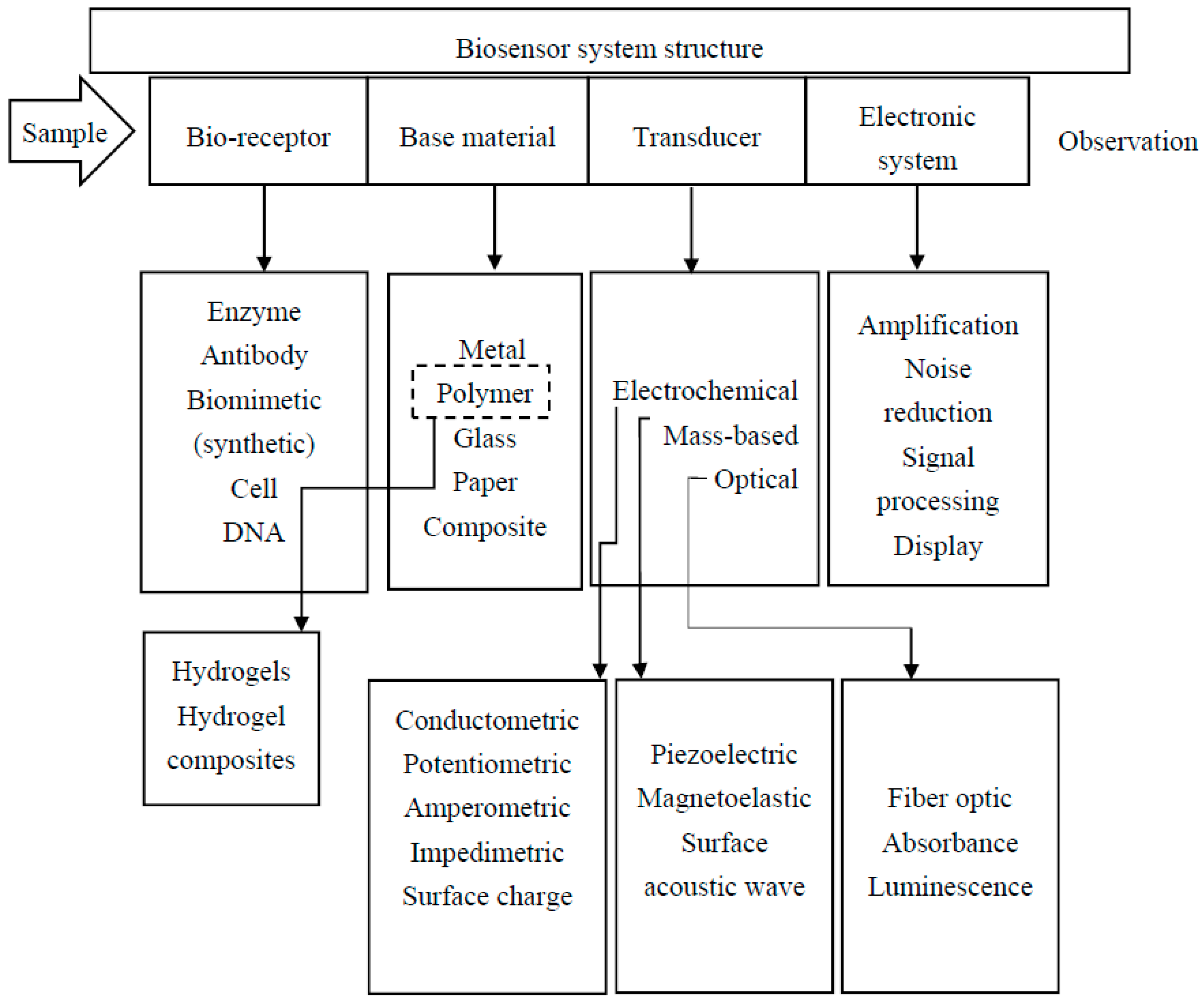
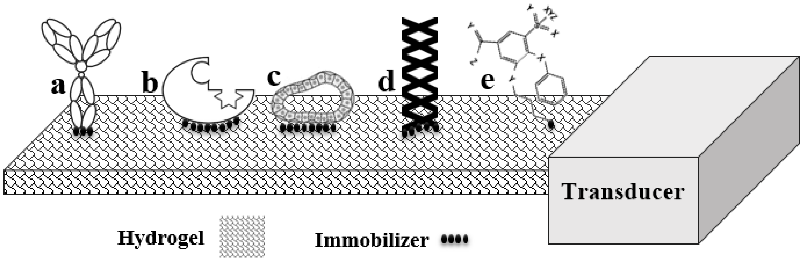
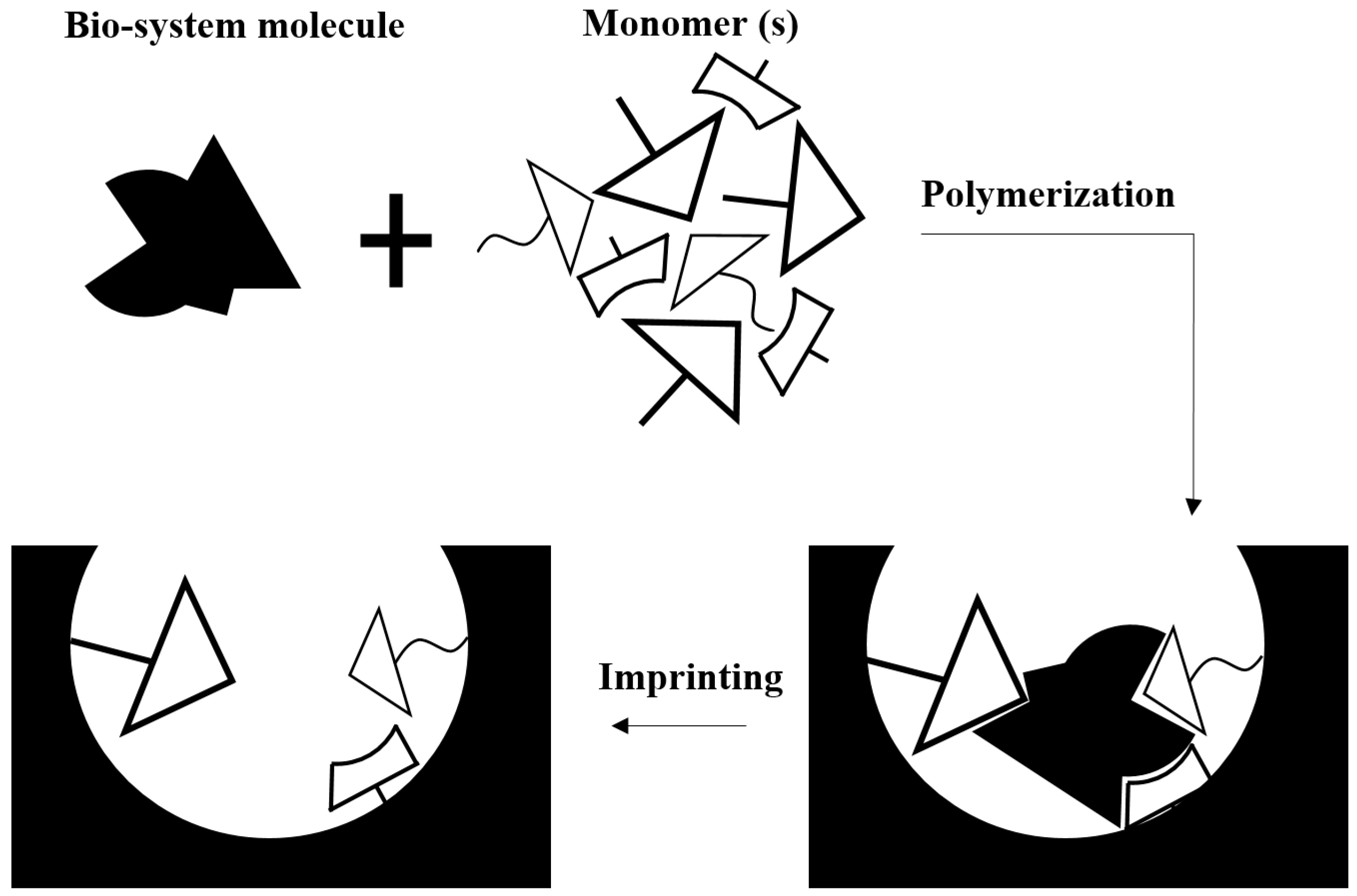
Table 1.
Advantages and disadvantages of different bioreceptors for hydrogel based biosensors.
| Bioreceptor | Advantage | Disadvantage |
|---|---|---|
| Antibody [13,14,45] | The immunogen need not be purified prior to detection. | Expensive and time-consuming method. Miniaturized immune-PCR detection methods have not yet been commercialized. |
| Enzymes [17,18,46,47,48] | Variety of reaction products arising from the catalytic process. | Stability problems have been reported. The detection limits can be very low due to signal amplification. |
| Nucleic acids [49,50] | Target molecule can be recognized by shape and sequence. A wide range of biomolecules can be detected. High binding affinity, simple synthesis method and easy storage have been reported. | It is not easy to design donor/acceptor labeling strategies. They are sensitive to pyrimidine specific nucleases that are abundant in biofluids. |
| Cells or cellular structures [7,26,29,31] | Can be used over prolonged periods of time as cells are closed systems. | |
| Biomimetic [32,33,34,35,36,37] | Known as an effective, accessible and inexpensive strategy. Physically, very stable (solid-like). | Molecular imprint probes do not have the same flexibility and selectivity as actual bioreceptors. |
| The molecular imprinted polymers can survive in destructive environments. |
Table 2.
Strategies for small biomolecule detection using hydrogel based biosensors
| Glucose | |||
|---|---|---|---|
| Hydrogel | Transduction Strategy | Technical Specification | Ref. |
| Polyaniline | Electrochemical | Sensitivity = 96.1 μA·mM−1·cm−2 | [248] |
| Response time = 3 s | |||
| Linear range = 0.01–8 mM | |||
| Polyaniline-PEG | Electrochemical | N/A | [249] |
| PEG | Optical | Linear range = 0–600 mg/dL | [250] |
| Response time = 10 min | |||
| PVA-Vinyl pyridine | Electrochemical | Sensitivity = 600 nA·mM−1·L−1 | [251] |
| Response time = 11 s | |||
| Chitosan | Electrochemical | Linear range = 5 μM–2.5 mM | [146] |
| Response time = 7 s | |||
| Chitosan-graphene oxide | Electrochemical | Linear range = 0.02–6.78 mM | [252] |
| Sensitivity = 10 μA·mM−1·cm−2 | |||
| Polypyrrole | Electrochemical | Linear range = up to 15 mM | [253] |
| PEG (injectable) | Optical | Linear range = up to 370 mg·dL−1 | [254] |
| Response time = 11 min | |||
| Polyvinylpyrrolidone | Optical | N/A | [255] |
| Alginate | Optical | Sensitivity = 0.80 ± 0.11 μs·dL·mg−1 | [256] |
| Linear range = 2.6–350 mg/dL | |||
| HEMA | Electrochemical | Linear range = 10 μM–40 mM | [257] |
| Lactate | |||
| BH40 (Hyper-branched) | Electrochemical | Response time = 7 s | [167] |
| Linear range = up to 580 mg/L | |||
| Polycarbamoyl sulfonate | Electrochemical | Response time = 2 s | [258] |
| Linear range = 10–400 μM | |||
| Albumin-mucin | Electrochemical | Response time = 90 s | [259] |
| Linear range = 0.7 μM–1.5 mM | |||
| Chitosan | Electrochemical | Sensitivity = 0.32 A·M−1·cm−2 | [260] |
| Response time = 5 s | |||
| Urea | |||
| Polyaniline | Electrochemical | Sensitivity = 85 mA·M−1·cm−2 | [261] |
| Response time = 15 s | |||
| Poly aniline | Electrochemical | Sensitivity = 878 μA·M−1·cm−2 | [262] |
| Aniline-co-o-phenylenediamine | Electrochemical | sensitivity = 31.12 mV/log [M] | [263] |
| Linear range = 3.16 × 10⁻⁴–3.16 × 10⁻² M | |||
| Cholesterol | |||
| Poly(thionine) | Electrochemical | Linear range = 25–125 μM | [264] |
| Sensitivity = 0.18 μA·mM−1·cm−2 | |||
| Polypyrrole | Electrochemical | Linear range = 5 × 10⁻⁴–1.5 × 10⁻² M | [265] |
| Response time = 30 s | |||
| Agarose | Electrochemical | Sensitivity = 6.9 nA·μM−1 | [266] |
| Response time = 120 s | |||
| Polyaniline | Electrochemical | Sensitivity = 0.042 μA·mg·dL−1 | [267] |
| Response time = 240 s | |||
| Polymethacrylate | Optical | Response time = 120 s | [268] |
© 2017 by the authors. Licensee MDPI, Basel, Switzerland. This article is an open access article distributed under the terms and conditions of the Creative Commons Attribution (CC BY) license (http://creativecommons.org/licenses/by/4.0/).
Share and Cite
MDPI and ACS Style
Tavakoli, J.; Tang, Y. Hydrogel Based Sensors for Biomedical Applications: An Updated Review. Polymers 2017, 9, 364. https://0-doi-org.brum.beds.ac.uk/10.3390/polym9080364
AMA Style
Tavakoli J, Tang Y. Hydrogel Based Sensors for Biomedical Applications: An Updated Review. Polymers. 2017; 9(8):364. https://0-doi-org.brum.beds.ac.uk/10.3390/polym9080364
Chicago/Turabian StyleTavakoli, Javad, and Youhong Tang. 2017. "Hydrogel Based Sensors for Biomedical Applications: An Updated Review" Polymers 9, no. 8: 364. https://0-doi-org.brum.beds.ac.uk/10.3390/polym9080364
Note that from the first issue of 2016, this journal uses article numbers instead of page numbers. See further details here.