In Vitro Propagation Strategies of Medicinally Important Berry Crop, Lingonberry (Vaccinium vitis-idaea L.)

1
St. John’s Research and Development Centre, Agriculture and Agri-Food Canada, Bldg. 25, 308 Brookfield Road, St. John’s, NL A1E 6J5, Canada
2
Department of Biology, Memorial University of Newfoundland, 232 Elizabeth Avenue, St. John′s, NL A1B 3X9, Canada
*
Author to whom correspondence should be addressed.
Agronomy 2020, 10(5), 744; https://0-doi-org.brum.beds.ac.uk/10.3390/agronomy10050744
Submission received: 22 April 2020
/
Revised: 16 May 2020
/
Accepted: 18 May 2020
/
Published: 21 May 2020
Abstract
:Lingonberry (Vaccinium vitis-idaea L.) is a health-promoting small fruit crop rich in antioxidant metabolites that helps to reduce the incidence of degenerative diseases. Being heterozygous, lingonberries cannot preserve genetic characteristics through seed propagation. Conventional vegetative propagation, although it produces true-to-type plants, is not economically viable. In vitro propagation can multiply plants much faster than conventional methods. A liquid cultures system under a bioreactor micropropagation system is of significant importance to increase the multiplication rates of in vitro-produced shoots. Enhanced vegetative growth and variation in biochemical constituents are observed in micropropagated plants. Clonal fidelity, although it may be a serious problem for commercial micropropagation, can be verified efficiently by molecular markers. The current review provides detailed and updated information on lingonberry micropropagation along with conventional methods and their effects on morphological, molecular and biochemical characteristics in micropropagated plants, filling the gap in literature.
1. Introduction
Lingonberries (Vaccinium vitis-idaea L.; family Ericaceae) are evergreen dwarf, rhizomatous, circumboreal woody shrubs [1]. They grow on heath areas on rocky places and dry peat soils and are economically important berry species to northern regions of the world [2]. There are many common names of lingonberries depending on regional nomenclature, such as partridgeberry or redberry in Newfoundland and Labrador; foxberry in Nova Scotia of Canada; airelle rouge in France, tytlebaer in Germany; puolukka in Finland; cowberry in Britain; kokemomo in Japan; and rock, mountain, dry ground or low bush cranberries and linberry in other parts of Canada and Alaska [3,4,5]. Throughout history, it has been grown as a fruit crop, a medicinal plant and a landscape ornamental ground cover [6]. The fruits can be consumed raw or used in juices, wines, pastries, jams, jellies, ice creams, cocktails and desserts [7,8].
Lingonberries gain significant importance in human diet for their rich source of vitamin C, omega-3 fatty acids, polyphenols and high antioxidant contents conferring health benefits. In lingonberries, 63–71% of the total phenolic contents are proanthocyanidins [9], which can defend against plant pathogens [10]. Flavonoids, phenolic acids, lignans and complex phenolic polymers (polymeric tannins) are the chemical substituents present in lingonberries and are richer sources of flavanols than many vegetables and fruits that are commonly used [11]. Lingonberry flavanols show antioxidative, anti- inflammatory, antibacterial, antiviral, antitumor, antifungal and vasoprotective activities [12,13]. Anthocyanins, which contribute to the red color of lingonberries, are one of the valuable phytochemicals showing protective effects against damage caused by radiation [14]. More than 116 anthocyanin and flavonoid compounds were isolated and identified primarily from lingonberry fruits or leaves [13,15]. Cyanidin-3-galactoside, cyanidin-3-arabinoside and cyanidin-3-glucoside are the three main anthocyanins in lingonberry [16,17,18]. Jin et al. [8] obtained 4.12 ± 0.18 mg g−1 of anthocyanins in lingonberry pomace, with 3.36 ± 0.14 mg g−1 of cyanidin-3-galactoside, 0.15 ± 0.01 mg g−1 of cyanidin-3-glucoside and 0.61 ± 0.03 mg g−1 of cyanidin-3-arabinoside. Lingonberry leaves and fruits are used to reduce cholesterol levels and treat kidney and bladder infections, stomach disorders and rheumatic diseases [3,19]. Lingonberry has highest content of resveratrol which are strong antioxidants with cancer chemopreventive activities [20], and they even help to reduce the threat of heart disease [21,22]. Lingonberry juice is helpful in protecting against urinary tract infection [23], and it possesses anti-inflammatory effects that protect the kidneys from ischemic-reperfusion injury [24]. Dietary supplementation with lingonberry reduces high-fat diet-induced inflammatory response and prevents kidney injury [24,25]. Lingonberry products, together with cranberry products, are well known as natural remedies for the treatment of urinary tract infections [26]. While the leaves of lingonberry have diuretic and urinary anti-septic properties, mainly related to their high content of tannins, arbutin (hydroquinone-ß-D-glucopyranoside) and arbutin derivatives [23].
Though the promising health benefits of lingonberries have inspired efforts to develop and magnify their commercial production [27,28], increasing their production throughout North America remains a challenge. Lingonberries are categorized under minor berries like black (Ribes nigrum L.) and red currants (R. rubrum L.), chokeberries (Prunus virginiana L.), cloudberries (Rubus chamaemorus L.), elderberries (Sambucus nigra L.) and gooseberries (R. uva-crispa L.) [29], and are not immediately available in the marketplace as blueberries (V. corymbosum L., V. angustifolium L., etc.) or cranberries (V. macrocarpon L.) are. Although lingonberries are native to the Canadian Pacific Northwest and northeastern Canada, they are not cultivated far and wide. Lingonberry is harvested mainly from native stands, but its high demands for industrial processing have led to the development of cultivars for commercial production. Currently, researchers at St. John’s Research and Development Centre of Agriculture and Agri-Food Canada in St. John’s, Newfoundland and Labrador, Canada, are developing high quality hybrids between European and Canadian lingonberries [3]. At present, commercial cultivation with European cultivars is available in small scale in Europe and North America, and most of the annual lingonberry harvest is from native stands [5]. In Newfoundland of Newfoundland and Labrador province Canada, lingonberries are grown in the wild, where 96,501 kg per year are commercially harvested [30]. The average yield of wild lingonberry was from 1900 to 3100 kg ha−1 over two growing seasons, 2011 and 2012, in Southern Labrador, Canada [31], and this is a relatively new endeavor in North America.
Being genetically heterozygous, it is not desirable to reproduce lingonberries from seeds because of the loss of their original form. Though propagation by vegetative means can conserve genetic integrity in lingonberries, conventional vegetative propagation is not economically feasible due to slow rhizome development and the fact that plants propagated by stem cuttings have brief life spans [32]. Micropropagated plants can proliferate more rapidly than conventional methods. Micropropagation is used for the speedy establishment of plants and for early fruit production [33,34,35]. Lingonberry plants raised through micropropagation were superior to those obtained by stem cuttings for fruit yield, rhizome production and vigor [36,37]. The present review describes in-depth the in vitro propagation systems in lingonberries, along with the clonal fidelity and phenotypic variation in micropropagated plants.
2. Taxonomy and Distribution
Lingonberries belong to the genus Vaccinium L. of subfamily Vaccinioideae in family Ericaceae, which contains about 4250 species in 124 genera [38]. The plants are dwarf and consist of a tap root system with rootlets. Adventitious roots are grown along the nodes of the stems and rhizomes. The flowers are small and light pink, with the inferior ovary producing dark red globose berries. Flowering occurs at the beginning of June and fruit matures at the end of August or the beginning of September. The fruits have an acidic taste (pH of 2.5) and contain tanins (7–21 mg g−1 fresh weight), anthocyanins (1–27 mg g−1 fresh weight) and total sugars (around 6%) [39]. The total rhizomes account for 80% of the total plant biomass [40]. Lingonberry plants are perennial eudicots, an evergreen shrub that extends from artic to north temperate regions in Eurasia and North America. They are categorized into two subspecies: ssp. vitis-idaea L. and ssp. minus (Lodd) [41]. Plant size is the main difference between the two subspecies, with ssp. minus being considerably smaller in height and leaf size (Figure 1, Table 1).
3. Propagation
Traditionally, lingonberries reproduce by seed or underground rhizomes. Stem cuttings of softwood or hardwood and rhizome divisions are used for vegetative propagation, based on the developmental stage and age of the material. In general, stem cuttings require 3 to 5 cm of a piece of plant material that re-grows into an independent plant. One of the important goals of the horticulture industry is to increase the acreage of lingonberry to meet the demand of the local fresh market. Healthy stocks used for propagating through conventional methods are not available. Besides, few plants are obtained from the mother plant in this method of propagation. With vegetative propagation, the plant takes several weeks to establish and it is labor intensive. Plant tissue culture provides an alternative for rapid propagation at a large scale. The way of maintaining or growing tissues in vitro to promote the differentiation and conservation of the structure and/or function is called tissue culture. Generally, it is practiced aseptically, where the plant parts are cultured under in vitro conditions (“in glass or plastic vessels”) providing an artificial environment. Plant tissue culture techniques are used in a wide range of settings for understanding basic problems in agriculture and horticulture. Due to advancement in its technology, it is successful in producing pathogen-free plants, the rapid multiplication of plants, the synthesis of useful metabolites and germplasm conservation. Methods have been developed to study the effects of nutrients and plant growth regulators (PGRs) on cell growth and differentiation.
4. Micropropagation
Micropropagation or in vitro propagation is the clonal propagation of plants by tissue, cell and organ culture methods. It involves the aseptic culture of explants of tissues and organs in closed vessels using defined culture media in a controlled environment. Haberlandt [46] was the first to study how to culture vegetative cells in simple nutrient solutions and was able to visualize the ability of regeneration. Immediately, the studies led to micropropagation, which has increased the production of agriculturally important plants, replacing cutting, grafting and division methods. Millions of flowering and ornamental plants that have been produced by micropropagation caused interest in adopting this technique in various crop plants globally. The technique gives a definitive answer to the problems of seed propagation; it multiplies plants in a small space more rapidly than traditional propagation methods, produces disease-free plants and is very suitable for germplasm conservation. The in vitro culture of plants has become an integral part of subjects like morphology, physiology, biochemistry, molecular biology and genetic engineering. Research into the micropropagation of lingonberry plants resulted in superior plants to those obtained by stem cuttings for berry yield, rhizome development and vigor in lingonberry cultivar Sanna [36]. A micropropagation program was initiated in 1999 at St. John’s Research and Development Centre of Agriculture and Agri-Food Canada in St. John’s, Canada, which developed a new protocol for lingonberry germplasm micropropagation [47]. The economic potential of micropropagation relies on true-to-type plants and the quality, cost-effectiveness and market value of the plant. Crop improvement methods adopt micropropagation technology, unlike conventional methods, because of the incidence of plant disease and abiotic stress. There are three methods of micropropagation: (1) axillary shoot proliferation, (2) adventitious shoot regeneration and (3) somatic embryogenesis.
4.1. Axillary Shoot Proliferation
In axillary shoot proliferation, shoots proliferate directly from the node via the axillary branching of buds from the original explants. This method is considered a convenient route for micropropagation because it does not include the callus stage. In this method, the regeneration of new buds does not occur, since bud meristems already exist in the axils of leaves and in the shoot tip. These bud meristems do not develop until the stem elongates and grows due to apical dominance. The explant (either apical or lateral short stem tip) contains many axillary buds in a condensed form and grows extensively when the shoot tips are excised and cultured in an appropriate medium containing cytokinins. This process is continued until the initial explant transforms into a mass of branches (Figure 2). This shoot multiplication cycle is repeated when the excised shoots are placed on a fresh medium.
Axillary bud culture was adopted for the mass propagation of various plants. Plants that are propagated in this method are genetically stable, as the development and growth of new shoots occurs from pre-existing meristems [48]. In vitro shoot proliferation has been reported in lingonberries [47,49,50,51,52] (Table 2).
Studies of Debnath and McRae [47] reported that both European lingonberry cultivars (ssp. vitis-idaea) and Canadian wild clones (ssp. minus) can be micropropagated from the shoot tips and nodal explants on a nutrient medium containing 12.3 μM N6-[2-isopentenyl]adenine (2iP) or 5.7 μM zeatin. They also reported that zeatin produced two to three times more shoots than 2iP in V. vitis-idaea ssp. vitis-idaea cultivar Regal. Similar results were also reported by Ostrolúcka et al. [55], where zeatin was more effective in improving shoot multiplication than 2iP. Another study by Gajdosova et al. [59] reported that the segmentation of regenerated microshoots and further culturing in fresh medium increased the shoot proliferation intensity. Shoot proliferation and multiplication are largely based on media formulations. Vaccinium culture in vitro prefers media with low ionic concentrations [60]. Woody Plant Medium [61] was found to be suitable for the shoot proliferation of the Vaccinium species. 2iP at 15 mg L−1 in Woody Plant Medium (WPM) produced shoots in vitro for Alaska lingonberries [62]. However, excessive levels of 2iP increased somaclonal variation in in vitro-derived regenerated plants [63]. Other studies showed the natural cytokinin zeatin was more effective than 2iP for the initiation [50] and proliferation of shoots in lingonberries [47]. Although cytokinin plays a role in shoot proliferation, excessive concentrations of cytokinins or auxins result in the abnormal morphology of regenerated shoots [60]. In a culture medium, carbohydrate is a crucial component [64]. The concentrations and types of carbohydrates affect lingonberry shoot proliferation in vitro. Sucrose and glucose are better than sorbitol to induce shoot proliferation in lingonberries, the optimum concentration being 10–20 g L−1 in lingonberry ssp. vitis-idaea and ssp. minus [52]. As lingonberries are acidophilic plants, the shoot proliferation intensity may depend on the pH of the medium. Shoot multiplication in vitro was effective in V. vitis-idaea ssp. minus at pH 5.0 [58].
4.2. Adventitious Shoot Regeneration
Adventitious shoot regeneration allows the forming of new organs from somatic cells. Although regeneration is a natural phenomenon during the life of a plant, it can be achieved at higher frequencies in tissue culture. Adventitious shoots can be developed either from explants or from calli developed on explants. Regeneration is achieved through organogenesis through unipolar organ (shoots or roots) formation, as well as by somatic embryogenesis, where the somatic embryos’ root and shoot meristem are developed [65]. Shoot organogenesis in lingonberries can be started with bud formation on explants, the elongation of the buds into shoots and the rooting of the shoots to form whole plants (Figure 3) [58].
Regeneration is a part of many micropropagation protocols; for example, haploid plants require adventitious shoot regeneration to form whole plants from somatic cells. Knowledge of adventitious organ formation is advancing in research due to the discovery of auxin and cytokinin. Auxin and cytokinin were helpful in forming adventitious roots and shoots at a high frequency. There are many other factors as well, including the genotype, culture medium, physical environment and explant development stage affecting adventitious shoot regeneration. Adventitious shoot regeneration has been reported in lingonberry [33,53,54]. In all cases, PGRs are involved.
An efficient method for the regeneration of adventitious shoots on excised leaves of micropropagated lingonberries was first developed by Debnath and McRae [53] on a basal medium (BM-A). The study showed that zeatin was more effective than thidiazuron (TDZ) or 2iP in inducing shoot regeneration. Although zeatin induced multiple shoot formation at 5–40 µM, the maximum morphogenic response was observed at 20 to 30 µM. The media containing TDZ promoted callus formation but suppressed shoot elongation [53].
Debnath [54] developed an improved shoot organogenesis from the hypocotyl segments of in vitro-grown lingonberry seedlings, in which the effects of TDZ on adventitious bud and shoot formation from the apical, central and basal segments of the hypocotyl were studied. Callus, bud, and shoot formation occurred more on the apical segments than the basal ones. In this study, a highly regenerative callus was obtained at 5–10 µM TDZ. The effect of TDZ on inhibiting shoot elongation was reduced by transferring the shoot cultures to a shoot-proliferating medium containing 1–2 µM of zeatin supplemented with 20 g L−1 of sucrose. TDZ (2.2 mg L−1) and zeatin (2.19 mg L−1) were found effective in inducing shoot regeneration in V. vitis-idaea ssp. vitis-idaea cvs. Red Pearl and Koralle on an Anderson culture medium [55]. A concentration of 20 μM zeatin along with 1 μM α-naphthaleneacetic acid was effective in promoting shoot regeneration in the lingonberry cultivar Red Pearl [56].
A two-step procedure to improve the efficiency of adventitious shoot regeneration from leaves of the in vitro-derived lingonberry cultivar Erntedank was studied by Debnath [33]. In this research, the leaves were cultured on basal media with different concentrations (0, 0.1, 1, 5, or 10 µM) of TDZ to study the effect of TDZ on adventitious shoot regeneration. After 8 weeks, the cultures on media with 5 µM TDZ were transferred to basal media with TDZ (0.1 or 1 µM), zeatin (1 or 2 µM) or basal media void of plant growth regulators (control) to study the effect of plant growth regulators on shoot elongation. An amount of 1 to 5 µM of TDZ supported bud and shoot regeneration but strongly suppressed shoot elongation. TDZ-initiated cultures, when transferred to the media containing 1–2 µM of zeatin, produced shoots after one additional subculture. Both thidiazuron and zeatin were found effective for shoot regeneration in V. vitis-idaea ssp. vitis-idaea cv. Red Pearl and Koralle on Anderson’s medium [55]. Gajdosova et al. [59] reported that lingonberry cultivar Red Pearl was tested for adventitious shoot induction from leaf tissues on culture medium containing 2.19 mg L−1 zeatin. After five weeks of cultivation, the explants were transferred to the medium containing 0.5 mg L−1 zeatin. The callus formation started on leaf explants cultured on media with 2.19 mg L−1 of zeatin. The adventitious buds began to appear on the callus surfaces when the callus was transferred to medium with 0.5 mg L−1 of zeatin, and the number of adventitious shoots regenerated was considerably high after three subcultures.
Successful regeneration in vitro depends also on the response of individual species and cultivar to the growth regulator type. In lingonberry, zeatin was more effective for the cultivar Red Pearl than for Koralle, showing the genotype-specific importance of cytokinins on regeneration [55]. Debnath [37] reported that clones of lingonberry belonging to two different subspecies differed in their shoot multiplication and development potential based on the growth regulators used. The research showed that TDZ concentration appeared to increase the shoot number in the clones tested, whereas adventitious buds were formed for shoots grown on media with high cytokinin concentrations. The in vitro response varying with the clone was also reported by Debnath and McRae [47]. The studies on diverse genotypes, can help to further characterize genotypic variation of lingonberry responses to in vitro conditions.
Explant orientation and polarity can profoundly affect the regeneration capacity of lingonberries. More callus growth and a higher bud and shoot number per explant were found to regenerate from the apical segments of the hypocotyls than from the central or basal segments in lingonberries [54]. The callus and bud regeneration percentage was higher when lingonberry leaves were cultured with their adaxial surface down (in contact with the medium) than those cultured with their abaxial side down [37]. However, shoot regeneration appeared on both sides of the leaves in lingonberry [33,53]. The explant response varies between the genotypes across propagation methods, as reported in two cultivars of lingonberry where in vitro-derived Erntedank plants had a better shoot growth than Regal plants [34]. The shoot regeneration system is beneficial for enhancing the vegetative growth of lingonberry plants, which may be of great advantage for growers concerned with the rapid establishment of plants for early fruit production.
4.3. Somatic Embryogenesis
In somatic embryogenesis, both the regeneration and organization are bipolar, where progenitor cells divide simultaneously to form a shoot and a root meristem that produce a group of cells termed pro-embryonic masses [66]. The differentiation and organization of a somatic embryo happen directly in the explant or from the callus. Like adventitious shoot regeneration, somatic embryogenesis depends on the explant type, the nutrient composition of the medium and the subculture establishment for a synchronized development of somatic embryo. In the Vaccinium species, somatic embryogenesis was successful only in blueberries [67].
4.4. Rooting and Acclimatization
The rooting of lingonberry microshoots can be achieved either in vitro or ex vitro. Meiners et al. [56] rooted the in vitro-derived shoots of lingonberry cultivar Red Pearl (ssp. vitis-idaea) on WPM containing 2.5–10 μM of indole-3-butyric acid (IBA). Ex vitro rooting is more common and successful for rooting lingonberry microshoots. While Miners et al. [56] reported the ex vitro rooting of Red Pearl lingonberry microshoots in a humidity chamber (98% relative humidity) without any PGR treatment, Arigundam et al. [58] treated leaf culture-derived wild lingonberry (ssp. minus) shoots with 39.4 mM of IBA powder, planted in two peats: one perlite (v/v) medium and maintained at 24 ± 2 °C and 95% humidity for a 16 h photoperiod (55 µmol m−2 s−1 photosynthetic photon flux, PPF) for rooting. For the ex vitro rooting of lingonberry ssp. vitis-idaea, IBA in talcum powder at 0.1–3% was used by Hosier et al. [49], and 2.07 mM KIBA solution (potassium salt of IBA) was used by Jaakola et al. [51]. Rooted plants can be acclimatized by the gradual lowering of humidity over 2–3 weeks and can be grown in a greenhouse at 20 ± 2 °C, 85% relative humidity, and 16 h photoperiod at a maximum PPF of 90 µmol m−2s−1 (Figure 4) [47,58].
5. Liquid Culture for Bioreactor Micropropagation
Although conventionally micropropagation is carried out on a semi-solid medium, the use of liquid medium is more efficient for large-scale micropropagation [68]. Liquid media are more advantageous than semi-solid in many plant species as they can enhance in vitro shoot proliferation, regeneration, somatic embryogenesis, rooting, culture growth, developmental process and micro-tuberization [69,70,71]. In liquid culture systems, the tissue is always in contact with the medium, stimulating the uptake of nutrients and phytohormones, which in turn leads to the uniform culturing of shoots and provides more rapid growth, yielding larger flesh and dry weight [72]. In the beginning, liquid culture medium was used to produce somatic embryos, microtubers and organ cultures in vitro both in agitated and non-agitated vessels and in bioreactors [73,74,75]. The continuous shaking of the liquid medium is known to provide aeration to the cultures, and this promote a larger growth and multiplication rate of the shoots [72]. The amount of sugars, inorganic ions and phytohormones present in the liquid medium shows a greater direct impact on in vitro shoot growth than on those grown on agar [72]. Agar medium contains inorganic impurities and limits diffusion rates [76]. In liquid media, explants can be cultured in partial immersion (temporary immersion), where gaseous exchange is not hindered and explants can use nutrients efficiently [68]. However, vitrification can be observed in in vitro-raised shoots as they are generally very sensitive to liquid media. This leads to the poor survival of tissue culture plants when they are transferred to ex vitro conditions. In vitro-derived plants are susceptible to environmental stress when grown ex vitro [77,78]. This is because of the absence of agar concentration in the culture medium causing vitrification in the tissues [79,80,81,82]. Hyper-hydricity has been proposed to define hyper-hydric malformations affecting in vitro-derived shoots in culture [83]. The phenomenon was previously known and described under the term vitrification [84]. Translucency, glassiness, vitrescence and glauciness are other less-used terms for this physiological disorder [85]. Vitreous, vitrified or hyper-hydric shoots look turgid with a watery surface and are hypo-lignified. The problems of hyper-hydricity can be avoided by exposing the explant alternately to liquid and air in the vessel. Further, liquid culture systems offer the renewal of media without changing containers, as the ease of container cleaning may alter the culture period. Moreover, conventional micropropagation techniques are limited due to high cost of production at the industrial level. Liquid culture systems are helpful in overcoming this limitation due to cost reduction, as they can be less labor intensive and require less time for sub-culturing. Other advantages of liquid media include the easy replenishment of culture media, escaping or minimizing the numbers of subcultures and reducing the unit cost, as gelling agent is not needed as an addition in the media [86,87].
5.1. Bioreactors
Bioreactors for in vitro culture range from 0.5–500 L in size and can be a jelly jar or a reformed microbial fermenter. Bioreactors are vessels widely used for the culture of organisms, including plants, cells and microbes to produce cells or metabolites [88,89]. Bioreactor technology is also applicable to plant propagation. The application for plant propagation using the shake culture technique was initially reported in Begonia by Takayama and Misawa [90]. Thereafter, the technique has been used in many plant species [91,92,93]. Bioreactor micropropagation consists of culturing explants in liquid media with forced aeration, leading to the formation and multiplication of propagules including plantlets, microtubers, microcorms or bulblets. The major advantages of bioreactor micropropagation include [94]:
- The easy production and scale-up of a large number of plantlets in the minimum time.
- Easy culture handling (inoculation and harvesting) with a reduced labor cost.
- The close contact of explants in liquid cultures, allowing the quick uptake of nutrients for rapid growth.
- Sufficient oxygen supply due to forced aeration for improved growth rate and multiplication.
- The movement of cultures in bioreactors, resulting in the removal of apical dominance and rapid shoot proliferation and plantlet formation.
Bioreactors used for micropropagation can culture explants in continuously submerged conditions or can be immersed partially or temporarily in the medium. There are many types of bioreactors used in plant biotechnology; few have been provided with light conditions for culturing explants [95]. Some other bioreactors have been fitted with pipes through which light was emitted [96]. Bioreactors used for in vitro propagation can be mechanically or pneumatically agitated, non-agitated or temporary immersion types [93]. The utility of a bioreactor depends on its cost-effectiveness and convenience of operation along with maintaining the culture quality. Temporary immersion (TIB) and stationary bioreactors (SB) have been used for the in vitro culture of lingonberries [58].
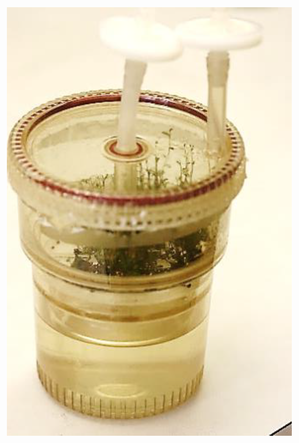
5.1.1. Temporary Immersion Bioreactor (TIB)
Various TIB types are available, where the design is chosen based on the requirement of the specific plant culture sensitivity to hyperhydricity and culture costs [91,97]. The TIB system is very useful for semi-automated in vitro propagation. It helps in controlling contamination, adequately supplying oxygen and nutrients and mixing. It requires less frequent sub-culturing, and it has the benefits of ease of medium changes and limited damage due to shearing. The RITA® (recipient for automated temporary immersion) [98] is a kind of TIB that consists of two compartments (Figure 5). While the upper compartment holds the explants, the liquid culture medium is kept in the lower compartment. They are joined together and the medium is lifted into the upper chamber via the application of overpressure to the lower chamber. The liquid medium drops to the lower chamber when the overpressure falls. Details of the system for mass micropropagation have been reviewed elsewhere [68,92,93]. Most reports claimed better multiplication rates in TIBs than on semi-solid media, although few observed no differences [99,100,101]. In the lingonberry, Arigundam et al. [58] reported the in vitro shoot proliferation of wild V. vitis-idaea ssp. minus clones in SB and TIB systems containing a liquid medium with 9.1 μM zeatin or 1.8 μM TDZ. The shoot proliferation was 2–3 times less on a semi-solid medium than in a liquid medium, although 10–25% of hyperhydric shoots were observed in bioreactors. However, the rooting of these shoots was not affected, as most of the bioreactor-cultured microshoots rooted in a peat—a perlite medium with 90–95% survivability under greenhouse conditions [58]. This indicated that in lingonberry, the hyperhydricity was reversible, confirming the previous report with strawberry [102].
5.1.2. Stationary Bioreactor (SB)
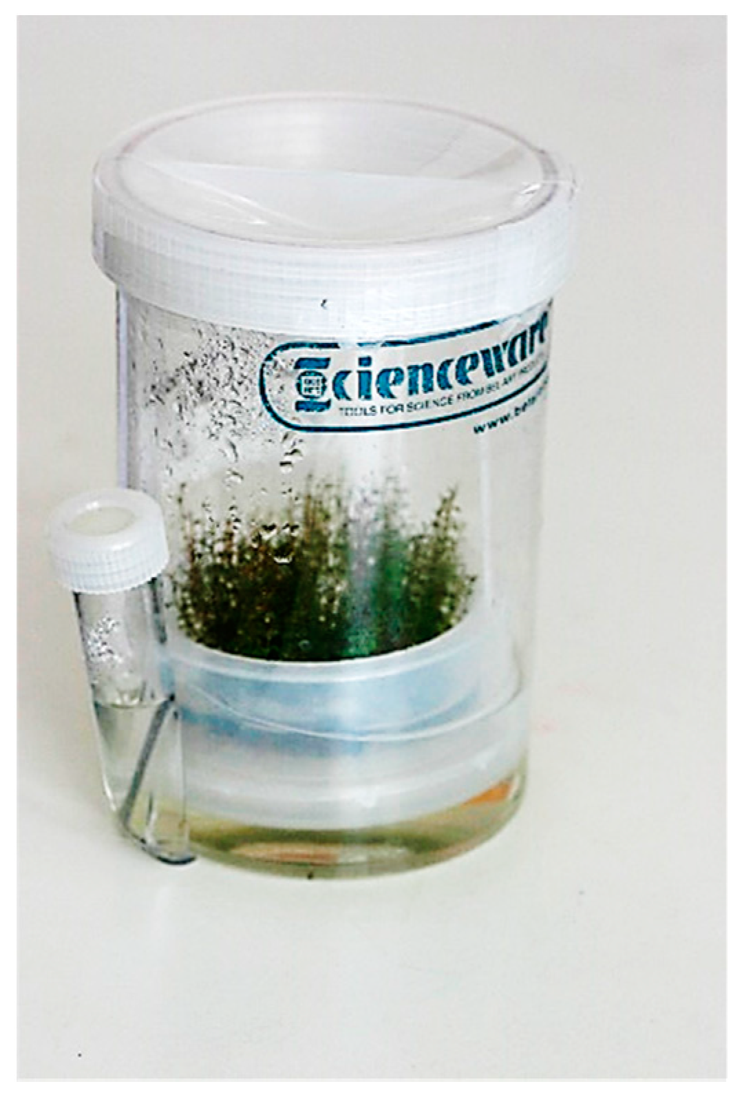
Stationary bioreactor is an effective system for the in vitro culture of lingonberries [58]. The Growtek is a low-cost SB system that has been designed, patented and commercialized for in vitro propagation [103]. The system is suitable for the increased proliferation rates of shoots. It saves time for incubation, minimizes contamination and helps in transferring plantlets, easily avoiding root injury. It has been applied for in vitro seed germination, molecular pharming, secondary metabolite production, culturing solid-state fungi and bioremediation. The SB contains a rotating and floating explant holder and a side tube that can be used for changing medium, feeding culture and monitoring contents (Figure 6). Compared to agar-gelled and liquid media using other culture vessels, Dey [103] reported that the Growtek SB bioreactor produced 1.2–23.3 times more shoots with reduced root injury (32–48%), contamination (12–18%) and incubation time (16–42%). Similar results were also reported in lingonberries by Arigundam et al. [58], where shoot multiplication was much less on a semi-solid medium than in SB or TIB with a liquid medium. The healthier shoots can be obtained in a Growtek vessel due to the presence of a more appropriate vapor/gas phase inside the vessel. Growtek can be used both in static and agitated conditions and covers the qualities of both liquid and gelled media systems. The unique combination of these features can be effectively used for the biosynthesis of secondary metabolites in hairy roots and for in vitro propagation, including somatic embryogenesis [103]. In the Growtek system, the higher thread rim depth helps improve the suitable gas exchange, thereby causing better shoot and root growth and vigor.
6. Variation in Micropropagated Plants
The occurrence of variation is great concern in a tissue culture system for commercial micropropagation. The variation in micropropagated plants is related to the source of the tissue or regeneration system [104]. The effect of variation in micropropagated plants can be determined morphologically, biochemically, cytologically and genetically. In vitro culture in lingonberries enhances vigorous vegetative growth in regenerated plants, including increased branching and more leaf and rhizome production compared to the stem cutting (SC) plants. Hosier et al. [49] found a better rhizome production in lingonberry tissue culture (TC) plants compared to SC plants after nine months of growth in a greenhouse. The field performance of lingonberry ssp. vitis-idaea cv. Sanna TC plants showed a greater fruit yield, rhizome production and total plant weight than SC plants after three years of growth [36]. Similar results were also reported by Debnath [105], where TC plants derived from the proliferated shoots of nodal explants and the regenerated shoots from leaf explants were compared with those of SC plants in Regal, Splendor and Erntedank lingonberries under greenhouse conditions. After three years of growth, the TC plants were superior to their respective SC plants for stem, leaf and rhizome numbers per plant [105]. In another study, the leaf culture-derived TC plants of the cultivars Regal and Erntedank produced smaller and less vigorous plants that had more stems, branches, leaves and rhizomes than conventional cuttings [34]. The in vitro shoots of the lingonberry cultivar Red Pearl rooted better than cutting plants [56]. Cultures in vitro on a nutrient medium containing PGR might induce the juvenile characteristics of lingonberry TC plants, enhancing vegetative production [34]. Foley and Debnath [106] reported that the berries from Splendor and Erntedank lingonberry TC plants had more antioxidant activity than those of berries from SC plants, although SC plants had more berries, with a larger diameter and yield per plant. In another study, the leaves from Regal, Splendor and Erntedank TC plants exhibited higher antioxidant enzyme activities than those of SC plants [107]. However, working with the ssp. vitis-idaea cultivars Koralle, Erntedank and Sussi, Serres et al. [108] did not observe any difference between SC and TC plants for the number of rhizomes and branches per plant. The enhanced vegetative growth of the lingonberry TC plants did not stay in their next SC propagation cycle, suggesting the possibility of the disappearance of the in vitro culture-derived juvenile phase in the second cycle [109].
7. Clonal Fidelity and Micropropagation
Plant tissue culture has become an important tool for the rapid micropropagation of valuable crops. However, true-to-type propagules and genetic stability are major concerns in commercial micropropagation, since the in vitro culture of plant cells, tissues, organs and undifferentiated calli are associated with genetic changes during the process [110] which result in physiological and somaclonal variation [111]. Somaclonal variations are both heritable (genetic) and non-heritable (epigenetic). All of these phenomena are dependent on factors like genotype, the availability of chimeral tissues, the type and origin of explants, the type of medium, the concentrations of PGRs and the duration and environment of the culture (light, temperature, etc.) that could limit the broader utility of the micropropagation systems [112]. Although somaclonal variation is a valuable source for selecting new genotypes, it is disadvantageous for germplasm preservation. The multiplication of elite genotypes is needed to preserve the specific genotypes. A genetic purity assessment is extremely important in order to develop proper programs of conservation. The genetic uniformity of TC plants can be verified at the morphological, physiological, biochemical and genetic levels. Molecular markers are reliable tools to validate the clonal fidelity of micropropagated plants as, unlike other markers, they are not influenced by the environment. The identification of genotypes can help in preventing the misidentification of varieties during nursery propagation. DNA-based markers are developed based on DNA polymorphism [113]. These markers detect nucleotide sequence variation at a particular location in the genome. DNA markers produce DNA “fingerprints” that are resolved by electrophoresis in agarose or acrylamide gels and identified by staining or labelling. Different sized molecules are allowed to pass through the gel, and the mobility varies depending on their molecular weight. The patterns of DNA fragments are visualized using a fluorescent dye specific for DNA, such as ethidium bromide.
There are a number of molecular markers that can be used for identifying the clonal fidelity of micropropagated plants of the Vaccinium species [109]. Random amplified polymorphic DNA (RAPD), amplified fragment length polymorphism (AFLP), restriction fragment length polymorphism (RFLP), inter simple sequence repeat (ISSR), simple (short) sequence repeat (SSR), DNA amplified fingerprinting (DAF), sequence characterized amplified region (SCAR), short tandem repeat (STR), sequence-tagged sites (STSs), expressed sequence tag (EST)- polymerase chain reaction (PCR) and cleaved amplified polymorphic sequences (CAPS) derived from expressed sequence tag polymerase chain reaction (EST-PCR) markers are used to regularly check the genetic purity. A great advantage of these markers is that DNA-based markers can compare different genetic material regardless of environmental influences [114]. The marker selection is important, as each marker possesses some strengths and drawbacks. The development of PCR [115] for amplifying DNA brought a range of new technologies that overcame the technical limitations of RFLPs. PCR-based DNA markers, including RAPDs, AFLPs and ISSRs, use non-specific or arbitrary DNA sequences, and SSRs and STSs use primers with a known sequence and can target a specific locus.
Inter simple sequence repeats (ISSRs) markers were first developed by Zietkiewicz and co-workers [116], where 3‘ anchored primers were utilized to amplify the inter SSR sequences or sequence-flanking SSRs. ISSRs have been extensively used in most of the studies and proved highly efficient for the assessment of genetic fidelity. ISSRs have several benefits, as they permit the detection of polymorphism in microsatellite loci without prior knowledge of the flanking sequence; these make them easier to use. They require a high annealing temperature and a longer primer sequence, which enables the study of a high degree of polymorphism between individuals within a population of closely related genotypes. They cost less and require a very small amount of DNA for amplification. ISSRs have been successfully used for genetic diversity analysis in lingonberries [35,117].
EST-PCR markers have been developed from EST libraries, which were derived from the floral buds of cold acclimated and non-acclimated highbush blueberry plants [118]. These markers were originally developed to fingerprint and estimate genetic similarity among the clones of lowbush blueberry. Besides this, EST-PCR products are effective in studies of intra-population genetic variation [119]. EST-PCR markers are useful for many species due to their codominant, reproducible polymorphic amplification products. With the availability of EST databases, efficient and cost-effective EST-derived SSR markers from wild and cultivated species have been developed [120]. These markers are codominant and abundant; they can identify allelic variability and distinguish between mostly related genotypes. The transferability of SSR markers across species made the markers the best choice for genotype identification. SSRs are highly polymorphic and generated by the amplification of a microsatellite in a PCR reaction with the use of 20–25 nt-long primers that are complementary to the flanking regions of the simple sequence repeat. The great advantage of SSR markers is that multiple SSRs can be analyzed in one PCR reaction.
Although Alam et al. [121] used Genotyping-by-Sequencing (GBS)—a high-throughput technology for rapid DNA or RNA base pair sequencing—to discern V. vitis-idaea ssp. minus wild clones, only EST-PCR, EST-SSR and ISSR markers have been used for monitoring micropropagated lingonberries [58].
Arigundam et al. [58] studied clonal fidelity in SB and TIB-derived lingonberry micropagules grown under greenhouse conditions. They used EST-PCR, EST-SSR and ISSR markers and observed monomorphic banding patterns in both the TC and SC plants of a V. vitis-idaea ssp. minus wild clone, confirming the clonal fidelity in micropropagated lingonberry plants [58].
8. Conclusions
In vitro propagation is in practice for the mass propagation of various plant species and is currently a multi-billion-dollar industry globally. The mass propagation of lingonberries can be achieved through axillary shoot proliferation, adventitious shoot regeneration or somatic embryogenesis. Axillary shoot proliferation is a simple and reliable propagation method for maintaining clonal fidelity. Shoot regeneration from leaves or hypocotyl segments is a rapid method of lingonberry micropropagation, provided the genetic integrity of the mother plant is retained in the micropropagules. As the proliferation of shoots involves the continuation of the growth of organized tissues, it is a better choice over adventitious shoot regeneration or somatic embryogenesis for producing true-to-type plants, although Arigundam et al. [58] claimed to have produced genetically uniform plants through adventitious shoot regeneration in lingonberries. Somatic embryogenesis has not been successful in lingonberries, but the process is suitable for mechanization and can used in a bioreactor system using liquid media. However, the possibility of getting abnormalities is greater in adventitious shoot regeneration and somatic embryogenesis, as the microshoots or plants are formed from unorganized cells or tissues. In vitro-derived variations, although not desirable for commercial micropropagation, can explore novel somaclones that can be used as commercial cultivars [122]. It is important to ensure that the genetic stability of in vitro-propagated plants and molecular markers are reliable for monitoring the clonal fidelity of TC plants. It is better to use more than one type of molecular marker for more genome coverage. The possibility of the occurrence of a somaclonal variation can be lowered by applying less PGR in the culture media; frequent sub-culturing and the choice of genotype are also factors. The increased rhizome production and vegetative growth of lingonberry TC plants are beneficial to lingonberry growers for a quick establishment, the spreading of the planting area and improved berry production, as well as for an improved return on their investment.
Bioreactor micropropagation using liquid media has significant potential for the commercial propagation of lingonberries. The application of the technology varies from genotype to genotype [58]. Automation and the close contact of liquid medium in bioreactors are very efficient and cost-effective for in vitro propagation. However, the appearance of hyperhydricity (vitrification) is a concern in liquid culture that needs to minimized with the proper optimization of immersion time in TIB systems.
A wide range of chemical composition in lingonberry leaves and fruits has been documented. A higher amount of phenolic compounds and antioxidant activity in the fruits and leaves of micropropagated lingonberries have highlighted their positive effect on human health. However, limited research on the mechanism behind the contribution of antioxidant activity to the total phenolic content and anthocyanin of lingonberry species has reduced the speed of development of commercially and nutritionally important traits. Knowledge of the genetic basis of the phenolic compounds in lingonberry needs to be expanded to increase the levels of phytochemicals in the leaves and fruits of lingonberries. From the economical point of view, exploring different propagation methods in various cultivars for mass production with health-promoting compounds and comparing them in different geographical locations can enhance lingonberry production with high-quality fruits.
Author Contributions
Conceptualization, writing, reviewing and editing by S.C.D.; writing and draft preparation by U.A. Both authors have agreed on the final version of the manuscript. The authors declare that the content of this paper has not been published or submitted for publication elsewhere. All authors have read and agreed to the published version of the manuscript.
Funding
This review received no external funding.
Acknowledgments
This is an Agriculture and Agri-Food Canada (AAFC) St. John’s Research and Development Centre contribution. The authors’ research in this area is supported by AAFC St. John’s Research and Development Centre, Newfoundland and Labrador, Canada.
Conflicts of Interest
The authors declare no conflict of interest.
References
- Luby, J.J.; Ballington, J.R.; Draper, A.D.; Pliszka, K.; Austin, M.E. Blueberries and cranberries (Vaccinium). Acta Hortic. 1991, 290, 391–456. [Google Scholar] [CrossRef]
- Nestby, R.; Hykkerud, A.L.; Martinussen, I. Review of botanical characterization, growth preferences, climatic adaptation and human health effects of Ericaceae and Empetraceae wild dwarf shrub berries in boreal, alpine and arctic areas. J. Berry Res. 2019, 9, 515–547. [Google Scholar] [CrossRef] [Green Version]
- Debnath, S.C.; Keske, C. Technological advances in the propagation and improvement of Newfoundland and Labrador berries. In Growing a Sustainable Food System for Newfoundland and Labrador; Keske, C., Ed.; ISER Books, Food Futures; Memorial University: St. John’s, NL, Canada, 2018; pp. 381–409. [Google Scholar]
- Vander Kloet, S.P. The Genus Vaccinium in North America; Agriculture Canada Publications 1828: Ottawa, ON, Canada, 1988; p. 201. [Google Scholar]
- Penhallegon, R. Lingonberry production guide for the Pacific Northwest. Oregon State Univ. Extn. Serv. Publ. PNW 583-E. 2006, p. 12. Available online: https://catalog.extension.oregonstate.edu/sites/catalog/files/project/pdf/pnw583.pdf (accessed on 1 May 2020).
- Jun, W.; Dierking, S.; Beerenobst, W. European Vaccinium species. Acta Hortic. 1993, 241, 299–304. [Google Scholar] [CrossRef]
- Launert, E. Edible and Medicinal Plants of Britain and North Europe; Hamlyn: London, UK, 1981; p. 194. [Google Scholar]
- Jin, Y.; Liu, Z.; Liu, D.; Shi, G.; Liu, D.; Yang, Y.; Gu, H.; Yang, L.; Zhou, Z. Natural antioxidant of rosemary extract used as an additive in the ultrasound-assisted extraction of anthocyanins from lingonberry (Vaccinium vitis-idaea L.) pomace. Ind. Crop. Prod. 2019, 138, 111425. [Google Scholar] [CrossRef]
- Petri, K.; Liisa, N.; Riitta, P.; Benita, W.; Tiina, L.; Jukka, W.; Eeva, M.; Marina, H. Lingonberry (Vaccinium vitis-idaea) and European cranberry (Vaccinium microcarpon) proanthocyanidins: Isolation, identification, and bioactivities. J. Agric. Food Chem. 2011, 59, 3373–3384. [Google Scholar] [CrossRef]
- Burdulis, D.; Äarkinas, A.; Jasutien, I.; Stackevi, E.; Nikolajevas, L.; Janulis, V. Comparative study of anthocyanin composition, antimicrobial and antioxidant activity in bilberry (Vaccinium myrtillus L.) and blueberry (Vaccinium corymbosum L.) fruits. Acta Pol. Pharm. 2009, 66, 399–408. [Google Scholar]
- Puupponen-pimiä, R.; Nohynek, L.; Alakomi, H.L.; Oksman-Caldentey, K.M. Bioactive berry compounds—Novel tools against human pathogens. Appl. Microbiol. Biotechnol. 2005, 67, 8–18. [Google Scholar] [CrossRef]
- Negi, P.S. Plant extracts for the control of bacterial growth: Efficacy, stability and safety issues for food application. Int. J. Food Microbiol. 2012, 156, 7–17. [Google Scholar] [CrossRef]
- Su, Z. Anthocyanins and flavonoids of Vaccinium L. Pharm Crop. 2012, 3, 7–37. [Google Scholar] [CrossRef] [Green Version]
- Fan, Z.L.; Wang, Z.Y.; Zuo, L.L.; Tian, S.Q. Protective Effect of anthocyanins from lingonberry on radiation-induced damages. Int. J. Environ. Res. Public Health 2012, 9, 4732–4743. [Google Scholar] [CrossRef] [Green Version]
- Kalt, W.; Mackinnon, S.; McDonald, J.; Vinqvist, M.; Craft, C.; Howell, A. Phenolics of Vaccinium berries and other fruit crops. J. Sci. Food Agric. 2008, 88, 68–76. [Google Scholar] [CrossRef]
- Dudonné, S.; Dubé, P.; Anhê, F.F.; Pilon, G.; Marette, A.; Lemire, M.; Harris, C.; Dewailly, E.; Desjardins, Y. Comprehensive analysis of phenolic compounds and abscisic acid profiles of twelve native Canadian berries. J. Food Compos. Anal. 2015, 44, 214–224. [Google Scholar] [CrossRef]
- Isaak, C.K.; Petkau, J.C.; O, K.; Debnath, S.C.; Siow, Y.L. Manitoba lingonberry (Vaccinium vitis-idaea) bioactivities in ischemia-reperfusion injury. J. Agric. Food Chem. 2015, 63, 5660–5669. [Google Scholar] [CrossRef] [PubMed]
- Lee, J.; Finn, C.E. Lingonberry (Vaccinium vitis-idaea L.) grown in the Pacific Northwest of North America: Anthocyanin and free amino acid composition. J. Funct. Foods 2012, 4, 213–218. [Google Scholar] [CrossRef]
- Novelli, S. Developments in berry production and use. Agric. Agri-Food Can. 2003, 21, 16. [Google Scholar]
- Rimando, A.M.; Kalt, W.; Magee, J.B.; Dewey, J.; Ballington, J.R. Resveratrol, pterostilbene, and piceatannol in Vaccinium berries. J. Agric. Food Chem. 2004, 52, 4713–4719. [Google Scholar] [CrossRef]
- Bomser, J.; Madhavi, D.L.; Singletary, K.; Smith, M.A. In vitro anticancer activity of fruit extracts from Vaccinium species. Planta Med. 1996, 62, 212–216. [Google Scholar] [CrossRef]
- Wang, S.Y.; Feng, R.; Bowman, L.; Penhallegon, R.; Ding, M.; Lu, Y. Antioxidant activity in lingonberries (Vaccinium vitis-idaea L.) and its mitogen activated protein kinases activation. J. Agric. Food Chem. 2005, 53, 3156–3166. [Google Scholar] [CrossRef] [Green Version]
- Ieri, F.; Martini, S.; Innocenti, M.; Mulinacci, N. Phenolic distribution in liquid preparations of Vaccinium myrtillus L. and Vaccinium vitis idaea L. Phytochem. Anal. 2013, 24, 467–475. [Google Scholar] [CrossRef]
- Isaak, C.K.; Wang, P.; Prashar, S.; O, K.; Brown, D.C.W.; Debnath, S.C.; Siow, Y.L. Supplementing diet with Manitoba lingonberry juice reduces kidney ischemia-reperfusion injury. J. Sci. Food Agric. 2017, 97, 3065–3076. [Google Scholar] [CrossRef]
- Kowalska, K.; Olejnik, A.; Wasielica, J.Z.; Olkowicz, M. Inhibitory effects of lingonberry (Vaccinium vitis-idaea L.) fruit extract on obesity-induced inflammation in 3T3-L1 adipocytes and RAW 264.7 macrophages. J. Funct. Foods 2019, 371–380. [Google Scholar] [CrossRef]
- Peron, G.; Sut, S.; Pellizzaro, A.; Brun, P.; Voinovich, D.; Castagliuolo, I.; Dall’Acqua, S. The antiadhesive activity of cranberry phytocomplex studied by metabolomics: Intestinal PAC-A metabolites but not intact PAC-A are identified as markers in active urines against uropathogenic Escherichia coli. Fitoterapia 2017, 122, 67–75. [Google Scholar] [CrossRef] [PubMed]
- Finn, C.E.; Mackey, T. Growth, yield, and fruit quality of 10 lingonberry (Vaccinium vitis-idaea) cultivars and selections in the Pacific Northwest, USA. Acta Hortic. 2006, 715, 289–294. [Google Scholar] [CrossRef] [Green Version]
- Lee, J.; Finn, C.E. Anthocyanins and other polyphenolics in American elderberry (Sambucus canadensis) and European elderberry (S. nigra) cultivars. J. Sci. Food Agric. 2007, 87, 2665–2675. [Google Scholar] [CrossRef] [PubMed] [Green Version]
- Hjalmarsson, I.; Ortiz, R. Lingonberry: Botany and horticulture. Hortic. Rev. 2001, 27, 79–123. [Google Scholar] [CrossRef]
- Heidenreich, C. The Lowdown on Lingonberries. New York Berry News. Volume 9. 19 June 2010. Available online: http://www.fruit.cornell.edu/berry/production/pdfs/Lingonberries.pdf (accessed on 20 April 2020).
- Li, J. Fruit Yield and Composition of Native Fruits, Partridgeberry (Vaccinium vitis-idaea L.) and Bakeapple (Rubus chamaemorus L.). Master’s Thesis, Dalhousie University, Halifax, NS, Canada, 2013; p. 109. Available online: http://hdl.handle.net/10222/31130 (accessed on 1 January 2020).
- Holloway, P.S. Rooting of lingonberry, Vaccinium vitis-idaea, stem cuttings. Plant Propag. 1985, 31, 7–9. [Google Scholar]
- Debnath, S.C. A two-step procedure for adventitious shoot regeneration from in vitro-derived lingonberry leaves: Shoot induction with TDZ and shoot elongation using zeatin. HortScience 2005, 40, 189–192. [Google Scholar] [CrossRef] [Green Version]
- Debnath, S.C. Influence of propagation method and indole-3-butyric acid on growth and development of in vitro and ex vitro-derived lingonberry plants. Can. J. Plant Sci. 2006, 86, 235–243. [Google Scholar] [CrossRef] [Green Version]
- Debnath, S.C. Inter simple sequence repeat (ISSR) to assess genetic diversity within a collection of wild lingonberry (Vaccinium vitis-idaea L.) clones. Can. J. Plant Sci. 2007, 87, 337–344. [Google Scholar] [CrossRef] [Green Version]
- Gustavsson, B.A.; Stanys, V. Field performance of ‘Sanna’ lingonberry derived by micropropagation vs. stem cuttings. HortScience 2000, 35, 742–744. [Google Scholar] [CrossRef] [Green Version]
- Debnath, S.C. Micropropagation of lingonberry: Influence of genotype, explant orientation, and overcoming TDZ-induced inhibition of shoot elongation using zeatin. HortScience 2005, 40, 185–188. [Google Scholar] [CrossRef]
- Christenhusz, M.J.M. The number of known plants species in the word and its annual increase. Phytotaxa 2016, 261, 201–217. [Google Scholar] [CrossRef] [Green Version]
- Hall, I.V.; Shay, J.M. The biological flora of Canada. 3. Vaccinium vitis-idaea L. var. minus Lodd. Supplementary Account. Can. Field-Nat. 1981, 95, 434–464. [Google Scholar]
- Holloway, P.S. Studies on Vegetative and Reproductive Growth of Lingonberry (Vaccinium vitis-idaea L.). Ph.D. Thesis, University of Minnesota, Minneapolis, MN, USA, 1981. [Google Scholar]
- Hultén, E. On the races in the Scandinavian flora. Svensk Botanisk Tidskrift Bd. 1949, 43, 383–406. [Google Scholar]
- Fernald, M.L. Gray’s Manual of Botany, 8th ed.; D. Van Nostrand, Co.: New York, NY, USA, 1970; p. 1632. [Google Scholar]
- Hultén, E. The Circumpolar Plants II. Dicotyledons; Almqvist and Wiksell: Stockholm, Sweden, 1970. [Google Scholar]
- Hultén, E. Atlas of the Distribution of Vascular Plants in NW Europe, 2nd ed.; Generalstabens litografiska anstalts förlag: Stockholm, Sweden, 1971. [Google Scholar]
- Welsh, S.L. Anderson’s Flora of Alaska and Adjacent Parts of Canada; Brigham Young University Press: Provo, UT, USA, 1974. [Google Scholar]
- Haberlandt, G. Kulturversuche mit isolierten Pflanzenzellen. Sitzungsber. Math. Naturwiss. Kl. Kais. Akad. Wiss. Wien 1902, 111, 69–92. [Google Scholar]
- Debnath, S.C.; McRae, K.B. In vitro culture of lingonberry (Vaccinium vitis-idaea L.): The influence of cytokinins and media types on propagation. Small Fruits Rev. 2001, 1, 3–19. [Google Scholar] [CrossRef]
- Hu, C.; Wang, P.J. Techniques for Propagation and Breeding. In Handbook of Plant Cell Culture Crop Species; Sharp, W.R., Envans, D.A., Ammirato, P.V., Yamada, Y., Eds.; MacMillan: New York, NY, USA, 1983; Volume 1, pp. 177–227. [Google Scholar]
- Hosier, M.A.; Flatebo, G.; Read, P.E. In vitro propagation of lingonberry. HortScience 1985, 20, 384–385. [Google Scholar]
- Reed, B.M.; Abdelnour-Esquivel, A. The use of zeatin to initiate in vitro cultures of Vaccinium species and cultivars. HortScience 1991, 26, 1320–1322. [Google Scholar] [CrossRef] [Green Version]
- Jaakola, L.; Tolvanen, A.; Laine, K.; Hohtola, A. Effect of N6-isopentenyladenine concentration on growth initiation in vitro and rooting of bilberry and lingonberry microshoots. Plant Cell Tissue Org. Cult. 2001, 66, 73–77. [Google Scholar] [CrossRef]
- Debnath, S.C. Effects of carbon source and concentration on development of lingonberry (Vaccinium vitis-idaea L.) shoots cultivated in vitro from nodal explants. In Vitro Cell. Dev. Biol. Plant 2005, 41, 145–150. [Google Scholar] [CrossRef]
- Debnath, S.C.; McRae, K.B. An efficient adventitious shoot regeneration system on excised leaves of micropropagated lingonberry (Vaccinium vitis-idaea L.). J. Hortic. Sci. Biotechnol. 2002, 77, 744–752. [Google Scholar] [CrossRef]
- Debnath, S.C. Improved shoot organogenesis from hypocotyl segments of lingonberry (Vaccinium vitis-idaea L.). In Vitro Cell. Dev. Biol. Plant. 2003, 39, 490–495. [Google Scholar] [CrossRef]
- Ostrolúcka, M.G.; Libiaková, G.; Ondrušková, E.; Gajdošová, A. In vitro propagation of Vaccinium species. Acta Universitias Latviensis 2004, 676, 207–212. [Google Scholar]
- Meiners, J.; Schwab, M.; Szankowski, I. Efficient in vitro regeneration systems for Vaccinium species. Plant Cell Tissue Org. Cult. 2007, 89, 169–176. [Google Scholar] [CrossRef]
- Paprštein, F.; Sedlák, J. In vitro multiplication of lingonberry—Short Communication. HortScience 2015, 42, 102–106. [Google Scholar] [CrossRef] [Green Version]
- Arigundam, U.; Variyath, A.M.; Yaw, L.S.; Marshall, D.; Debnath, S.C. Liquid culture for efficient in vitro propagation of adventitious shoots in wild Vaccinium vitis-idaea ssp. minus (lingonberry) using temporary immersion and stationary bioreactors. Sci. Hortic. 2020, 264, 1091–1099. [Google Scholar]
- Gajdošová, A.; Ostrolucká, M.G.; Libiaková, G.; Ondrušková, E. Protocol for micropropagation of Vaccinium vitis-idaea L. In Protocols for Micropropagation of Woody Trees and Fruits; Jain, S.M., Häggman, H., Eds.; Springer: Berlin/Heidelberg, Germany; New York, NY, USA, 2007; pp. 447–464. [Google Scholar]
- George, E.F. Plant Propagation by Tissue Culture, Part 2: In Practice; Exegetics Ltd.: Edington, UK, 1996. [Google Scholar]
- Zimmerman, R.H.; Broome, O.C. Blueberry micropropagation. In Proceedings of the Conference on Nursery Production of Fruit Plants through Tissue Culture—Applications and Feasibility, Beltsville, MD, USA, 21–22 April 1980; p. 1080. [Google Scholar]
- Talbot, V.L.; Holloway, P.S. On-farm tissue culture production of lingonberries. Acta Hortic. 2002, 574, 405–408. [Google Scholar] [CrossRef]
- Marcotrigiano, M.; Mc Glew, S.P. A two-stage micropropagation system for cranberries. J. Am. Soc. Hortic. Sci. 1991, 116, 911–916. [Google Scholar] [CrossRef] [Green Version]
- Kozai, T. Micropropagation under photoautotrophic conditions. In Micropropagation; Debergh, P.C., Zimmerman, R.H., Eds.; Springer: Dordrecht, The Netherlands, 1991; pp. 447–469. [Google Scholar] [CrossRef]
- Ammirato, P.V. Embryogenesis. In Handbook of Plant Cell Culture; Evans, D.A., Sharp, W.R., Ammirato, P.V., Yamada, Y., Eds.; Macmillian Publishing Company: New York, NY, USA, 1985. [Google Scholar]
- Evans, D.A.; Sharp, W.R.; Ammirato, P.V.; Yamada, Y. Techniques for propagation and breeding. In Handbook of Plant Cell Culture; Evans, D.A., Sharp, W.R., Ammirato, P.V., Yamada, Y., Eds.; Macmillian Publishing Company: New York, NY, USA, 1983; Volume 1, p. 970. [Google Scholar]
- Gosh, A.; Igamberdiev, A.U.; Debnath, S.C. Thidiazuron-induced somatic embryogenesis and changes of antioxidant properties in tissue cultures of half-high blueberry plants. Sci. Rep. 2018, 8, 169–178. [Google Scholar] [CrossRef] [Green Version]
- Etienne, H.; Berthouly, M. Temporary immersion systems in plant micropropagation. Plant Cell Tissue Org. Cult. 2002, 69, 215–231. [Google Scholar] [CrossRef]
- Ascough, G.D.; Fennel, C.W. The regulation of plant growth and development in liquid culture. S. Afr. J. Bot. 2004, 70, 181–190. [Google Scholar] [CrossRef] [Green Version]
- Hvoslef-Eide, A.K.; Preil, W. Liquid Culture Systems for In Vitro Plant Propagation; Springer: Dordrecht, The Netherlands, 2005. [Google Scholar]
- Pullman, G.S.; Johnson, S.; Bucalo, K. Douglas fir embryogenic tissue initiation. Plant Cell Tissue Org. Cult. 2009, 96, 75–84. [Google Scholar] [CrossRef]
- Adelberg, J. Agitated, thin-films of liquid media for efficient micropropagation. In Engineering for Plant Tissue Culture, Frontiers of Biotechnology; Dutta Gupta, S., Ibaraki, Y., Eds.; Springer: Heidleburg, Germany, 2006; Volume 6, pp. 101–117. [Google Scholar]
- Smart, N.J.; Fowler, M.W. An airlift column bioreactor suitable for large scale cultivation of plant cell suspensions. J. Exp. Bot. 1984, 35, 531–537. [Google Scholar] [CrossRef]
- Attree, S.M.; Pomery, M.K.; Fowke, L.C. Production of vigorous, desiccation tolerant white spruce [Picea glauca (Moench.) Voss.] synthetic seeds in a bioreactor. Plant Cell Rep. 1994, 13, 601–606. [Google Scholar] [CrossRef] [PubMed]
- Tautorus, T.E.; Dunstan, D.I. Scale up of embryogenic plant suspension cultures in bioreactors. In Somatic Embryogenesis in Woody Plants; Jain, M., Gupta, P.K., Newton, R.J., Eds.; Kluwer Academy Publication: Dordrecht, The Netherlands, 1995; Volume 1, pp. 265–269. [Google Scholar]
- Leifert, C.; Murphy, K.P.; Lumsden, P.J. Mineral and carbohydrate nutrition of plant cell and tissue cultures. Crit. Rev. Plant Sci. 1995, 14, 83–109. [Google Scholar] [CrossRef]
- Robert, M.L.; Herrera-Herrera, J.L.; Herrera-Herrera, G.; Herrera Alamillo, M.A.; Fuentes-Carrillo, P. A new temporary immersion bioreactor system for micropropagation. Methods Mol. Biol. 2006, 318, 121–129. [Google Scholar] [CrossRef]
- Shaik, S.; Dewir, Y.H.; Singh, N.; Nicholas, A. Micropropagation and bioreactor studies of the medicinally important plant Lessertia (Sutherlandia) frutescens L. S. Afr. J. Bot. 2010. 76, 180–186. [CrossRef] [Green Version]
- Hakkaart, F.A.; Versluijs, J.M. Some factors affecting glassiness in carnation meristem tip cultures. Neth. J. Plant Pathol. 1983, 89, 7–53. [Google Scholar] [CrossRef]
- John, A. Vitrification in Sitka spruce cultures. In Plant Tissue Culture and its Agricultural Applications; Withers, L., Alderson, P.G., Eds.; Butterworth: London, UK, 1986; pp. 167–174. [Google Scholar]
- Densco, I. Factors influencing vitrification of carnation and conifers. Acta Hortic. 1987, 212, 167–176. [Google Scholar] [CrossRef]
- Kerers, C.; Prat, R.; Gaspar, T.H. Vitrification of carnation in vitro: Changes in the cell wall mechanical properties, cellulose and lignin content. Plant Growth Regul. 1987, 5, 59–66. [Google Scholar] [CrossRef]
- Debergh, P.; Aitken-Christie, J.; Cohen, D.; Grout, B.; Von Arnold, S.; Zimmerman, R.; Ziv, M. Reconsideration of the term vitrification as used in micropropagation. Plant Cell Tissue Org. Cult. 1992, 30, 140–165. [Google Scholar] [CrossRef]
- Debergh, P.; Harbaoui, Y.; Lemeur, R. Mass propagation of globe artichoke (Cynara scolymus): Evaluation of different hypotheses to overcome vitrification with special reference to water potential. Physiol. Plant. 1981, 53, 181–187. [Google Scholar] [CrossRef]
- Gaspar, T.H.; Kevers, C.; Debergh, P.; Maene, L.; Pâques, M.; Boxus, P. Vitrification: Morphological, physiological, and ecological aspects. In Cell and Tissue Culture in Forestry Forest Sciences; Bonga, J.M., Durzan, D.J., Eds.; Springer: Dordrecht, The Netherlands, 1987; Volume 24–26, pp. 152–166. [Google Scholar]
- Gupta, P.K.; Timmis, R. Mass propagation of conifer trees in liquid cultures-progress towards commercialization. Plant Cell Tissue Org. Cult. 2005, 81, 339–346. [Google Scholar] [CrossRef]
- Rizvi, M.Z.; Kukreja, A.K.; Khanuja, S.P.S. In vitro culture of Chlorophytum borivilianum Sant. et Fernand. in liquid culture medium as a cost-effective measure. Curr. Sci. 2007, 92, 87–90. [Google Scholar]
- Takayama, S. Bioreactors for plant cell tissue and organ cultures. In Fermentation and Biochemical Engineering Handbook, 2nd ed.; Vogel, H.C., Todaro, C.L., Eds.; Noyes Publications: Westwood, NJ, USA, 1997; pp. 46–70. [Google Scholar] [CrossRef]
- Takayama, S. Bioreactors, Airlift. In The Encyclopedia of Cell Technology; Spier, R.E., Griffiths, B., Scragg, A.H., Eds.; John Wiley and Sons, Inc.: New York, NY, USA, 2000; Volume 2, pp. 201–218. [Google Scholar]
- Takayama, S.; Misawa, M. Mass propagation of Begonia hiemalis plantlet by shake culture. Plant Cell Physiol. 1981, 22, 461–467. [Google Scholar] [CrossRef]
- Ziv, M. Bioreactor technology for plant micropropagation. Hortic Rev. 2000, 24, 1–30. [Google Scholar]
- Paek, K.Y.; Chakrabarty, D.; Hahn, E.J. Application of bioreactor systems for large scale production of horticultural and medicinal plants. In Liquid Culture Systems for In Vitro Plant Propagation; Hvoslef-Eide, A.K., Preil, W., Eds.; Springer: Dordrecht, The Netherlands, 2005; pp. 95–116. [Google Scholar] [CrossRef]
- Debnath, S.C. Adventitious shoot regeneration in a bioreactor system and EST-PCR based clonal fidelity in lowbush blueberry (Vaccinium angustifolium Ait.). Sci. Hortic. 2011, 128, 124–130. [Google Scholar] [CrossRef]
- Takayama, S.; Akita, M. The types of bioreactors used for shoots and embryos. Plant Cell Tissue Org. Cult. 1994, 39, 147–156. [Google Scholar] [CrossRef]
- Ikeda, H. Culture vessel for photoautotrophic cultures. Japanese Patent Kokai Tokyo Koho 60-237984, 1985. (In Japanese). [Google Scholar]
- Inoue, M. Culture instrument for photoautotrophic organisms. Japanese Patent Kokai Tokyo Koho 59-21682, 1984. (In Japanese). [Google Scholar]
- Kozai, T. Closed systems for high quality transplants using minimum resources. In Plant Tissue Culture Engineering—Focus on Biotechnology; Gupta, S.D., Ibaraki, Y., Eds.; Springer: Dordrecht, The Netherlands, 2008; Volume 6, pp. 275–312. [Google Scholar] [CrossRef]
- Alvard, D.; Cote, F.; Teisson, C. Comparison of methods of liquid medium culture for banana micropropagation: Effects of temporary immersion of explants. Plant Cell Tissue Org. Cult. 1993, 32, 55–60. [Google Scholar] [CrossRef]
- Hanhineva, K.; Kokko, H.; Käarenlampi, S. Shoot regeneration from leaf explants (Fragaria x Ananassa) cultivars in temporary immersion bioreactor system. In Vitro Cell. Dev. Biol. Plant. 2005, 41, 826–831. [Google Scholar] [CrossRef]
- Jo, U.A.; Murphy, N.H.; Hahn, E.J.; Paek, K.Y. Micropropagation of Alocasia amazonica using semisolid and liquid cultures. In Vitro Cell. Dev. Biol. Plant. 2008, 44, 26–32. [Google Scholar] [CrossRef]
- Stanly, C.; Bhatt, A.; Keng, C.L. A comparative study of Curcuma sedoaria and Zingiber zerumbet plantlet production using different micropropagation systems. Afr. J. Biotechnol. 2010, 9, 4326–4333. [Google Scholar]
- Debnath, S.C. Characteristics of strawberry plants propagated by in vitro bioreactor culture and ex vitro propagation method. Eng. Life Sci. 2009, 9, 239–246. [Google Scholar] [CrossRef]
- Dey, S. Cost-effective mass cloning of plants in liquid media using a novel Growtek bioreactor. In Liquid Culture Systems for In Vitro Plant Propagation; Hvoslef-Eide, A.K., Preil, W., Eds.; Springer: Dordrecht, The Netherlands, 2005; pp. 127–141. [Google Scholar] [CrossRef]
- Leva, A.R.; Petruccelli, R.; Rinaldi, L.M.R. Somaclonal variation in tissue culture: A case study with olive. In Recent Advances in Plant In Vitro Culture; Leva, A., Rinaldi, L.M.R., Eds.; IntechOpen: London, UK, 2012; Chapter 7. [Google Scholar] [CrossRef] [Green Version]
- Debnath, S.C. Morphological development of lingonberry as affected by in vitro and ex vitro propagation methods and source propagule. HortScience 2005, 40, 760–763. [Google Scholar] [CrossRef] [Green Version]
- Foley, S.L.; Debnath, S.C. Influence of in vitro and ex vitro propagation on anthocyanin content and anti-oxidant activity of lingonberries. J. Hortic. Sci. Biotech. 2007, 82, 114–118. [Google Scholar] [CrossRef]
- Vyas, P.; Debnath, S.C.; Igamberdiev, A.U. Metabolism of glutathione and ascorbate in lingonberry cultivars during in vitro and ex vitro propagation. Biol. Plant. 2013, 57, 603–612. [Google Scholar] [CrossRef]
- Serres, R.; Klueh, J.; Stang, E. Influence of source propagule on rhizome production from lingonberry cuttings. Acta Hortic. 1993, 346, 178–182. [Google Scholar] [CrossRef]
- Debnath, S.C.; Vyas, P.; Goyali, J.C.; Igamberdiev, A.U. Morphological and molecular analyses in micropropagated berry pants acclimatized under ex vitro condition. Can. J. Plant Sci. 2012, 92, 1065–1073. [Google Scholar] [CrossRef]
- Dale, A.; Hughes, B.R.; Donnelly, D. The role of micropropagation in producing specific pathogen-tested plants. HortScience 2008, 43, 74–77. [Google Scholar] [CrossRef] [Green Version]
- Larkin, P.J.; Scowcroft, W.R. Somaclonal variation—A novel source of variability from cell cultures for plant improvement. Theory Appl. Genet. 1981, 60, 197–214. [Google Scholar] [CrossRef]
- Graham, J. Fragaria Strawberry. In Biotechnology of Fruit and Nut Crops; Litz, R., Ed.; Biotechnology in Agriculture Series No. 29; CAB International: Wallingford, UK, 2005; pp. 456–474. [Google Scholar]
- Kan, Y.W.; Dozy, A.M. Polymorphism of DNA sequence adjacent to the human 3- globin structural gene: Relationship to sickle mutation. Proc. Natl. Acad. Sci. USA 1978, 75, 5631–5635. [Google Scholar] [CrossRef] [PubMed] [Green Version]
- Weising, K.; Nybom, H.; Wolff, K.; Meyer, W. DNA Fingerprinting in Plants and Fungi; CRC Press Inc.: Boca Raton, FL, USA, 1995; p. 322. [Google Scholar]
- Saiki, R.K.; Sharf, S.; Faloona, F.; Mullis, K.B.; Horn, G.T.; Erlich, H.A.; Arnheim, N. Enzymic amplification of b-globin genomic sequences and restriction site analysis for diagnosis of sickle cell anemia. Science 1985, 230, 1350–1354. [Google Scholar] [CrossRef] [PubMed]
- Zietkiewicz, E.; Rafalski, A.; Labuda, D. Genome fingerprinting by simple sequence repeat (SSR)-anchored polymerase chain reaction amplification. Genomics 1994, 20, 176–183. [Google Scholar] [CrossRef] [PubMed]
- Debnath, S.C.; Sion, M. Genetic diversity, antioxidant activities, and anthocyanin contents in lingonberry. Int. J. Fruit Sci. 2009, 9, 185–199. [Google Scholar] [CrossRef]
- Rowland, L.J.; Mehra, S.; Dhanaraj, A.; Ogden, E.L.; Slovin, J.P.; Ehlenfeldt, M.K. Development of EST-PCR markers for DNA fingerprinting and genetic relationship studies in blueberry. J. Am. Soc. Hortic. Sci. 2003, 128, 682–690. [Google Scholar] [CrossRef] [Green Version]
- Wei, H.; Fu, Y.; Arora, R. Utilization of iritron-flanking EST specific markers in the phylogenetic analysis and parentage identification of rhododendron species and hybrids. J. Am. Soc. Hortic. Sci. 2006, 131, 814–819. [Google Scholar] [CrossRef]
- Hemant, K.Y.; Ranjan, A.; Asif, M.; Mantri, S.; Sawant, S.; Tuli, R. EST-derived SSR markers in Jatropha curcas L.: Development, characterization, polymorphism, and transferability across the species/genera. Tree Genet. Genomes 2011, 7, 207–219. [Google Scholar] [CrossRef]
- Alam, Z.; Julissa Roncal, J.; Peña-Castillo, L. Genetic variation associated with healthy traits and environmental conditions in Vaccinium vitis-idaea. BMC Genomics 2018, 19, 4. [Google Scholar] [CrossRef] [Green Version]
- Jain, S.M. Tissue culture-derived variation in crop improvement. Euphytica 2001, 118, 153–166. [Google Scholar] [CrossRef]
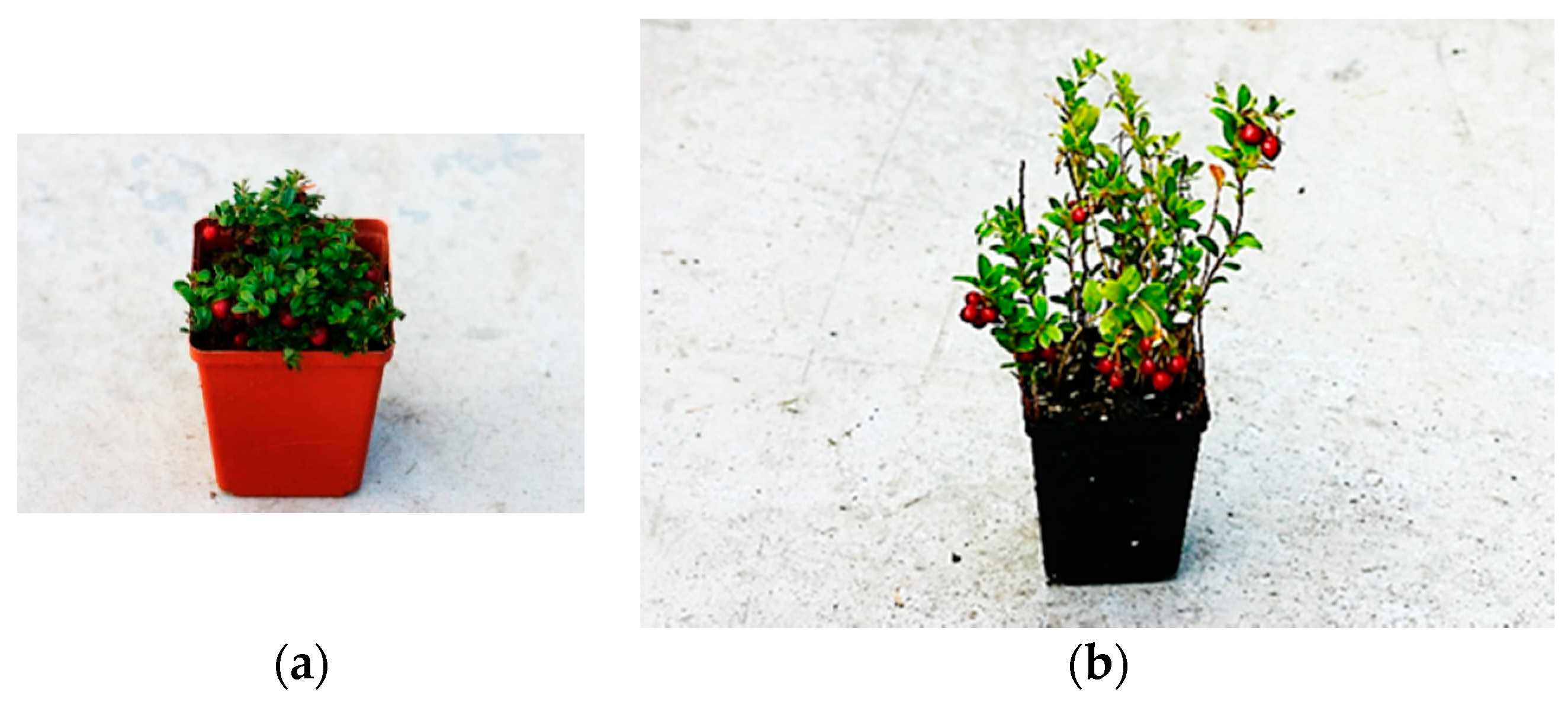
Figure 1.
Greenhouse grown lingonberry plants. (a) Vaccinium vitis-idaea ssp. minus; (b) V. vitis-idaea ssp. vitis-idaea.
Figure 1.
Greenhouse grown lingonberry plants. (a) Vaccinium vitis-idaea ssp. minus; (b) V. vitis-idaea ssp. vitis-idaea.

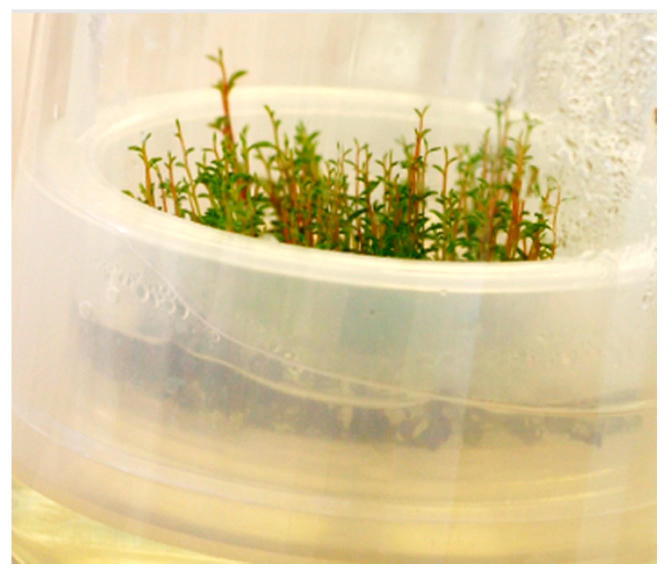
Figure 2.
Shoot proliferation in lingonberry.

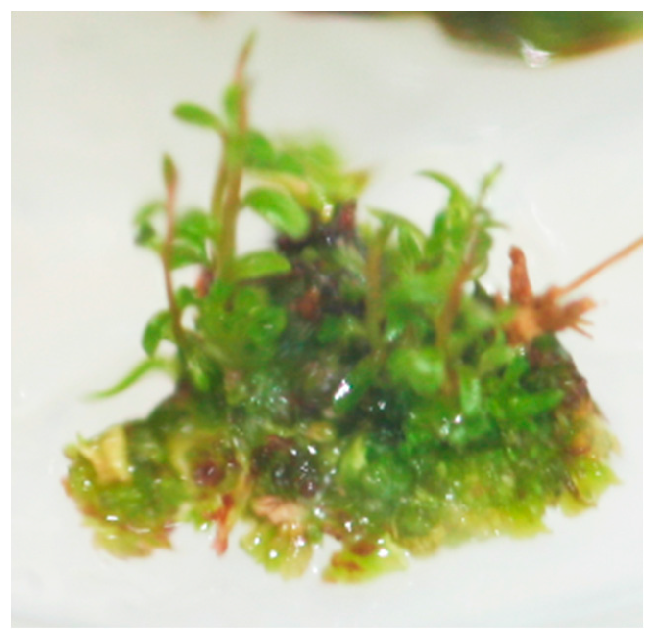
Figure 3.
In vitro shoot regeneration in lingonberry.

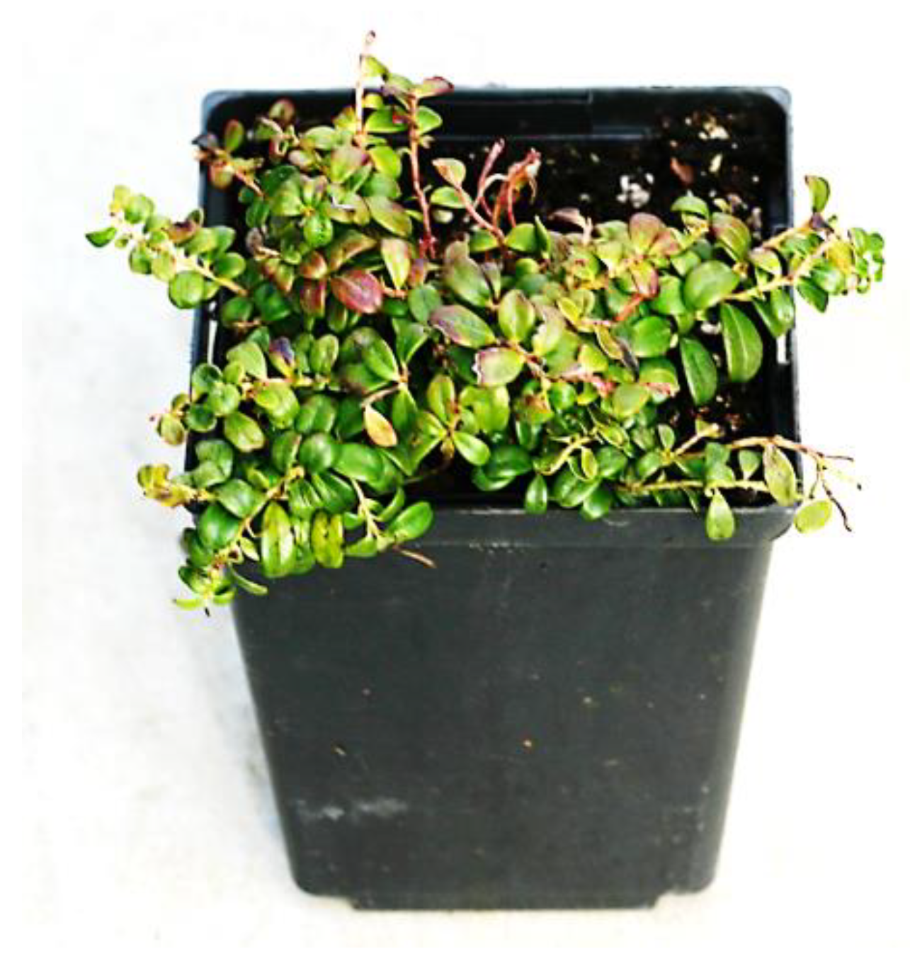
Figure 4.
Greenhouse-grown lingonberry tissue culture plant.

Figure 5.
In vitro culture of lingonberry in a recipient for automated temporary immersion (RITA) bioreactor.
Figure 5.
In vitro culture of lingonberry in a recipient for automated temporary immersion (RITA) bioreactor.

Figure 6.
In vitro culture of lingonberry in a Growtek vessel.


Table 1.
Differences between the two subspecies of Vaccinium vitis-idaea.
| Character | V. vitis-idaea Ssp. Minus | V. vitis-idaea Ssp. vitis-idaea |
|---|---|---|
| Variety [42] | North American | European |
| Race [43] | Small arctic | Large low land |
| Distribution [44] | Iceland, Greenland, North America, northern Asia, Scandinavia | Europe, Asia |
| Plant height | Generally up to 20 cm | Exceeds 30 cm |
| Leaf size [45] (Welsh 1974) | Length: 1.0 cm; width: 0.5 cm | Length: 2.5 cm; width: 1.0 cm |
| Crop per year [3] | One crop | Two crops |
Table 2.
Examples of axillary shoot proliferation (ASP) and adventitious shoot regeneration (ASR) in Vacinium vitis-idaea ssp. vitis-idaea and V. vitis-idaea ssp. minus.
Table 2.
Examples of axillary shoot proliferation (ASP) and adventitious shoot regeneration (ASR) in Vacinium vitis-idaea ssp. vitis-idaea and V. vitis-idaea ssp. minus.
| Subspecies | Genotype | Method | Explant | Medium 1 | PGR Used 2 | Findings | Reference |
|---|---|---|---|---|---|---|---|
| Ssp. vitis-idaea | Regal, Splendor, Erntedank | ASP | Shoot tips, nodes | Modified MS, BM-B, C, D | 2Ip (12.3 μM), zeatin (5.7 μM) | Modified MS medium was better than WPM for shoot multiplication. | [47] |
| Ssp. minus | Two wild clones | ASP | Shoot tips, nodes | Modified MS | 2iP (12.3 μM) | Nodal explants produced 4 to 6 healthy axillary shoots. | [47] |
| Ssp. vitis-idaea | Wild population | ASP | Seeds, nodes | Modi-fied MS | 2iP (9.8–78.4 μM) | 2iP at 24.6 µM was best for shoot initiation. | [51] |
| Ssp. vitis-idaea | Regal, Splendor, Erntedank, ECL1 (wild clone) | ASR | Leaves | BM-A | Zeatin, TDZ, 2iP | Zeatin (20 to 30 µM) was more efficient than TDZ or 2iP. | [53] |
| Ssp. vitis-idaea; ssp. minus | Splendor, Erntesegen; wild clone | ASR, ASP | Hypo-cotyls | BM-D | TDZ (5–10 μM, ASR); zeatin (1–2 μM, ASP) | TDZ) induced shoot regeneration; zeatin promoted shoot elongation. | [54] |
| Ssp. vitis-idaea | Red Pearl; Koralle | ASR | Leaves | AN | TDZ (2.2 mg L−1), zeatin (2.19 mg L−1) | Both zeatin and TDZ were effective. | [55] |
| Ssp. vitis-idaea; ssp. minus | EL1 (ssp. vitis-idaea wild cline); NL1 (ssp. minus wild clone) | ASP | Shoot tip | BM-D | TDZ (0.1 to 1 µM), Zeatin (1 µM) | TDZ supported shoot proliferation but inhibited shoot elongation. | [52] |
| Ssp. vitis-idaea | Regal; Erntedank | ASR | Leaves | BM-D | Zeatin (5 μM) | Adventitious shoots regenerated directly or from the leaf callus. | [34] |
| Ssp. vitis-idaea | Red Pearl | ASR | Leaves | WPM | TDZ (1–20 µM), zeatin (5–20 µM) | Zeatin was superior to TDZ. | [56] |
| Ssp. vitis-idaea | Koralle; Linnea; Run Bielawskie | ASP | Shoot tips | WPM; AN; half-MS | Zeatin (0.5–2 mg L−1) | Highest shoot multiplication rate was for Runo Bielawskie on WPM medium with 2 mg L−1 of zeatin. WPM was the better than AN and half MS. | [57] |
| Ssp. minus | Three wild clones | ASR | Leaves | BM-D | Zeatin (9.1 µM), TDZ (1.8 µM) | Larger callus formation with zeatin; zeatin was better for regeneration percentage and shoot vigor. | [58] |
| Ssp. minus | Three wild clones | ASP | Nodes | BM-D (semi-solid and liquid) | Zeatin (9.1 µM), and TDZ (1.8 µM) | Zeatin was more effective for shoot elongation; shoot proliferation was affected by interactions among propagation methods, clones and PGR types. | [58] |
1 MS = Murashige and Skoog medium; BM = Basal medium; AN = Anderson’s medium; WPM = Woody Plant Medium. 2 PGR = plant growth regulator; 2ip = N6-[2-isopentenyl]adenine; TDZ = thidiazuron.
© 2020 by the authors. Licensee MDPI, Basel, Switzerland. This article is an open access article distributed under the terms and conditions of the Creative Commons Attribution (CC BY) license (http://creativecommons.org/licenses/by/4.0/).
Share and Cite
MDPI and ACS Style
Debnath, S.C.; Arigundam, U. In Vitro Propagation Strategies of Medicinally Important Berry Crop, Lingonberry (Vaccinium vitis-idaea L.). Agronomy 2020, 10, 744. https://0-doi-org.brum.beds.ac.uk/10.3390/agronomy10050744
AMA Style
Debnath SC, Arigundam U. In Vitro Propagation Strategies of Medicinally Important Berry Crop, Lingonberry (Vaccinium vitis-idaea L.). Agronomy. 2020; 10(5):744. https://0-doi-org.brum.beds.ac.uk/10.3390/agronomy10050744
Chicago/Turabian StyleDebnath, Samir C., and Usha Arigundam. 2020. "In Vitro Propagation Strategies of Medicinally Important Berry Crop, Lingonberry (Vaccinium vitis-idaea L.)" Agronomy 10, no. 5: 744. https://0-doi-org.brum.beds.ac.uk/10.3390/agronomy10050744
Note that from the first issue of 2016, this journal uses article numbers instead of page numbers. See further details here.