Article

Improved Conservation of Coffee (Coffea arabica L.) Germplasm via Micropropagation and Cryopreservation

by
Yanelis Castilla Valdés
1,2,
Mukund R. Shukla
1,
María Esther González Vega
2 and
Praveen K. Saxena
1,*
1
Gosling Research Institute for Plant Preservation, Department of Plant Agriculture, University of Guelph, Guelph, ON N1G 2W1, Canada
2
Department of Genetics and Plant Breeding, National Institute of Agricultural Sciences (INCA), Carretera a Tapaste, km 3 1/2, San José de las Lajas 32700, Cuba
*
Author to whom correspondence should be addressed.
Submission received: 24 August 2021 / Revised: 11 September 2021 / Accepted: 13 September 2021 / Published: 16 September 2021

Abstract

:
Coffee (Coffea spp.) is an important tropical agricultural crop that has significant economic and social importance in the world. The ex situ conservation of plant genetic resources through seeds is not feasible due to the sensitivity of coffee seed to desiccation and low temperatures. The cryopreservation of zygotic embryos may allow for an efficient and long-term storage of coffee germplasm. This study describes the cryopreservation methods for conserving zygotic embryos of Coffea arabica L. for the long-term conservation of currently available germplasm. Zygotic embryos were successfully cryopreserved in liquid nitrogen at −196 °C under controlled environmental conditions with either droplet-vitrification or encapsulation–vitrification protocols without dehydration. Zygotic embryos had the highest regrowth (100%) following droplet-vitrification cryopreservation using the Plant Vitrification Solution 3 (PVS3) for 40 min at 23 °C. In the case of encapsulation–vitrification using PVS3 for 40 min at 23 °C, the embryo regeneration response was 78%. Plantlets were recovered following shoot multiplication using a temporary immersion system (TIS) and in vitro rooting. The prolific rooting of shoots was observed after 4 weeks of culture in the liquid medium with plugs made of the inert substrate Oasis® in vitro Express (IVE) compared to the semi-solid medium. The successful cryopreservation of coffee zygotic embryos using droplet vitrification and encapsulation–vitrification followed by micropropagation in temporary immersion culture system has not been reported earlier and together these technologies are anticipated to further facilitate the initiatives for the conservation and distribution of coffee germplasm.

1. Introduction

Coffee (Coffea spp.) plays an important economic role worldwide, as it represents a major source of foreign income in about 80 coffee-producing countries [1]. Global coffee production reached 163.7 million 60 kg bags as of 2019/2020 (https://0-www-statista-com.brum.beds.ac.uk/statistics/263311/worldwide-production-of-coffee/ (accessed on 10 September 2021). The majority of the world’s coffee, around 80%, is produced by Brazil, Colombia, and Costa Rica [2,3]. In Cuba, the coffee crop constitutes a priority in the agricultural sector because of its status as a preferred beverage of consumption and the high economic significance of the crop for export revenue [4].
The traditional varieties of C. arabica in Latin America are mostly propagated by seed, and the vegetative propagation is less frequently used [5]. These varieties typically have a very narrow genetic base, since they are derived from genealogical selections based on very few individuals [2]. C. arabica is predominantly self-pollinated and thus, the progenies arising from the seeds are very uniform [6]. Therefore, seeds are considered a good starting material in the in vitro establishment and propagation of this species [7]. However, the seed viability of C. arabica decreases rapidly after 4–6 months at ambient temperatures [8], and the loss of germination during storage is one of the major issues for the propagation and conservation of coffee genetic resources [9]. The coffee seeds are cold-sensitive, and desiccation does not improve their longevity [10]. However, zygotic embryos culture is an effective way to recover the plant material from the seeds with low viability in storage [9].
Cryopreservation, the storage of living explants at cryogenic temperatures (below −140 °C), is a complementary alternative to ex situ germplasm collection, which enables plant genetic resources to be conserved long term, safely, and cost effectively, with minimal requirements of space and routine maintenance [11,12,13,14]. Moreover, cryopreservation eliminates the need of regular seed collection and renewal, thus reducing the risk of genetic erosion caused by pests, diseases, weather conditions, pollution, and genetic variations [15].
Several studies have been reported for coffee zygotic embryo cryopreservation with varying results. Abdelnour et al. reported a 41.6 to 95.8% survival rate for zygotic embryos after the desiccation cryopreservation of three Coffea genotypes following dehydration for 30 min [16]. Higher zygotic embryo germination (98%) was found after dehydration in silica gel for 1 h to achieve 23% moisture content followed by cryopreservation [17]. However, a very low germination (3.3%) of C. arabica zygotic embryos was reported by Martinez et al. [18]. The germination percentage in the range of 30–66% was observed in two separate studies of the cryopreservation of C. canephora and C. arabica zygotic embryos with the vitrification method, using Plant Vitrification Solution 2 (PVS2) and Plant Vitrification Solution 3 (PVS3) [19,20]. Similarly, de Freitas et al. reported a successful vitrification protocol for the zygotic embryo of C. arabica with higher germination percentage (90%) after dehydration with PVS2 for 100 min at 0 °C followed by liquid nitrogen (LN) treatment [21].
Vitrification has the advantage of allowing the explant freezing within a short period and requires a precise control of the duration of dehydration. Therefore, it may limit the treatment of a large number of samples [22,23,24,25]. However, it has been possible to overcome this issue in recent methods of plant cryopreservation using V-cryoplates [26].
Several stress-related factors are associated with the cryoinjury, which occurs during the treatment with liquid nitrogen due to the failure of tissues in tolerating the extreme conditions of dehydration and temperature [27]. The adverse effects of dehydration and oxidation may be minimized by incorporating antioxidants in cryopreservation protocols. Many antioxidants have been found to improve the recovery of cryopreserved plant tissues [22,28,29,30,31]. Antioxidant application is considered a potential protective measure for minimizing cell damage at low temperatures presumably by modulating the physiological and biological properties of the membranes and the cytoplasm [32].
The main objective of the present study was to evaluate the droplet-vitrification and encapsulation–vitrification methods for the cryopreservation of zygotic embryos of C. arabica L. We examined the effect of PVS3 exposure time and temperature during droplet-vitrification protocol. In addition, we evaluated the effect of the addition of melatonin during the preculture and regrowth stages in the encapsulation–vitrification-based cryopreservation method. An additional objective of this study was to develop an efficient micropropagation technique for shoot multiplication and in vitro rooting using the rocker-based temporary immersion system for the propagation of plants from cryopreserved tissues.

2. Materials and Methods

2.1. Plant Material

Coffea arabica L. mature fruits were obtained from the greenhouse of the Edmund C. Bovey building, Department of Plant Agriculture, University of Guelph, Guelph, ON, Canada. The fruits were placed in closed paper bags for five days to remove the pericarp, mesocarp, and endocarp. The seed moisture content (MC) was measured according to the formula MC = (Fw − Dw/Fw)×100, where Fw is the fresh weight of 30 seeds and Dw is the dry weight of 30 seeds after desiccation at 100 °C for 48 h in an oven. The seeds were surface sterilized with 2.4% (v/v) commercial bleach (Clorox; 5.4% sodium hypochlorite) for 90 min followed by three rinses in sterile-distilled water, each rinse lasting 3 min. Sterilized seeds were transferred to a 125 mL Erlenmeyer flask containing 0.5% boric acid solution and kept on a rotary shaker (150 rpm, Thermo Fisher Scientific, Ottawa, Canada) under standard growth room condition for 72 h. All cultures were kept in the culture room at 23 ± 2 °C under a 16/8 h photoperiod provided by cool white florescent lamps (Osram Sylvania, Mississauga, ON, Canada) at a light intensity of 40 µmolm−2 s−1. Seeds were rinsed with sterile distilled water and embryo extracted from the seeds in sterile conditions (Figure 1A). Extracted embryos were placed in Petri dishes (60 mm × 15 mm) containing 10 mL of semi-solid germination medium consisting of MS (Murashige and Skoog) [33] basal salts with vitamins (Phytotechnology Laboratory, Shawnee Mission, KS), 3% (w/v) sucrose, and 2.2 gL−1 Phytagel (Sigma-Aldrich, Oakville, Canada). The pH was adjusted to 5.75 using 0.1 N NaOH or HCL prior to autoclaving at 121 °C and 118 kPa for 20 min.

2.2. Encapsulation–Vitrification

All cryoprotectant solutions used in different steps of the protocols were prepared in liquid MS medium (salts and vitamins, pH 5.7) and sterilized by vacuum filtration through 0.2 μm filters (Nalgene™, ThermoFisher Scientific®, Burlington, ON, Canada). All the chemicals used in the present study were purchased from Sigma-Aldrich®, Oakville, ON, Canada, unless otherwise mentioned.
Embryos cultured on the germination medium for 24 h were used for encapsulation according to the protocol described by Dereuddre et al. [34]. Briefly, the zygotic embryos were suspended in 3% sodium alginate (Sigma-Aldrich, Oakville, ON, Canada) in liquid MS medium without calcium salt at pH 5.7. Alginate droplets, each containing one explant, were transferred into MS medium with 100 mM CaCl2 solution (pH 5.7) using a wide mouth, 10 mL serological pipette (CorningTM, Thermo Fisher Scientific, Ottawa, ON, Canada) and left for 20 min at room temperature with gentle agitation to form beads that were ca. 4 mm in diameter (Figure 1C) [35]. Beads were desiccated over a filter paper for 30 min in sterile conditions using the laminar flow cabinet. Then, the beads were precultured in 0.3 M sucrose solution for 18 h and transferred into loading solution (2.0 M glycerol + 0.4 M sucrose) for 25 min at room temperature [36]. Afterwards, the beads were suspended in the Plant Vitrification Solution 3 (PVS3) for 45 min at room temperature on a rotary shaker (90 rpm). The PVS3 consisted of 50% glycerol + 50% sucrose (w/v) in basal culture medium [36]. For cryopreservation, the beads were placed in 2 mL cryovials filled with 1 mL fresh PVS3 solution. Cryovials were sealed and quickly immersed in liquid nitrogen (LN) for 1 h and rewarmed in a 40 °C water bath for 2 min. Rewarmed beads were transferred to unloading solution containing liquid MS medium with 1.2 M sucrose for 20 min and placed on germination medium for regrowth. Embryos in beads not exposed to LN were also directly immersed in unloading solution, placed on germination medium, and considered as the control. After cryopreservation, the embryos were placed on semi-solid basal medium as described earlier and kept in the dark at 23 ± 2 °C (Figure 1B). After seven days of cryopreservation, the embryos were sub-cultured on the MS basal medium supplemented with 2.2 µM 6-Benzylaminopurine (BA), 0.3 µM Naphthaleneacetic acid (NAA), 1.0 µM Gibberellic acid (GA3), 3% (w/v) sucrose, and 2.2 gL−1 Phytagel. Culture growth room conditions were same as described above. Further treatments for the optimization of cryopreservation techniques included (i) changing the preculture step by the desiccation of the beads for 16 h in capped Petri dishes and (ii) skipping the 30 min desiccation over filter paper and changing the preculture step with the desiccation of the beads for 15 h in the Petri dish. All materials were cultured in darkness for the initial five days after rewarming and then were moved to low light (5 ± 2 µmolm−2 s−1). The germination percentage was evaluated after seven days of inoculation on the medium. Germinated embryos became turgid and turned green. The embryos that continued growth and development for 3 weeks were included in the determination of “regrowth”.
An experiment was conducted with 10 µM melatonin in the preculture and postcryoculture medium to evaluate the advantage of its antioxidant properties in improving the survival and regrowth of the zygotic embryo. Embryos were cultured on the MS basal medium containing 10 µM melatonin (pH 5.7) for 24 h before encapsulation and then washed with MS liquid basal medium containing 10 µM melatonin for 3 min. The embryos encapsulated as described above were desiccated for 30 and 60 min before suspending them in the liquid preculture medium containing 0.5 M sucrose for 18 h. All embryos from the cryopreservation and non-cryopreservation treatments were cultured on the medium with and without 10 µM melatonin following the same protocol of encapsulation cryopreservation as described above. Embryos cultured without melatonin were considered as the control for preculture and postcryoculture treatments with melatonin.
After cryopreservation, the embryos were placed on semi-solid basal medium as described earlier and kept in the dark at 23 ± 2 °C (Figure 1B). After seven days of cryopreservation, the embryos were sub-cultured on MS basal medium supplemented with 2.2 µM 6-benzylaminopurine (BA), 0.3 µM naphthaleneacetic acid (NAA), 1.0 µM gibberellic acid (GA3), 3% (w/v) sucrose, and 2.2 gL−1 Phytagel and kept in the culture growth room as described above.

2.3. Droplet Vitrification

Extracted embryos were placed on the germination medium for 24 h and kept in the dark at 23 ± 2 °C. Before conducting cryopreservation experiments, different steps of the protocol (preculture, osmoprotection, and dehydration) were evaluated without freezing the samples in LN. The standard conditions followed were according to Nishizawa et al. [36] and included preculture (0.3 M sucrose for 18 h), osmoprotection (2.0 M glycerol + 0.4 M sucrose at 23 ± 2 °C for 20 min), and vitrification (PVS3 with 50% glycerol +50% sucrose (w/v) at 23 ± 2 °C for 40 min). The zygotic embryos were kept on a rotary shaker (90 rpm, ThermoFisher Scientific) between the steps involved in cryopreservation. After the vitrification step, the embryos were placed in individual drops of PVS3 solution on the aluminum foil strips (6 × 30 mm; Western Plastics, Mississauga, ON, Canada) (Figure 1D). Foil strips with embryos were quickly immersed in LN for at least 1 h. Based on the preliminary results of the experiment with PVS3 at 40 min, the embryos were exposed in PVS3 for 30, 40, and 50 min at 4 °C and 23 °C to optimize the exposure time.
For rewarming, the foil strips were plunged into preheated (40 ± 1 °C) 1.2 M sucrose solution for 30 s, after which an equivalent volume of unloading solution was added at room temperature. Foils were removed with forceps, and the zygotic embryos were kept in the unloading solution for 20 min on a rotary shaker (90 rpm). Zygotic embryos not exposed to LN were also directly immersed in unloading solution and considered as the control. Then, the zygotic embryos were dried on a sterile filter paper, plated on the germination medium, and kept in the dark for 72 h in the culture room at 23 ± 2 °C. Later, the embryo cultures were transferred to standard growth conditions as described above. The zygotic embryos that became turgid and green after seven days were counted as germinated and those that continued growth and development for 3 weeks were counted as the embryos showing regrowth.

2.4. Shoot Multiplication, In Vitro Rooting, and Acclimatization

The rocker-based Temporary Immersion System (TIS; Culture shift; VRE System, Grassie, ON, Canada) was used for shoot multiplication and rooting experiments. This rocker is programmable for controlling the immersion interval as well as the rocking speed with a separate timer for the controlling light duration [37].
Seedlings developed from the cryopreserved zygotic embryos were multiplied to increase the number of shoots by sub-culturing 10–15 shoot explants per bioreactor vessel containing 50 mL of the liquid medium [38]. The multiplication medium consisted of MS basal medium supplemented with 2.2 µM BA, 0.3 µM NAA, 1.0 µM GA3, and 3% (w/v) sucrose with a pH of 5.7. Liquid cultures were placed on the TIS with an immersion interval of 25 S and a 7 S transfer time in between the two immersion periods. Plantlets were grown under cool white fluorescent lamps (EiKO®, Barrie, ON, Canada) with an intensity of 40–60 µmolm−2 s−1 at 22 °C with a 16/8 h photoperiod. After 5 weeks, shoots were sub-cultured into fresh bioreactor vessels each containing 50 mL of multiplication medium. Explant cultures were sub-cultured at 5-week intervals until enough shoots were produced for the in vitro rooting experiments.
The in vitro rooting efficiency of micropropagated coffee shoots was evaluated using liquid rooting medium with Oasis® IVE foam (Oasis® Grower Solutions, Kent, OH, USA) using the TIS and the semi-solid medium (2.2 gL−1 Phytagel) as the control. The rooting medium (pH 5.7) consisted of 3% sucrose, 1/2 strength MS salts with vitamins, and 20 µM IBA. The bioreactor vessels with Oasis® IVE foam contained 250 mL of the rooting medium, and the same volume was used for the semi-solid medium.
Rooted plantlets were transplanted into cell trays (4 × 10) filled with Sunshine professional growing medium #4 (Sun Gro Horticulture Canada Ltd., Brantford, ON, Canada). Plantlets were initially acclimatized in the mist bed at 24 °C for 16 h light and 20 °C for 8 h darkness, with 85% humidity for two weeks and later transferred to the greenhouse at 24 °C for 16 h light and 20 °C for 8 h darkness with light intensity at 110 µmolm−2 s−1. The survival rate (surviving plants/total rooted plants) was recorded after 4 weeks of growth in the greenhouse.

2.5. Differential Scanning Calorimetry (DSC) Analysis

The differential scanning calorimetry technique was used to identify and quantify the events of crystallization and vitreous transition in cryopreservation. The phase transitions in zygotic embryos of coffee were investigated using the differential scanning calorimeter (DSC, Mettler Toledo, Leicester, UK), calibrated with zinc (422.81 °C, 115.57 Jg−1) and indium (156.85 °C, 29.03 Jg−1) standards. Zygotic embryos were sealed in 40 µL aluminum pans. Samples were placed in the DSC at 22 °C and cooled at a rate of −10 °C min−1 to −80 °C, at which point the sample was held isothermally for 5 min before rewarming to 25 °C at a rate of 10 °C min−1. The samples tested included (i) control zygotic embryos without any treatment, (ii) zygotic embryos precultured in 0.3 M sucrose for 18 h, (iii) zygotic embryos precultured in 0.3 M sucrose for 18 h and exposed to LS for 20 min, and (iv) zygotic embryos precultured in 0.3 M sucrose for 18 h and exposed to LS for 20 min followed by PVS3 for 40 min. Each treatment was replicated three times. Thermograms were analyzed using STARe thermal analysis software (Mettler Toledo, Leicester, UK).

2.6. Statistical Analysis

Experiments were performed in three independent replications with each replication consisting of 10 to 30 zygotic embryos. Results were expressed as a proportion of germinated zygotic embryos/preserved zygotic embryos, and data were analyzed by proportion comparison analysis, using the program SPSS V. 21.0 (IBM Canada Ltd., Markham, ON, Canada). Figures present proportion values with Standard Error of the proportion. Values followed by different letters are significantly different at p < 0.05.

3. Results

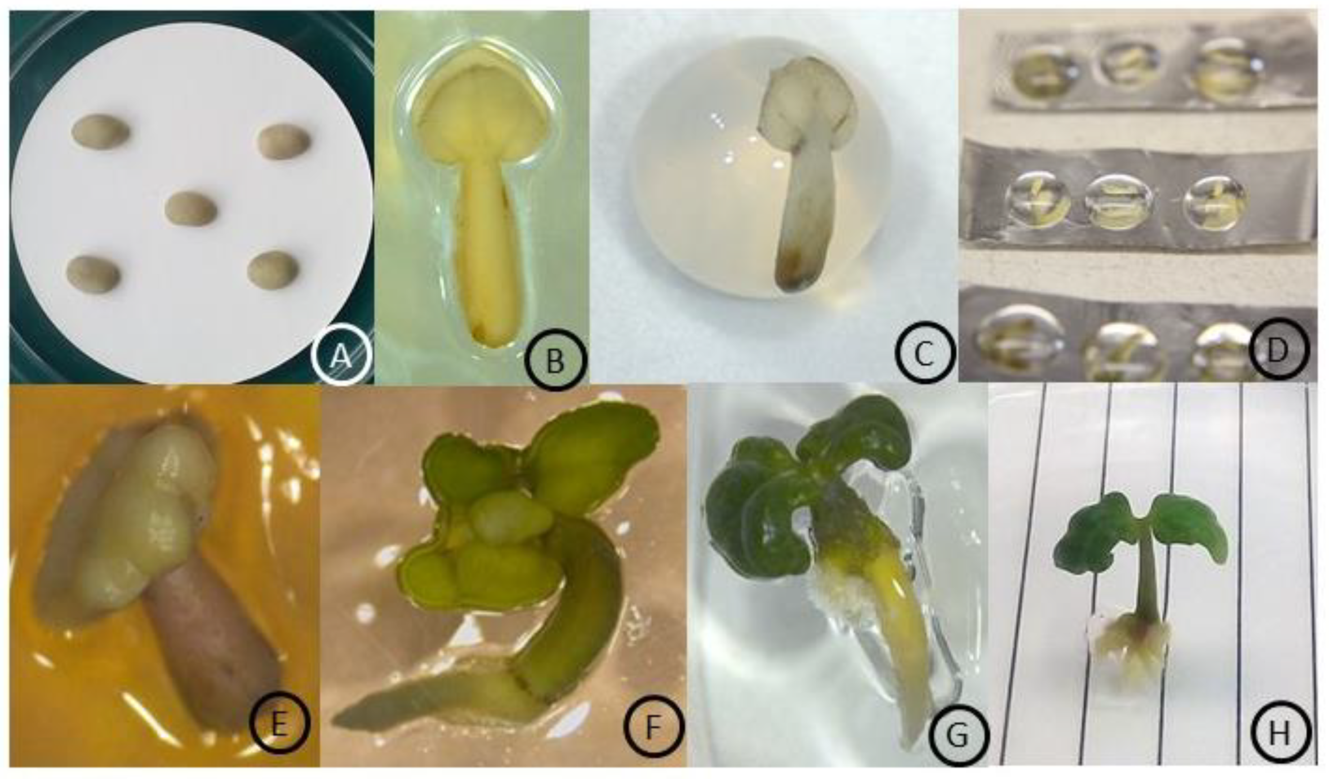
3.1. Zygotic Embryo Culture

The viability of coffee zygotic embryos was tested through the germination of intact seeds and extracted zygotic embryos. Surface-sterilized coffee seeds and the extracted zygotic embryos showed no contamination on the culture medium, indicating the efficiency of the sterilization method. Both the seeds with 40% moisture content and the extracted zygotic embryos showed 100% germination (Figure 1A,B).

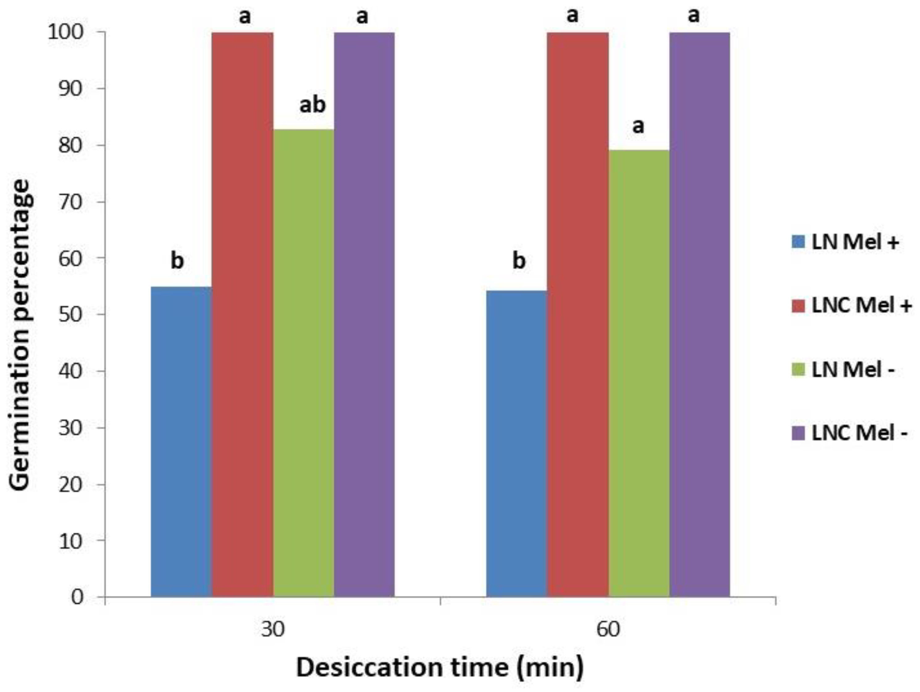
3.2. Cryopreservation Using Encapsulation–Vitrification

The germination percentage of the embryo after cryopreservation (+LN) by encapsulation–vitrification following 30 and 60 min desiccation over a filter paper in the laminar flow cabinet was 83 and 79%, respectively (Figure 2). However, the difference for germination percentage over the desiccation period was not significant. The addition of melatonin in the preculture and postcryoculture steps did not increase the germination percentage of cryopreserved zygotic embryos in both desiccation exposure times (Figure 2). The addition of melatonin in the preculture and postcryoculture was not found to be beneficial in promoting germination percentage (54–55%) after cryopreservation with both desiccation exposure times. However, healthier growth and root development were observed in the seedlings recovered following the cryopreservation treatment of zygotic embryos cultured on the melatonin-supplemented medium compared to the control treatment (Figure 1G–H). The values for the regrowth of zygotic embryos after 3 weeks of cryopreservation treatment corresponded with the values of germination, since zygotic embryos that survived continued further development.

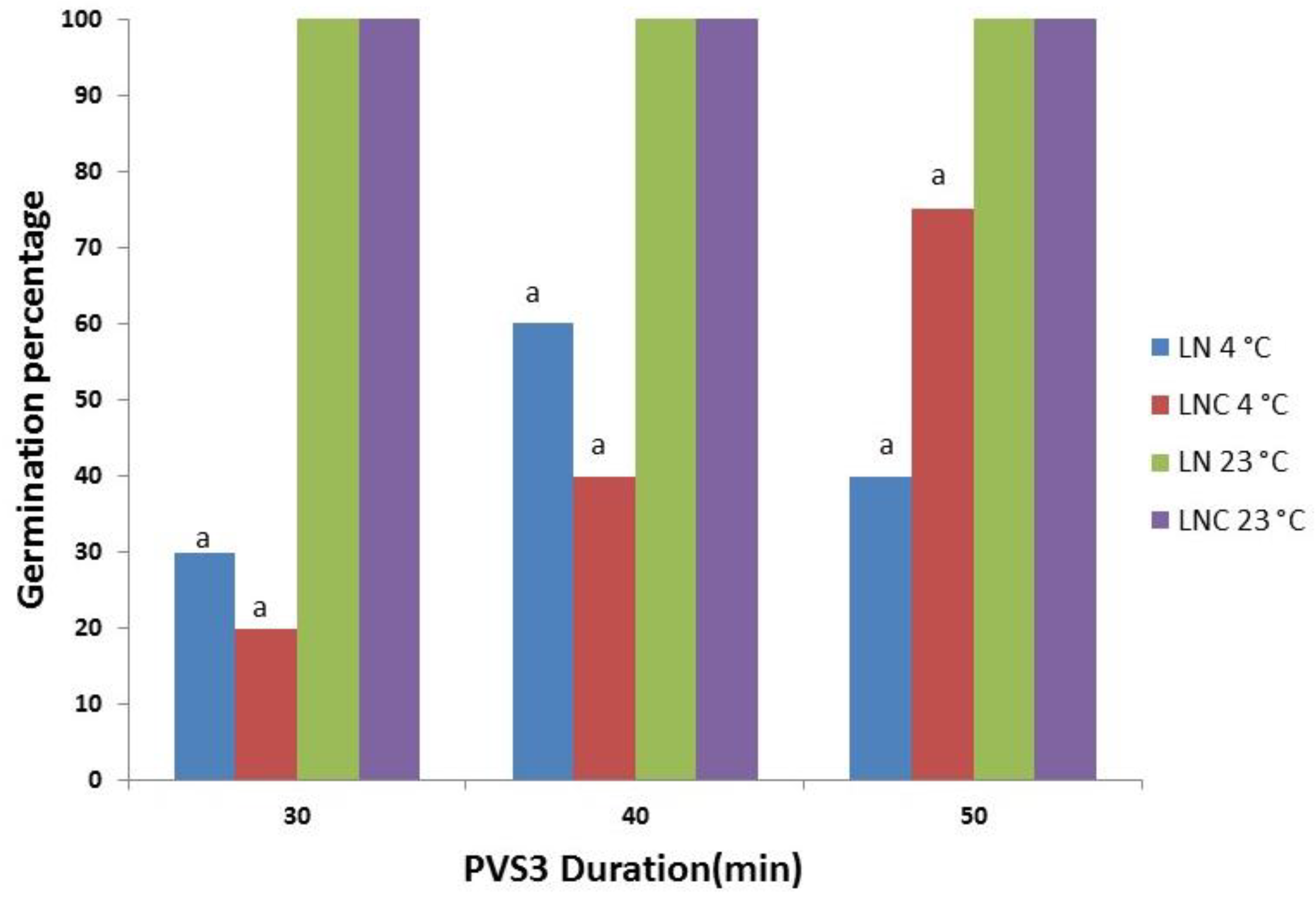
3.3. Cryopreservation Using Droplet Vitrification

Embryos not stored in LN that were treated with all of the solutions used showed 100% germination, regardless of their immersion time in PVS3 at room temperature. Droplet-vitrification cryopreserved embryos showed 100% germination after dehydration with PVS3 for 30, 40, and 50 min at 23 °C followed by LN exposure (Figure 1E). However, the differences with PVS3 exposure time (30, 40, and 50 min) were found to be non-significant for the germination of zygotic embryos after cryopreservation at 23 °C (Figure 3). The highest percentage of embryo germination (60%) was observed after dehydration in PVS3 for 40 min at 4 °C followed by LN treatment compared to 30 as well as 50 min exposure treatments at 4 °C. For controls (-LN), the highest germination percentage (75%) was observed when embryos were exposed to PVS3 for 50 min at 4 °C (Figure 3). The results for different PVS3 exposure at 4 °C during the cryopreservation of zygotic embryos did not show higher germination using the droplet-vitrification method as well as in control treatment compared to the treatment at 23 °C. All the zygotic embryos that survived after 3 weeks of cryopreservation treatment continued further development and were able to produce healthy seedlings (Figure 1F).

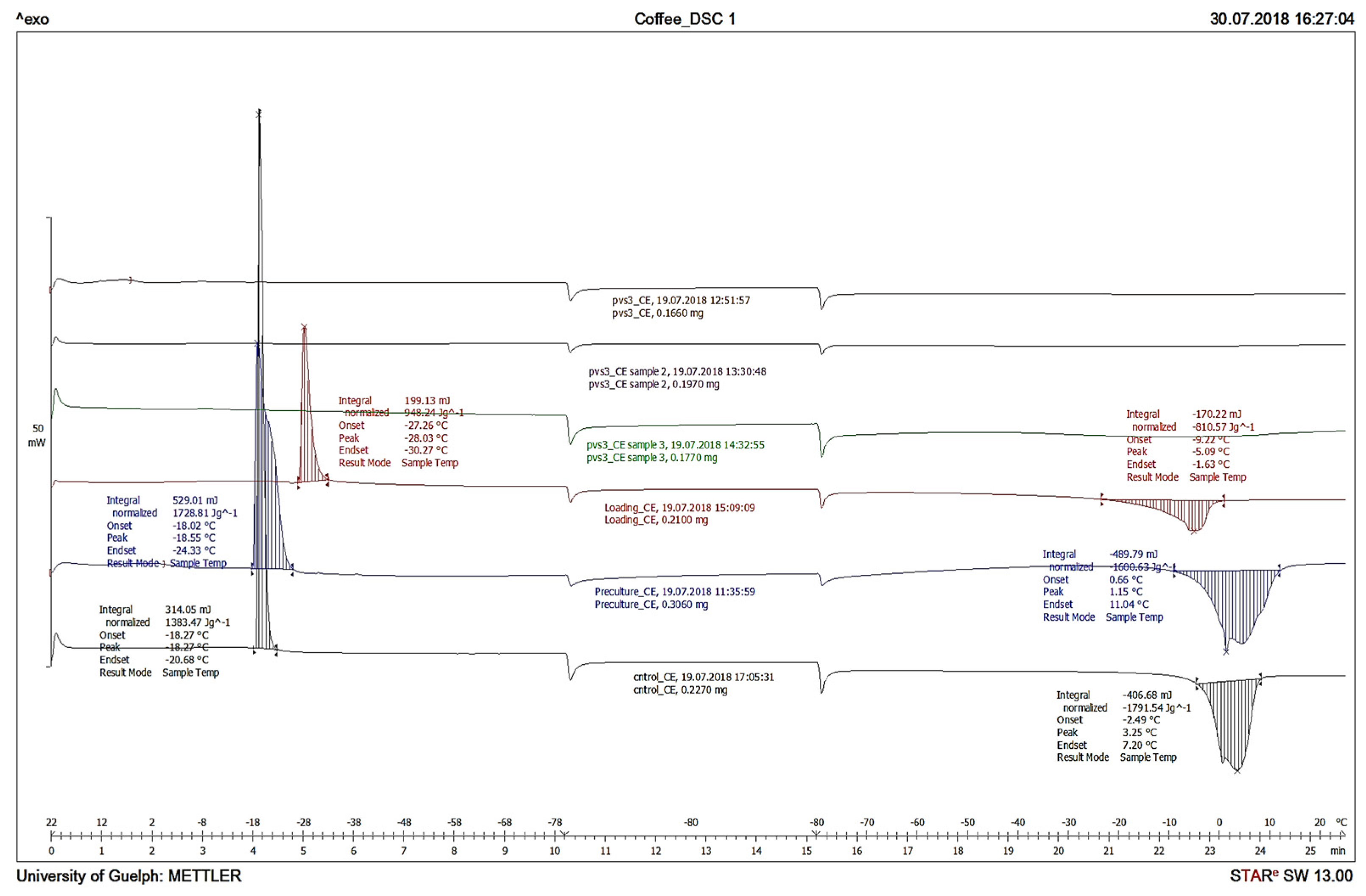
3.4. Differential Scanning Calorimetry

The DSC analysis confirmed the absence of ice crystallization in the coffee zygotic embryos during cryopreservation by the droplet-vitrification method, which showed 100% germination (Figure 4). Thermograms were interpreted for critical cryopreservation parameters such as crystallization and melting during onset temperatures. Exothermal and endothermal peaks were observed in zygotic embryos without any treatment (control), with preculture treatment and with loading treatments indicating ice crystallization during cooling and ice melting during rewarming. However, no crystallization or melting events were detected when zygotic embryos were treated with PVS3 for 40 min (Figure 4). These studies showed that the PVS3 exposure for 40 min was optimal for zygotic embryo cryopreservation.

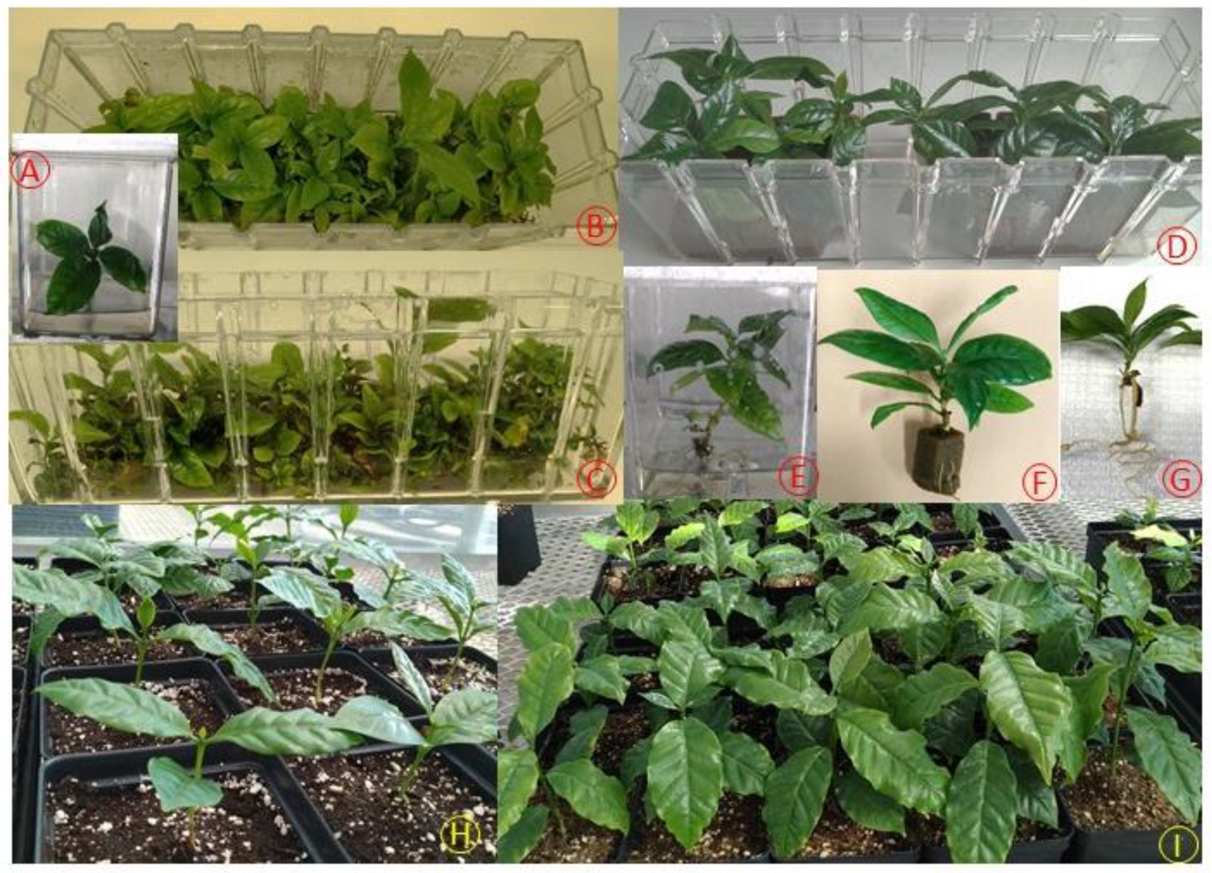
3.5. Shoot Multiplication and Acclimatization

In liquid TIS, shoots multiplied with good shoot growth compared to semi-solid medium using the same multiplication medium (Figure 5A–C). The multiplication rate was significantly higher in liquid TIS (4.2 shoot per explant) compared to semi-solid medium (1.3 shoots per explant). Shoots grown in TIS were significantly longer than those in semi-solid medium. Leaves of shoots from liquid medium were larger and darker green in color compared to those from shoots grown on the semi-solid medium. The results of root initiation experiments showed that 90% of the shoots developed roots in the liquid medium with half-strength MS basal salt mixture containing 20 μM IBA (Figure 5D–G). All shoots showed two to three primary roots with an average root length of 5.5 cm after 6 weeks (Figure 5G). Shoots cultured in the semi-solid medium did not develop roots until 4 weeks, and only 15% of the shoots were observed to have roots in the sixth week. After two weeks of acclimatization in the mist bed, rooted plantlets transplanted in the greenhouse showed a survival rate of 95% after one month of growth in ex vitro conditions (Figure 5H,I).

4. Discussion

The objective of this study was to develop an integrated strategy for the long-term conservation and large-scale propagation of coffee germplasm. Our approach consisted of two major components: cryopreservation using zygotic embryos and the development of an efficient micropropagation system. Generally, seed-based propagation in coffee is most common due to the occurrence of little variability among the seedling populations [39]. In this study, we optimized the droplet-vitrification as well as encapsulation–vitrification methods for the cryopreservation of zygotic embryos as a strategy for the long-term conservation of coffee germplasm. A micropropagation method was also developed for the multiplication of shoots from cryopreserved coffee embryos using a rocker-based temporary immersion system.
Our results demonstrated that 100% embryo germination was possible after exposure to LN following the optimization of the basic steps of the cryopreservation procedure. Cryopreservation through the encapsulation–vitrification method was found to be highly effective with 30 min desiccation over a filter paper in the laminar flow cabinet followed by 40 min in PVS3 before immersion in LN. More than 80% embryo germination was observed after cryopreservation using the encapsulation–vitrification method. The germination percentage of the zygotic embryos with an extended period of desiccation (60 min) did not show further improvement. A lower zygotic embryo germination percentage with a longer desiccation period suggests that the injury due to desiccation stress may be the reason for low germination. Additional reasons may include a detrimental effect of the alginate matrix that can suppress the growth of regenerating plantlets out of the beads or insufficient dehydration of alginate-coated shoot tips [40]. The desiccation injury has been shown to reduce the survival of somatic embryos of C. canephora after cryopreservation using the encapsulation–dehydration method [41]. The encapsulation–dehydration of shoot apices of two species, C. racemosa and C. sessiliflora, also showed low survival rates of 27% and 37%, respectively, after cryopreservation following a progressive increase in the sucrose concentration from 0.5 to 1 M [42]. Encapsulation–dehydration and encapsulation–vitrification methods have been applied successfully for the long-term storage of Hladnikia pastinacifolia Rchb. germplasm through cryopreservation although encapsulation–vitrification was faster and resulted in normal regrowth [43].
Tropical species are usually low-temperature sensitive and present one of the most difficult challenges in developing preconditioning treatments to stimulate physiological responses that can enhance the tolerance to dehydration and cryogenic procedures [29,44,45]. In most cases, the critical storage temperature is much higher, between 8 and 15 °C, for tropical cold-sensitive species, in comparison with that of the species from temperate climates [31]. During the in vitro preservation of C. arabica shoots, the temperature of 12 °C was lethal for the explants, and 100% of explants survived when they were exposed to higher temperature of 12 to 27 °C [46]. Low-temperature (0 °C) exposure during vitrification with PVS2 showed 73% viability and 93% germination of zygotic embryos of C. arabica when the zygotic embryos were exposed for 100 to 250 min [21]. In the present study, the zygotic embryos were exposed to PVS3 solution for 30 to 50 min at 4 °C to reduce the cryoprotectant solution toxicity; the results showed no beneficial effect of reducing temperature of PVS3 treatment for the cryopreserved as well as non-cryopreserved zygotic embryo germination compared to room temperature (23 °C). The permeability of the cryoprotectant (PVS) increases at room temperature, but it can increase the risk of tissue damage compared to that at low temperatures [47,48]. This indicated that the PVS3 solution was not toxic to the zygotic embryo during the process of cryopreservation, and the low germination percentage might be due to the cold sensitivity of the embryos, which were not cold hardened before the cryopreservation process. However, cold hardening can be replaced by preculturing the explants on a sucrose-enriched medium to provide sufficient cryoprotection during the cryopreservation process and stimulate 100% germination of zygotic embryo after cryopreservation [25,49]. Generally, the explants of some species may be particularly sensitive to the toxic effects of penetrating cryoprotectant compounds such as ethylene glycol and dimethyl sulfoxide (DMSO), which are essential components of PVS2 [49,50]. For such sensitive materials, modification of the traditional PVS2 solution, treatment conditions, or the use of two-component cryoprotectant solutions composed of only glycerol and sucrose, such as in PVS3, may be beneficial [40,49]. Cryopreservation through the droplet-vitrification method had 100% germination of zygotic embryos when exposed to PVS3 solution for 30 min at 23 °C followed by LN exposure. A low zygotic embryo germination rate of 66% was reported for C. arabica and C. canephora after vitrification cryopreservation, using PVS3 for 60 min at room temperature [20]. This was the first attempt to use the droplet-vitrification method for the cryopreservation of Coffea spp., which showed 100% survival of zygotic embryo compared to other vitrification-based methods of cryopreservation [20,21].
Melatonin in the preculture or postcryoculture medium has been found to be very effective in enhancing the survival as well as regrowth of explants after cryopreservation in many species, including American Elm, St. John Wort, and tobacco [50,51]. However, the melatonin effect is a dose-dependent response that is mainly based on explant type and plant species [52,53]. The melatonin content in green fruit (6.8 µg/g DW) and roasted grains (9.6 µg/g DW) of coffee is quite high, and it is possible that zygotic embryos may have an endogenous level of melatonin [54]. In this case, the exogenous application of melatonin may have little or an inhibitory effect on germination of zygotic embryos (Figure 2). Lupinus albus showed a 2.5-fold increase in melatonin levels in a cold environment as compared plants growing at 24 °C, indicating that the biosynthesis of melatonin is upregulated in stress situations [54]. Cryopreservation is a process associated with extreme cold stress [29], which can increase the endogenous levels of melatonin [55].
We developed a nodal explant-based micropropagation method, which allowed the clonal propagation of shoots developed from cryopreserved embryos. Our earlier study demonstrated the beneficial effects of a rocker-based temporary immersion bioreactor system (TIS) on shoot multiplication as well as the root formation of hazelnut cultivars [37,56]. The results of this study using rocker-based TIS also showed that the liquid medium can significantly improve the effectiveness of in vitro shoot multiplication and overall plantlet growth (Figure 5B,C). Similarly, the TIS was found to be suitable for embryo development and shoot multiplication in coffee species [57,58,59,60]. The micropropagation method developed in this study relied on the use of BA as a cytokinin along with GA3. BA is the most commonly used cytokinin for shoot development and multiplication in a range of species [37,56]. The combination of cytokinin and gibberellic acid is known to improve shoot growth in the in vitro grown cultures, which is likely due to increased cell elongation [61,62].
The successful in vitro rooting was observed in TIS with the use of the inert substrate Oasis® IVE foam. The foam placed in the liquid medium can hold shoot explants in an upright position during the rooting stage [63]. The main advantage of Oasis® IVE foam is that it allows the shoot explants to easily interact with the liquid rooting medium while also acting as a well-aerated substrate to enhance oxygen availability and cellular respiration of the roots [64]. The use of inert substrates (e.g., foam, rockwool, vermiculite, and perlite) to enhance aeration has been reported to be a very effective technique for improving in vitro rooting in many woody species compared with semi-solid medium-based rooting [65]. Therefore, the use of liquid medium in the TIS along with Oasis® IVE foam may have improved the interaction of rooting medium and oxygen with the shoot explants resulting in enhanced root development and healthy plantlet formation. Rooting can be successfully achieved with the exogenous application of auxin. In our study, a maximum of 90% of the shoots formed roots in the presence of IBA at 20 µM, while significantly lower rooting was observed on semi-solid medium. Similarly, Oasis® IVE foam in combination with 1/2 strength DKW, 10 µM IBA also improved rooting in hazelnut shoot explants [37]. The coffee plantlets resulting from TIS showed higher survival rates during acclimatization and greenhouse transplant.

5. Conclusions

In conclusion, this study further highlights the utility and wide applicability of the droplet-vitrification and encapsulation–vitrification methods for the cryopreservation of coffee zygotic embryo as well as the efficacy of a rocker-based TIS for micropropagation. The optimized cryopreservation protocol is simple and can be widely adopted even with limited resources. The availability of these methods of cryopreservation strengthens the ex situ preservation of coffee genetic resources and can also complement the in situ conservation of germplasm. The droplet-vitrification method for cryopreservation with high survival of the explants may be useful for the conservation of other related plant species. Our results demonstrate that efficient shoot multiplication and rooting can be obtained using rocker-based TIS compared to traditional micropropagation methods and may facilitate mass production and the field transplanting of coffee germplasms. Collectively, these accomplishments may have significant potential for improved coffee germplasm conservation and the production of high-quality plants at commercial scale.

Author Contributions

M.R.S. and P.K.S. conceptualized and designed in vitro propagation and cryopreservation studies. Y.C.V. conducted the experiments and collected the growth data. Y.C.V. collected and analyzed the data. M.R.S., M.E.G.V. and P.K.S. organized and supervised the study. Y.C.V. prepared the manuscript. All authors have read and agreed to the published version of the manuscript.

Funding

Award of research grants from the Gosling Foundation, Guelph, ON, Canada and Emerging Leaders in the Americas Program (ELAP).

Institutional Review Board Statement

Not applicable.

Informed Consent Statement

Not applicable.

Data Availability Statement

No new data were created or analyzed in this study. Data sharing is not applicable to this article.

Acknowledgments

We thank Shuping Li for technical help in micropropagation experiments.

Conflicts of Interest

The authors declare no conflict of interest.

References

  1. Bramel, P.; Krishnan, S.; Horna, D.; Lainoff, B.; Montagnon, C. Global Conservation Strategy for Coffee Genetic Resources; World Coffee Research-Crop Trust: Munich, Germany, 2017; pp. 5–7. Available online: https://worldcofferesearch.org/documents/42/Coffee_Strategy_Low_Res.pdf (accessed on 10 September 2021).
  2. Aguilar, M.E.; Ortiz, J.L.; Mesen, F.; Jimenez, L.D.; Altmann, F. Cafe Arabica Coffea arabica L. In Step Wise Protocols for Somatic Embryogenesis of Important Woody Plants; Forestry Sciences; Jain, S.M., Gupta, P., Eds.; Springer International Publishing AG: Basel, Switzerland, 2018; Volume 85, pp. 39–62. [Google Scholar] [CrossRef]
  3. ICO. Coffee Production Worldwide from 2003/04 to 2019/20 (in Million 60 Kilogram Bags)*. Statista, Statista Inc. Available online: https://0-www-statista-com.brum.beds.ac.uk/statistics/263311/worldwide-production-of-coffee/ (accessed on 31 January 2021).
  4. González, M.E.; Rosales, P.R.; Castilla, Y.; Lacerra, J.A.; Ferrer, M. Efecto del Bioenraiz® como estimulante de la germinación y el desarrollo de plántulas de cafeto (Coffea arabica L.). Cultiv. Trop. 2015, 36, 73–79. [Google Scholar]
  5. Wintgens, J.N.; Zamarripa, C.A. Coffee propagation. In Coffee: Growing, Processing, Sustainable Production: A Guidebook for Growers, Processors, Traders and Researchers; WILEY-VCH Verlag GMBH & Co. KGaA: Weinheim, Germany, 2004; pp. 91–140. [Google Scholar]
  6. Roubik, D. The value of bees to the coffee harvest. Nature 2002, 417, 708. [Google Scholar] [CrossRef]
  7. Ebrahim, N.; Shibli, R.; Makhadmeh, I.; Shatnawi, M.; Abu-Ein, A. In vitro propagation and in vivo acclimatization of three coffee cultivars (Coffea arabica L.) from Yemen. World Appl. Sci. J. 2007, 2, 142–150. [Google Scholar]
  8. Eira, M.T.S.; Amaral da Silva, E.A.; de Castro, R.D.; Dussert, S.; Walters, C.; Bewley, J.D.; Hilhorst, H.W.M. Coffee seed physiology. Braz. J. Plant Physiol. 2006, 18, 149–163. [Google Scholar] [CrossRef] [Green Version]
  9. Isaac, E.; Hernández, C.; González, J.L.; González, M.E.; Fung, Y.; Ferrer, A.E. Effect of electromagnetic field on mineral content and chemical group during in vitro establishment and multiplication phases of coffee seedlings. Rev. Cubana Quím. 2016, 28, 692–702. [Google Scholar]
  10. Dussert, S.; Chabrillange, N.; Engelmann, F.; Anthony, F.; Hamon, S. Cryopreservation of coffee (Coffea arabica L.) seeds: Importance of the precooling temperature. CryoLetters 1997, 18, 269–276. [Google Scholar]
  11. Benson, E.E. Cryopreservation of phytodiversity: A critical appraisal of theory & practice. Crit. Rev. Plant Sci. 2008, 27, 141–219. [Google Scholar] [CrossRef]
  12. Engelmann, F. Use of biotechnologies for the conservation of plant biodiversity. Cell. Dev. Biol. Plant 2011, 47, 5–16. [Google Scholar] [CrossRef]
  13. Wang, M.R.; Chen, L.; Teixeira da Silva, J.A.; Volk, G.M.; Wang, Q.C. Cryobiotechnology of apple (Malus spp.): Development, progress and future prospects. Plant Cell Rep. 2018, 37, 689–709. [Google Scholar] [CrossRef]
  14. Bettoni, J.C.; Bonnart, R.; Volk, G.M. Challenges in implementing plant shoot tip cryopreservation technologies. Plant Cell Tissue Organ Cult. 2021, 144, 21–34. [Google Scholar] [CrossRef]
  15. Popova, E.; Shukla, M.; Hoon, H.; Saxena, P.K. Plant Cryopreservation for Biotechnology and Breeding. In Advances in Plant Breeding Strategies: Breeding, Biotechnology and Molecular Tools; Al-Khayri, J.M., Jain, M., Johnson, D.V., Eds.; Springer International Publishing: Basel, Switzerland, 2015; pp. 63–93. [Google Scholar] [CrossRef]
  16. Abdelnour-Esquivel, A.; Villalobos, V.; Engelmann, F. Cryopreservation of zygotic embryos of Coffea spp. CryoLetters 1992, 13, 297–302. [Google Scholar]
  17. Pinto, M.S.; Paiva, R.; Correa, D.P.; Almeida, P.A.; Theresan, R.; Coutinho, L. Cryopreservation of coffee zygotic embryos: Dehydration and osmotic rehydration. Ciência e Agrotecnologia 2016, 40, 380–389. [Google Scholar] [CrossRef] [Green Version]
  18. Martínez, M.; González-Arnao, M.T.; Urra, C.; Rojas, R.; Cuba, M.; García, D. Estudios preliminares para la crioconservación de embriones cigóticos de Coffea arabica variedad 9722. Cultiv. Trop. 1996, 17, 79–81. [Google Scholar]
  19. Castilla, Y.; Dussert, S.; González, M.E.; Engelmann, F. Crioconservación de embriones cigóticos de Coffea canephora mediante vitrificación: Primeras aproximaciones. In Proceedings of the 8th International Congress on Plant Biotechnology and Agriculture Bioveg 2011, Ciego de Ávila, Cuba, 2–6 May 2011; ISBN 978-959-16-1286-1. [Google Scholar]
  20. Castilla, Y.; Martínez-Montero, M.E.; González, M.E.; Engelmann, F. Nuevos resultados en la crioconservación de dos especies de cafeto (Coffea spp) mediante el método de vitrificación. In Proceedings of the 9th International Congress on Plant Biotechnology and Agriculture Bioveg 2013, Ciego de Ávila, Cuba, 7–10 May 2013; ISBN 978-959-16-2045-3. [Google Scholar]
  21. De Freitas, R.T.; Paiva, R.; Silva, T.; Pedrosa, D.; Valquiria, M.; de Souza, A.C.; Veiga, S.D. Cryopreservation of Coffea arabica L. zygotic embryos by vitrification. Not. Bot. Horti Agrobot. 2016, 44, 445–451. [Google Scholar] [CrossRef] [Green Version]
  22. Matsumoto, T.; Niino, T. The development of plant vitrification solution 2 and recent PVS2-based vitrification protocols. Acta Hortic. 2014, 1039, 21–28. [Google Scholar] [CrossRef]
  23. Matsumoto, T. Cryopreservation of plant genetic resources: Conventional and new methods. Rev. Agric. Sci. 2017, 5, 13–20. [Google Scholar] [CrossRef] [Green Version]
  24. Carra, A.; Carimi, F.; Bettoni, J.C.; Pathirana, R. Progress and Challenges in the Application of Synthetic Seed Technology for Ex Situ Germplasm Conservation in Grapevine (Vitis spp.). In Synthetic Seeds; Faisal, A., Alatar, A.A., Eds.; Springer Nature: Basel, Switzerland, 2019; pp. 439–467. [Google Scholar] [CrossRef]
  25. Sakai, A.; Engelmann, F. Vitrification, encapsulation-vitrification and droplet-vitrification: A review. CryoLetters 2007, 28, 151–172. [Google Scholar] [PubMed]
  26. Martínez, M.E.; Harding, K. Cryobionomics: Evaluating the concept in plant cryopreservation. In Plant Omics: The Omics of Plant Science; Barh, D., Khan, M.S., Davies, E., Eds.; Springer: New Delhi, India, 2015; pp. 655–678. [Google Scholar] [CrossRef]
  27. Lazo-Javelera, M.F.; Tiznado-Hernández, M.E.; Vargas-Arispuro, I.; Valenzuela-Soto, E.; Rocha-Granados, M.C.; Mar-tínez-Montero, M.E.; Rivera-Domínguez, M. Data on antioxidant activity in grapevine (Vitis vinifera L.) following cryopreservtion by vitrification. Data Brief 2015, 5, 549–555. [Google Scholar] [CrossRef] [Green Version]
  28. Reed, B.M. Antioxidants and cryopreservation, the new normal? Acta Hort. 2014, 1039, 41–48. [Google Scholar] [CrossRef]
  29. Bettoni, J.; Kretzschmar, A.A.; Bonnart, R.; Shepherd, A.; Volk, G.M. Cryopreservation of 12 Vitis species using apical shoot tips derived from plants grown in vitro. HortScience 2019, 54, 976–981. [Google Scholar] [CrossRef] [Green Version]
  30. Uchendu, E.E.; Muminova, M.; Gupta, S.; Reed, B.M. Antioxidant and antistress compounds improve regrowth of cryopreserved Rubus shoot tips. Cell. Dev. Biol. Plant 2010, 46, 386–393. [Google Scholar] [CrossRef]
  31. Uchendu, E.E.; Keller, E.R.J. Melatonin-loaded alginate beads improve cryopreservation of yam (Dioscorea alata and D. cayenensis). Cryoletters 2016, 37, 77–87. [Google Scholar]
  32. Ha, J.S.; Lee, J.W.; Seo, S.H.; Ahn, C.Y.; Rho, G.J.; Lee, H.G.; Oh, H.M. Optimized cryopreservation of Ettlia sp. using short cold acclimation and controlled freezing procedure. J. Appl. Phycol. 2019, 31, 2277–2287. [Google Scholar] [CrossRef]
  33. Murashige, T.; Skoog, F. A revised medium for rapid growth and bioassays with tobacco tissue cultures. Physiol. Plant. 1962, 15, 473–497. [Google Scholar] [CrossRef]
  34. Dereuddre, J.; Scottez, C.; Arnaud, Y.; Duron, M. Resistance of alginate-coated axillary shoot tips of pear tree (Pyrus communis L. cv. Beurre Hardy) in vitro plantlets to dehydration and subsequent freezing in liquid nitrogen. C. R. Acad. Sci. Paris 1990, 310, 317–322. [Google Scholar]
  35. Fabre, J.; Dereuddre, J. Encapsulation-dehydration: A new approach to cryopreservation of Solanum shoot tips. CryoLetters 1990, 11, 413–426. [Google Scholar]
  36. Nishizawa, S.; Sakai, A.; Amano, Y.; Matsuzawa, T. Cryopreservation of asparagus (Asparagus officinalis L.) embryogenic suspension cells and subsequent plant regeneration by vitrification. Plant Sci. 1993, 91, 67–73. [Google Scholar] [CrossRef]
  37. Nicholson, J.; Shukla, M.R.; Saxena, P.K. In vitro rooting of Hybrid hazelnuts (Corylus avellana X Corylus americana) in a temporary immersion system. Botany 2020, 98, 343–352. [Google Scholar] [CrossRef]
  38. Shukla, M.R.; Singh, A.S.; Piunno, K.; Saxena, P.K.; Jones, A.M.P. Application of 3D printing to prototype and develop novel plant tissue culture systems. Plant Methods 2017, 13, 6. [Google Scholar] [CrossRef] [Green Version]
  39. Tran, H.T.M.; Lee, L.S.; Furtado, A.; Smyth, H.; Henry, R.J. Advances in genomics for the improvement of quality in coffee. Sci. Food Agric. 2016, 96, 3300–3312. [Google Scholar] [CrossRef] [Green Version]
  40. Rathwell, R.; Popova, E.; Shukla, M.R.; Saxena, P.K. Development of cryopreservation methods for cherry birch (Betula lenta L.), an endangered tree species in Canada. Can. J. For. Res. 2016, 46, 1284–1292. [Google Scholar] [CrossRef] [Green Version]
  41. Hatanaka, T.; Yasuda, T.; Yamaguchi, T.; Sakai, A. Direct regrowth of encapsulated somatic embryos of coffee (Coffea canephora) after cooling in liquid nitrogen. CryoLetters 1994, 15, 47–52. [Google Scholar]
  42. Mari, S.; Engelmann, F.; Chabrillange, N.; Huet, C.; Michaux-Ferriere, N. Histo-cytological studies of apices of coffee (Coffea racemosa and C. sessiliflora) in vitro plantlets during their cryopreservation using the encapsulation-dehydration technique. CryoLetters 1995, 16, 289–298. [Google Scholar]
  43. Takagi, H. Recent developments in cryopreservation of shoot apices of tropical species. In Cryopreservation of Tropical Plant Germplasm: Current Research Progress and Applications; Engelmann, F., Takagi, H., Eds.; Japan International Research Center for Agricultural Sciences (JIRCAS): Tsukuba, Japan; International Plant Genetic Resources Institute (IPGRI): Rome, Italy, 2000; pp. 178–193. [Google Scholar]
  44. Ciringer, T.; Martín, C.; Sajna, N.; Kaligaric, N.; Ambrozik-Dolinsek, J. Cryopreservation of an endangered Hladniki apastinacifolia Rchb. by shoot-tip encapsulation-dehydration and encapsulation-vitrification. Cell. Dev. Biol. Plant 2018, 54, 565–575. [Google Scholar] [CrossRef]
  45. Malik, S.K.; Chaudhury, R. Cryopreservation techniques for conservation of tropical horticultural species using various explants. In Conservation and Utilization of Horticultural Genetic Resources; Rajasekharan, P., Rao, V., Eds.; Springer Nature: New York, NY, USA, 2019; pp. 579–594. [Google Scholar] [CrossRef]
  46. Jouve, L.; Engelmann, F.; Noirot, M.; Charrier, A. Evaluation of biochemical markers (sugar, proline, malonedialdehyde and ethylene) for cold sensitivity in microcuttings of two coffee species. Plant Sci. 1993, 91, 109–116. [Google Scholar] [CrossRef]
  47. Volk, G.M.; Harris, J.L.; Rotindo, K.E. Survival of mint shoot tips after exposure to cryoprotectant solution components. Cryobiology 2006, 52, 305–308. [Google Scholar] [CrossRef]
  48. Bettoni, J.C.; Souza, J.A.; Volk, G.M.; Dalla Costa, M.; da Silva, F.N.; Kretzschmar, A.A. Eradication of latent viruses from apple cultivar ’Monalisa’ shoot tips using droplet-vitrification cryotherapy. Sci. Hortic. 2019, 250, 12–18. [Google Scholar] [CrossRef]
  49. Kim, H.H.; Popova, E.; Shin, D.J.; Yi, J.Y.; Kim, C.H.; Lee, J.S.; Yoon, M.K.; Engelmann, F. Cryobanking of Korean Allium Germplasm Collections: Results from a 10 Year Experience. Cryoletters 2012, 33, 45–57. [Google Scholar]
  50. Uchendu, E.E.; Shukla, M.R.; Reed, B.M.; Saxena, P.K. Melatonin enhances the recovery of cryopreserved shoot tips of American elm (Ulmus americana L.). J. Pineal Res. 2013, 55, 435–442. [Google Scholar] [CrossRef]
  51. Uchendu, E.E.; Shukla, M.R.; Reed, B.M.; Saxena, P.K. An efficient method for cryopreservation of St Jhon’s Wort and tobacco: Role of melatonin. ISHS Acta Hortic. 2013, 1039, 233–241. [Google Scholar]
  52. Erland, L.A.E.; Saxena, P.K. Melatonin in plant morphogenesis. Cell. Dev. Biol. Plant 2018, 54, 3–24. [Google Scholar] [CrossRef]
  53. Campos, C.M.; Ávila, R.G.; Dázio de Souza, K.R.; Azevedo, L.M.; Alves, J.D. Melatonin reduces oxidative stress and promotes drought tolerance in young Coffea arabica L. plants. Agric. Water Manag. 2019, 211, 37–47. [Google Scholar] [CrossRef]
  54. Ramakrishna, A.; Giridhar, P.; Sankar, K.U.; Ravishankar, G.A. Melatonin and serotonin profiles in beans of Coffea species. J. Pineal Res. 2012, 52, 470–476. [Google Scholar] [CrossRef]
  55. Arnao, M.B.; Hernández, J. Growth conditions determine different melatonin levels in Lupinus albus L. J. Pineal Res. 2013, 55, 149–155. [Google Scholar] [CrossRef] [PubMed]
  56. Latawa, J.; Shukla, M.R.; Saxena, P.K. An efficient temporary immersion system for micropropagation of hybrid hazelnut. Botany 2016, 94, 1–8. [Google Scholar] [CrossRef]
  57. Etienne-Barry, D.; Bertrand, B.; Vasquez, N.; Etienne, H. Direct sowing of Coffea arabica somatic embryos mass-produced in a bioreactor and regeneration of plants. Plant Cell Rep. 1999, 19, 111–117. [Google Scholar] [CrossRef]
  58. Albarrán, J.; Bertrand, B.; Lartaud, M.; Etienne, H. Cycle characteristics in a temporary immersion bioreactor affect regeneration, morphology, water and mineral status of coffee (Coffea arabica) somatic embryos. Plant Cell Tissue Organ Cult. 2005, 81, 27–36. [Google Scholar] [CrossRef]
  59. Etienne, H.; Dechamp, E.; Barry-Etienne, D.; Bertrand, B. Bioreactors in coffee micropropagation. Braz. J. Plant Physiol. 2006, 18, 45–54. [Google Scholar] [CrossRef]
  60. Gatica-Arias, A.M.; Arrieta-Espinoza, G.; Espinoza Esquivel, A.M. Plant regeneration via indirect somatic embryogenesis and optimization of genetic transformation in Coffea arabica L. cvs. Caturra and Catuaí. Electron. J. Biotechnol. 2008, 11, 101–112. [Google Scholar]
  61. Shukla, M.R.; Jones, A.M.P.; Sullivan, J.A.; Liu, C.; Gosling, S.; Saxena, P.K. In vitro conservation of American elm (Ulmus americana): Potential role of auxin metabolism in sustained plant proliferation. Can. J. For. Res. 2012, 42, 686–697. [Google Scholar] [CrossRef]
  62. Shukla, M.R.; Kibler, A.; Turi, C.E.; Erland, L.A.E.; Sullivan, J.A.; Murch, S.J.; Saxena, P.K. Selection and Micropropagation of an Elite Melatonin Rich Tulsi (Ocimum sanctum L.) Germplasm Line. Agronomy 2021, 11, 207–231. [Google Scholar] [CrossRef]
  63. Naylor-Adelberg, J.A.; Adelberg, J.W.; Rapaka, V.K. A rooting matrix and tissue culture vessel system yielded larger, faster growing Echeveria plants. Acta Hortic. 2016, 1113, 53–58. [Google Scholar] [CrossRef]
  64. Adelberg, J. Bioreactors and “smart vessels” for large-scale propagation. Acta Hortic. 2017, 1187, 123–138. [Google Scholar] [CrossRef]
  65. Economou, A.S. From microcutting rooting to microplant establishment: Key points to consider for maximum success in woody plants. Acta Hortic. 2013, 988, 43–56. [Google Scholar] [CrossRef]
Figure 1. In vitro germination of coffee (C. arabica L.) seed and embryos. Sterilized seed (A) were used to extract the zygotic embryo (B) for encapsulation–vitrification (C) and droplet-vitrification cryopreservation (D) methods. Zygotic embryo survived after cryopreservation using droplet-vitrification protocol (E) and shoots developed after 3 weeks (F). Zygotic embryos cryopreserved by encapsulation–vitrification survived and continued to grow after 3 weeks of cryopreservation in both melatonin-rich medium (G) and control medium (H).
Agronomy 11 01861 g001
Figure 2. Germination of coffee (C. arabica L.) zygotic embryos after encapsulation–vitrification with desiccation for 30 or 60 min over filter paper in the laminar flow cabinet, with preculture and postcryoculture on the medium containing melatonin (10 µM). Results are presented as percentages of germination of cryopreserved zygotic embryos (LN) and non-cryopreserved zygotic embryos (LNC). Data with different letters indicate significant difference at p < 0.05.
Agronomy 11 01861 g002
Figure 3. Germination of coffee (C. arabica L.) zygotic embryos after droplet vitrification with 30, 40, or 50 min in Plant Vitrification Solution 3 (PVS3) at 4 °C and 23 °C. Results are presented as the percentage of germination of cryopreserved zygotic embryos (LN) and non-cryopreserved zygotic embryos (LNC). Data with different letters indicate significant differences at p < 0.05.
Agronomy 11 01861 g003
Figure 4. Combined differential scanning calorimeter (DSC) thermograms of coffee (C. arabica L.) zygotic embryos for control (without any treatment), after treatment with preculture solution, after treatment with loading solution, and three samples after treatment with Plant Vitrification Solution 3 (PVS 3) for 40 min.
Agronomy 11 01861 g004
Figure 5. Shoot multiplication and plant development from cryopreserved zygotic embryos of coffee (C. arabica L.). Cryopreserved embryos were cultured on shoot development medium and 6-week-old shoots (A) were transferred to a liquid-based temporary immersion system. Shoot multiplication was observed after 8 weeks (top view (B) and side view (C)) in the bioreactor vessel. Single shoots were transferred to rooting medium using Oasis® IVE foam placed into the TIS (D) and on the semi-solid medium (E). Six-week-old rooted shoots (F,G) were transferred to the greenhouse for acclimatization and observed for plant growth after one (H) and three months (I).
Agronomy 11 01861 g005
Publisher’s Note: MDPI stays neutral with regard to jurisdictional claims in published maps and institutional affiliations.

