Article

Plants from Urban Parks as Valuable Cosmetic Ingredients: Green Extraction, Chemical Composition and Activity

1
Faculty of Pharmacy and Biochemistry, University of Zagreb, A. Kovačića 1, 10000 Zagreb, Croatia
2
Department of Chemistry, Faculty of Sciences, University of Girona, C/M. Aurèlia Campmany 69, 17003 Girona, Spain
3
Istituto Zooprofilattico Sperimentale della Lombardia e dell’Emilia Romagna “Bruno Ubertini”, Via Antonio Bianchi 7/9, 25124 Brescia, Italy
*
Author to whom correspondence should be addressed.
Submission received: 10 December 2021 / Revised: 4 January 2022 / Accepted: 8 January 2022 / Published: 15 January 2022

Abstract

:
The research on the possibilities of using biowaste from urban green areas is scarce. In this work, four plants, widely distributed in urban parks in Central Europe (Lotus corniculatus, Medicago lupulina, Knautia arvensis and Plantago major) were extracted using eco-friendly solvents based either on aqueous cyclodextrin solutions (hydroxypropyl-β-cyclodextrin or γ-cyclodextrin) or natural deep eutectic solvents based on glycerol, betaine and glucose. Metal content was determined using total reflection X-ray fluorescence (TXRF). The content of selected metabolites was determined using UV-VIS spectrophotometric methods and HPLC. Skin-related bioactivity was assessed using tyrosinase and elastase inhibition assays. The selected plants contained metals beneficial for skin health, such as zinc and calcium, while having a low content of toxic heavy metals. The extracts contained the bioactive phenolics such as quercetin, kaempferol, luteolin and apigenin. L. corniculatus was the most potent tyrosinase inhibitor, while K. arvensis showed the most pronounced elastase inhibitory activity. The employed solvents actively contributed to the observed bioactivity. The results indicate that the biowaste obtained from urban parks represents an ecologically acceptable alternative to conventional cultivation for the preparation of ecologically acceptable, high-value cosmetic products.

1. Introduction

Public green spaces, including urban parks, form a significant portion of modern cities. They contain relatively well-preserved vegetation and are an important source of biodiversity in urbanized zones. Being grassland spotted with occasional trees, they closely resemble savanna-type ecosystems [1]. Biowaste produced in urban parks is produced as a consequence of plants’ normal growth and is composed of large masses of organic matter [2]. Despite its large quantities, the resulting biomass is only occasionally used economically, mostly for compost manufacture or energy production in biogas stations or incinerators [3]. Even though utilizing biowaste to obtain products of high economic value can reduce production costs and contribute to the sustainability of the production [4], the research on possibilities of using biowaste from urban green areas in other industry areas is lacking. However, plant biowaste contains phenolic compounds that can be used for production of high-value products such as food supplements and cosmetics [2].
In recent years, interest in natural cosmetic formulations has increased due to their presumed efficacy and safety. The production, development and marketing of natural cosmetics is one of the fastest-growing industries [5]. An increasing number of cosmetic products contains ingredients derived from nature [6]. In such products, they may act as antioxidants [7], and anti-ageing or skin-whitening agents [6]. However, the natural origin of the product is just one of the preferences of the modern consumer. As a consequence of the ever-increasing concerns about the environmental impact, new products are being developed using green and sustainable technologies and materials [8], including environmentally friendly solvents for extraction of plant bioactive principles [9].
The ideal solvent for green extraction should have a low environmental impact, including being biodegradable, obtained from renewable resources and easy to recycle without any deleterious effect on the environment. In addition, it should be safe for human consumption. Deep eutectic solvents (DES) meet most of those demands [10]. Some of the constituents of DES are commonly used in cosmetics. Glycerol, a natural, viscous hygroscopic polyol, is one of the most widely used ingredients in skincare. It acts as a humectant and skin protectant, as well as a hair conditioning and oral-care agent [11]. Glycerol-containing DES may easily be incorporated into the final product, deeming DES extraction very desirable from the energy-saving point of view [12]. Other common DES components that may be included in the final cosmetic products include various sugars, such as glucose. Another common cosmetic ingredient is betaine, a quaternary ammonium salt of natural origin that may serve as a humectant and skin-conditioning agent in topical formulations [13].
Cyclodextrins (CDs) and their aqueous solutions are another example of eco-friendly solvents. CDs are cyclic oligosaccharides consisting of 6, 7 or 8-glucopyranose units. They are characterized by a hydrophilic exterior and hydrophobic cavity capable to encapsulate a wide range of phytochemicals by the inclusion complex formation [14,15]. They are one of the most prominent current examples of nanotechnology-based systems that enhance the performances of phytochemicals in skincare. Not only can they can improve the solubility and stability of phytochemicals in water, but they can also facilitate their permeation in deeper layers of the skin [16]. The use of CDs aqueous solutions as an extraction medium offers an additional advantage of dissolving both hydrophilic and hydrophobic compounds as the former would be dissolved in the aqueous phase, while the latter would be solubilised by the supramolecular cyclodextrin encapsulation [14].
Lotus corniculatus L., Medicago lupulina L. (Fabaceae), Knautia arvensis (L.) Coult (Caprifoliaceae) and Plantago major L. (Plantaginaceae) are examples of plants widely present in urban parks and discarded as biowaste after park mowing. Those plants are rich in bioactive compounds and are still used as a part of traditional medicine or diet. For example, L. corniculatus is used for myriad ailments including skeletomuscular problems, abdominal and stomach pain, and as a sedative and diuretic. It is rich in bioactive phenolic compounds including quercetin, kaempferol derivatives, as well as phytoestrogen genistein [17]. M. lupulina is a rich source of various bioactive and nutritional compounds. The adult plants and their microgreens are edible and contain significant amounts of primary metabolites [18], as well as phenolic compounds that were shown to be responsible for an excellent antioxidant, antiradical and reducing activity of the species [19]. Furthermore, P. major possesses many bioactive properties that are anti-inflammatory, anti-nociceptive, antidiabetic and antioxidant, but it is probably best known for its wound-healing properties [20]. K. arvensis is also rich in valuable phenolic secondary metabolites, mostly caffeic acid derivatives such as cryptochlorogenic and chlorogenic acid but also flavonoids such as isovitexin derivatives [21].
This work was aimed to optimize green extraction of bioactive phenols from the aerial parts of four plants widely inhabiting urban parks by using two types of green extraction solutions: natural eutectic solvents and cyclodextrins. The chemical composition and biological activity of the prepared extracts were determined using selected assays targeting the activities relevant to cosmetic applications.

2. Materials and Methods

2.1. Plant Material, Chemicals and Apparatus

M. lupulina (flowering aerial parts), L. corniculatus (flowering aerial parts), K. arvensis (flowering aerial parts) and P. major (leaves) were collected in the recreational area in the city of Zagreb (Lake Jarun, 45°48′ N 15°90′ E). The identity was confirmed by the authors. The enzymes, elastase from the porcine pancreas and mushroom tyrosinase, and the HPLC standards were purchased from Sigma-Aldrich (St. Louis, MO, USA). The purity of standards was ≥98.5%. A stock solution of Ga 1000 mg/L (in 5% nitric acid, TraceCERT®, a standard for ICP, Fluka) was used to prepare the internal Ga standard solution. Other reagents and chemicals were of analytical grade. Extraction was performed using Bandelin SONOREX® Digital 10 P DK 156 BP ultrasonic bath (Berlin, Germany). A microplate reader (FLUOstar Omega, BMG Labtech, Ortenberg, Germany) was used for UV-VIS spectroscopic measurements. Agilent 1200 series HPLC instrument equipped with an autosampler, DAD detector, and a Zorbax Eclipse XDB-C18 (5 μm, 25 mm × 4.6 mm) column (Agilent Technologies, Santa Clara, CA, USA) was used for determination of the selected secondary metabolites. Total reflection X-ray fluorescence (TXRF) analysis of plant material was performed using a commercial benchtop TXRF system equipped with Mo X-ray tube (S2 PICOFOX, Bruker AXS Microanalysis GmbH, Berlin, Germany) operating at 600 µA and 50 kV, a multilayer monochromator. Evaluation of the TXRF spectra and the calculation of the net peak areas of the analytes were performed using the commercial software linked to the Mo systems (Spectra Plus 7.5.3.0; Bruker AXS Microanalysis GmbH, Berlin, Germany).

2.2. Determination of Metal Content in Plant Material

For the TXRF analysis, the dried plant material was ground into a fine powder using an agate mortar and sifted through a sieve with a diameter of 63 μm. Then, 20 mg of sample was suspended in 1 mL of de-ionized water containing 10 μg of Ga as an internal standard (IS) and the mixture was homogenized by ultrasonication (5 min sonication in an ultrasonic bath). Finally, a drop (10 μL) of the prepared mixture was deposited onto the carrier made of quartz glass and dried using an infrared lamp for the TXRF analysis. Characteristic radiation emitted by the elements present in the sample was detected using a silicon drift detector with a resolution of less than 149 eV at Mn-Kα. Samples were irradiated for 600 s live time. Quantification was performed by internal standardization using the following expression described in Equation (1):
C i = ( N i C i s S i s N i s S i ) ,
where Ci: analyte concentration, Ni: analyte net peak area, Cis: IS concentration, Sis: instrumental sensitivity for the IS, Nis: IS net peak area, Si: instrumental sensitivity for the analyte.

2.3. Preparation of the Extracts Using Cyclodextrin Solvents

In 25 mL of water, 0.75 g of hydroxypropyl-β-cyclodextrin (HPβCD) or 0.34 g of γ-cyclodextrin (γCD) was dissolved. To the resulting solution, 2 g of plant material was added. The mixture was placed in an ultrasonic bath at 25 °C and ultrasonication strength of 360 W. After 20 min, the suspensions were filtered, and the filtrates were stored at −20 °C in the dark until use.

2.4. Preparation of the Extracts Using Eutectic Solvents

DES were prepared using the heating method [13,22]. To 40 g of glycerol, 8 g of betaine and 2 g of glucose was added. The mixture was kept at 75 °C for 24 h until a clear mixture was obtained. The prepared solvent was diluted with water in proportion 8:2 (DES80) or 4:6 (DES40). To 25 g of thus prepared solvents, 2 g of plant material was added and the mixture was placed in the ultrasonic bath. After 20 min at 25 °C and ultrasonication power of 360 W, the suspensions were filtered, and the filtrates stored at −20 °C in the dark until use.

2.5. Spectrophotometric Determination of Total Phenolic Content

Total phenolic content (TP) was determined using the modified Folin–Ciocalteu method, by mixing 80 μL of Folin–Ciocalteu reagent, 80 μL of 10% sodium carbonate solution and 80 μL of the extract solution [23]. After 1 h, absorbance at 700 nm was measured. TP content was calculated from the calibration curve of caffeic acid.

2.6. Spectrophotometric Determination of Total Flavonoid Content

Total flavonoid content (TF) was determined using modified Folin–Ciocalteu colorimetric method [24], by mixing 120 μL extract solution and 120 μL of 0.2% AlCl3 solution. After 1 h, absorbance at 420 nm was measured. TF was calculated from the calibration curve of quercetin.

2.7. Spectrophotometric Determination of Total Phenolic Acid Content

Total phenolic acid content (TPA) was determined using a modified method described by Nicolle et al. [25]. To 50 μL of 0.5 M HCl, nitrite-molibdate reagent (prepared using 10 g of sodium nitrite and 10 g of sodium molybdate made up to 100 mL with distilled water), 50 μL of 8.5% NaOH and 100 μL of the extract solution were added. TPA was calculated from the calibration curve of caffeic acid.

2.8. HPLC Analysis of Phenolic Constituents

For flavonoid aglycon analysis, the extracts were subjected to acid hydrolysis. To 1 mL of the corresponding extract solution 400 μL 6 M HCl was added. The obtained mixtures were heated for 2 h at the boiling temperature in a water bath and then filtered to a 5 mL volumetric flask. The precipitate on filter paper was washed with 60% ethanol and the filtrate was added to the flask contents to the volume. The solvents A and B were mixtures of water, methanol, and formic acid in the proportions 93:5:2 (v:v:v) and 3:95:2 (v:v:v) respectively [26]. They were applied in the following order: 0 min 20% B, 10 min 40% B, 35 min 50% B. Separation was performed at 40 °C and flow of 1.0 mL/min. The content of flavonoid aglycons was determined at 270 nm.
Prior to the injection, all the extracts and the standards (0.2 mg/mL) were filtered through a 0.45 μm PTFE syringe filter. To construct calibration curves, varying volumes of standard solutions were injected using an autosampler. The peak assignment and identification were based on a comparison of retention times of peaks in sample chromatogram and UV spectra (200–500 nm) with those of the standards. Calibration curve parameters, limit of detection (LOD), and limit of quantification and (LOQ) of the analyzed compounds are reported in Table 1.

2.9. Tyrosinase Inhibitory Activity

Tyrosinase inhibitory activity (TyInh) was determined by mixing 120 μL extract solution and 40 μL of tyrosinase solution dissolved in 16 mM pH 6.8 phosphate buffer. The solution was incubated at room temperature for 10 min. Following that, L-DOPA (60 μL, 0.19 mg/mL) dissolved in the same buffer was added. The absorbance was measured at 492 nm after 10 min [27]. The reaction mixture containing buffer or kojic acid solution (80 μL) instead of the extract served as the negative and positive control, respectively. TyInh was calculated according to Equation (2) as follows:
T y I n h   ( % ) = A c o n t r o l A s a m p l e A c o n t r o l × 100 ,
where Acontrol is the absorbance of the negative control and Asample is the absorbance of the respective extract. TyInh IC50 was calculated as the concentration of the extract that inhibits 50% of tyrosinase activity and was expressed as μL of extract/mL of solution (μL extract/mL).

2.10. Elastase Inhibitory Activity

To a 100 μL of plant extract solution in Tris-HCl buffer (0.1 M, pH 8.0), 1 mM N-succinyl-(Ala)3-nitroanilide (SANA) in the same buffer was added. After 10 min at 25 °C, 25 µL of elastase solution was added and the absorbance was measured at 410 nm [28]. The reaction mixture containing buffer or ursolic acid solution (100 μL) instead of the extract served as the negative and positive control, respectively. Elastase inhibitory activity (ElInh) was calculated according to Equation (3) as:
E l I n h   ( % ) = A c o n t r o l A s a m p l e A c o n t r o l × 100 ,
where Acontrol is the absorbance of the negative control and Asample is the absorbance of the respective extract. The concentration of the extract, which inhibits 50% of elastase activity (ElInh IC50), was calculated and expressed as μL extract/mL.

2.11. Statistical Analysis

The measurements were performed in triplicate. The results are presented as mean ± standard deviation. The IC50 values were calculated using regression analysis. Analyses were performed using one-way ANOVA followed by the Tukey post hoc test for comparisons between the extracts. Unless otherwise noted, p-values < 0.05 were considered statistically significant. Statistical analyses were performed using GraphPad Prism 8.0 (Prism GraphPad 8, GraphPad Software, Inc., San Diego, CA, USA).

3. Results and Discussion

3.1. Mineral Content of the Plants

Due to air and soil pollution, plant material collected in urban areas may represent a potential health hazard to the consumers who use them, especially due to the presence of toxic heavy metals present in the environment. However, some minerals present in medicinal and other plants can also present beneficial effects on the skin. Thus, the analysis of the mineral composition in the collected plant material was performed to assess not only its safety but also its potential contribution to the skin-related effects of the selected plants. The results of determination of metal content are presented in Table 2.
Park plants contained several minerals that may beneficially affect the skin. The most abundant among them was calcium, the metal that modulates keratinocyte differentiation and proliferation and acts as one of the regulators of wound healing. Calcium is involved as Factor IV in the haemostatic phase of wound repair, as well as in epidermal cell migration and regeneration in the later stages of healing [29]. Although all the tested plants contained high calcium concentrations, P. major was particularly abundant in this mineral.
One of the most important metals for skin health, and rather abundant in the collected park plants, is zinc. Its deficit is connected with numerous skin disorders, such as alopecia, pellagra, and delayed wound healing [30]. Furthermore, low zinc concentrations adversely affect the differentiation and proliferation of keratinocytes. Finally, it has been repeatedly shown that topical application of zinc-containing products may accelerate wound healing [31]. Among the tested plants, K. arvensis contained the highest zinc content, almost twice as much as M. lupulina. All the tested plants contained strontium, the metal with salts that are often used in dentistry for reducing the sensitivity of gums in periodontal disease. Dermal use of strontium salts, namely strontium chloride hexahydrate, may cause a reduction in inflammation related to lower TNF levels [32].
The presence of iron in the cosmetic product may have a dual nature. On one hand, iron regulates many processes in the skin, such as wound healing, inflammatory response, and maturation of skin collagen positively contributing to the skin-related effects of the product. On the other hand, high iron levels negatively affect the stability of the product’s constituents and consequently shorten its shelf-life [31]. As it may be observed in Table 2, M. lupulina was especially abundant in iron, while K. arvensis contained a relatively low content of this metal. The content of toxic heavy metals, such as chromium, lead, nickel and cadmium was either low or non-detectable, and was thus significantly below the legal limits for cosmetic products [33]. It is important to say that while the results in Table 2 present the content of metals in dry plant material, the content in the prepared extract will be even lower due to the dilution during the extraction, as well as the limited solubility of the minerals in the employed solvents.

3.2. Extract Preparation

Plant extracts contain complex mixtures of secondary and primary metabolites that may show synergistic effects by acting simultaneously on multiple targets. However, due to their differing chemical properties (e.g., polarities and chemical stability), different plant metabolites may require different conditions for effective extraction. Thus, the extraction conditions may strongly influence both the composition and the activity of prepared extracts [34]. To find the green solvents that are best suited for the composition and the activity of the prepared extracts, the extraction was performed using two groups of solvents of different physicochemical characteristics: CDs and DES’s. The CD group consisted of hydroxypropyl-β-CD (HPβCD) and γCD aqueous solutions. The selected CDs were chosen due to the different solubility in water as well as different sizes of their intramolecular cavity. In addition, they show excellent biocompatibility and pronounced complexation and solubilisation efficiency of the targeted phytochemicals [14]. For the preparation of DES, glycerol, glucose and betaine were used, and their eutectic mixture was further diluted with water in two different proportions (DES80 and DES40) allowing for two different polarities of the final extraction solution. Due to their safety in topical applications as well as their eco-friendliness, all the employed solvents are suitable for direct use in cosmetic products [13]. It is important to note that the preparation of solvents was rather cost-effective. Namely, the prepared green solvents contained a large amount of water and other low-cost chemicals. This is especially true for the prepared DESs. For example, betaine price is as low as USD 1.2 per kg [35], while glucose [36] and the glycerol [37] prices are even lower, being around USD 0.3 per kg. On the other hand, CD solvents are somewhat more expensive, with HPβCD and γCD costing USD 41 and 241 per kg, respectively [38]. However, the content of the CDs within the solvent mixture is rather low, rendering the extraction acceptable from an economic point of view. Using the aforementioned solvents, a total of 16 extracts were prepared. The prepared extracts and their abbreviations are presented in Table 3.

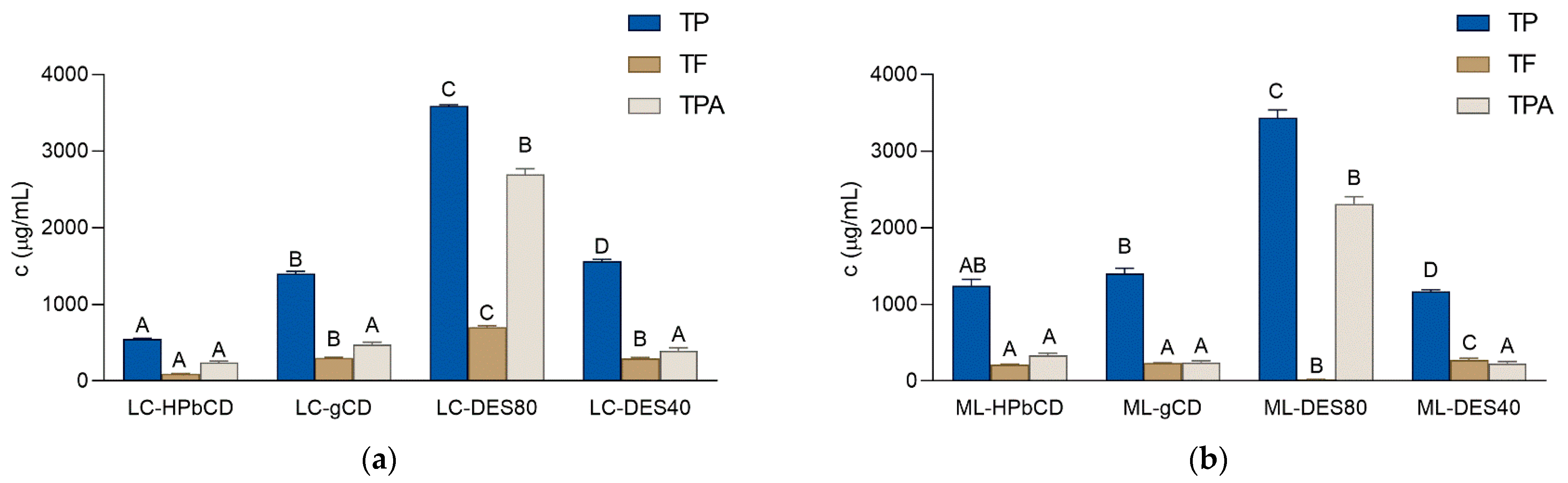
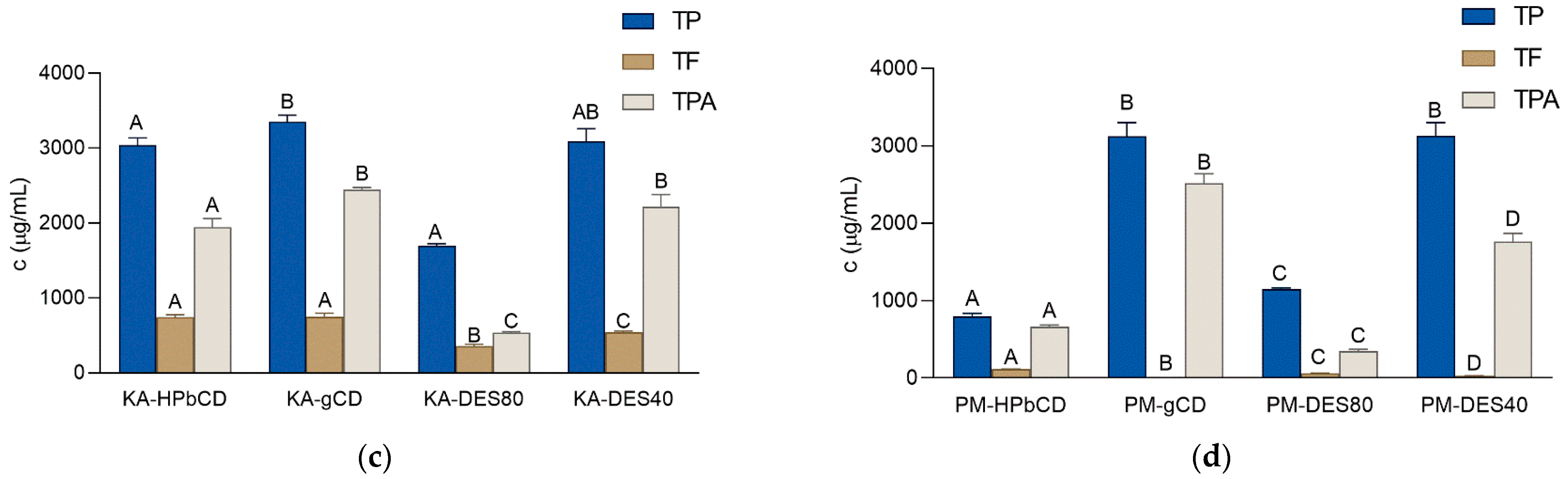
3.3. Phytochemical Composition of the Extracts

TP, TF and TPA content of the extracts are presented in Figure 1. To estimate the effects and influence of individual constituents on the observed effects of the extracts, HPLC analysis was used (Table 4). Of the many plant metabolites, phenolic compounds, including flavonoids and phenolic acids, are among the most appreciated due to their antioxidant and anti-inflammatory properties. They are increasingly being used in the cosmetic industry due to their ability to suppress aging and the various processes that can cause it [39]. Their antioxidant potential and the myriad biological activities that they display render phenolic-rich plants and natural extracts excellent preservatives and functional ingredients in natural cosmetics [40]. The most abundant in phenolics were the extracts of L. corniculatus and M. lupulina prepared using DES80. Interestingly, the same solvent was the least appropriate for the extraction of TP from the remaining two plants, indicating a less polar nature of phenolics in the two plants from the Fabaceae family. The extracts of K. arvensis and P. major that were the richest in phenolic compounds were those prepared by γCD and DES40.
Flavonoids are the most diversified class of polyphenols, widely distributed in leaves, fruits and flowers of terrestrial plants. All flavonoids display antioxidant activity and thus may protect both the cosmetic product and the skin from the deleterious influence of free radicals. Furthermore, they may shield the skin from the harmful influence of UV radiation [41] and act as depigmentation agents [42]. All the aforementioned characteristics render them valuable ingredients in cosmetic and dermatologic products. TF content of the investigated extracts is presented in Figure 1. Among the prepared extracts, LC-DES80, as well as the extracts of K. arvensis prepared using both CDs were particularly rich in flavonoids. There was no clear connection between the solvent used for extraction and the TF content of the extracts. For example, while the DES80 was the best suited for the extraction of TF from L. corniculatus, it was also the solvent that extracted the least flavonoids from M. lupulina. Another important group of phenolic compounds universally present in plants are phenolic acids. They have a high antioxidant capacity and the ability to remove free radicals [43]. They show photoprotective, anti-inflammatory and depigmenting properties. In addition, they may display anti-aging activity based on their ability to reduce the activity of matrix-degrading enzymes such as collagenase [44]. Similar to TP, DES80 was able to extract the most TPA from L. corniculatus and M. lupulina (Figure 1). On the other hand, γCD was the solvent most capable to extract TPA from K. arvensis and P. major.
Flavonoids in plant material are usually present in the form of glycosides, where specific flavonoid aglycon is bound to one or more sugar molecules. HPLC analysis of all the glycosides of a given aglycone may be a challenging task due to the necessity of finding all the appropriate standards. Nevertheless, the type and the quantity of the aglycon that remains upon the hydrolysis of the glycoside is the feature that predominantly affects the pharmacodynamic effects of the flavonoids [45]. Therefore, this work was focused on assessing only the content of the selected flavonoid aglycones (apigenin, kaempferol, luteolin and quercetin) in the prepared extracts upon the acid hydrolysis. The selected compounds may play a notable role in skincare and protection. Apigenin, for example, inhibits UVA-induced cytotoxicity in vitro and prevents signs of skin aging in vivo [46]. Furthermore, kaempferol and quercetin display antimicrobial and anti-inflammatory activity [41]. Finally, luteolin modulates the process of skin aging and shows therapeutic potential in the wound healing process, skin cancer, and inflammatory skin diseases, including atopic dermatitis and psoriasis [47].
As it may be observed in Table 4, M. lupulina contained derivatives of all the analyzed flavonoid aglycons, apigenin, kaempferol, luteolin and quercetin, while K. arvensis was missing only luteolin. L. corniculatus contained only kaempferol and quercetin derivatives, and P. major contained only apigenin and luteolin. The presence of the flavonoid aglycones was not only species- but also extract-specific. For example, LC-HPbCD and ML-HPbCD extracts did not contain any of the analyzed flavonoids. On the other hand, KA-HPbCD was the only K. arvensis extract that contained kaemepferol and quercetin derivatives. Similarly, PM-HPbCD was the only P. major extract that contained apigenin and luteolin. This may be related to the cavity sizes of the employed CDs that were suitable for extraction of only specific flavonoid derivatives present in the plant species. On the other hand, the differences in the solubility of the flavonoid glycosides in DES80 and DES40 may be assigned to the different polarities of the two solvents, deeming them appropriate for dissolution of only specific derivatives. M. lupulina was previously found to be a rich source of apigenin, kaempferol, luteolin and quercetin derivatives [26]. Apigenin, quercetin and kaempferol derivatives were also previously found in K. arvensis [48] and L. croniculatus [49], as well as luteolin and apigenin derivatives in P. major [20]. Thus, the results presented herein are in line with the previously published results.

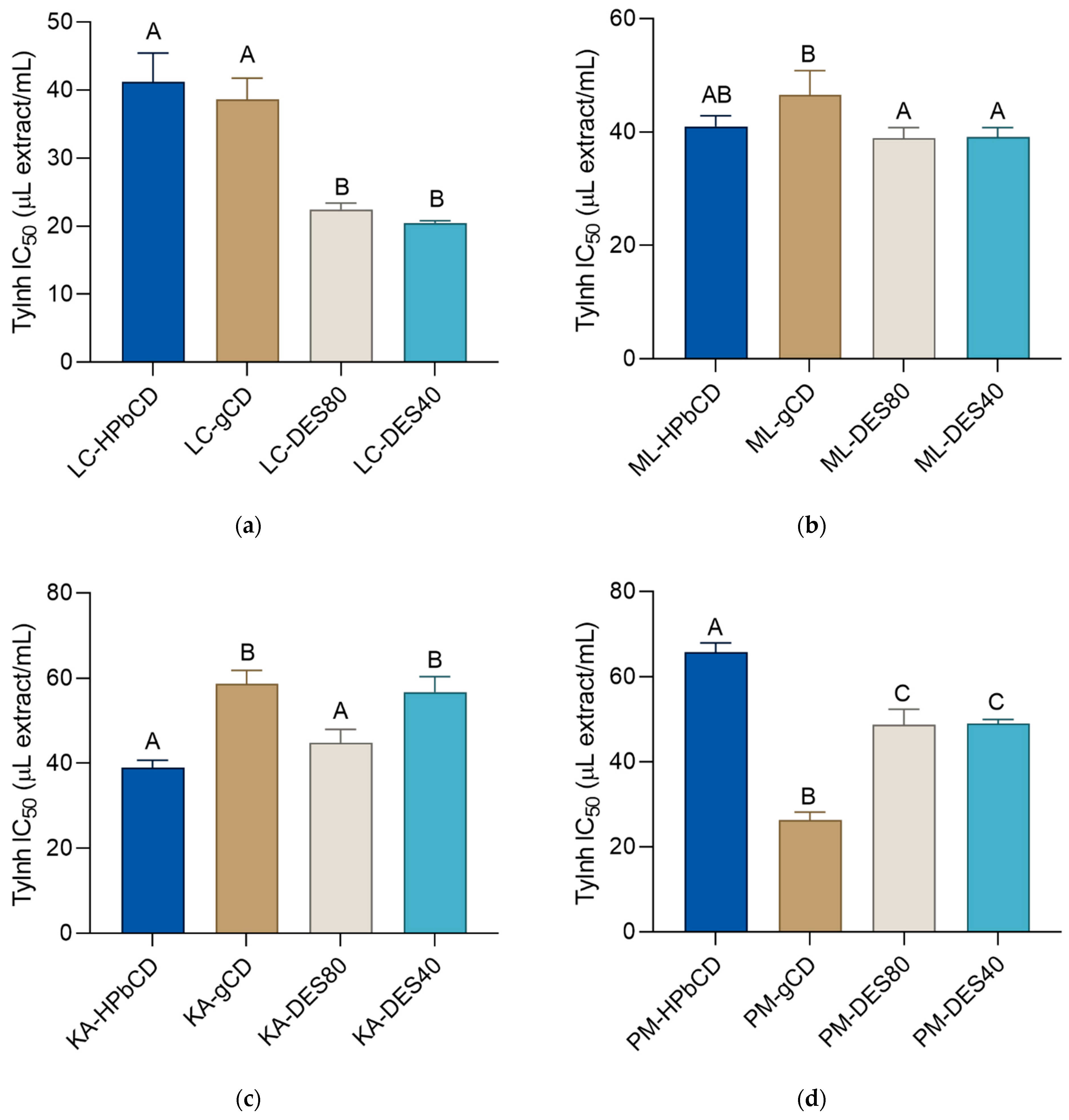
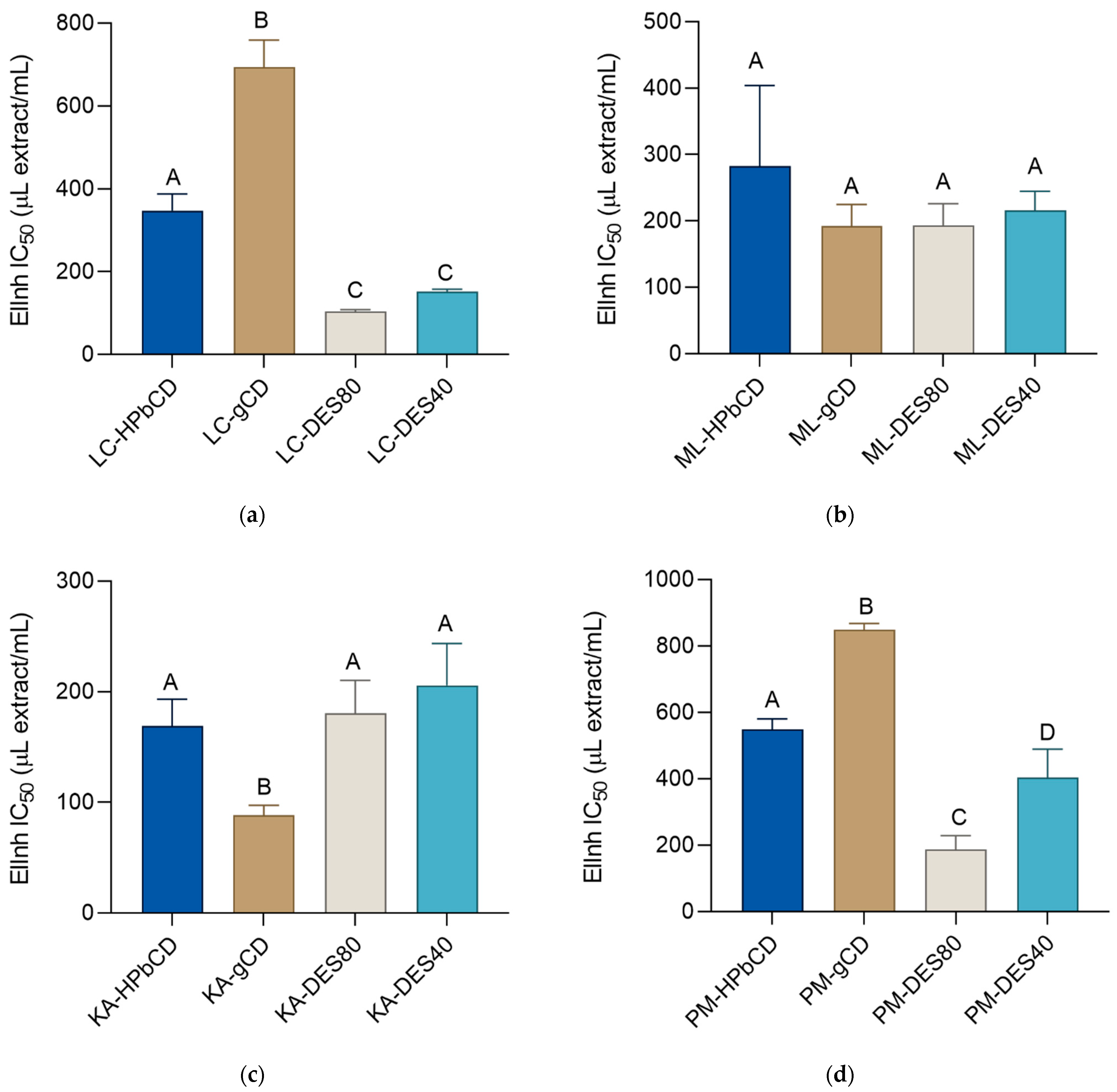
3.4. Tyrosinase- and Elastase-Inhibiting Activity of the Extracts

The investigation of the skin-related activity of the prepared extracts was conducted using two enzymatic methods: inhibition of elastase and tyrosinase. The two assays were selected because these enzymes affect two different aspects of the skin: pigmentation and structure. Since it is not possible to evaporate the DES solvents, the unit for IC50 values of all the extracts is expressed as μL of the extract per mL of the reaction solution (μL extract/mL). When examining tyrosinase- and elastase-inhibiting activity of the extracts, the activity of standard inhibitors, kojic and ursolic acid, respectively, was also assessed and it was expressed as μg/mL. However, it is important to note that a direct comparison of the extracts’ IC50 values with the IC50 values of standard enzyme inhibitors was not possible due to different measurement units. However, as this IC50 value of the standards was numerically equal to the IC50 value of the tested 1 mg/mL standard solution concentration, the activity of the standard is reported and presented for informative purposes.
Melanin is a photoprotective substance responsible for the pigmentation of human skin. However, despite the beneficial effects of melanin, its uneven accumulation may represent an aesthetic and even psychological problem for the affected individual. This is a characteristic of many skin disorders, such as melasma, solar lentigo, liver spots or freckles [50,51]. The enzyme tyrosinase catalyzes the first, rate-limiting step of melanogenesis, the oxidation of tyrosine to dopaquinone. Consequently, tyrosinase inhibitors reduce the production of melanin and prevent hyperpigmentation of the skin [52]. The tyrosinase-inhibiting activity of the extracts is presented in Figure 2a–d. The IC50 value of the standard tyrosinase inhibitor, kojic acid, was determined to be 4.11 ± 0.23 μg/mL. Thus, it may be noted that the numerical IC50 value of the inhibitor was notably lower than those of the tested extracts.
As it may be observed, L. corniculatus extracts prepared using the eutectic mixtures (LC-DES40 and LC-DES80) were especially potent tyrosinase inhibitors, followed by PM-gCD. While the IC50 values of those extracts did not exceed 30 μL extract/mL, the activity of the other samples was only rarely lower than 50 μL extract/mL. The only exceptions are PM-HPbCD, KA-gCD and KA-DES40. One study reported that water extracts of L. corniculatus were more efficient than methanol or ethyl-acetate extracts, with IC50 values below 20 kojic acid equivalents [53]. However, the results are not comparable with those presented herein due to the different units used for the expression of the activity. The glycerol extract of M. lupulina also displayed tyrosinase-inhibiting activity; however, its reported activity of over 250 μL extract/mL was significantly lower than the activity of the extracts presented within the frame of this work. The activity of K. arvensis and P. major was not assessed before, and this is, to the best of our knowledge, the first report of the anti-tyrosinase activity of those plants.
There was no clear rule determining the role the solvent played in the tyrosinase-inhibiting activity of the extracts. For example, the activity of the LC-DES extracts was clearly more pronounced than the activity of LC-CD extracts. However, in the case of the other plants, the difference in the activity between the extracts was either relatively small (M. lupulina), or a relationship between the type of solvent and the activity of the extract was not straightforward (K. arvensis, P. major). It is important to note that the activity of the prepared extracts did not originate solely from the extracted plants’ phytochemical principles. The extraction solvents showed a degree of activity as well. According to its IC50 values, the most active was DES80 (81.6 ± 2.0 μL solvent/mL), followed by HPβCD (108.2 ± 15.7 μL solvent/mL). Even though their activities were considerably lower than the activities of the prepared extracts, it is clear that the solvents played an important part in the overall observed activity of the extracts. The good activity of DES 80 may, at least to some degree, be explained by its high glycerol content. It has been previously noted that a high concentration of that polyol may effectively inhibit tyrosinase activity [26] The activities of DES40 (385.6 ± 47.9 μL solvent/mL) and γCD (558.8 ± 151.6 μL solvent/mL) were considerably less pronounced.
Elastase is a protease from the chymotrypsin family that degrades elastin, extracellular matrix proteins responsible for skin shape and firmness. Chronic exposure to UV radiation leads to a significant increase in elastase activity due to the denaturation of collagen and elastin. The elasticity of the skin is also significantly reduced, which results in the appearance of wrinkles and photoaging of the skin [28,54,55]. Increased elastase activity is related to various skin problems and diseases that include psoriasis, delayed wound-healing, wrinkle formation and premature skin-aging [54]. Numerous clinical studies have confirmed that plant secondary metabolites may act as elastase inhibitors and protect the skin against aging and structural damage to the extracellular matrix [28,54,55].
As it may be observed in Figure 3a–d, KA-gCD with IC50 of 88.4 ± 8.9 µL extract/mL showed the most prominent elastase-inhibiting properties, followed by LC-DES80 with IC50 of 103.6 ± 4.5 µL extract/mL. In general, L. corniculatus and P. major extracts prepared using the DES were more active in the elastase-inhibiting assay than the extracts prepared using CDs. The activity among the M. lupulina extracts, on the other hand, was alike, regardless of the extraction solvent. The IC50 value of the standard elastase inhibitor, ursolic acid, was determined to be 99.2 ± 2.5 μg/mL. Even though the IC50 values of the extracts may not be directly compared to the standard antioxidants due to the different measurement units, it is interesting to note that the activity of the extracts and the standards solution of ursolic acid was, at least numerically, rather similarly pronounced. This indicates a promising activity of the extracts in this assay. A 2020 study reported that P major subsp. major was a weak elastase inhibitor [56], yet it is difficult to compare the results across the studies due to different measurement units and different subspecies. The glycerol extract of M. lupulina was also demonstrated to be an elastase inhibitor; however, its reported activity of over 600 μL extract/mL was significantly lower than the activity of the extract presented within the frame of this work. This emphasizes the suitability of the utilized solvents for the extraction of bioactive principles of M. lupulina. The anti-elastase activity of K. arvensis and L. corniculatus was not assessed before, and this is, to the best of our knowledge, the first report of the anti-elastase activity of those plants.
The elastase-inhibitory activity of the extraction solvents was also assessed. The most active was DES80 with an IC50 value of 285.1 ± 40.7 μL solvent/mL. This indicates the important contribution that this solvent plays in the overall activity of the –DES80 extracts. The good activity of DES80 may be explained by the high glycerol concentrations that the solvent contains. Namely, it has been demonstrated that high glycerol concentration can negatively affect elastase activity [26]. DES40, with its IC50 being 2342.2 ± 156.1 μL solvent/mL, displayed only a negligible activity in this assay. It is interesting to note that, except for PM-DES40/PM-DES80, the activities of –DES40 extracts of the most plants were statistically equal to those of -DES80 extracts. This indicates that the dissolved bioactive principles are primarily responsible for the observed elastase-inhibiting activities of the DES40 extracts. The IC50 values of the two remaining solvents HPβCD and γCD were 915.4 ± 29.8 μL solvent/mL and 489.1 ± 66.0 μL solvent/mL, respectively.

4. Conclusions

The results presented herein indicate that the selected plants from urban parks contained a high content of bioactive phenolics and metals that display properties beneficial for skin health. Simultaneously, the content of toxic heavy metals was low. The eco-friendly, low-cost solvents used in this work were not only suited for the extraction of metabolites with anti-tyrosinase and –elastase properties but also actively contributed to the observed activity. The activity of the prepared extract was species- and solvent-dependent. In general, L. corniculatus was both a potent tyrosinase- and elastase-inhibitor, while K. arvensis showed a pronounced elastase inhibitory activity. The obtained results indicate that the biowaste obtained from urban parks represents a potentially viable and ecologically and economically acceptable alternative to conventional cultivation for the preparation of high-value cosmetic products.

Author Contributions

Conceptualization, M.Z.K.; methodology, M.M., M.J., J.J., E.M. and M.Z.K.; validation, M.M., J.J. and M.Z.K.; investigation, M.M., M.J., L.J., J.J., E.M. and M.Z.K.; resources, E.M., R.D., E.S. and M.Z.K.; writing—original draft preparation, M.M., J.J. and M.Z.K.; writing—review and editing, M.J., J.J., E.M. and M.Z.K.; supervision, E.M. and M.Z.K. All authors have read and agreed to the published version of the manuscript.

Funding

This research is funded by the Croatian Science Foundation project Bioactive plant principles extraction using green solvents—a step towards green cosmeceuticals, Grant No. IP-2018-01-6504. The APC was funded by COST Action CA18130.

Acknowledgments

This research is part of the Croatian Science Foundation project Bioactive plant principles extraction using green solvents—a step towards green cosmeceuticals, Grant No. IP-2018-01-6504. This article/publication is based upon work from COST Action CA18130 ENFORCE TXRF, supported by COST (European Cooperation in Science and Technology)”. COST (European Cooperation in Science and Technology) is a funding agency for research and innovation networks. Our Actions help connect research initiatives across Europe and enable scientists to grow their ideas by sharing them with their peers. This boosts their research, careers, and innovations. http://www.cost.eu/ accessed on 10 December 2021.

Conflicts of Interest

The authors declare no conflict of interest. The funders had no role in the design of the study; in the collection, analyses, or interpretation of data; in the writing of the manuscript, or in the decision to publish the results.

References

  1. Feltynowski, M.; Kronenberg, J.; Bergier, T.; Kabisch, N.; Łaszkiewicz, E.; Strohbach, M.W. Challenges of Urban Green Space Management in the Face of Using Inadequate Data. Urban For. Urban Green. 2018, 31, 56–66. [Google Scholar] [CrossRef]
  2. Ravindran, R.; Hassan, S.S.; Williams, G.A.; Jaiswal, A.K. A Review on Bioconversion of Agro-Industrial Wastes to Industrially Important Enzymes. Bioengineering 2018, 5, 93. [Google Scholar] [CrossRef] [PubMed] [Green Version]
  3. Długoński, A.; Szumański, M. Use of Recreational Park Bio-Waste as Locally Available Energy Resource. Ecol. Chem. Eng. A 2016, 23. [Google Scholar] [CrossRef]
  4. Góralczyk-Bińkowska, A.; Jasińska, A.; Długoński, A.; Płociński, P.; Długoński, J. Laccase Activity of the Ascomycete Fungus Nectriella Pironii and Innovative Strategies for Its Production on Leaf Litter of an Urban Park. PLoS ONE 2020, 15, e0231453. [Google Scholar] [CrossRef] [Green Version]
  5. Nicoletti, M. Nutraceuticals and Botanicals: Overview and Perspectives. Int. J. Food Sci. Nutr. 2012, 63, 2–6. [Google Scholar] [CrossRef]
  6. Zaid, A.N.; Al Ramahi, R. Depigmentation and Anti-Aging Treatment by Natural Molecules. Curr. Pharm. Des. 2019, 25, 2292–2312. [Google Scholar] [CrossRef]
  7. Hoang, H.T.; Moon, J.-Y.; Lee, Y.-C. Natural Antioxidants from Plant Extracts in Skincare Cosmetics: Recent Applications, Challenges and Perspectives. Cosmetics 2021, 8, 106. [Google Scholar] [CrossRef]
  8. Costa, R.; Santos, L. Delivery Systems for Cosmetics—From Manufacturing to the Skin of Natural Antioxidants. Powder Technol. 2017, 322, 402–416. [Google Scholar] [CrossRef]
  9. Izadiyan, P.; Hemmateenejad, B. Multi-Response Optimization of Factors Affecting Ultrasonic Assisted Extraction from Iranian Basil Using Central Composite Design. Food Chem. 2016, 190, 864–870. [Google Scholar] [CrossRef]
  10. Chemat, F.; Abert Vian, M.; Ravi, H.K.; Khadhraoui, B.; Hilali, S.; Perino, S.; Fabiano Tixier, A.-S. Review of Alternative Solvents for Green Extraction of Food and Natural Products: Panorama, Principles, Applications and Prospects. Molecules 2019, 24, 3007. [Google Scholar] [CrossRef] [Green Version]
  11. Becker, L.C.; Bergfeld, W.F.; Belsito, D.V.; Hill, R.A.; Klaassen, C.D.; Liebler, D.C.; Marks, J.G.; Shank, R.C.; Slaga, T.J.; Snyder, P.W.; et al. Safety Assessment of Glycerin as Used in Cosmetics. Int. J. Toxicol. 2019, 38, 6S–22S. [Google Scholar] [CrossRef]
  12. Chemat, F.; Vian, M.A.; Cravotto, G. Green Extraction of Natural Products: Concept and Principles. Int. J. Mol. Sci. 2012, 13, 8615–8627. [Google Scholar] [CrossRef] [PubMed] [Green Version]
  13. Jeong, K.M.; Ko, J.; Zhao, J.; Jin, Y.; Yoo, D.E.; Han, S.Y.; Lee, J. Multi-Functioning Deep Eutectic Solvents as Extraction and Storage Media for Bioactive Natural Products That Are Readily Applicable to Cosmetic Products. J. Clean. Prod. 2017, 151, 87–95. [Google Scholar] [CrossRef]
  14. Pinho, E.; Grootveld, M.; Soares, G.; Henriques, M. Cyclodextrins as Encapsulation Agents for Plant Bioactive Compounds. Carbohydr. Polym. 2014, 101, 121–135. [Google Scholar] [CrossRef] [PubMed] [Green Version]
  15. Suvarna, V.; Gujar, P.; Murahari, M. Complexation of Phytochemicals with Cyclodextrin Derivatives—An Insight. Biomed. Pharmacother. 2017, 88, 1122–1144. [Google Scholar] [CrossRef]
  16. Puglia, C.; Santonocito, D. Cosmeceuticals: Nanotechnology-Based Strategies for the Delivery of Phytocompounds. Curr. Pharm. Des. 2019, 25, 2314–2322. [Google Scholar] [CrossRef] [PubMed]
  17. Fumić, B.; Končić, M.Z.; Jug, M. Development of Cyclodextrin-Based Extract of Lotus Corniculatus as a Potential Substrate Reduction Therapy in Mucopolysaccharidoses Type III. J. Incl. Phenom. Macrocycl. Chem. 2018, 92, 369–379. [Google Scholar] [CrossRef]
  18. Butkutė, B.; Padarauskas, A.; Cesevičienė, J.; Pavilonis, A.; Taujenis, L.; Lemežienė, N. Perennial Legumes as a Source of Ingredients for Healthy Food: Proximate, Mineral and Phytoestrogen Composition and Antibacterial Activity. J. Food Sci. Technol. 2017, 54, 2661–2669. [Google Scholar] [CrossRef]
  19. Kicel, A.; Olszewska, M.A. Evaluation of Antioxidant Activity, and Quantitative Estimation of Flavonoids, Saponins and Phenols in Crude Extract and Dry Fractions of Medicago Lupulina Aerial Parts. Nat. Prod. Commun. 2015, 10, 483–486. [Google Scholar] [CrossRef] [Green Version]
  20. Adom, M.B.; Taher, M.; Mutalabisin, M.F.; Amri, M.S.; Abdul Kudos, M.B.; Wan Sulaiman, M.W.A.; Sengupta, P.; Susanti, D. Chemical Constituents and Medical Benefits of Plantago Major. Biomed. Pharmacother. 2017, 96, 348–360. [Google Scholar] [CrossRef]
  21. Moldoch, J.; Szajwaj, B.; Masullo, M.; Pecio, L.; Oleszek, W.; Piacente, S.; Stochmal, A. Phenolic Constituents of Knautia Arvensis Aerial Parts. Nat. Prod. Commun. 2011, 6, 1627–1630. [Google Scholar] [CrossRef] [Green Version]
  22. Dai, Y.; van Spronsen, J.; Witkamp, G.-J.; Verpoorte, R.; Choi, Y.H. Natural Deep Eutectic Solvents as New Potential Media for Green Technology. Anal. Chim. Acta 2013, 766, 61–68. [Google Scholar] [CrossRef]
  23. Singleton, V.L.; Orthofer, R.; Lamuela-Raventós, R.M. Analysis of Total Phenols and Other Oxidation Substrates and Antioxidants by Means of Folin-Ciocalteu Reagent. Methods Enzymol. 1999, 299, 152–178. [Google Scholar]
  24. Kumazawa, S.; Hamasaka, T.; Nakayama, T. Antioxidant Activity of Propolis of Various Geographic Origins. Food Chem. 2004, 84, 329–339. [Google Scholar] [CrossRef]
  25. Nicolle, C.; Carnat, A.; Fraisse, D.; Lamaison, J.-L.; Rock, E.; Michel, H.; Amouroux, P.; Remesy, C. Characterisation and Variation of Antioxidant Micronutrients in Lettuce (Lactuca Sativa Folium). J. Sci. Food Agric. 2004, 84, 2061–2069. [Google Scholar] [CrossRef]
  26. Jakupović, L.; Kalvarešin, M.; Bukovina, K.; Poljak, V.; Vujić, L.; Zovko Končić, M. Optimization of Two Eco-Friendly Extractions of Black Medick (Medicago Lupulina L.) Phenols and Their Antioxidant, Cosmeceutical, α-Glucosidase and α-Amylase Inhibitory Properties. Molecules 2021, 26, 1610. [Google Scholar] [CrossRef] [PubMed]
  27. Masuda, T.; Yamashita, D.; Takeda, Y.; Yonemori, S. Screening for Tyrosinase Inhibitors among Extracts of Seashore Plants and Identification of Potent Inhibitors from Garcinia Subelliptica. Biosci. Biotechnol. Biochem. 2005, 69, 197–201. [Google Scholar] [CrossRef] [Green Version]
  28. Bose, B.; Choudhury, H.; Tandon, P.; Kumaria, S. Studies on Secondary Metabolite Profiling, Anti-Inflammatory Potential, in Vitro Photoprotective and Skin-Aging Related Enzyme Inhibitory Activities of Malaxis Acuminata, a Threatened Orchid of Nutraceutical Importance. J. Photochem. Photobiol. B 2017, 173, 686–695. [Google Scholar] [CrossRef] [PubMed]
  29. Lansdown, A.B.G. Calcium: A Potential Central Regulator in Wound Healing in the Skin. Wound Repair Regen 2002, 10, 271–285. [Google Scholar] [CrossRef] [PubMed]
  30. Ogawa, Y.; Kinoshita, M.; Shimada, S.; Kawamura, T. Zinc and Skin Disorders. Nutrients 2018, 10, 199. [Google Scholar] [CrossRef] [Green Version]
  31. Coger, V.; Million, N.; Rehbock, C.; Sures, B.; Nachev, M.; Barcikowski, S.; Wistuba, N.; Strauß, S.; Vogt, P.M. Tissue Concentrations of Zinc, Iron, Copper, and Magnesium During the Phases of Full Thickness Wound Healing in a Rodent Model. Biol. Trace Elem. Res. 2019, 191, 167–176. [Google Scholar] [CrossRef] [Green Version]
  32. Berksoy Hayta, S.; Durmuş, K.; Altuntaş, E.E.; Yildiz, E.; Hisarciklıo, M.; Akyol, M. The Reduction in Inflammation and Impairment in Wound Healing by Using Strontium Chloride Hexahydrate. Cutan. Ocul. Toxicol. 2018, 37, 24–28. [Google Scholar] [CrossRef]
  33. Pravilnik o Zdravstvenoj Ispravnosti Predmeta Široke Potrošnje. Available online: https://narodne-novine.nn.hr/clanci/sluzbeni/2009_10_125_3093.html (accessed on 11 June 2021).
  34. Brglez Mojzer, E.; Knez Hrnčič, M.; Škerget, M.; Knez, Ž.; Bren, U. Polyphenols: Extraction Methods, Antioxidative Action, Bioavailability and Anticarcinogenic Effects. Molecules 2016, 21, 901. [Google Scholar] [CrossRef]
  35. Zou, H.; Wu, Z.; Xian, M.; Liu, H.; Cheng, T.; Cao, Y. Not Only Osmoprotectant: Betaine Increased Lactate Dehydrogenase Activity and l-Lactate Production in Lactobacilli. Bioresour. Technol. 2013, 148, 591–595. [Google Scholar] [CrossRef] [PubMed]
  36. Lange, J.-P. Performance Metrics for Sustainable Catalysis in Industry. Nat. Catal. 2021, 4, 186–192. [Google Scholar] [CrossRef]
  37. Catalysts|Free Full-Text|New Glycerol Upgrading Processes|HTML. Available online: https://0-www-mdpi-com.brum.beds.ac.uk/2073-4344/11/1/103/htm (accessed on 3 January 2022).
  38. PharmaCompass—Grow Your Pharma Business Digitally. Available online: https://www.pharmacompass.com/ (accessed on 3 January 2022).
  39. Panzella, L.; Moccia, F.; Nasti, R.; Marzorati, S.; Verotta, L.; Napolitano, A. Bioactive Phenolic Compounds From Agri-Food Wastes: An Update on Green and Sustainable Extraction Methodologies. Front. Nutr. 2020, 7, 60. [Google Scholar] [CrossRef]
  40. Pandey, A.; Jatana, G.K.; Sonthalia, S. Cosmeceuticals. In StatPearls; StatPearls Publishing: Treasure Island, FL, USA, 2020. [Google Scholar]
  41. Kurek-Górecka, A.; Górecki, M.; Rzepecka-Stojko, A.; Balwierz, R.; Stojko, J. Bee Products in Dermatology and Skin Care. Molecules 2020, 25, 556. [Google Scholar] [CrossRef] [Green Version]
  42. Nagula, R.L.; Wairkar, S. Recent Advances in Topical Delivery of Flavonoids: A Review. J. Control Release 2019, 296, 190–201. [Google Scholar] [CrossRef] [PubMed]
  43. Gülçin, İ. Antioxidant Activity of Food Constituents: An Overview. Arch. Toxicol. 2012, 86, 345–391. [Google Scholar] [CrossRef]
  44. Taofiq, O.; González-Paramás, A.M.; Barreiro, M.F.; Ferreira, I.C.F.R. Hydroxycinnamic Acids and Their Derivatives: Cosmeceutical Significance, Challenges and Future Perspectives, a Review. Molecules 2017, 22, 281. [Google Scholar] [CrossRef]
  45. Chen, Z.; Zheng, S.; Li, L.; Jiang, H. Metabolism of Flavonoids in Human: A Comprehensive Review. Curr. Drug Metab. 2014, 15, 48–61. [Google Scholar] [CrossRef] [PubMed]
  46. Choi, S.; Youn, J.; Kim, K.; Joo, D.H.; Shin, S.; Lee, J.; Lee, H.K.; An, I.-S.; Kwon, S.; Youn, H.J.; et al. Apigenin Inhibits UVA-Induced Cytotoxicity in Vitro and Prevents Signs of Skin Aging in Vivo. Int. J. Mol. Med. 2016, 38, 627–634. [Google Scholar] [CrossRef] [Green Version]
  47. Gendrisch, F.; Esser, P.R.; Schempp, C.M.; Wölfle, U. Luteolin as a Modulator of Skin Aging and Inflammation. Biofactors 2021, 47, 170–180. [Google Scholar] [CrossRef] [PubMed]
  48. Kopyt’ko, Y.F.; Dargaeva, T.D.; Rendyuk, T.D. Composition of the Field Scabious (Knautia Arvensis L.). Pharm. Chem. J. 2020, 54, 725–733. [Google Scholar] [CrossRef]
  49. Fumić, B.; Jug, M.; Končić, M.Z. Optimization of Ultrasound-Assisted Extraction of Phenolic Antioxidants from Lotus Corniculatus. Croat. Chem. Acta 2019, 92, 369–377. [Google Scholar] [CrossRef] [Green Version]
  50. Hwang, J.A.; Park, N.H.; Na, Y.J.; Lee, H.K.; Lee, J.H.; Kim, Y.J.; Lee, C.S. Coumestrol Down-Regulates Melanin Production in Melan-a Murine Melanocytes through Degradation of Tyrosinase. Biol. Pharm. Bull. 2017, 40, 535–539. [Google Scholar] [CrossRef] [Green Version]
  51. Mukherjee, P.K.; Biswas, R.; Sharma, A.; Banerjee, S.; Biswas, S.; Katiyar, C.K. Validation of Medicinal Herbs for Anti-Tyrosinase Potential. J. Herb. Med. 2018, 14, 1–16. [Google Scholar] [CrossRef]
  52. Pillaiyar, T.; Manickam, M.; Namasivayam, V. Skin Whitening Agents: Medicinal Chemistry Perspective of Tyrosinase Inhibitors. J. Enzyme Inhib. Med. Chem. 2017, 32, 403–425. [Google Scholar] [CrossRef] [Green Version]
  53. Yerlikaya, S.; Baloglu, M.C.; Diuzheva, A.; Jekő, J.; Cziáky, Z.; Zengin, G. Investigation of Chemical Profile, Biological Properties of Lotus Corniculatus L. Extracts and Their Apoptotic-Autophagic Effects on Breast Cancer Cells. J. Pharm. Biomed. Anal. 2019, 174, 286–299. [Google Scholar] [CrossRef]
  54. Jabs, H.-U. Elastase—Ziel Einer Neuen Anti-Aging Strategie Bei Hautalterung, Elastizitätsverlust Und Faltenbildung. Ästhetische Dermatol. 2014, 2012, 2–4. [Google Scholar]
  55. Azmi, N.; Hashim, P.; Hashim, D.M.; Halimoon, N.; Majid, N.M.N. Anti–Elastase, Anti–Tyrosinase and Matrix Metalloproteinase–1 Inhibitory Activity of Earthworm Extracts as Potential New Anti–Aging Agent. Asian Pac. J. Trop. Biomed. 2014, 4, S348–S352. [Google Scholar] [CrossRef] [PubMed] [Green Version]
  56. Genc, Y.; Dereli, F.T.G.; Saracoglu, I.; Akkol, E.K. The Inhibitory Effects of Isolated Constituents from Plantago Major Subsp. Major L. on Collagenase, Elastase and Hyaluronidase Enzymes: Potential Wound Healer. Saudi Pharm. J. 2020, 28, 101–106. [Google Scholar] [CrossRef] [PubMed]
Figure 1. Total phenolic (TP), total flavonoid (TF) and total phenolic acid (TPA) content of the extracts (a) L. corniculatus, (b) M. lupulina, (c) K. arvensis, (d) P. major. Different uppercase letters indicate statistically significant difference between the phenols of the same type (indicated by the columns of the same color) (p < 0.05).
Agronomy 12 00204 g001aAgronomy 12 00204 g001b
Figure 2. Anti-tyrosinase activity of the extracts (a) L. corniculatus, (b) M. lupulina, (c) K. arvensis, (d) P. major. Different uppercase letters indicate statistically significant difference (p < 0.05).
Agronomy 12 00204 g002
Figure 3. Anti-elastase activity of the extracts (a) L. corniculatus, (b) M. lupulina, (c) K. arvensis (d) P. major. Different uppercase letters indicate statistically significant difference (p < 0.05).
Agronomy 12 00204 g003
Table 1. Calibration curve parameters for flavonoids and phenolic acid standards.
StandardEquationr2LOD
(μg/mL)
LOQ
(μg/mL)
Apigeniny = 3755.16x − 27.750.99980.0280.085
Kaempferoly = 2802.76x − 21.620.99980.0250.078
Luteoliny = 2787.17x + 46.600.99980.0250.076
Quercetiny = 2200.20x − 36.750.99980.0280.085
LOD = limit of detection; LOQ = Limit of quantification; y = Area under curve (mAU × s); x = amount of the standard (μg).
Table 2. The results of the determination of heavy metals in the plant.
Element
(mg/kg)
L. corniculatus
mg/kg
M. lupulina
mg/kg
K. arvensis
mg/kg
P. major
mg/kg
Ca15,285 ± 20620,101± 51314,766 ± 15628,481 ± 734
Zn64.1 ± 0.645.0 ± 1.890.0 ± 3.566.8 ± 5.4
Sr25.3 ± 1.029.1 ± 1.622.9 ± 1.740.6 ± 1.0
Fe1183 ± 523175 ± 234243 ± 40860 ± 143
Cr3.6 ± 0.48.5 ± 0.9n. d.n. d.
Pb4.6 ± 0.15.9 ± 1.11.3 ± 0.33.8 ± 1.3
Ni3.5 ± 0.64.5 ± 0.61.4 ± 0.22.0 ± 0.2
Cd 3.6 ± 0.48.5 ± 0.9n.d.n.d.
n.d.–not detected.
Table 3. The plants, extracts, and their abbreviations used in this study.
Plant SpeciesAbbreviations According to the Extraction Solvents
HPβCDγCDDES80DES40
L. corniculatusLC-HPbCDLC-gCDLC-DES80LC-DES40
M. lupulinaML-HPbCDML-gCDML-DES80ML-DES40
K. arvensisKA-HPbCDKA-gCDKA-DES80KA-DES40
Plantago majorPM-HPbCDPM-gCDPM-DES80PM-DES40
Extracts: -HPbCD = extract prepared using hydroxy propyl-β-cyclodextrin; -gCD = extract prepared using hydroxy propyl-γ-cyclodextrin; -DES80 = extract prepared using DES80; -DES40 = extract prepared using DES40. The details of the extraction procedure are given in Section 2.3 and Section 2.4.
Table 4. Quantity of individual phenolics in the extracts.
ExtractAP
µg/mL
KAE
µg/mL
LUT
µg/mL
QUE
µg/mL
LC-HPbCDn.d.n.d.n.d.n.d.
LC-gCDn.d.31.30n.d.48.19
LC-DES80n.d.13.44n.d.20.65
LC-DES40n.d.38.00n.d.62.00
ML-HPbCDn.d.n.d.n.d.n.d.
ML-gCD6.174.9414.479.40
ML-DES80n.d.n.d.n.d.n.d.
ML-DES409.175.6617.18n.d.
KA-HPbCDn.d.22.50n.d.32.98
KA-gCDn.d.n.d.n.d.n.d.
KA-DES804.93n.d.n.d.n.d.
KA-DES40n.d.n.d.n.d.n.d.
PM-HPbCD7.17n.d.12.92n.d.
PM-gCDn.d.n.d.n.d.n.d.
PM-DES80n.d.n.d.n.d.n.d.
PM-DES40n.d.n.d.n.d.n.d.
AP = Apigenin; KAE = Kaempferol; LUT = Luteolin; Que = Quercetin. n.d. = not detected. The extracts’ abbreviations are presented in Table 2.
Publisher’s Note: MDPI stays neutral with regard to jurisdictional claims in published maps and institutional affiliations.

