Review

Metal Ions Induce Liquid Condensate Formation by the F Domain of Aedes aegypti Ecdysteroid Receptor. New Perspectives of Nuclear Receptor Studies

Laboratory of Biochemistry and Molecular Biology, Department of Biochemistry, Molecular Biology and Biotechnology, Faculty of Chemistry, Wrocław University of Science and Technology, Wybrzeże Wyspiańskiego 27, 50-370 Wrocław, Poland
*
Author to whom correspondence should be addressed.
These authors contributed equally to this work.
Submission received: 11 December 2020 / Revised: 2 March 2021 / Accepted: 3 March 2021 / Published: 5 March 2021

Abstract

:
The superfamily of nuclear receptors (NRs), composed of ligand-activated transcription factors, is responsible for gene expression as a reaction to physiological and environmental changes. Transcriptional machinery may require phase separation to fulfil its role. Although NRs have a similar canonical structure, their C-terminal domains (F domains) are considered the least conserved and known regions. This article focuses on the peculiar molecular properties of the intrinsically disordered F domain of the ecdysteroid receptor from the Aedes aegypti mosquito (AaFEcR), the vector of the world’s most devastating human diseases such as dengue and Zika. The His-Pro-rich segment of AaFEcR was recently shown to form the unique poly-proline helix II (PPII) in the presence of Cu2+. Here, using widefield microscopy of fluorescently labeled AaFEcR, Zn2+- and Cu2+-induced liquid-liquid phase separation (LLPS) was observed for the first time for the members of NRs. The perspectives of this finding on future research on the F domain are discussed, especially in relation to other NR members.

Graphical Abstract

1. Characteristics of NRs

Nuclear receptors (NRs) are a specific superfamily of the most abundant class of ligand-activated transcription factors. These transcription factors play a critical role in the modulation of the expression of target genes by selective binding to appropriate genomic DNA response elements. They are responsible for the regulation of fundamental biological processes such as development, tissue and metabolic homeostasis, cell proliferation, metamorphosis, and reproduction in animals [1,2]. The activities of most NRs are regulated endogenously by small lipophilic ligands such as retinoids, steroids, thyroid hormones, or dietary lipids [3]. NRs are also the targets of other cellular signaling pathways that modify post-translationally receptors, or their co-modulators, in turn affecting their activities and functionality [4]. Their remarkable properties of transducing extracellular signals via the binding of specific ligands to directly modulate gene expression make them critical pharmaceutical and therapeutic drug targets [5]. Disrupted NR signaling pathways contribute to numerous diseases such as cancer or diabetes [5,6,7]. Although diversity in the overall shape, size, and plurality of specific ligands can be observed, the majority of the known members of NRs share a common modular structure (Figure 1) that generally consists of four domains [8,9].
The N-terminal domain (NTD) is responsible for ligand-independent transactivation (AF-1). The most characterized NTDs to date exhibit features of intrinsically disordered proteins (IDPs) [12,13,14,15,16,17,18,19]. Such a property is advantageous for their inter- and intramolecular interactions [20], as NTDs can undergo disorder-to-order (or partially order) transitions as a result of co-activator/co-repressor binding, interaction with DNA or posttranslational modifications (PTMs) [21,22,23]. An NTD is followed by a highly conserved DNA-binding domain (DBD). Next, the disordered and flexible hinge region serves as a linker between the DBD and the evolutionarily conserved ligand-binding domain (LBD), which is responsible for the ligand molecule binding and ligand-dependent activation of transcription (AF-2), but also mediates homo- or heterodimerization with another NR superfamily member. Some NRs own an additional hypervariable (in terms of length and sequence) C-terminal F domain (Figure 1) [1,3,10,11,24]. The C-terminal F domains of some NRs are not clearly defined. Amino acid residues, following the last helix (helix 12) of LBDs, define the F domains, and due to their short sequence, in many mammalian NRs, they are often considered as a part of the LBDs.

2. Transcription Factors in the Cellular Response to Mosquito-Borne Infections

Tropical and subtropical areas stand out in terms of their lush vegetation, various animal species, and changing weather conditions. Most of the world’s population live in these areas and struggle with the health problems that frequently occur there. Environmental conditions are favorable for insect reproduction, and therefore for insect-dependent disease transmission.
Caused by one of the 4 serotypes of the dengue virus, dengue is one of the greatest threats in the world. Annually, about 390 million people get infected, with 96 million of them developing symptoms [24,25]. Most people suffer from a characteristic rash, headache, and joint and muscle pain. Dengue fever may develop into much more severe states: dengue hemorrhagic fever (DHF) and dengue shock syndrome (DSS), which require immediate hospitalization and specific treatment [26]. Another pathogen, like dengue virus, belonging to the Flaviviridae family, is Zika virus—first reported in 1947 in Uganda [27], sequenced in 2007 [28]. Until its outbreak in 2015 in Brazil (more than 1.5 million infections), only several cases of Zika infection were reported [29]. Some patients undergo the disease with fever, rash, and joint and muscle pain, but most of them do not show any symptoms [30]. This is the actual problem, because Zika virus can be transmitted sexually or passed from the mother to the fetus. The mother’s infection may result in neurological disorders for the child, like microencephaly [31]. Yet another mosquito-borne disease is one of the deadliest on Earth-malaria. In 2018 alone, 228 million cases were reported [32]. After the blood meal, Plasmodium falciparum sporozoites (formed through fertilization) are transferred to human hepatocytes. Later, merozoites invade human erythrocytes, in turn causing erythocytic schizogony [33]. Shivers, fever, profuse sweating and headache are the first symptoms of malaria infection. Without proper treatment, patients may experience anemia, convulsions and coma [34].

2.1. Human Cellular Response to Viral Infection Involves Changes in Gene Expression

The regulation of gene expression requires the balanced orchestration of adjustable complexes of many specific transcription factors, modulators and signal transduction. Modulation of gene expression profiles by NRs is an allosteric process where binding ligands and specific DNA sequences initiate the recruitment of diverse transcription factors, co-activators, co-repressors and elements of the chromatin remodeling machinery to transactivate or transrepress the expression of target genes. Symptoms of dengue, Zika or malaria diseases are extensive and caused by a series of metabolic pathways. Some of them involve transcription factors to maintain the balanced orchestration of gene expression.
CREB3 (cyclic AMP-responsive element-binding protein 3), which enhances the transcription of genes involved in secretory pathways, takes part in the cellular response to Zika and dengue virus infections [35,36]. After infection with Zika virus, the host’s cells are forced to produce viral proteins, and therefore the secretory demands increase and cause endoplasmic reticulum stress. Transcriptome analyses of human cells infected with Zika virus, conducted by Moni et al., revealed upregulation of pathways concerning endoplasmic reticulum functioning [37]. To maintain endoplasmic reticulum homeostasis after infection with Zika virus, the expression level of the CREB3L2 gene (with other genes associated with this pathway) can be three times higher [37].

2.2. Transcription Factors Involved in the Mosquito’s Response to Pathogens

What is interesting is that the parasite itself is able to trigger a mechanism that will enable it to survive in the host’s cells [38,39,40]. The role of the transcription factors engaged in these mechanisms cannot be ignored. In Anopheles, the expression of antimicrobial peptides (AMPs), i.e., gambicin or attacin, is controlled by the Rel1 and Rel2 transcription factors. Their translocation to the nucleus depends on the activation of the Toll and Imd pathways, respectively [41]. In mosquitos, after ingestion, gametocytes turn into gametes, and after fertilization a zygote occurs. It develops into sporozoites, which are present in the salivary glands of mosquitos. Before transmitting Plasmodium to human, A. gambiae shows an immune response to the presence of the parasite. Disrupted parasite development is achieved by an increased level of reactive nitric oxide (NO). A high level of NO is caused by NO synthase (NOS), the transcription of which is mediated by STAT-A in A. gambiae. Interestingly, the mRNA level of STAT-A is controlled by its ortholog—STAT-B [41]. The so-called JAK-STAT mechanism has also been described as a part of the immune response of A. aegypti to dengue infection [42]. Most of the physiological effects described above are related to changes in the activity of transcription factors. It was recently shown that the development of P. falciparum parasites in A. gambiae females is linked to physiological processes controlled by 20-hydroxyecdysone (20E), which is a physiological ligand of the insect ecdysteroid receptor (EcR) during egg development [43]. The analysis of both eggs and parasite counts in individual females unveiled an unexpected positive correlation between mosquito and parasite fitness that may be dependent on 20E signaling [43]. However, the knowledge of the functional roles of NRs at the molecular level in such events in humans and insects is still obscure and limited.

3. Liquid-Liquid Phase Separation in Gene Expression

Recently, liquid-liquid phase separation (LLPS) emerged as a mechanism in which cells organize their interior, separate, and then segregate biochemical processes [44,45,46,47]. This thermodynamically driven process leads to the formation of barrier-free cellular bodies known as membraneless organelles (MLOs) [48,49]. MLOs are self-regulated liquid condensates, the components of which can diffuse freely and be exchanged rapidly with the surrounding milieu [45,50,51]. Interestingly, observations of molecule kinetics lead to the conclusion that although MLOs are composed of a number of different molecules [52], only a small fraction of them is needed to maintain the integrity of the condensates [50]. The majority of the MLOs’ components diffuse into the condensate due to interactions with scaffold molecules. The composition of these so-called client molecules changes, and therefore the functional properties of MLOs also change [50]. LLPS leads to the formation of MLOs in response to changes in the surrounding milieu [44]. Their formation may be triggered by stress factors [51] or be due to biochemical changes of molecules such as PTMs [53], the presence of ligands [54], and changes in the concentration of certain molecules [55]. The fact that pathogens utilize LLPS for invading host cells can also be seen to be interesting. For example, upon infection in the cytoplasm, viral ribonucleoprotein complexes alter the translation of cellular proteins by interfering with stress granules [56]. The intrinsically disordered regions (IDRs) of the EBNA2 and EBNALP transcription factors of the Epstein-Barr virus participate in nuclear LLPS in newly infected cells [57]. Following transcription of viral RNA, the N protein from the SARS-CoV-2 phase separates with RNA and forms liquid condensates which are a precursor for new virions [58,59]. This mechanism is also common for other types of viruses [60,61,62].

Liquid-Liquid Phase Separation in Transcriptional Regulation by NRs

LLPS is important for the regulation of various aspects of gene expression, including transcription [63,64,65,66,67,68,69]. One of the first indications that transcription may depend on LLPS was provided by Hnisz et al., who developed a model describing the unique properties of macromolecular complexes formed on super-enhancers [65]. Sabari et al. showed that transcription co-activators condensate to super-enhancers and form liquid condensates [67]. Further research revealed that some transcription factors that possess IDRs can undergo spontaneous LLPS, in turn concentrating the transcriptional machinery and allowing the transcription to proceed [63]. This finding sheds new light on the mechanism by which transcription factors and transcription coactivators regulate gene expression. To date, little is known about the ability of NRs to induce LLPS. It was shown that the NTD of the androgen receptor (AR) can form liquid condensates in a concentration-dependent manner [70]. The AR is a substrate for Speckle-type POZ protein (SPOP) [71]. The interaction of AR and SPOP leads to proteasomal degradation of AR. Both proteins were shown to localize in liquid-like nuclear compartments. Mutations in SPOP, which alter its activity, are associated with some types of solid tumors in humans. It is important that the cancer-associated mutation disrupts the substrate-associated phase separation, in turn leading to an aberrant distribution of the protein between MLOs [70]. It has also been shown that the NTD of RXRγ can form liquid condensates in a temperature-dependent manner [19]. In fact, many proteins undergo phase separation in relation to changes of temperature [44,72]. One of the best known examples are proteins which form stress granules when the temperature decreases [51,73]. The NTD of RXRγ, however, undergoes a phase transition when heated above a critical temperature [19]. These properties were attributed to the fact that this domain is enriched in Pro and Gly residues encoded in Pro-Xn-Gly motifs (Xn residues are preferably nonpolar) with residues that are largely nonpolar [19,74]. The glucocorticoid receptor (GR) was also found to be distributed in the nucleus in a specific dotted pattern [75]. GR foci contain some transcription co-activators, including some subunits of the Mediator complex, which indicates that these condensates are an indispensable part of the transcription regulation machinery [76]. Similar observations were made for the estrogen receptor (ER) [63]. It is interesting that the AR and RXRγ induce LLPS by the NTD [19,70], whereas the GR and ER do not. In the case of GR removal of the C-terminal, the LBD drastically affected foci formation [76]. Likewise, the ER also forms liquid condensates when bounded in a ligand-dependent manner [63]. These examples show that NRs may influence gene expression using the LLPS to regulate their activity. However, due to insufficient evidence, the regions or domains responsible for provoking the process cannot be generalized. This is a new challenge in the study of NRs. At present, we also do not know if NRs in liquid condensates serve as scaffold molecules responsible for the formation of a condensate or as clients recruited to the preformed condensates similarly to client UBQLN2 recruited to stress granules [77]. In either case, NRs need to have specific features in their sequences which determine their ability to partition into liquid condensates. Their determination can contribute to a better understanding of the NRs’ functionality as transcription factors.

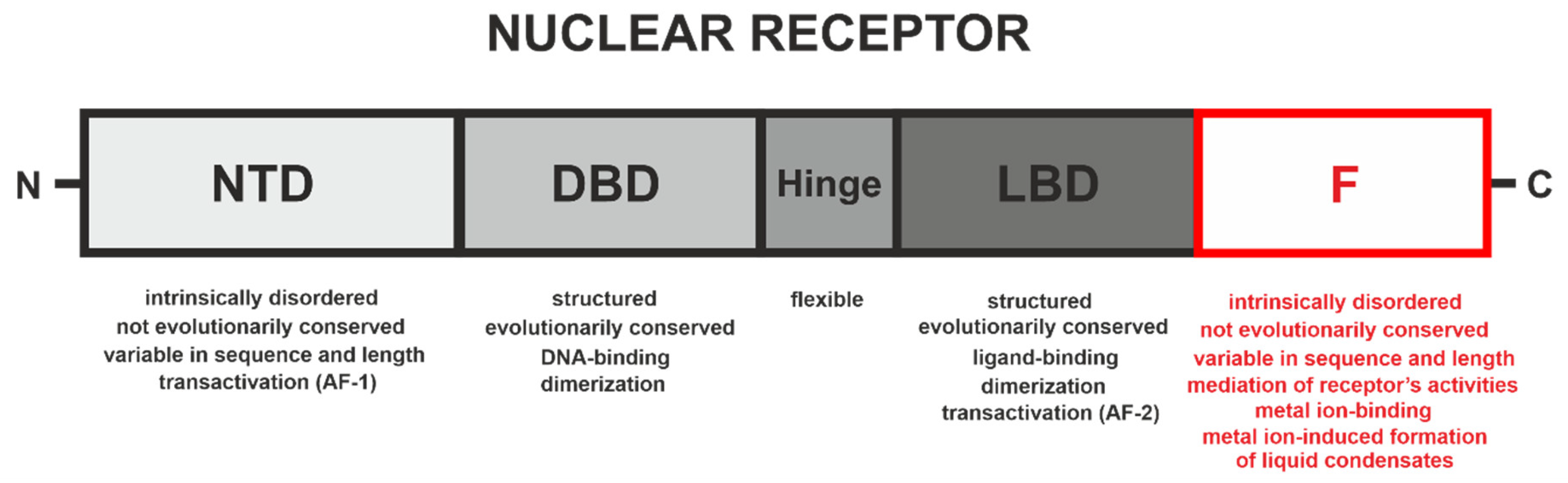
4. F Domains of Nuclear Receptors

When compared to the F domain of A. aegypti EcR (AaEcR) (ca. 107 aa) or the EcR from D. melanogaster (ca. 190 aa), the F domains of mammalian NRs are relatively short (from several to several dozen amino acid residues) [78]. As was mentioned above, the presence of F domains is not conserved in the NR superfamily, and the sequences of known F domains are highly variable in terms of length and sequence (for example see Figure 2). This remarkably variety is also visible among the members of the EcR family (Figure S1 in the Supplementary Materials). These short sequences are often considered as short extensions of the last helix in LBDs. Patel and Skafar extensively described the activities of the F domains of such NRs as ERα, ERβ, GR, PR, AR, MR, RXRα or RARα [78]. Although the F domains of the described NRs possess different lengths and they are not evolutionarily conserved in mammals (Figure 2 and Figure S2 in the Supplementary Materials), and the fact that only part of them seem to be intrinsically disordered (Figure S3 in the Supplementary Materials), they all appear to regulate the activities of receptors. They seem to affect dimerization, interactions with other proteins, and the stabilization of ligand binding, and thus contribute to the regulation of transcriptional activation [78].
Concerning the F domain role in the functioning of NRs, two papers during the last five years are worth paying attention to. Bianchetti and co-workers showed that the F domain of the glucocorticoid receptor-α (GRα) forms a steric hindrance to the well-known canonical LBD dimer assembly [80]. According to the authors, the currently accepted GRα homodimer structure and experimental investigations of the alternative architectures should be reexamined [80]. In the case of the F domain of the ERα, it was shown that this region governs the species-specific tamoxifen-mediated transcriptional activity of the ERα [81]. According to our current knowledge, none of the described F domains of NRs have the propensity to form metal ion-induced PPII [10] or undergo LLPS.

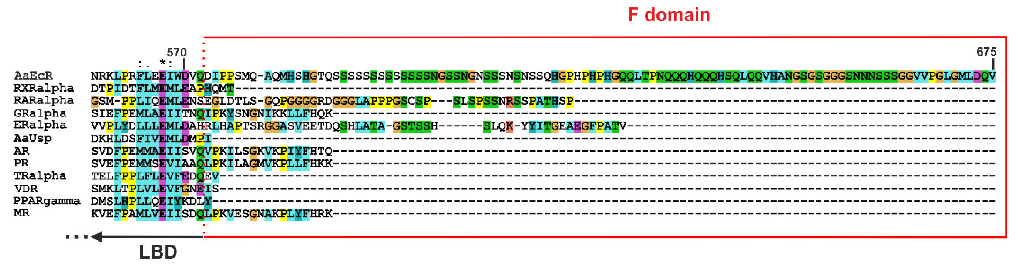
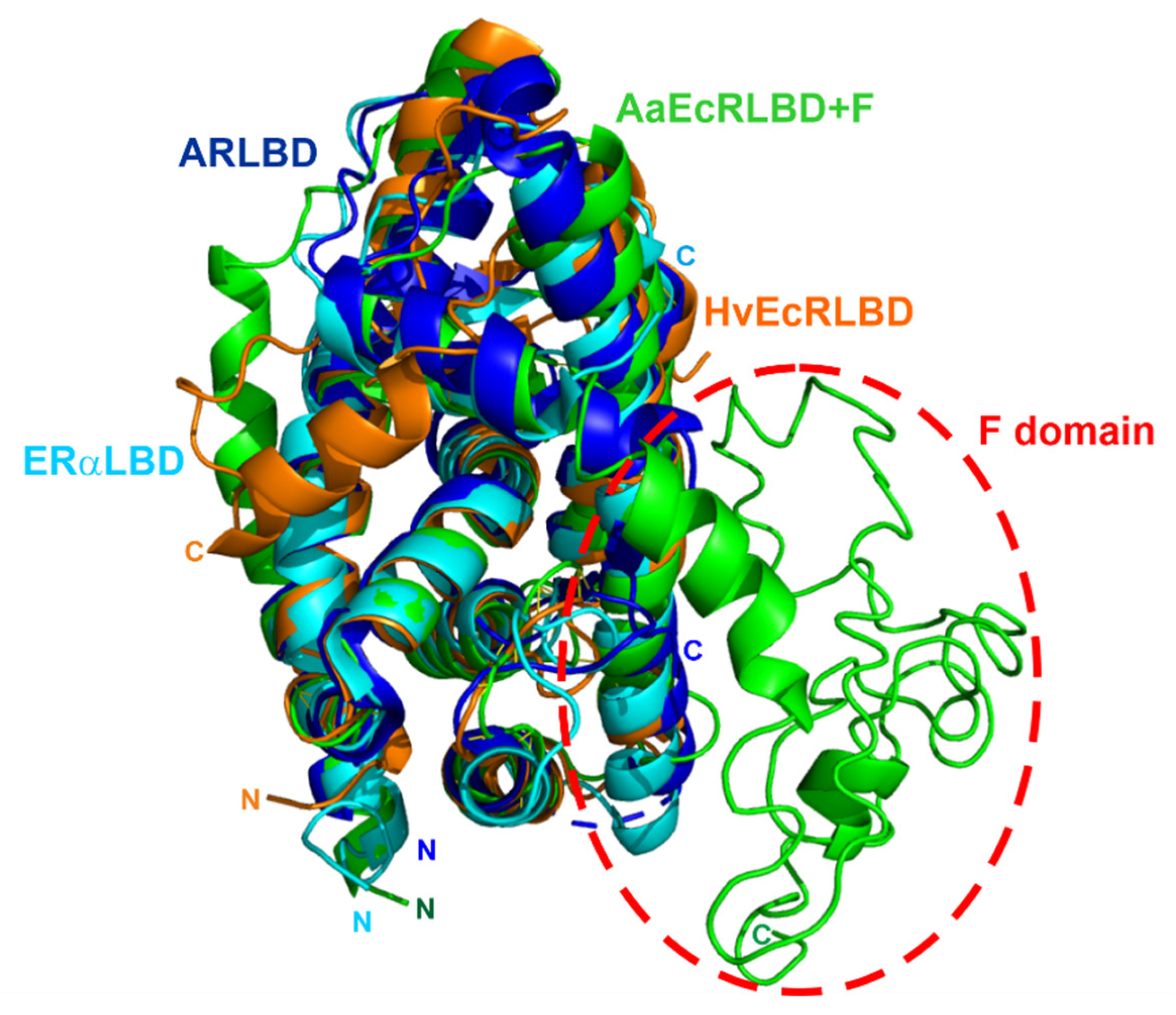
4.1. F Domain of the Ecdysteroid Receptor (EcR) from A. aegypti

A. aegypti mosquitoes have been the main vectors of the world’s most devastating human diseases in recent years, such as dengue, Zika, yellow fever and chikungunya [82,83,84,85]. Many crucial processes in mosquitos’ reproduction follow the formation of an active heterodimeric complex of the 20E receptor (EcR) and Ultraspiracle protein (Usp) [86]. The EcR of A. aegypti (AaEcR) contains one of the longest F domains in known members of NRs (Figure 2), and it clearly stands out structurally from the evolutionary conserved overall canonical folds of NR LBDs and their C-terminal extensions (Figure 3).
It was previously shown that recombinant AaFEcR is mainly disordered with residual ordered secondary structures and takes the conformation of a premolten globule (PMG) [11]. It can undergo both induced folding (in the presence of trifluoroethanol) and unfolding (in the presence of guanidinium chloride). The increased content of His and Pro residues (compared to SwissProt database [11]) is not accidental, as it strongly resembles the amino acid motif present in His-Pro-rich glycoproteins (HPRGs) that is known to bind Zn2+ and Cu2+ ions [88]. Mass spectrometry results clearly showed the formation of AaFEcR-Zn2+ and AaFEcR-Cu2+ complexes and their stoichiometry. Coordination of Cu2+ and Zn2+ ions to this domain did not induce any statistically relevant changes in its overall secondary structure [11]. Further studies were conducted on two model peptides (Ac-HGPHPHPHG-NH2 and Ac-QQLTPNQQQHQQQHSQLQQVHANG-NH2) contained in the sequence of AaFEcR. The most fascinating result was observed for the interaction between the Ac-HGPHPHPHG-NH2 peptide and Cu2+ ions, which resulted in the formation of the rare and unique polyproline type II helical structure (PPII) [10]. Our findings were the first to show the Cu2+ binding-induced formation of PPII—a structure, which had not been reported for any NR superfamily member before. PPII helixes responsible for protein-protein and protein-DNA interactions [89,90,91,92]. However, the ion binding-dependent functional potential of AaFEcR remains to be solved.

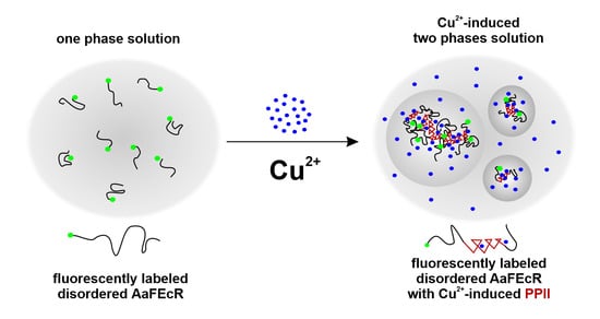
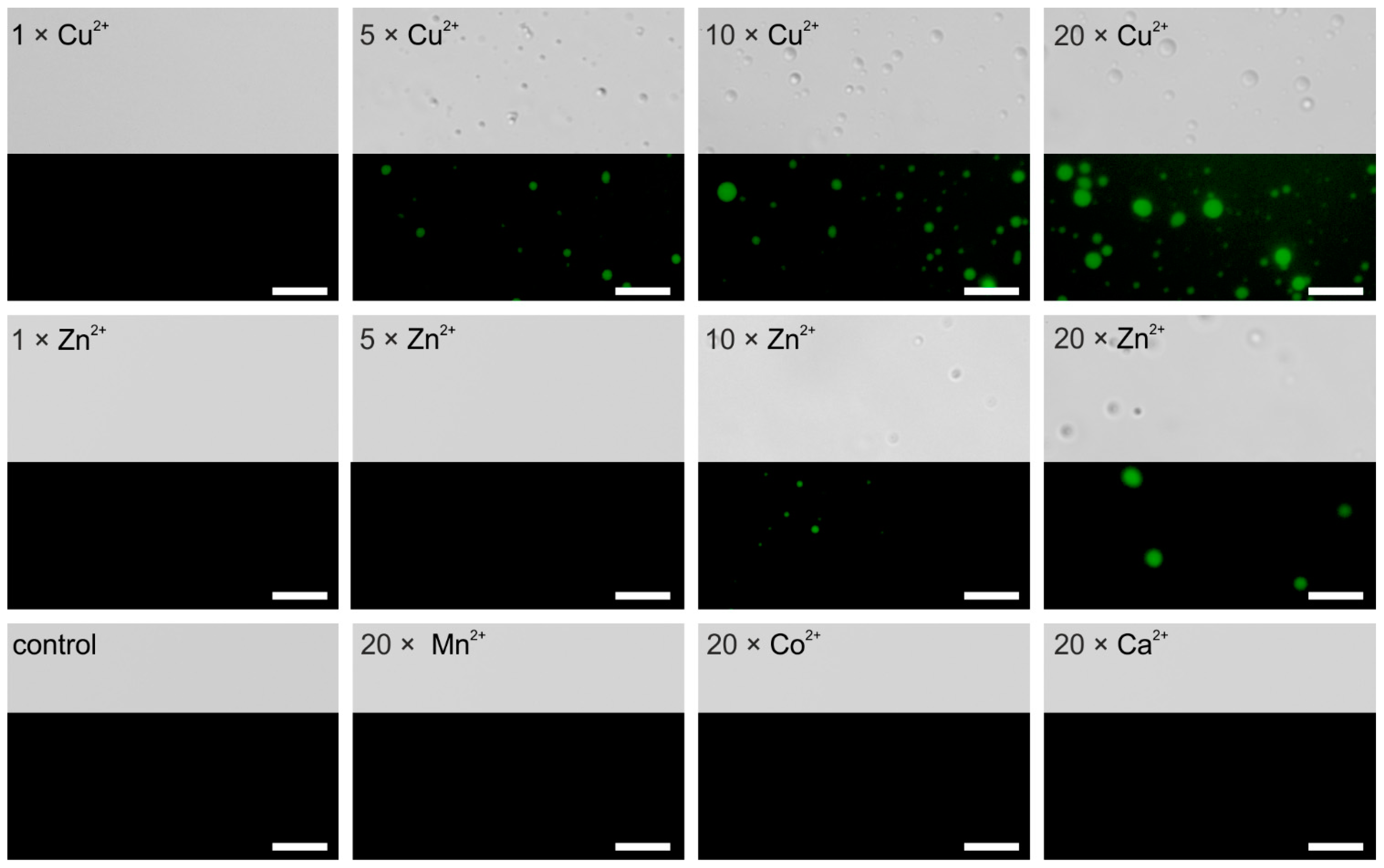
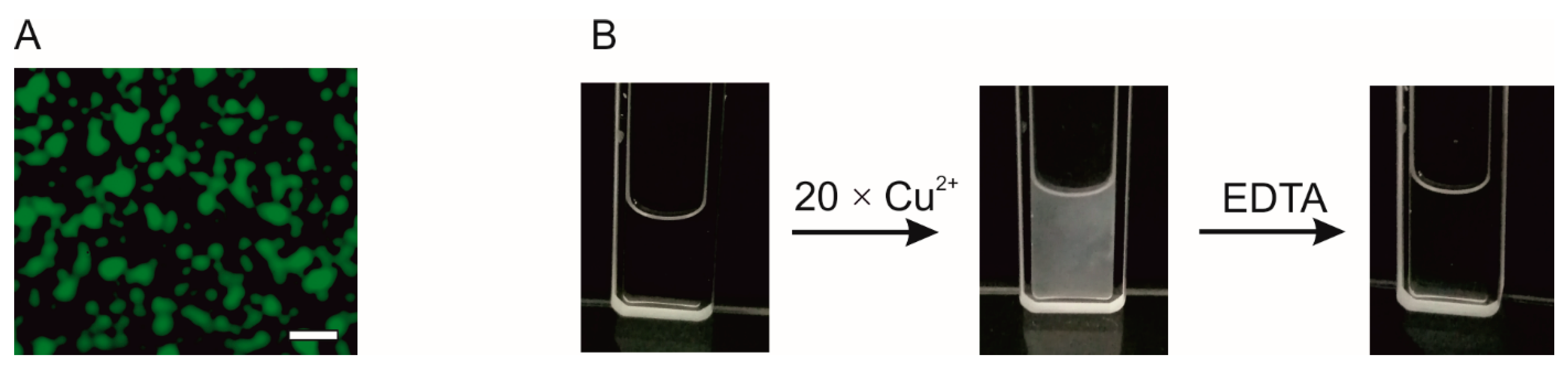
4.2. AaFEcR Provokes LLPS

The EcR is an important transcription regulator in arthropods. The physiological function of AaFEcR at the molecular level, especially in the activities of the full-length AaEcR, is still enigmatic. To investigate if the functionality of AaEcR may depend on the propensity of the AaFEcR formation of liquid condensates, bioinformatic analysis was initially performed. In the case of AaEcR, in silico sequence analysis performed using catGRANULE, PSP [93] and FuzDrop [94] predictors indicates that certain fragments may provoke LLPS especially in the N- and C-terminal fragments (Figure 4A). Comparative analysis of the EcR sequences between the receptor sequences from A. aegypti and D. melanogaster, which possess one of the longest F domains, indicates that both N-terminal domains can probably lead to the formation of liquid condensates (Figure 4B). In both cases, central fragments of the proteins containing folded DBDs and LBDs are characterized by negative scores, which suggests that these protein fragments may lack the ability for LLPS. The predictions obtained for the hinge and F regions gave interesting results. For the hinge region of EcR from D. melanogaster, catGRANULE and FuzDrop predict no propensity for provoking LLPS. For AaFEcR the analysis gave opposite results, i.e., a strong tendency for LLPS was predicted by FuzDrop and a lack of tendency was predicted by catGRANULE. For the sequence related to AaFEcR, high positive scores were obtained in the case of both predictors, whereas for the region of D. melanogaster the analysis was also ambiguous and inconclusive. As stated before, the F regions are the most variable fragments of NRs and they may have remarkable different molecular properties.
For further examination, in vitro analysis of LLPS using the isolated AaFEcR domain was performed. The fluorescently labeled domain was examined microscopically in different conditions. At first, we performed a series of experiments to test if the domain could form condensates in a ligand-independent manner. We tested different concentrations of the protein (ranging from 10–230 µM) in a Tris buffer containing 150 mM sodium chloride; next we tested the F domain (70 µM) in a buffer containing different sodium chloride concentrations ranging from 150–700 mM. Finally, the protein at 70 µM was analyzed in the presence of various buffer additives such as glycerol, polyethylene glycol 300 and 8000, ficol, hexanediol (for more details refer to Materials and Methods section in the Supplementary Materials). The formation of liquid condensates was not observed in any of the tested samples (data not shown). Inspired by our previous studies [10,11], the metal ions were included in the analyses. We found here that for the protein analyzed at 70 µM (1 mg/mL), 5–20× molar excess of Cu2+ and 10–20× molar excess of Zn2+ ions can induce the formation of condensates (Figure 5). Obtained results indicate that Cu2+ ions has a stronger effect since the ions can provoke the formation of condensates at lower molecular excess, whereas the effect of Zn2+ ions is weaker since its concentration to provoke phase separation needs to be higher. Other tested metal ions such as Co2+, Ca2+, and Mn2+ have no effect and, even in a larger molecular excess (20×), the solution remains in a one-phase regime (Figure 5).
The condensates formed by the F domain in the presence of Cu2+ ions are spherical and dynamic (see Video S1 in the Supplementary Materials). Several minutes after formation, they settle on the glass slide and can fuse together forming larger droplets (Figure 6A). That behavior indicates that they have a liquid nature [95]. To further examine the type of the phase transition, we performed an experiment in which to the sample containing F domain in the presence of 20× molar excess of Cu2+ ions, EDTA was added. The addition of the chelating agent to divalent ions dissolved the condensates. The absorbance measured at 340 nm for the unlabeled F domain, 70 µM and 20× Cu2+ ions increased to 0.342. When EDTA was added to the sample containing protein condensates, the absorbance dropped to 0.020 and the solution became clear (Figure 6B). That observation indicates that the formation of the condensates is reversible. Taking into account the spherical shape of the condensates, the fact that after several minutes of their appearance in solution they wetted the glass slide and the fact that their formation is reversible, we conclude that the condensates possess a liquid nature and are formed via the LLPS.
In our previous paper [10], it was shown that Cu2+ ions induce the formation of the PPII helix in the AaFEcR domain. The presented observation indicates that this type of secondary structure can probably accompany LLPS.
Presently, the knowledge regarding metal ions-induced LLPS is scattered [96,97] and this is one of the first reports of the Zn2+ and Cu2+-induced LLPS of disordered proteins. Nonetheless, the putative role of the Cu2+-induced PPII in these processes at the molecular level is waiting to be revealed. At present, however, we know little about the physiological consequences of the formation of metal-induced liquid condensates via LLPS by the F domain. Ion binding by AaFEcR, leading to the formation of liquid condensates, may enhance the assembly of AaEcR transcriptional complexes. AaFEcR may serve as a scaffold for metal ion-gated liquid condensates and may also allow various client molecules to come into the condensate, in turn contributing to the formation of a complete transcriptional machinery. It is also possible that metal ion-induced LLPS of AaFEcR leads to changes in the specificity or affinity of the interaction of the full-length AaEcR with DNA, appropriate ligands, or the receptor’s partners [10,11]. The physiological levels of Cu2+ and Zn2+ ions required to introduce the effector functions in EcR are unknown. In addition, not fully understood are the changes in mosquito cells’ homeostasis upon infection. However, it can be expected that the presence of pathogens may alter to some extent mosquito cells’ physiology and infection may trigger some stress-responsive mechanisms. Stress-induced changes of cell physiology reflect a changed pattern of gene expression [98]. The relation between the pathogen infection and metal ions-induced formation of liquid condensates is currently a puzzle. Yet, understanding of this phenomenon would enable a deeper insight into the mechanism that controls the life cycle and the basic developmental processes of A. aegypti.

5. Summary and Perspectives on Future Research

Mosquito transmitted diseases stand out for their universality and scale, but they have still not yet been overcome. There is no successful vaccine against Zika virus. The vaccine against all 4 serotypes of the dengue virus (Dengvaxia), which was approved by the FDA in 2019, is not recommended for people who have not been infected with the virus in the past [99]. The RTS,S malaria vaccine only provides partial protection [100,101]. Considering vaccine imperfection and the fact that the drug treatment of diseases is not 100% effective, it is essential to find other ways to protect exposed people. Researchers should either focus on reducing the mosquito population, or try to strengthen the immune reaction of people to mosquito-borne viruses or parasite invasion. The reduction of the mosquito population can be influenced by the EcR signaling pathway. By a deep understanding of the LLPS formation by its IDRs, we could change its transcriptional machinery gathering pattern. It is assumed that the multivalent macromolecular interactions are one of the necessary conditions for the formation of biomolecular condensates. In many cases, LLPS is driven by multivalent interactions between IDPs or IDRs and/or their protein or nucleic acid specific partners [102]. One can speculate that EcR from A. aegypti may use its intrinsically disordered F domain with the Cu2+-induced PPII helical structure as the platform for multiple interactions with modulatory proteins controlling gene expression under the control of specific ligands. By modulating the availability of metal ions, the LLPS formation could be disrupted in the appropriate signaling pathway. The 20-hydroxyecdysone signaling is strongly believed to be a promising target for the chemical control of malaria vectors [103]. It also regulates the physiological processes associated with vector competence and the abundance of mosquito vectors [103]. Thus, in a controlled manner, we could vastly and safely reduce the mosquito population and reduce the number of dengue, Zika fever and malaria infections. Undoubtedly, transcription factors play an important role in both the reproduction of mosquitoes and the immune response to Zika, dengue and P. falciparum inflammation. Various pathways are under their control, and therefore transcription factors, together with NRs, can be treated as an attractive target to strengthen an organism’s immune response. Deepening the knowledge about each element of the NRs’ structure will reveal the detailed mechanism of their action. A significant part of previous studies of the NRs’ structure was conducted using in vitro methods and recombinant proteins. Single-molecule in vivo studies of full-length NRs, supported by the recent technological advances in live-cell microscopy, should be applied to this new area of science. We believe that detailed studies of the mechanisms of LLPS in the activities of NRs during transcriptional events will significantly contribute to the development of new strategies to combat not only mosquito-transmitted diseases, but also cancer and other pathological disorders.

Supplementary Materials

The following are available online at https://0-www-mdpi-com.brum.beds.ac.uk/2073-4409/10/3/571/s1, Materials and Methods, Figure S1: The F domains of the insects’ ecdysone receptors vary in sequence and length, Figure S2: The F domains of the representative members of the mammalian nuclear receptors and AaFEcR vary in sequence and length, Figure S3: In silico analyses of disorder occurrence in the F domains of the representative nuclear receptors, Video S1: The condensates formed by the F domain in the presence of Cu2+ ions.

Author Contributions

A.W. and A.T. contributed equally to this work; A.W., A.T., and M.O. conducted the experiments; A.W., A.T., A.O. and M.O. wrote the paper. All authors have read and agreed to the published version of the manuscript.

Funding

The work was supported by a subsidy from the Polish Ministry of Science and Higher Education for the Faculty of Chemistry of Wrocław University of Science and Technology.

Institutional Review Board Statement

Not applicable.

Informed Consent Statement

Not applicable.

Data Availability Statement

Data available within the article and its Supplementary Materials.

Acknowledgments

The authors wish to thank Karolina Partyk (Laboratory of Biochemistry and Molecular Biology, Wrocław University of Science and Technology) for her excellent technical assistance.

Conflicts of Interest

The authors declare no conflict of interest.

References

  1. Aranda, A.; Pascual, A. Nuclear hormone receptors and gene expression. Physiol. Rev. 2001, 81, 1269–1304. [Google Scholar] [CrossRef]
  2. Altucci, L.; Gronemeyer, H. Nuclear receptors in cell life and death. Trends Endocrinol. Metab. 2001, 12, 460–468. [Google Scholar] [CrossRef]
  3. Aagaard, M.M.; Siersbæk, R.; Mandrup, S. Molecular basis for gene-specific transactivation by nuclear receptors. Biochim. Biophys. Acta Mol. Basis Dis. 2011, 1812, 824–835. [Google Scholar] [CrossRef] [PubMed] [Green Version]
  4. Pawlak, M.; Lefebvre, P.; Staels, B. General molecular biology and architecture of nuclear receptors. Curr. Top. Med. Chem. 2012, 12, 486–504. [Google Scholar] [CrossRef] [PubMed] [Green Version]
  5. Mcewan, I.J.; Kumar, R. Nuclear Receptors: From Structure to the Clinic; Springer: Berlin, Germany, 2015; ISBN 978-3-319-18728-0. [Google Scholar]
  6. Schulman, I.G. Nuclear receptors as drug targets for metabolic disease. Adv. Drug Deliv. Rev. 2010, 62, 1307–1315. [Google Scholar] [CrossRef] [Green Version]
  7. Weikum, E.R.; Liu, X.; Ortlund, E.A. The nuclear receptor superfamily: A structural perspective. Protein Sci. 2018, 27, 1876–1892. [Google Scholar] [CrossRef] [PubMed]
  8. Rastinejad, F.; Huang, P.; Chandra, V.; Khorasanizadeh, S. Understanding nuclear receptor form and function using structural biology. J. Mol. Endocrinol. 2013, 51, T1–T21. [Google Scholar] [CrossRef] [Green Version]
  9. Huang, P.; Chandra, V.; Rastinejad, F. Structural Overview of the Nuclear Receptor Superfamily: Insights into Physiology and Therapeutics. Annu. Rev. Physiol. 2010, 72, 247–272. [Google Scholar] [CrossRef] [PubMed] [Green Version]
  10. Rowińska-Żyrek, M.; Więch, A.; Wątły, J.; Wieczorek, R.; Witkowska, D.; Ożyhar, A.; Orłowski, M. Copper(II)-binding induces a unique polyproline type II helical structure within the ion-binding segment in the intrinsically disordered F-domain of ecdysteroid receptor from Aedes aegypti. Inorg. Chem. 2019, 58, 11782–11792. [Google Scholar] [CrossRef] [PubMed]
  11. Więch, A.; Rowińska-Żyrek, M.; Wątły, J.; Czarnota, A.; Hołubowicz, R.; Szewczuk, Z.; Ożyhar, A.; Orłowski, M. The intrinsically disordered C-terminal F domain of the ecdysteroid receptor from Aedes aegypti exhibits metal ion-binding ability. J. Steroid Biochem. Mol. Biol. 2019, 186, 42–55. [Google Scholar] [CrossRef] [PubMed]
  12. Nocula-Ługowska, M.; Rymarczyk, G.; Lisowski, M.; Ożyhar, A. Isoform-specific variation in the intrinsic disorder of the ecdysteroid receptor N-terminal domain. Proteins Struct. Funct. Bioinform. 2009, 76, 291–308. [Google Scholar] [CrossRef]
  13. Pieprzyk, J.; Zbela, A.; Jakób, M.; Ożyhar, A.; Orłowski, M. Homodimerization propensity of the intrinsically disordered N-terminal domain of Ultraspiracle from Aedes aegypti. Biochim. Biophys. Acta Proteins Proteom. 2014, 1844, 1153–1166. [Google Scholar] [CrossRef]
  14. Wärnmark, A.; Wikström, A.; Wright, A.P.H.; Gustafsson, J.Å.; Härd, T. The N-terminal Regions of Estrogen Receptor α and β Are Unstructured in Vitro and Show Different TBP Binding Properties. J. Biol. Chem. 2001, 276, 45939–45944. [Google Scholar] [CrossRef] [PubMed] [Green Version]
  15. Reid, J.; Kelly, S.M.; Watt, K.; Price, N.C.; McEwan, I.J. Conformational analysis of the androgen receptor amino-terminal domain involved in transactivation. Influence of structure-stabilizing solutes and protein-protein interactions. J. Biol. Chem. 2002, 277, 20079–20086. [Google Scholar] [CrossRef] [PubMed] [Green Version]
  16. Dziedzic-Letka, A.; Rymarczyk, G.; Kapłon, T.M.; Górecki, A.; Szamborska-Gbur, A.; Wojtas, M.; Dobryszycki, P.; Ożyhar, A. Intrinsic disorder of Drosophila melanogaster hormone receptor 38 N-terminal domain. Proteins Struct. Funct. Bioinform. 2011, 79, 376–392. [Google Scholar] [CrossRef] [PubMed]
  17. Belorusova, A.Y.; Osz, J.; Petoukhov, M.V.; Peluso-Iltis, C.; Kieffer, B.; Svergun, D.I.; Rochel, N. Solution Behavior of the Intrinsically Disordered N-Terminal Domain of Retinoid X Receptor α in the Context of the Full-Length Protein. Biochemistry 2016, 55, 1741–1748. [Google Scholar] [CrossRef] [Green Version]
  18. Wycisk, K.; Tarczewska, A.; Kaus-Drobek, M.; Dadlez, M.; Hołubowicz, R.; Pietras, Z.; Dziembowski, A.; Taube, M.; Kozak, M.; Orłowski, M.; et al. Intrinsically disordered N-terminal domain of the Helicoverpa armigera Ultraspiracle stabilizes the dimeric form via a scorpion-like structure. J. Steroid Biochem. Mol. Biol. 2018, 183, 167–183. [Google Scholar] [CrossRef]
  19. Sołtys, K.; Ożyhar, A. Ordered structure-forming properties of the intrinsically disordered AB region of hRXRγ and its ability to promote liquid-liquid phase separation. J. Steroid Biochem. Mol. Biol. 2020, 198, 105571. [Google Scholar] [CrossRef] [PubMed]
  20. Uversky, V.N. Natively unfolded proteins: A point where biology waits for physics. Protein Sci. 2002, 11, 739–756. [Google Scholar] [CrossRef] [Green Version]
  21. Simons, S.S.; Edwards, D.P.; Kumar, R. Minireview: Dynamic Structures of Nuclear Hormone Receptors: New Promises and Challenges. Mol. Endocrinol. 2014, 28, 173–182. [Google Scholar] [CrossRef] [Green Version]
  22. Liu, J.; Perumal, N.B.; Oldfield, C.J.; Su, E.W.; Uversky, V.N.; Dunker, A.K. Intrinsic Disorder in Transcription Factors. Biochemistry 2006, 45, 6873–6888. [Google Scholar] [CrossRef] [Green Version]
  23. Dunker, A.K.; Uversky, V.N. Drugs for ‘protein clouds’: Targeting intrinsically disordered transcription factors. Curr. Opin. Pharmacol. 2010, 10, 782–788. [Google Scholar] [CrossRef]
  24. Brady, O.J.; Gething, P.W.; Bhatt, S.; Messina, J.P.; Brownstein, J.S.; Hoen, A.G.; Moyes, C.L.; Farlow, A.W.; Scott, T.W.; Hay, S.I. Refining the Global Spatial Limits of Dengue Virus Transmission by Evidence-Based Consensus. PLoS Negl. Trop. Dis. 2012, 6. [Google Scholar] [CrossRef]
  25. Bhatt, S.; Gething, P.W.; Brady, O.J.; Messina, J.P.; Farlow, A.W.; Moyes, C.L.; Drake, J.M.; Brownstein, J.S.; Hoen, A.G.; Sankoh, O.; et al. The global distribution and burden of dengue. Nature 2013, 496, 504–507. [Google Scholar] [CrossRef]
  26. Bakshi, A.S. Dengue Fever, DHF and DSS. Apollo Med. 2007, 4, 111–117. [Google Scholar] [CrossRef]
  27. Dick, G.W.A.; Kitchen, S.F.; Haddow, A.J. Zika virus. I. Isolations and serological specificity. Trans. R. Soc. Trop. Med. Hyg. 1952, 46, 509–520. [Google Scholar] [CrossRef]
  28. Kuno, G.; Chang, G.J.J. Full-length sequencing and genomic characterization of Bagaza, Kedougou, and Zika viruses. Arch. Virol. 2007, 152, 687–696. [Google Scholar] [CrossRef] [PubMed]
  29. Faye, O.; Freire, C.C.M.; Iamarino, A.; Faye, O.; de Oliveira, J.V.C.; Diallo, M.; Zanotto, P.M.A.; Sall, A.A. Molecular Evolution of Zika Virus during Its Emergence in the 20th Century. PLoS Negl. Trop. Dis. 2014, 8, 36. [Google Scholar] [CrossRef] [Green Version]
  30. Sikka, V.; Chattu, V.K.; Popli, R.K.; Galwankar, S.C.; Kelkar, D.; Sawicki, S.G.; Stawicki, S.P.; Papadimos, T.J. The Emergence of Zika Virus as a Global Health Security Threat: A Review and a Consensus Statement of the INDUSEM Joint working Group (JWG). J. Glob. Infect. Dis. 2018, 8, 3–15. [Google Scholar] [CrossRef]
  31. Oliveira Melo, A.S.; Malinger, G.; Ximenes, R.; Szejnfeld, P.O.; Alves Sampaio, S.; Bispo De Filippis, A.M. Zika virus intrauterine infection causes fetal brain abnormality and microcephaly: Tip of the iceberg? Ultrasound Obstet. Gynecol. 2016, 47, 6–7. [Google Scholar] [CrossRef]
  32. World Health Organization. WHO Malaria Report 2019; World Health Organization: Geneva, Switzerland, 2019; ISBN 9789241565721. [Google Scholar]
  33. Aly, A.S.I.; Vaughan, A.M.; Kappe, S.H.I. Malaria Parasite Development in the Mosquito and Infection of the Mammalian Host. Annu. Rev. Microbiol. 2009, 63, 195–221. [Google Scholar] [CrossRef] [Green Version]
  34. Scotland’s National Health Information Service Malaria. Available online: https://www.nhsinform.scot/illnesses-and-conditions/infections-and-poisoning/malaria#symptoms-of-malaria (accessed on 15 August 2020).
  35. Sager, G.; Gabaglio, S.; Sztul, E.; Belov, G.A. Role of host cell secretory machinery in zika virus life cycle. Viruses 2018, 10, 559. [Google Scholar] [CrossRef] [PubMed] [Green Version]
  36. Sampieri, L.; Di Giusto, P.; Alvarez, C. CREB3 Transcription Factors: ER-Golgi Stress Transducers as Hubs for Cellular Homeostasis. Front. Cell Dev. Biol. 2019, 7, 1–13. [Google Scholar] [CrossRef] [Green Version]
  37. Moni, M.A.; Lio, P. Genetic Profiling and Comorbidities of Zika Infection. J. Infect. Dis. 2017, 216, 703–712. [Google Scholar] [CrossRef]
  38. Malaguarnera, L.; Musumeci, S. The immune response to Plasmodium falciparum malaria. Lancet Infect. Dis. 2002, 2, 472–478. [Google Scholar] [CrossRef]
  39. Tripathi, A.K.; Sha, W.; Shulaev, V.; Stins, M.F.; Sullivan, D.J. Plasmodium falciparum-infected erythrocytes induce NF-κB regulated inflammatory pathways in human cerebral endothelium. Blood 2009, 114, 4243–4252. [Google Scholar] [CrossRef] [Green Version]
  40. Belachew, E.B. Immune Response and Evasion Mechanisms of Plasmodium falciparum Parasites. J. Immunol. Res. 2018, 2018. [Google Scholar] [CrossRef] [PubMed] [Green Version]
  41. Clayton, A.M.; Dong, Y.; Dimopoulos, G. The anopheles innate immune system in the defense against malaria infection. J. Innate Immun. 2014, 6, 169–181. [Google Scholar] [CrossRef]
  42. Souza-Neto, J.A.; Sim, S.; Dimopoulos, G. An evolutionary conserved function of the JAK-STAT pathway in anti-dengue defense. Proc. Natl. Acad. Sci. USA 2009, 106, 17841–17846. [Google Scholar] [CrossRef] [Green Version]
  43. Werling, K.; Shaw, W.R.; Itoe, M.A.; Westervelt, K.A.; Marcenac, P.; Paton, D.G.; Peng, D.; Singh, N.; Smidler, A.L.; South, A.; et al. Steroid Hormone Function Controls Non-competitive Plasmodium Development in Anopheles. Cell 2019, 177, 315–325.e14. [Google Scholar] [CrossRef] [Green Version]
  44. Hyman, A.A.; Weber, C.A.; Jülicher, F. Liquid-Liquid Phase Separation in Biology. Annu. Rev. Cell Dev. Biol. 2014, 30, 39–58. [Google Scholar] [CrossRef] [PubMed] [Green Version]
  45. Mitrea, D.M.; Kriwacki, R.W. Phase separation in biology; functional organization of a higher order. Cell Commun. Signal. 2016, 14, 1. [Google Scholar] [CrossRef] [PubMed] [Green Version]
  46. Shin, Y.; Brangwynne, C.P. Liquid phase condensation in cell physiology and disease. Science 2017, 357, eaaf4382. [Google Scholar] [CrossRef] [Green Version]
  47. Itakura, A.K.; Futia, R.A.; Jarosz, D.F. It Pays To Be in Phase. Biochemistry 2018, 57, 2520–2529. [Google Scholar] [CrossRef] [PubMed]
  48. Gomes, E.; Shorter, J. The molecular language of membraneless organelles. J. Biol. Chem. 2019, 294, 7115–7127. [Google Scholar] [CrossRef] [Green Version]
  49. Uversky, V.N. Protein intrinsic disorder-based liquid–liquid phase transitions in biological systems: Complex coacervates and membrane-less organelles. Adv. Colloid Interface Sci. 2017, 239, 97–114. [Google Scholar] [CrossRef]
  50. Banani, S.F.; Rice, A.M.; Peeples, W.B.; Lin, Y.; Jain, S.; Parker, R.; Rosen, M.K. Compositional Control of Phase-Separated Cellular Bodies. Cell 2016, 166, 651–663. [Google Scholar] [CrossRef] [Green Version]
  51. Molliex, A.; Temirov, J.; Lee, J.; Coughlin, M.; Kanagaraj, A.P.; Kim, H.J.; Mittag, T.; Taylor, J.P. Phase Separation by Low Complexity Domains Promotes Stress Granule Assembly and Drives Pathological Fibrillization. Cell 2015, 163, 123–133. [Google Scholar] [CrossRef] [Green Version]
  52. Posey, A.E.; Holehouse, A.S.; Pappu, R.V. Phase Separation of Intrinsically Disordered Proteins. Methods Enzymol. 2018, 611, 1–30. [Google Scholar] [CrossRef]
  53. Owen, I.; Shewmaker, F. The Role of Post-Translational Modifications in the Phase Transitions of Intrinsically Disordered Proteins. Int. J. Mol. Sci. 2019, 20, 5501. [Google Scholar] [CrossRef] [Green Version]
  54. Nair, S.J.; Yang, L.; Meluzzi, D.; Oh, S.; Yang, F.; Friedman, M.J.; Wang, S.; Suter, T.; Alshareedah, I.; Gamliel, A.; et al. Phase separation of ligand-activated enhancers licenses cooperative chromosomal enhancer assembly. Nat. Struct. Mol. Biol. 2019, 26, 193–203. [Google Scholar] [CrossRef]
  55. Courchaine, E.M.; Lu, A.; Neugebauer, K.M. Droplet organelles? EMBO J. 2016, 35, 1603–1612. [Google Scholar] [CrossRef] [PubMed] [Green Version]
  56. White, J.P.; Lloyd, R.E. Regulation of stress granules in virus systems. Trends Microbiol. 2012, 20, 175–183. [Google Scholar] [CrossRef] [PubMed]
  57. Peng, Q.; Wang, L.; Qin, Z.; Wang, J.; Zheng, X.; Wei, L.; Zhang, X.; Zhang, X.; Liu, C.; Li, Z.; et al. Phase Separation of Epstein-Barr Virus EBNA2 and Its Coactivator EBNALP Controls Gene Expression. J. Virol. 2020, 94. [Google Scholar] [CrossRef]
  58. Perdikari, T.M.; Murthy, A.C.; Ryan, V.H.; Watters, S.; Naik, M.T.; Fawzi, N.L. SARS-CoV-2 nucleocapsid protein undergoes liquid-liquid phase separation stimulated by RNA and partitions into phases of human ribonucleoproteins. BioRxiv 2020. [Google Scholar] [CrossRef]
  59. Cubuk, J.; Alston, J.J.; Incicco, J.J.; Singh, S.; Stuchell-Brereton, M.D.; Ward, M.D.; Zimmerman, M.I.; Vithani, N.; Griffith, D.; Wagoner, J.A.; et al. The SARS-CoV-2 nucleocapsid protein is dynamic, disordered, and phase separates with RNA. BioRxiv 2020. [Google Scholar] [CrossRef]
  60. Alenquer, M.; Vale-Costa, S.; Etibor, T.A.; Ferreira, F.; Sousa, A.L.; Amorim, M.J. Influenza A virus ribonucleoproteins form liquid organelles at endoplasmic reticulum exit sites. Nat. Commun. 2019, 10, 1629. [Google Scholar] [CrossRef] [PubMed] [Green Version]
  61. Monette, A.; Niu, M.; Chen, L.; Rao, S.; Gorelick, R.J.; Mouland, A.J. Pan-retroviral Nucleocapsid-Mediated Phase Separation Regulates Genomic RNA Positioning and Trafficking. Cell Rep. 2020, 31, 107520. [Google Scholar] [CrossRef]
  62. Heinrich, B.S.; Maliga, Z.; Stein, D.A.; Hyman, A.A.; Whelan, S.P.J. Phase Transitions Drive the Formation of Vesicular Stomatitis Virus Replication Compartments. mBio 2018, 9. [Google Scholar] [CrossRef] [Green Version]
  63. Boija, A.; Klein, I.A.; Sabari, B.R.; Dall’Agnese, A.; Coffey, E.L.; Zamudio, A.V.; Li, C.H.; Shrinivas, K.; Manteiga, J.C.; Hannett, N.M.; et al. Transcription Factors Activate Genes through the Phase-Separation Capacity of Their Activation Domains. Cell 2018, 175, 1842–1855.e16. [Google Scholar] [CrossRef] [PubMed] [Green Version]
  64. Boehning, M.; Dugast-Darzacq, C.; Rankovic, M.; Hansen, A.S.; Yu, T.; Marie-Nelly, H.; McSwiggen, D.T.; Kokic, G.; Dailey, G.M.; Cramer, P.; et al. RNA polymerase II clustering through carboxy-terminal domain phase separation. Nat. Struct. Mol. Biol. 2018, 25, 833–840. [Google Scholar] [CrossRef]
  65. Hnisz, D.; Shrinivas, K.; Young, R.A.; Chakraborty, A.K.; Sharp, P.A. A Phase Separation Model for Transcriptional Control. Cell 2017, 169, 13–23. [Google Scholar] [CrossRef] [Green Version]
  66. Sawyer, I.A.; Bartek, J.; Dundr, M. Phase separated microenvironments inside the cell nucleus are linked to disease and regulate epigenetic state, transcription and RNA processing. Semin. Cell Dev. Biol. 2019, 90, 94–103. [Google Scholar] [CrossRef]
  67. Sabari, B.R.; Dall’Agnese, A.; Boija, A.; Klein, I.A.; Coffey, E.L.; Shrinivas, K.; Abraham, B.J.; Hannett, N.M.; Zamudio, A.V.; Manteiga, J.C.; et al. Coactivator condensation at super-enhancers links phase separation and gene control. Science 2018, 361, eaar3958. [Google Scholar] [CrossRef] [Green Version]
  68. Tarczewska, A.; Greb-Markiewicz, B. The Significance of the Intrinsically Disordered Regions for the Functions of the bHLH Transcription Factors. Int. J. Mol. Sci. 2019, 20, 5306. [Google Scholar] [CrossRef] [Green Version]
  69. Lu, Y.; Wu, T.; Gutman, O.; Lu, H.; Zhou, Q.; Henis, Y.I.; Luo, K. Phase separation of TAZ compartmentalizes the transcription machinery to promote gene expression. Nat. Cell Biol. 2020, 22, 453–464. [Google Scholar] [CrossRef] [PubMed]
  70. Bouchard, J.J.; Otero, J.H.; Scott, D.C.; Szulc, E.; Martin, E.W.; Sabri, N.; Granata, D.; Marzahn, M.R.; Lindorff-Larsen, K.; Salvatella, X.; et al. Cancer Mutations of the Tumor Suppressor SPOP Disrupt the Formation of Active, Phase-Separated Compartments. Mol. Cell 2018, 72, 19–36.e8. [Google Scholar] [CrossRef] [PubMed] [Green Version]
  71. An, J.; Wang, C.; Deng, Y.; Yu, L.; Huang, H. Destruction of Full-Length Androgen Receptor by Wild-Type SPOP, but Not Prostate-Cancer-Associated Mutants. Cell Rep. 2014, 6, 657–669. [Google Scholar] [CrossRef] [Green Version]
  72. Brangwynne, C.P.; Tompa, P.; Pappu, R.V. Polymer physics of intracellular phase transitions. Nat. Phys. 2015, 11, 899–904. [Google Scholar] [CrossRef]
  73. Protter, D.S.W.; Parker, R. Principles and Properties of Stress Granules. Trends Cell Biol. 2016, 26, 668–679. [Google Scholar] [CrossRef] [Green Version]
  74. Quiroz, F.G.; Chilkoti, A. Sequence heuristics to encode phase behaviour in intrinsically disordered protein polymers. Nat. Mater. 2015, 14, 1164–1171. [Google Scholar] [CrossRef]
  75. Stortz, M.; Presman, D.M.; Bruno, L.; Annibale, P.; Dansey, M.V.; Burton, G.; Gratton, E.; Pecci, A.; Levi, V. Mapping the Dynamics of the Glucocorticoid Receptor within the Nuclear Landscape. Sci. Rep. 2017. [Google Scholar] [CrossRef] [Green Version]
  76. Stortz, M.; Pecci, A.; Presman, D.M.; Levi, V. Unraveling the molecular interactions involved in phase separation of glucocorticoid receptor. BMC Biol. 2020, 18, 59. [Google Scholar] [CrossRef]
  77. Dao, T.P.; Kolaitis, R.-M.; Kim, H.J.; O’Donovan, K.; Martyniak, B.; Colicino, E.; Hehnly, H.; Taylor, J.P.; Castañeda, C.A. Ubiquitin Modulates Liquid-Liquid Phase Separation of UBQLN2 via Disruption of Multivalent Interactions. Mol. Cell 2018, 69, 965–978.e6. [Google Scholar] [CrossRef] [Green Version]
  78. Patel, S.R.; Skafar, D.F. Modulation of nuclear receptor activity by the F domain. Mol. Cell. Endocrinol. 2015, 418, 298–305. [Google Scholar] [CrossRef]
  79. Sievers, F.; Wilm, A.; Dineen, D.; Gibson, T.J.; Karplus, K.; Li, W.; Lopez, R.; McWilliam, H.; Remmert, M.; Söding, J.; et al. Fast, scalable generation of high-quality protein multiple sequence alignments using Clustal Omega. Mol. Syst. Biol. 2011, 7, 539. [Google Scholar] [CrossRef]
  80. Bianchetti, L.; Wassmer, B.; Defosset, A.; Smertina, A.; Tiberti, M.L.; Stote, R.H.; Dejaegere, A. Alternative dimerization interfaces in the glucocorticoid receptor-α ligand binding domain. Biochim. Biophys. Acta Gen. Subj. 2018, 1862, 1810–1825. [Google Scholar] [CrossRef]
  81. Arao, Y.; Korach, K.S. The F domain of estrogen receptor α is involved in species-specific, tamoxifen-mediated transactivation. J. Biol. Chem. 2018, 293, 8495–8507. [Google Scholar] [CrossRef] [Green Version]
  82. Guzman, M.G.; Halstead, S.B.; Artsob, H.; Buchy, P.; Farrar, J.; Nathan, M.B.; Pelegrino, J.L.; Simmons, C.; Yoksan, S. Dengue: A continuing global threat Europe PMC Funders Author Manuscripts. Nat. Rev. Microbiol. 2010, 8, S7–S16. [Google Scholar] [CrossRef] [PubMed] [Green Version]
  83. Marchette, N.J.; Garcia, R.; Rudnick, A. Isolation of Zika virus from Aedes aegypti mosquitoes in Malaysia. Am. J. Trop. Med. Hyg. 1969, 18, 411–415. [Google Scholar] [CrossRef]
  84. Barrett, A.D.T.; Higgs, S. Yellow fever: A disease that has yet to be conquered. Annu. Rev. Entomol. 2007, 52, 209–229. [Google Scholar] [CrossRef] [Green Version]
  85. Tsetsarkin, K.A.; Chen, R.; Weaver, S.C. Interspecies transmission and chikungunya virus emergence. Curr. Opin. Virol. 2016, 16, 143–150. [Google Scholar] [CrossRef] [PubMed] [Green Version]
  86. Raikhel, A.S.; Kokoza, V.; Zhu, J.; Martin, D.; Wang, S.F.; Li, C.; Sun, G.; Ahmed, A.; Dittmer, N.; Attardo, G. Molecular biology of mosquito vitellogenesis: From basic studies to genetic engineering of antipathogen immunity. Insect Biochem. Mol. Biol. 2002, 32, 1275–1286. [Google Scholar] [CrossRef]
  87. Zhang, Y. I-TASSER server for protein 3D structure prediction. BMC Bioinform. 2008, 9, 40. [Google Scholar] [CrossRef] [Green Version]
  88. Ranieri-Raggi, M.; Moir, A.; Raggi, A. The Role of Histidine-Proline-Rich Glycoprotein as Zinc Chaperone for Skeletal Muscle AMP Deaminase. Biomolecules 2014, 4, 474–497. [Google Scholar] [CrossRef] [Green Version]
  89. Adzhubei, A.A.; Sternberg, M.J.E.; Makarov, A.A. Polyproline-II helix in proteins: Structure and function. J. Mol. Biol. 2013, 425, 2100–2132. [Google Scholar] [CrossRef] [PubMed]
  90. Kay, B.K.; Williamson, M.P.; Sudol, M. The importance of being proline: The interaction of proline-rich motifs in signaling proteins with their cognate domains. FASEB J. 2000, 14, 231–241. [Google Scholar] [CrossRef] [PubMed]
  91. Hicks, J.M.; Hsu, V.L. The Extended Left-Handed Helix: A Simple Nucleic Acid-Binding Motif. Proteins Struct. Funct. Genet. 2004, 55, 330–338. [Google Scholar] [CrossRef] [PubMed]
  92. Siligardi, G.; Drake, A.F. The importance of extended conformations and, in particular, the PII conformation for the molecular recognition of peptides. Biopolymers 1995, 37, 281–292. [Google Scholar] [CrossRef] [PubMed]
  93. Vernon, R.M.; Chong, P.A.; Tsang, B.; Kim, T.H.; Bah, A.; Farber, P.; Lin, H.; Forman-Kay, J.D. Pi-Pi contacts are an overlooked protein feature relevant to phase separation. eLife 2018, 7. [Google Scholar] [CrossRef]
  94. Horvath, A.; Miskei, M.; Ambrus, V.; Vendruscolo, M.; Fuxreiter, M. Sequence-based prediction of protein binding mode landscapes. PLoS Comput. Biol. 2020, 16, e1007864. [Google Scholar] [CrossRef] [PubMed]
  95. Alberti, S.; Gladfelter, A.; Mittag, T. Considerations and Challenges in Studying Liquid-Liquid Phase Separation and Biomolecular Condensates. Cell 2019, 176, 419–434. [Google Scholar] [CrossRef] [PubMed] [Green Version]
  96. Hong, K.; Song, D.; Jung, Y. Behavior control of membrane-less protein liquid condensates with metal ion-induced phase separation. Nat. Commun. 2020, 11, 5554. [Google Scholar] [CrossRef]
  97. Monette, A.; Mouland, A.J. Zinc and Copper Ions Differentially Regulate Prion-Like Phase Separation Dynamics of Pan-Virus Nucleocapsid Biomolecular Condensates. Viruses 2020, 12, 1179. [Google Scholar] [CrossRef]
  98. Rao, A.K.; Ziegler, Y.S.; McLeod, I.X.; Yates, J.R.; Nardulli, A.M. Effects of Cu/Zn superoxide dismutase on estrogen responsiveness and oxidative stress in human breast cancer cells. Mol. Endocrinol. 2008, 22, 1113–1124. [Google Scholar] [CrossRef] [PubMed] [Green Version]
  99. FDA First FDA-Approved Vaccine for the Prevention of Dengue Disease in Endemic Regions. Available online: https://www.fda.gov/news-events/press-announcements/first-fda-approved-vaccine-prevention-dengue-disease-endemic-regions (accessed on 1 May 2020).
  100. Alonso, P.L.; Sacarlal, J.; Aponte, J.J.; Leach, A.; Macete, E.; Milman, J.; Mandomando, I.; Spiessens, B.; Guinovart, C.; Espasa, M.; et al. Efficacy of the RTS,S/AS02A vaccine against Plasmodium falciparum infection and disease in young African children: Randomised controlled trial. Lancet 2004, 364, 1411–1420. [Google Scholar] [CrossRef]
  101. Centers for Disease Control and Prevention How to Reduce Malaria’s Impact. Available online: https://www.cdc.gov/malaria/malaria_worldwide/reduction/vaccine.html (accessed on 1 May 2020).
  102. Fung, H.Y.J.; Birol, M.; Rhoades, E. IDPs in macromolecular complexes: The roles of multivalent interactions in diverse assemblies. Curr. Opin. Struct. Biol. 2018, 49, 36–43. [Google Scholar] [CrossRef]
  103. Ekoka, E.; Maharaj, S.; Nardini, L.; Dahan-Moss, Y.; Koekemoer, L.L. 20-Hydroxyecdysone (20E) signaling as a promising target for the chemical control of malaria vectors. Parasites Vectors 2021, 14, 86. [Google Scholar] [CrossRef] [PubMed]
Figure 1. Scheme of the overall structural and functional organization of nuclear receptors (NRs). Nuclear receptors contain at least four distinct domains: a N-terminal domain (NTD), a DNA-binding domain (DBD), a hinge region, and a ligand-binding domain (LBD). Some NRs possess an additional C-terminal F domain. The contribution of each domain to the NR’s activities is different. The most important functions and properties located in the individual domains are listed below the scheme [1]. The F domain is marked in red [10,11].
Cells 10 00571 g001
Figure 2. The F domains of various nuclear receptors are not evolutionarily conserved. The used abbreviations and the UniProt accession numbers (in parentheses) are as follows: A. aegypti EcR (AaEcR, GenBank: AAA87394.1), human retinoid X receptor α (RXRα, UniProt: P19793), human all-trans retinoic receptor (RARα, UniProt: P10276), human glucocorticoid receptor α (GRα, UniProt: P04150), human estrogen receptor α (ERα, UniProt: P03372), A. aegypti Usp protein (AaUsp, UniProt: Q9GSG7), human androgen receptor (AR, P10275), human progesterone receptor (PR, UniProt: P06401), human thyroid hormone receptor (TR, UniProt: P10827-2), human vitamin D receptor (VDR, UniProt: P11473), human peroxisome proliferator-activated receptor γ (PPARγ, UniProt: P37231), and human mineralocorticoid receptor (MR, UniProt: P08235). Residue numbering corresponds to the sequence of AaEcR (GenBank: AAA87394.1). Sequences corresponding to the F domain are boxed in red. All representative amino acid sequences were compared using the ClustalΩ tool [79].
Cells 10 00571 g002
Figure 3. Superposition of the corresponding Cα atoms of the 3D structures of the LBDs of the exemplary representatives of NR LBDs with the 3D model of AaEcRLBD+F. The figure shows the superposition of the corresponding Cα atoms of ARLBD (blue) (PDB accession code 4oea), ERαLBD (cyan) (PDB accession code 1g50), EcRLBD from Heliothis virescens (HvEcRLBD) (orange) (PDB accession code 2r40) and the 3D model of AaEcRLBD with the F domain (AaEcRLBD+F). The 3D model of the depicted structure of AaEcRLBD+F (green) was generated using the I-TASSER server for predicting the 3D structure of the protein (http://zhanglab.ccmb.med.umich.edu/I-TASSER/(accessed on 14 August 2020)) [87] using the sequence of the full-length AaEcR (GenBank: AAA87394.1). All structures were visualized using PyMOL (PyMOL Molecular Graphics System, Version 2.0.4, Schrödinger, LLC, New York, NY, USA).
Cells 10 00571 g003
Figure 4. In silico analyses of the phase separation propensity of AaEcR. (A) Analysis performed for the sequence of EcR from A. aegypti. The propensity profiles refer to a scheme depicting the modular structure of the protein (above). Values above 0 indicate a putative tendency for provoking LLPS predicted by catGRANULE (black) and PSP (red) predictors, whereas values above 0.5 indicate a probable tendency predicted by FuzDrop (green). (B) Averaged propensity score values obtained for fragments of the EcR from A. aegypti (GenBank: AAA87394.1), and D. melanogaster (Uniprot: P34021). The black (A. aegypti) and grey (D. melanogaster) bars show the analysis performed using the catGRANULE predictor, asterisks show the results of an analysis performed using FuzDrop. The used predictors are available at: catGRANULE: http://s.tartaglialab.com/update_submission/329388/68a2757da8 (accessed on 15 September 2020); PSP: http://abragam.med.utoronto.ca/~JFKlab/Software/psp.htm (accessed on 15 September 2020); FuzDrop: http://protdyn-fuzpred.org/ (accessed on 26 January 2021).
Cells 10 00571 g004
Figure 5. Metal ions-induced formation of liquid condensates by AaFEcR. The representative images of condensates observed using widefield microscopy with Differential Interference Contrast (DIC) (top) and fluorescence (bottom). The 70 µM solution of fluorescently labelled recombinant AaFEcR in 10 mM Tris-HCl, 150 mM NaCl, and pH 7.5 buffer was analyzed. The protein solution was supplemented with various metal ions in molar excess as indicated on the image. Scale bar 5 µm.
Cells 10 00571 g005
Figure 6. AaFEcR domain containing condensates exhibit liquid properties. (A) shows condensates formed by 70 µM fluorescently labeled F domain in 10 mM Tris-HCl, 150 mM NaCl, and pH 7.5 buffer supplemented with 20× molar excess of Cu2+ ions. The condensates wet the glass slide. (B) The changes of the F domain solution turbidity caused by the addition of 20× molar excess of Cu2+ ions and EDTA. Scale bar 5 µm.
Cells 10 00571 g006
Publisher’s Note: MDPI stays neutral with regard to jurisdictional claims in published maps and institutional affiliations.

