
3D-Printed Collagen Scaffolds Promote Maintenance of Cryopreserved Patients-Derived Melanoma Explants

 , , ,
, , ,  , and
, and 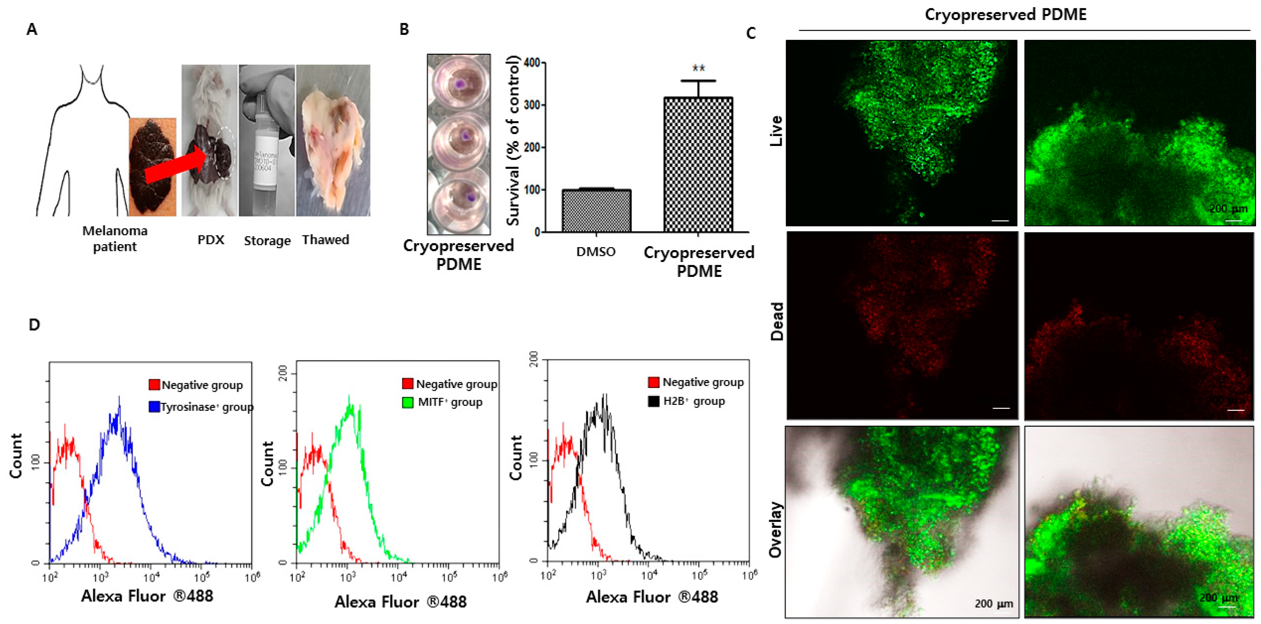
Abstract
:1. Introduction
2. Materials and Methods
2.1. Cell Line and Reagents
2.2. PDME, PDX Model and Cryopreservation
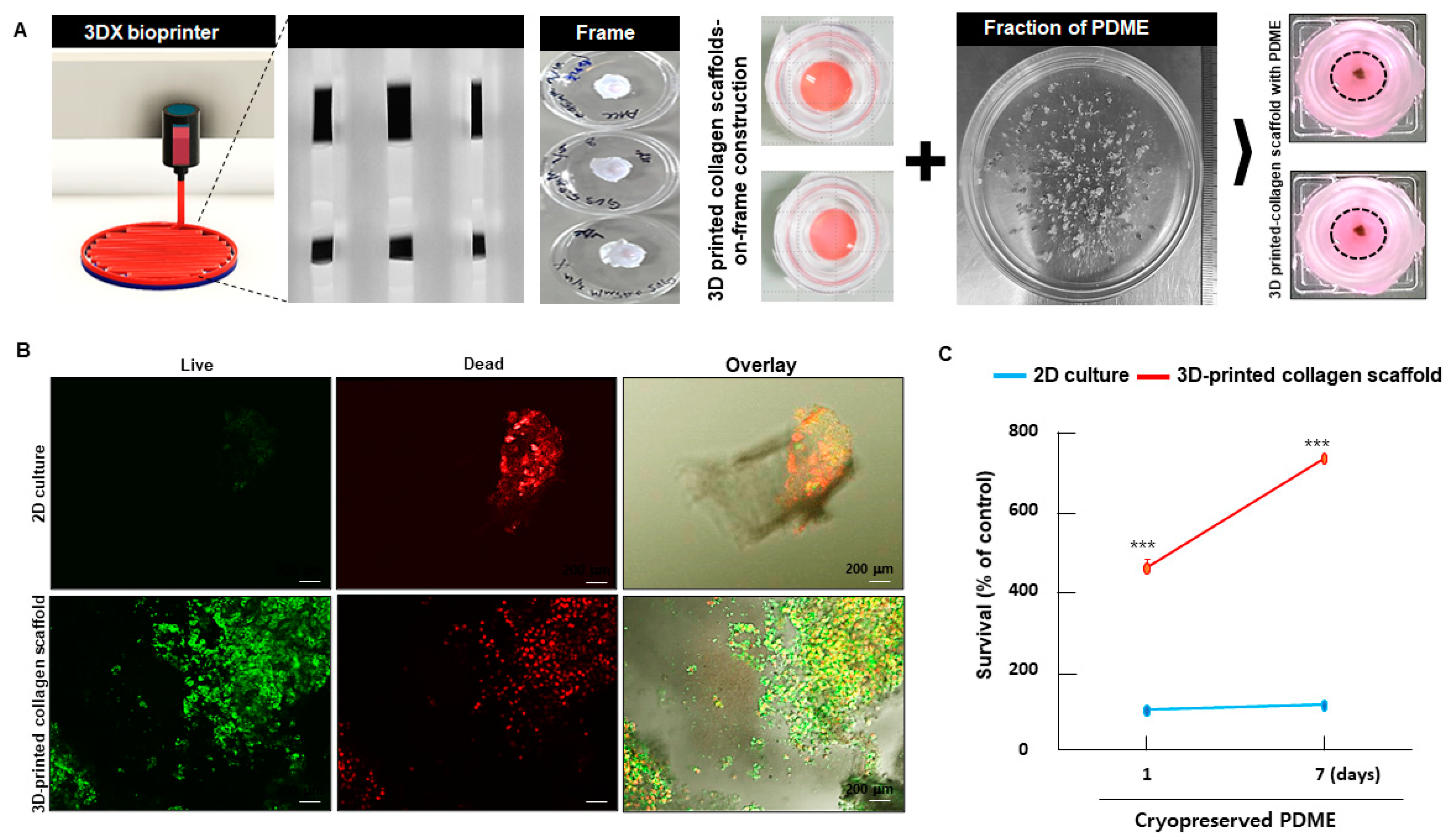
2.3. Fabrication of 3D-Printed Collagen Scaffold-on-Frame Construction and Loaded with Cryopreserved PDME
2.4. The Viability and Proliferation Assay
2.5. LIVE/DEAD Staining
2.6. Flow Cytometric Analysis
2.7. Immunohistochemistry (IHC) and Immunofluorescence Staining (IFS)
2.8. Western Blot Analysis
2.9. Statistical Analysis
3. Results
3.1. The Viability and Characterization of Cryopreserved PDME after Thawing
3.2. Differences in 2D vs. 3D-Printed Collagen-Scaffolds-on-Frame Construction for Short-Term Maintenance of Cryopreserved PDME
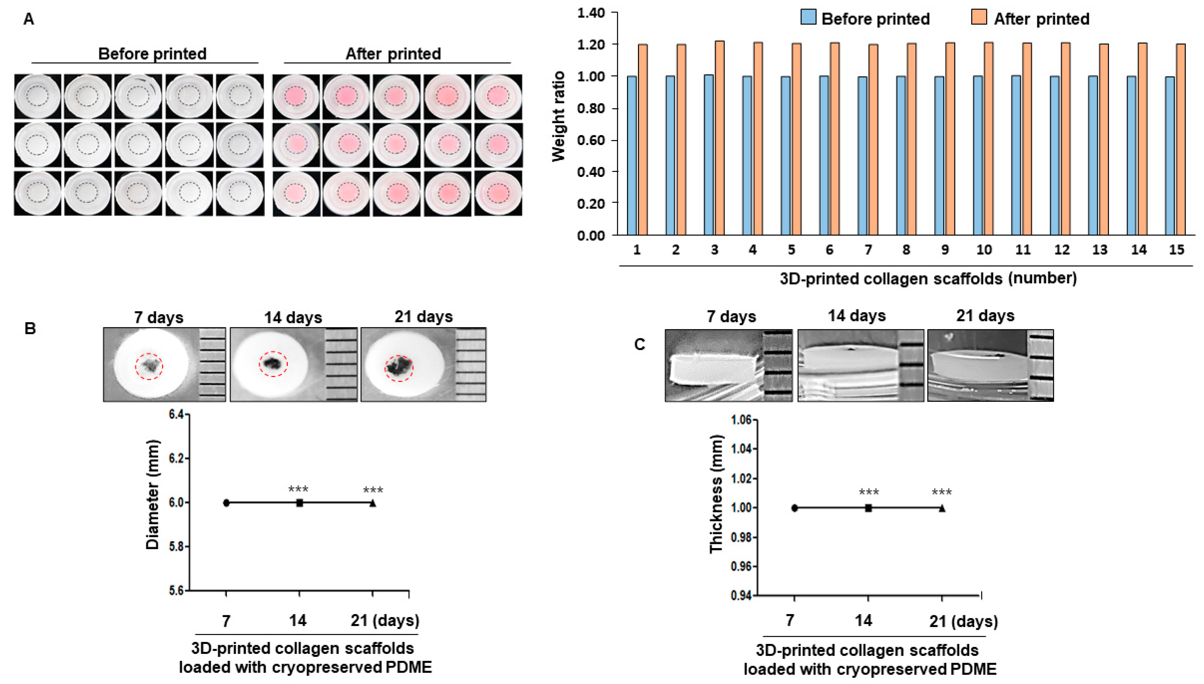
3.3. The Uniformity of Morpholgy and the Shape stability of 3D-Printed Collagen-Scaffolds-on-Frame Construction for Subsequent Long-Term Maintenance of Cryopreserved PDME
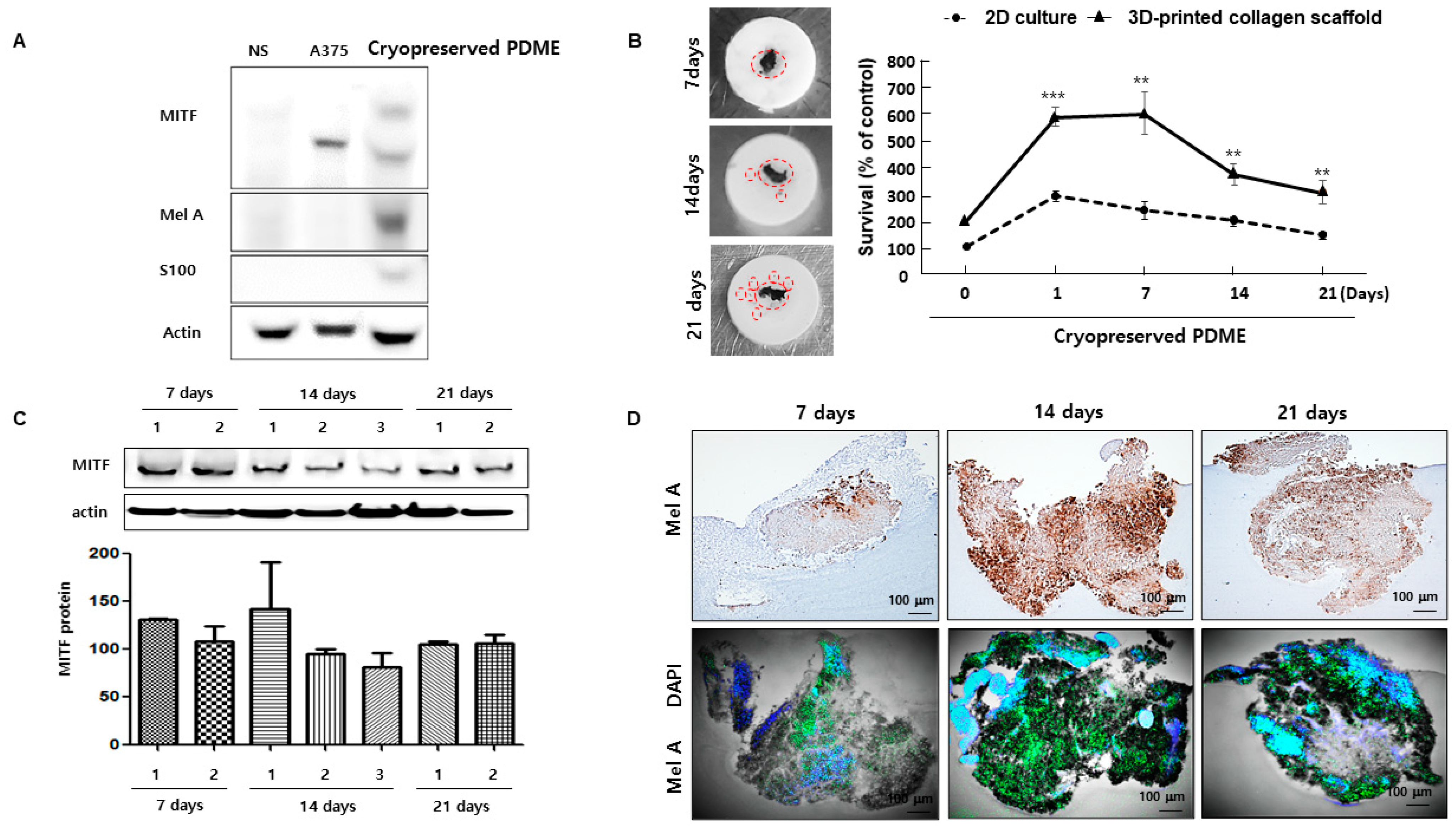
3.4. 3D-Printed Collagen Scaffolds Help to Maintain the Functional Health of Cryopreserved PDME
4. Discussion
5. Conclusions
Supplementary Materials
Author Contributions
Funding
Institutional Review Board Statement
Informed Consent Statement
Data Availability Statement
Acknowledgments
Conflicts of Interest
References
- Rebecca, V.W.; Somasundaram, R.; Herlyn, M. Pre-clinical modeling of cutaneous melanoma. Nat. Commun. 2020, 11, 2858–2867. [Google Scholar] [CrossRef] [PubMed]
- Efimenko, M.; Ignatev, A.; Koshechkin, K. Review of medical image recognition technologies to detect melanomas using neural networks. BMC Bioinform. 2020, 21, 270–277. [Google Scholar] [CrossRef]
- Jones, O.T.; Ranmuthu, C.K.I.; Hall, P.N.; Funston, G.; Walter, F.M. Recognizing Skin Cancer in Primary Care. Adv. Ther. 2020, 37, 603–616. [Google Scholar] [CrossRef] [Green Version]
- Ramón, Y.; Cajal, S.; Sesé, M.; Capdevila, C.; Aasen, T.; De, M.A.L.; Diaz, C.S.J.; Hernández, L.J.; Castellví, J. Clinical implications of intratumor heterogeneity: Challenges and opportunities. J. Mol. Med. 2020, 98, 161–177. [Google Scholar] [CrossRef] [PubMed] [Green Version]
- Kozar, I.; Margue, C.; Rothengatter, S.; Haan, C.; Kreis, S. Many ways to resistance: How melanoma cells evade targeted therapies. Biochim. Biophys. Acta Rev. Cancer. 2019, 1871, 313–322. [Google Scholar] [CrossRef]
- Xiao, M.; Rebecca, V.W.; Herlyn, M. A Melanoma Patient-Derived Xenograft Model. J. Vis. Exp. 2019, 147. [Google Scholar] [CrossRef]
- Kageyama, K.; Ozaki, S.; Sato, T. Generation of a Liver Orthotopic Human Uveal Melanoma Xenograft Platform in Immunodeficient Mice. J. Vis. Exp. 2019, 153. [Google Scholar] [CrossRef]
- Ice, R.J.; Chen, M.; Sidorov, M.; Le, H.T.; Woo, R.W.L.; Rodriguez, B.A.; Luu, T.; Jian, D.; Kim, K.B.; Leong, S.P.; et al. Drug responses are conserved across patient-derived xenograft models of melanoma leading to identification of novel drug combination therapies. Br. J. Cancer 2020, 122, 648–657. [Google Scholar] [CrossRef]
- Yoshida, G.J. Applications of patient-derived tumor xenograft models and tumor organoids. J. Hematol. Oncol. 2020, 13, 4. [Google Scholar] [CrossRef]
- Datta, P.; Dey, M.; Ataie, Z.; Unutmaz, D.; Ozbolat, I.T. 3D bioprinting for reconstituting the cancer microenvironment. NPJ Precis. Oncol. 2020, 4, 18. [Google Scholar] [CrossRef] [PubMed]
- Murphy, S.V.; De, C.P.; Atala, A. Opportunities and challenges of translational 3D bioprinting. Nat. Biomed. Eng. 2020, 4, 370–380. [Google Scholar] [CrossRef]
- Quílez, C.; de Aranda, I.G.; García, M.; López, V.; Montero, A.; Valencia, L.; Velasco, D. Bioprinting for Skin. Methods Mol. Biol. 2020, 2140, 217–228. [Google Scholar] [PubMed]
- Kang, D.; Hong, G.; An, S.; Jang, I.; Yun, W.S.; Shim, J.H.; Jin, S.W. Bioprinting of Multiscaled Hepatic Lobules within a Highly Vascularized Construct. Small 2020, 16, e1905505. [Google Scholar] [CrossRef] [PubMed]
- Aughton, K.; Shahidipour, H.; Djirackor, L.; Coupland, S.E.; Kalirai, H. Characterization of Uveal Melanoma Cell Lines and Primary Tumor Samples in 3D Culture. Transl. Vis. Sci. Technol. 2020, 9, 39. [Google Scholar] [CrossRef]
- Jeong, Y.M.; Cheng, X.W.; Kim, W. Substance P Administered after Myocardial Infarction Upregulates Microphthalmia-Associated Transcription Factor, GATA4, and the Expansion of c-Kit+ Cells. Stem Cells Int. 2020, 2020, 1835950. [Google Scholar] [CrossRef]
- Ivanics, T.; Bergquist, J.R.; Liu, G.; Kim, M.P.; Kang, Y.; Katz, M.H.; Perez, M.V.R.; Thomas, R.M.; Fleming, J.B.; Truty, M.J. Patient-derived xenograft cryopreservation and reanimation outcomes are dependent on cryoprotectant type. Lab. Investig. 2018, 98, 947–956. [Google Scholar] [CrossRef] [PubMed]
- Chacón, M.; Pfluger, Y.; Angel, M.; Waisberg, F.; Enrico, D. Uncommon Subtypes of Malignant Melanomas: A Review Based on Clinical and Molecular Perspectives. Cancers 2020, 12, 2362. [Google Scholar] [CrossRef]
- Osidak, E.O.; Kozhukhov, V.I.; Osidak, M.S.; Domogatsky, S.P. Collagen as Bioink for Bioprinting: A Comprehensive Review. Int. J. Bioprint. 2020, 6, 270. [Google Scholar]
- Sapudom, J.; Pompe, T. Biomimetic tumor microenvironments based on collagen matrices. Biomater. Sci. 2018, 6, 2009–2024. [Google Scholar] [CrossRef]
- Kapałczyńska, M.; Kolenda, T.; Przybyła, W.; Zajączkowska, M.; Teresiak, A.; Filas, V.; Ibbs, M.; Bliźniak, R.; Łuczewski, Ł.; Lamperska, K. 2D and 3D cell cultures—A comparison of different types of cancer cell cultures. Arch. Med. Sci. 2018, 14, 910–919. [Google Scholar] [CrossRef]
- Cruz, R.N.; Lineros, J.; Rodríguez, C.S.; Martínez, L.M.; Rodríguez, J.A. Establishment of Two Dimensional (2D) and Three-Dimensional (3D) Melanoma Primary Cultures as a Tool for In Vitro Drug Resistance Studies. Methods Mol. Biol. 2019, 1913, 119–131. [Google Scholar]
- Fontoura, J.C.; Viezzer, C.; Dos, S.F.G.; Ligabue, R.A.; Weinlich, R.; Puga, R.D.; Antonow, D.; Severino, P.; Bonorino, C. Comparison of 2D and 3D cell culture models for cell growth, gene expression and drug resistance. Mater. Sci. Eng. C Mater. Biol. Appl. 2020, 107, 110264. [Google Scholar] [CrossRef] [PubMed]
- Khot, M.I.; Levenstein, M.A.; de Boer, G.N.; Armstrong, G.; Maisey, T.; Svavarsdottir, H.S.; Andrew, H.; Perry, S.L.; Kapur, N.; Jayne, D.G. Characterising a PDMS based 3D cell culturing microfluidic platform for screening chemotherapeutic drug cytotoxic activity. Sci. Rep. 2020, 10, 15915. [Google Scholar] [CrossRef] [PubMed]
- Wan, X.; Li, Z.; Ye, H.; Cui, Z. Three-dimensional perfused tumour spheroid model for anti-cancer drug screening. Biotechnol. Lett. 2016, 38, 1389–1395. [Google Scholar] [CrossRef] [Green Version]
- Boussadia, Z.; Lamberti, J.; Mattei, F.; Pizzi, E.; Puglisi, R.; Zanetti, C.; Pasquini, L.; Fratini, F.; Fantozzi, L.; Felicetti, F.; et al. Acidic microenvironment plays a key role in human melanoma progression through a sustained exosome mediated transfer of clinically relevant metastatic molecules. J. Exp. Clin. Cancer. Res. 2018, 37, 245. [Google Scholar] [CrossRef] [PubMed]
- Miskolczi, Z.; Smith, M.P.; Rowling, E.J.; Ferguson, J.; Barriuso, J.; Wellbrock, C. Collagen abundance controls melanoma phenotypes through lineage-specific microenvironment sensing. Oncogene 2018, 37, 3166–3182. [Google Scholar] [CrossRef] [Green Version]
- Zhu, H.; Liu, Q.; Miao, L.; Musetti, S.; Huo, M.; Huang, L. Remodeling the fibrotic tumor microenvironment of desmoplastic melanoma to facilitate vaccine immunotherapy. Nanoscale 2020, 7, 3400–3410. [Google Scholar] [CrossRef] [PubMed]
- van Kempen, L.C.; Rijntjes, J.; Mamor-Cornelissen, I.; Vincent-Naulleau, S.; Gerritsen, M.J.; Ruiter, D.J.; van Dijk, M.C.; Geffrotin, C.; van Muijen, G.N. Type I collagen expression contributes to angiogenesis and the development of deeply invasive cutaneous melanoma. Int. J. Cancer 2008, 122, 1019–1029. [Google Scholar] [CrossRef]


Publisher’s Note: MDPI stays neutral with regard to jurisdictional claims in published maps and institutional affiliations. |
© 2021 by the authors. Licensee MDPI, Basel, Switzerland. This article is an open access article distributed under the terms and conditions of the Creative Commons Attribution (CC BY) license (http://creativecommons.org/licenses/by/4.0/).
Share and Cite
Jeong, Y.-M.; Bang, C.; Park, M.; Shin, S.; Yun, S.; Kim, C.M.; Jeong, G.; Chung, Y.-J.; Yun, W.-S.; Lee, J.H.; et al. 3D-Printed Collagen Scaffolds Promote Maintenance of Cryopreserved Patients-Derived Melanoma Explants. Cells 2021, 10, 589. https://0-doi-org.brum.beds.ac.uk/10.3390/cells10030589
Jeong Y-M, Bang C, Park M, Shin S, Yun S, Kim CM, Jeong G, Chung Y-J, Yun W-S, Lee JH, et al. 3D-Printed Collagen Scaffolds Promote Maintenance of Cryopreserved Patients-Derived Melanoma Explants. Cells. 2021; 10(3):589. https://0-doi-org.brum.beds.ac.uk/10.3390/cells10030589
Chicago/Turabian StyleJeong, Yun-Mi, ChulHwan Bang, MinJi Park, Sun Shin, Seokhwan Yun, Chul Min Kim, GaHee Jeong, Yeun-Jun Chung, Won-Soo Yun, Ji Hyun Lee, and et al. 2021. "3D-Printed Collagen Scaffolds Promote Maintenance of Cryopreserved Patients-Derived Melanoma Explants" Cells 10, no. 3: 589. https://0-doi-org.brum.beds.ac.uk/10.3390/cells10030589