Review

Role of Transcriptional and Epigenetic Regulation in Lymphatic Endothelial Cell Development

Department of Stem Cell and Regenerative Biotechnology and Institute of Advanced Regenerative Science, Konkuk University, Seoul 05029, Korea
*
Author to whom correspondence should be addressed.
These authors contributed equally to this work.
Submission received: 26 April 2022 / Revised: 13 May 2022 / Accepted: 17 May 2022 / Published: 19 May 2022

Abstract

:
The lymphatic system is critical for maintaining the homeostasis of lipids and interstitial fluid and regulating the immune cell development and functions. Developmental anomaly-induced lymphatic dysfunction is associated with various pathological conditions, including lymphedema, inflammation, and cancer. Most lymphatic endothelial cells (LECs) are derived from a subset of endothelial cells in the cardinal vein. However, recent studies have reported that the developmental origin of LECs is heterogeneous. Multiple regulatory mechanisms, including those mediated by signaling pathways, transcription factors, and epigenetic pathways, are involved in lymphatic development and functions. Recent studies have demonstrated that the epigenetic regulation of transcription is critical for embryonic LEC development and functions. In addition to the chromatin structures, epigenetic modifications may modulate transcriptional signatures during the development or differentiation of LECs. Therefore, the understanding of the epigenetic mechanisms involved in the development and function of the lymphatic system can aid in the management of various congenital or acquired lymphatic disorders. Future studies must determine the role of other epigenetic factors and changes in mammalian lymphatic development and function. Here, the recent findings on key factors involved in the development of the lymphatic system and their epigenetic regulation, LEC origins from different organs, and lymphatic diseases are reviewed.

1. Introduction

The lymphatic system is involved in lipid reabsorption, fluid balance, and immune surveillance. In contrast to the blood circulation system, the lymphatic system, comprising the lymphatic vessels and lymph nodes (LNs), is blind-ended and unidirectional from the periphery to the heart. The vasculature of the blood circulatory system can only transport large molecules owing to the presence of the tightly connected zipper-like structures of the endothelial cell (EC) junctions [1]. The size limit of the molecules that can pass through the blood capillary is in the range of 5–12 nm; however, it can be 60 nm in some cases in the bone marrow [2]. Lymphatic EC (LEC) junctions in lymphatic capillaries exhibit button-like structures and conditional gating, which allows the transport of large molecules and cells with a size in the micrometer range [1,3,4,5]. This structural difference allows the lymphatic capillaries to complement the blood circulatory system by transporting and cycling back into circulation from the interstitial fluid the large and fat-soluble molecules which cannot be transported through the blood circulatory system. Furthermore, the physical separation of the blood and the lymph provides an environment that is minimally affected by the blood circulation and blood pressure exerted by the heart [6,7]. Lymphocytes and antigens in lymphatic organs must be attached to the epithelium of lymphatic organs to communicate through the signals involved in eliciting immune responses. The blood circulatory system transports oxygen and carbon dioxide. Thus, circulating blood creates a dynamic environment that is not optimal for the maturation of lymphocytes [8]. Hematopoietic stem cells reside in the bone marrow, thymus (where T-cell development occurs), LNs, tonsils, and spleen, providing an optimal environment for lymphocyte maturation and activation [7,9]. Therefore, the loss or impairment of lymphatic system function can lead to the development of various circulatory and immune-related diseases.
The major aims of treating various diseases are the restoration and enhancement of lymphatic function. Research on embryonic development is critical for devising therapeutic strategies for lymphatic diseases. The elucidation of the molecular mechanisms underlying the formation of the lymphatic system in the early developmental stages will enable the development of useful strategies for the reconstitution of the optimal functioning of the lymphatic system. Previous studies have utilized high-throughput sequencing (HTS) technology, genetic knockout (KO) and knock-in experiments, and lineage tracing to reveal the molecular interactions during lymphatic development. For example, genetic studies revealed that molecules such as VE-cadherin, a cell–cell adhesion molecule, and CCBE1, a factor involved in the activation of VEGFC, are essential for lymphangiogenesis [10,11,12]. Epigenetic factors are also critical mediators of the regulatory mechanisms of key transcription factors. Previous studies have reported the importance of epigenetic mechanisms in lymphatic development [5,13,14]. The mapping of chromatin dynamics using methods such as chromatin immunoprecipitation sequencing (ChIP-seq), DNase-seq, and assay for transposase-accessible chromatin-sequencing (ATAC-seq) has enabled detailed description of chromatin dynamics. Combination of transcriptome and epigenomic analyses, especially the analysis of chromatin conformation and histone modifications, could provide useful insights into the molecular mechanisms of lymphatic system development [13].
Additionally, recent studies have employed novel HTS and genetic alteration methods to identify key factors involved in LEC specification as well as to reveal the molecular characteristics of lymph capillaries, collecting vessels, valves, and various lymphatic organs, such as the LN, bone marrow, and spleen [14,15]. In contrast to the heterogeneity of the endothelium of the blood vessels, the organ-specific heterogeneity of LECs has only recently begun to be discussed. Additionally, the novel developmental origins of LECs have been recently discovered [3,14]. This review discusses transcriptional regulation, epigenetic regulation, and organ specificity during the development of the lymphatic system. Additionally, molecular alterations in clinical cases of lymphatic diseases have been outlined to provide insights into developing potential therapeutic strategies for lymphatic defects.

2. Diseases Associated with the Lymphatic System

Lymphatic vasculature dysfunction causes diverse pathological conditions, including lymphedema, inflammation, and neo-lymphangiogenesis, which can promote tumor metastasis [16]. Recent studies have reported the role of the lymphatic system in health and disease [3]. However, the genome-wide mechanisms of lymphatic disorders and the underlying pathogenesis have not been elucidated. This section discusses the current knowledge on lymphatic defects in humans and mice, along with their etiological and genetic factors.

2.1. Lymphedema

Congenital disorders in lymphatic network formation or lymphatic failure can potentially lead to lymphedema, owing to the stagnation of lymphatic circulation and the accumulation of fluid in the interstitial tissue [17]. Disfiguring and life-threatening diseases are characterized by leg swelling, tissue fibrosis, impaired immune responses, and fatty-acid degradation [18]. Lymphedema is categorized into primary (congenital) lymphedema and secondary lymphedema based on the etiology. The etiological factors for primary lymphedema are hereditary genetic mutations, while those for secondary lymphedema are infection, radiation damage, and postoperative complications. Various genetic mutations that induce primary lymphedema are summarized in Table 1 [3,19]. Mutations in key transcription factors, including FOXC2, SOX18, and GATA2, can cause primary lymphedema [20,21,22,23,24,25]. Most patients with primary lymphedema exhibit impaired lymphatic valve function and hypoplasia or hyperplasia of the lymphatic vessels. The most prevalent secondary lymphedema is caused by infection with parasites, such as Wuchereria bancrofti and Brugia malayi [26]. Infections from parasites that cause filariasis (known as elephantiasis) are common in tropical regions. Surgical removal of cancer tissue or radiation therapy can also induce secondary lymphedema by damaging lymphatic vessels and LNs. Approximately 20% of patients with breast cancer develop lymphedema because of the side effects of surgery or radiation [27]. Another potential molecular mechanism involved in the formation of lymphedema has been suggested by Díaz-Flores et al. Their recent studies have shown that during human LN development, intussusceptive lymphangiogenesis is induced by highly abundant and evenly distributed VEGFC [28]. In turn, intussusceptive lymphangiogenesis has been found to participate in the formation of the meshwork of processes in the LN sinuses. Their studies provided the foundation for the explanation of the role of intussusceptive lymphangiogenesis in clinical cases of lymphedema [28,29]. Furthermore, Ogino et al. showed that transplantation of adipose-derived stem cells accelerated LEC proliferation, increased lymphatic vessel numbers, and mitigated fibrosis of the surrounding interstitial tissue via intussusceptive lymphangiogenesis [30].

2.2. Lipid Homeostasis and Obesity

The lymphatic vasculature is involved in absorbing various nutrients and lipid molecules from the intestine. Lipid molecules are packaged into chylomicrons, which are absorbed in the gut villi and reabsorbed by mesenteric lymphatic vessels [50]. The lymphatic system is essential for regulating lipid metabolism and homeostasis. Dysfunctional mesenteric lymphatic vessels can lead to the accumulation of lipids in the abdominal cavity [51]. Mouse models of lymphatic disorders often exhibit accumulation of subcutaneous fat and abdominal chylous ascites or enhanced adipogenesis. Metabolic syndrome associated with obesity also leads to lymphatic anomalies [52,53,54]. The secretion of pro-inflammatory signals from the adipocytes can induce chronic inflammation and lymphatic dysfunction.

2.3. Inflammation

Lymphatic vessels enable the transportation of activated antigen-presenting cells to secondary lymph organ LNs during adaptive immune responses. In response to inflammatory stimuli, such as pro-inflammatory cytokines, activated immune cells exhibit upregulated expression of VEGFC [55,56] and enhanced lymphatic drainage [57]. The inhibition of VEGFR3 signaling results in lymphedema and prolonged immune responses after irradiation with UVB [58]. LECs also participate in the process of inflammatory response regulation by mediating antigen presentation and inducing CD4 T-cell tolerance [59,60]. Recent studies suggest that LECs present peptide:MHC-II complexes acquired from dendritic cells (DCs) [59,60,61] or participate in the process of antigen presentation of DCs by providing various peripheral tissue antigens (PTAs) to induce CD4 T-cell tolerance [61].
The role of lymphangiogenesis in transplant rejection is mediated through the CCL21/CCR7 pathway. In human kidney transplants, lymphatic vessels in the host tissue produce CCL21, attract CCR7-expressing DCs [62], and initiate adaptive immunity. However, the inhibition of VEGFR3 signaling downregulates CCL21 in transplanted LECs and impairs immune responses [63]. This suggests that lymphangiogenesis inhibitors are potential immunosuppressive agents.

2.4. Cancer

Skobe et al. demonstrated that lymphatic vessels serve as an avenue for metastasis during cancer progression [64]. Studies on animal models of cancer progression have reported that tumor cells secrete lymphangiogenic factors, including VEGFA, VEGFC, and VEGFD; induce active metastasis; and promote the circulation of cancer cells into LNs and other sites [65,66,67,68]. Treatment with VEGFA and VEGFC inhibitors decreased tumor metastasis into LNs and lungs in a mouse mammary-gland cancer model [69]. On the other hand, immune modulation by LV is also critical for the trafficking of DC and initiating anti-tumor adaptive immunity (i.e., T-cell responses), suggesting a dual function of lymphatics in tumor metastasis depending on tumor types and tumor progression [70,71].
In physiological conditions, the expression of VEGFR3 is restricted to LECs. However, VEGFR3 is expressed in malignant blood endothelial cells (BECs) during metastasis [72,73,74]. Moreover, VEGFR3 activity suppressed tumor angiogenesis in a mouse model. These studies indicate that the inhibition of lymphangiogenesis can potentially suppress tumor metastasis. On the other hand, recent studies have shown that VEGFC treatment with lymphatic expansion could enhance anti-tumor immunity and the efficacy of immunotherapy or radiotherapy for glioma, suggesting a dual function of the lymphatic system on tumor metastasis in a context-dependent manner [75,76,77].

2.5. Novel Functions of the Lymphatic System and Associated Diseases

Myocardial infarction (MI), a coronary artery disease, results from decreased blood flow caused by plaque formation in the interior arterial walls [78]. The accumulation of plaques containing lipids and cholesterols in the arteries leads to the narrowing of the arteries, which may result in heart injury, stroke, and atherosclerosis [79]. Recent studies have demonstrated that lymphangiogenesis induction can improve the prognosis of myocardial edema and inflammation caused by MI and delay atherosclerosis [80,81,82]. In the MI mouse model, VEGFC administration improved cardiac function by promoting lymphangiogenic responses [83]. Further validation was performed using a rat MI model. Sustained release of VEGFCC152S resulted in the maintenance of fluid balance and the alleviation of fibrosis and cardiac inflammation in the rat MI model [80]. These studies suggest that the VEGFC–VEGFR3 pathway is a potential therapeutic target for cardiac diseases.
The brain tissue has an alternative fluid drainage system called the glymphatic system [84]. Meningeal lymphatic vessels (mLVs) are located in the dura mater on the surface of the brain along the dural sinus. Similar to the lymphatic system in other tissues, the glymphatic system promotes waste clearance and drains cerebrospinal and interstitial fluids of the brain into the mLVs [85,86]. A recent study demonstrated that mLVs, and not the dural sinuses, are the primary drainage system of cerebrospinal fluid [87]. Moreover, mLVs are reported to be involved in the pathogenesis of neurodegenerative diseases, such as Alzheimer’s disease (AD), Parkinson’s (PD) disease, and stroke. mLVs may play an important role in the clearance of toxic amyloid-beta and inflammatory mediators, which are reported to cause AD [84,88]. Treatment with VEGFC promotes brain lymphangiogenesis and glymphatic perfusion [89]. PD is characterized by impaired dopaminergic neurons and α-synuclein aggregation [90]. The blockage of mLVs in A53T mice overexpressing human α-synuclein resulted in a PD phenotype [91]. Furthermore, a stroke animal model exhibited a dysfunctional meningeal lymphatic system [92]. These studies suggest that targeting the meningeal lymphatic system may serve as an effective therapeutic strategy for neurodegenerative diseases.
In the eyes, Schlemm’s canals (SCs) are endothelium-lined channels that express both blood and lymphatic endothelial markers [93,94]. Proper functioning of the SC is required for draining the aqueous humor from the intraocular chamber and balancing ocular pressure [95]. Glaucoma, which is characterized by damage to the optic nerve, can lead to irreversible blindness. Increased intraocular pressure is one of the etiological factors for glaucoma. Decreased aqueous humor drainage in the SC increases ocular pressure and consequently leads to optic neuropathy [96]. The modulation of PROX1, VEGFR3, and TIE2 signals, which mediate SC development, can be a potential novel therapeutic strategy for glaucoma.
Lymphatic anomalies in patients with Crohn’s disease are characterized by a leaky lymphatic system at the inflamed intestinal wall and impaired drainage of LNs [97]. Tertiary lymphoid organs and B cell–rich structures were observed along with mesenteric collecting vessels in patients with Crohn’s disease. The involvement of lymphatic vasculature in pathological conditions has not been completely elucidated. Thus, further studies are needed to understand the mechanisms of lymphatic development and to devise novel therapeutic strategies.

3. Key Transcription Factors of LECs and Epigenetic Regulation of Their Transcription

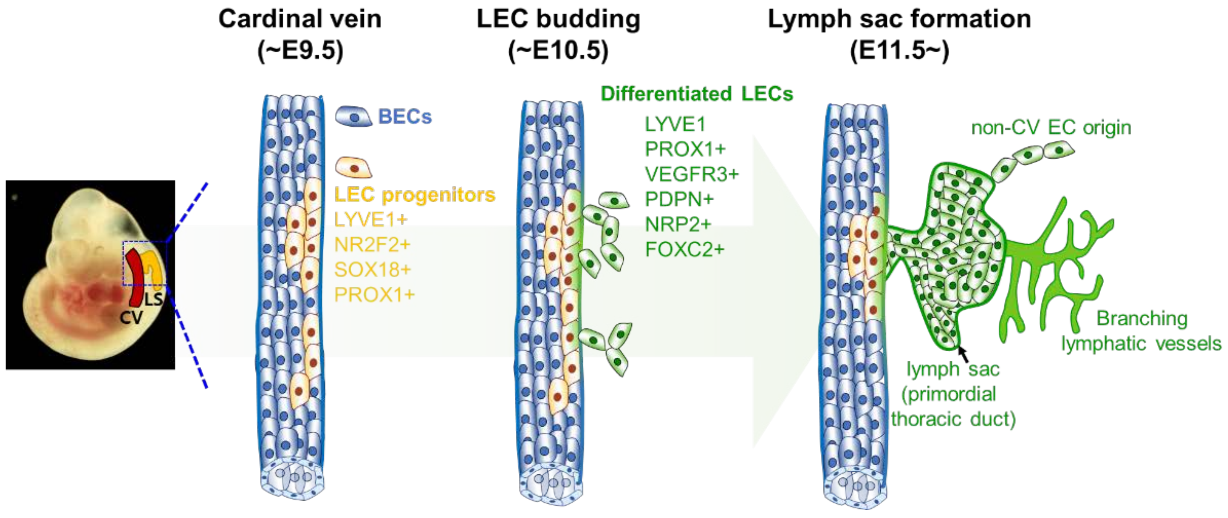
3.1. Historical Aspects of Lymphatic Vessel Development

In 1902, Florence Sabin injected Indian ink into the jugular area of pig embryos and observed the primitive lymphatic organs connect with the blood vasculature in the cardinal vein [98]. The primitive lymphatic organ, called the ‘lymph sac,’ was assumed to be the origin of the lymphatic vessels. This was the first study to suggest that the lymphatic vasculature originated from the cardinal vein. Sabin’s findings have been validated using lineage-tracing experiments with LEC-specific markers and the cardinal vein origin of lymphatic vasculature is considered to be a widely accepted concept. Studies based on this concept have examined the development of the lymphatic system from the lymph sac to explain the mechanism of LEC differentiation. Srinivasan et al. used a lineage-tracing mouse model to demonstrate that LECs are differentiated from a subset of the endothelium in the cardinal vein. [99]. The markers for cells involved in LEC lineage specification include PROX1, LYVE1, NRP2, VEGFR3, and PDPN (Figure 1) [100,101]. Various studies have demonstrated that PROX1, is the ‘master regulator’ of LEC specification and maintenance [102]. PROX1 activity in the cardinal vein ECs upregulates the transcription of LEC-specific genes and downregulates the transcription of BEC-specific genes [103]. Multiple Prox1 KO and overexpression studies have verified the critical role of PROX1 in lymphatic vessel development and maintenance [84,100,104]. Thus, the initiation of active Prox1 transcription in ECs indicates LEC lineage specification. This observation led to an in-depth investigation of the transcriptional regulation of Prox1.

3.2. Regulatory Networks of Epigenetic and Transcription Factors in Lymphatic Vessel Formation and Function

Transcriptome analysis and cellular imaging (for molecular colocalization) studies have revealed a high degree of correlation between PROX1 and other transcription factors involved in the development and maturation of lymphatic vasculature. Various transcription factors, including NR2F2, SOX18, GATA2, MAFB, FOXC2, NFATC1, and HHEX, are reported to be involved in the transcriptional regulation of PROX1. In turn, PROX1 is reported to be involved in the transcriptional regulation of LEC factors [104,105,106,107,108,109]. Additionally, several studies have reported the role of epigenetic modulators of transcription factors involved in lymph system development (Figure 2) [110,111,112,113,114]. The epigenetic factors that regulate chromatin conformational changes and histone modifications are indispensable to the optimal development of the lymphatic system. Analysis of gene expression and epigenetic modifications will provide valuable insights into the mechanisms underlying lymphatic development.
The binding motif of the transcription factor NR2F2 is located approximately 9.5 kb upstream of the open reading frame of mouse Prox1 [106]. This sequence is conserved among various mammals [106]. β-Galactosidase staining and immunofluorescence analyses have demonstrated the colocalization of NR2F2 with PROX1 and LYVE1 in the LECs [105,106]. Nr2f2 KO decreased the migration of PROX1-positive cells in the tissue around the cardinal vein of E11.5 and E13.5 mice. Additionally, the lymph sac was absent in Nr2f2 KO mice [106]. However, Nr2f2 deletion in PROX1-positive cells at E13.5 or later developmental stages did not result in major lymphatic defects. This indicates that NR2F2 promoted Prox1 expression only during early developmental stages [106]. BRG1, which regulates the expression of NR2F2 [110], is a catalytic ATPase that constitutes the SWITCH/sucrose nonfermentable SWI/SNF-like complex. The SWI/SNF complex is an ATP-dependent chromatin remodeling complex that inhibits DNA-histone interactions. In the absence of BRG1, the Nr2f2 promoter exhibits a highly compact structure, which results in the downregulation of Nr2f2 expression. Brg1 KO in endothelial cells recapitulated the genetic depletion of Nr2f2 with the downregulation of lymphatic markers and the upregulation of arterial markers [110].
SOX18 binds to Prox1 at two sites (1135–1130 bp and 813–808 bp) upstream of the transcription start site of Prox1 [104]. The results of the luciferase reporter-gene assay with the mlEnd cell line and in vivo immunofluorescence experiments revealed that both binding sites must be intact for SOX18 to drive the expression of the reporter gene during LEC development [104]. Prox1 expression was downregulated in the absence of functional SOX18. Conversely, the exogenously expressed Sox18 restored the expression of Prox1 [104]. Sox18-null mice completely lack LECs but contain other PROX1-positive cells, such as myocardial cells [104]. These results indicate that the transcription factor SOX18 specifically mediates the differentiation of BEC to LEC in the cardinal vein. DOT1L, a histone H3K79 methyltransferase, is involved in the transcriptional regulation of Sox18 [111]. RNA-sequencing (RNA-seq) analysis revealed that the expression of Sox18 is downregulated in LECs from E15.5 Dot1l KO mouse skin. Meanwhile, ChIP-seq revealed that H3K79 methylation was reduced in the gene body of Sox18 in these LECs [111]. Similar ChIP-seq results were obtained for other LEC development-related genes, such as Flt4, Ramp2, and Foxc2 [111].
MAFB promotes the transition of vascular ECs to LECs by upregulating the transcription of genes that promote LEC fate [107]. The binding motif of MAFB is in the first intron of Prox1. The induction of MAFB in primary LEC cell culture through VEGFR3, an upstream signal-transducing factor of MAFB, upregulated the expression of Prox1. MAFB is reported to bind to genes encoding other major factors regulating LEC specification, such as Klf4, Nr2f2, and Sox18. Short-interfering RNA-mediated Mafb knockdown downregulated the expression of Prox1. The role of MAFB in lymphatic development has been demonstrated using the back skin of Mafb KO E14.5 mouse embryos [107]. In these embryos, lymphatic vessel sprouting was delayed or incomplete.
Oscillatory shear stress (OSS) generated by lymph flow upregulates the expression levels of GATA2, FOXC2, and NFATC1, which regulate PROX1 transcription in LECs [109]. Physical stimuli promote the transcription of GATA2 [115]. GATA2, FOXC2, and NFATC1 may regulate PROX1 expression through their binding consensus sequences located close to each other at the first intron of PROX1 (approximately 11 kb upstream from the transcription start site) [109]. FOXC2 and NFATC1 can form a complex. The downregulation of FOXC2 or NFATC1 results in impaired lymphatic vessel development. However, GATA2 interacts directly with neither FOXC2 nor NFATC1. This indicates that GATA2 transforms the enhancer region of PROX1 into an ‘active’ state rather than directly inducing the transcription of PROX1. GATA2 regulates the opening of the PROX1 promoter region, whereas HDAC3 regulates the promoter region of Gata2 by inducing acetylation of H3K27 [112]. In the proposed model, HDAC3 is recruited to the intragenic enhancer of Gata2 in response to OSS. Next, TAL1, GATA2, ETS1/2, and HDAC3 form a complex and promote the recruitment of EP300. Finally, the EP300-mediated accumulation of H3K27ac promotes the expression of GATA2.
The binding site of HHEX is located 800 bp upstream of the transcription start site of human PROX1 [116]. The in vitro binding of HHEX to the PROX1 promoter region was confirmed using cultured human umbilical vein endothelial cells. This suggests that HHEX directly regulates the expression of PROX1. Hhex knockdown downregulated the expression of Prox1 in mouse and human LECs. Hhex KO in TIE2-positive cells impaired the proliferation, maturation, and sprouting of lymphatic vessels. HHEX is involved in the transcription of Vegfc and Flt4, which are involved in the development of lymphatic vessels. However, the binding of HHEX to the Vegfc and Flt4 promoters could not be confirmed. This suggests that HHEX may indirectly regulate the transcription of VEGFC and FTL4 and that the role of HHEX in lymphatic vessel development is mediated only by the activity of PROX1. Thus, the role of HHEX in LEC specification may be limited to the transcriptional regulation of PROX1.
The binding of PROX1 to target genomic DNAs induces the recruitment and transcriptional regulation of other co-factors [117,118]. Homeobox proteins share the helix-turn-helix (HTH) structure [119]. Similar to other homeobox proteins, the HTH structure of PROX1 enables it to directly bind to the major groove of DNA. The binding of PROX1 to DNA is followed by the recruitment of other epigenetic modifiers and transcription factors, such as the histone acetyltransferase EP300 [118]. Histone acetylation mediated by PROX1 and EP300 in the promoter region induces the unpacking of chromatin, the recruitment of other transcriptional factors, such as polymerase II, and the upregulation of the transcription of these genes. ChIP-seq analysis revealed that PROX1 binds to the promoter region of genes encoding proteins involved in lymphatic vessel development, such as VEGFR3, NRP2, SOX18, and PROX1 [106,115,120]. PROX1 targets CPT1A, which positively regulates the rate of fatty acid β-oxidation (FAO) [114]. Subsequently, the upregulated FAO induces acetyl coenzyme A production, which is utilized by EP300 for histone acetylation. The EP300/PROX1 complex promotes the histone acetylation of target gene promoters, including the promoter of PROX1. Thus, CPT1A is a part of the positive feedback loop of PROX1. PROX1 epigenetically regulates the expression of CYP7A1, which is involved in the bile acid synthesis pathway in the liver [121]. Co-immunoprecipitation (CoIP) studies with human hepatoblastoma cells (HepG2 cells) and GST-pull down assays performed using human embryonic kidney cells (HEK293T cells) revealed that the binding of PROX1 to the promoter represses the expression of target genes by recruiting the LSD1/NuRD complex, directly binding to the LSD1 unit, and promoting H3K4 hypomethylation. The role of CHD4, a subunit of the NuRD complex, in lymphatic development has been previously reported [113]. In the absence of VEGFC signaling, the Yap/Taz complex recruits the CHD4/NuRD complex to the PROX1 promoter and regulates PROX1 expression in a negative feedback loop through the deactivation of the Hippo signaling pathway [122]. This regulatory mechanism is critical for the patterning of the lymphatic plexus.
Previous studies on the transcriptional regulation of PROX1 have provided useful insights into the key factors involved in LEC specification. However, the elucidation of the comprehensive transcription regulation mechanism of transcription factors and other LEC-specifying genes throughout embryonic development is currently ongoing. In the myriad questions to be answered, findings suggest that the promoters and enhancers of LEC specification-related genes play important roles in LEC specification. The TFs with major roles in LEC fate determination and proliferation interact with the promoter of Prox1 gene, and the modification of promoter accessibility by epigenetic regulation is believed to be a key factor in lymphatic system development. Future studies could focus on the dynamics of epigenetic alterations, which will further aid in understanding the lymphatic system development process.

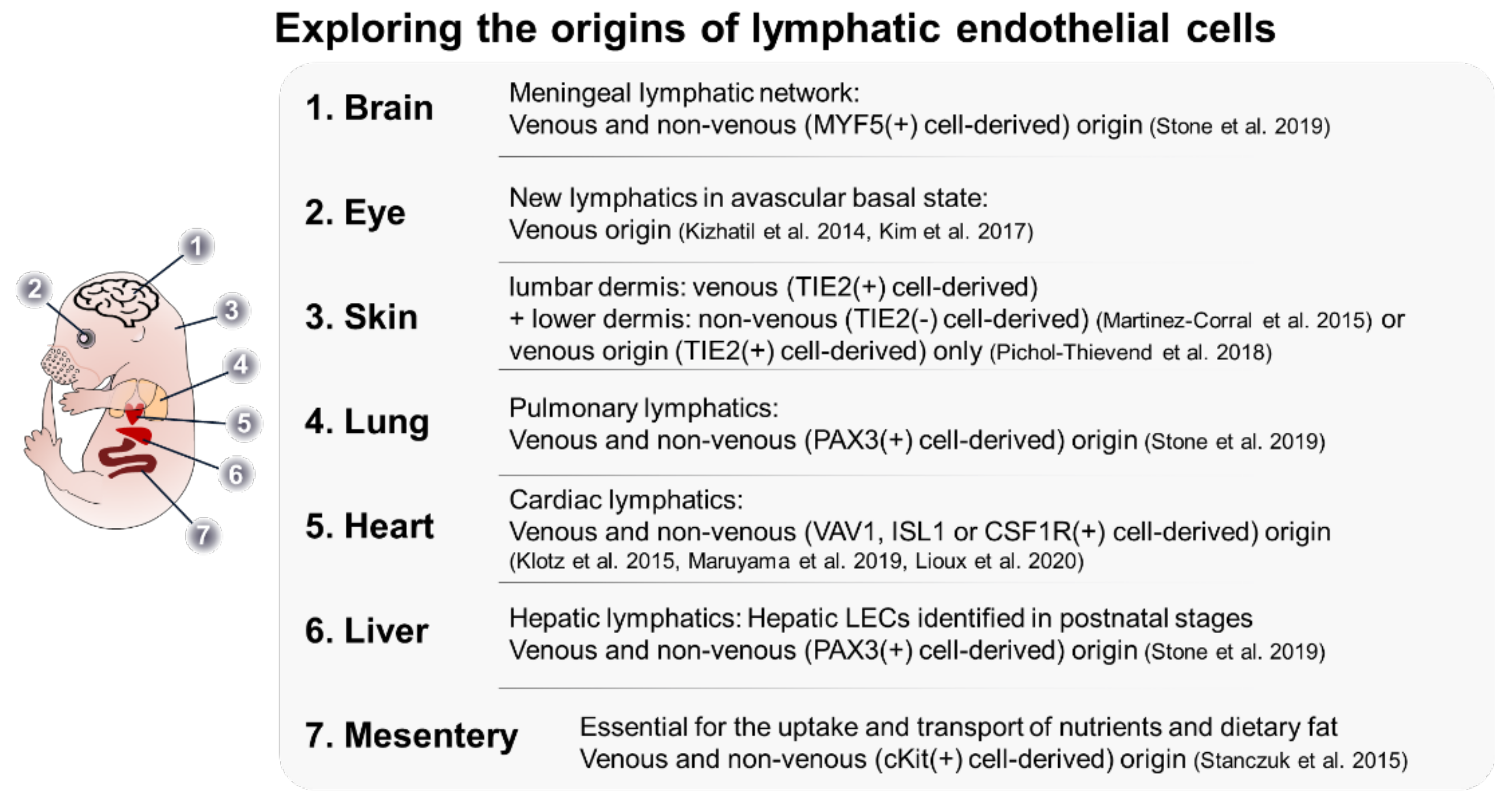
4. Heterogeneity in LEC Origin

In the theories based on Sabin’s research, the cardinal vein is the sole source of the entire lymphatic system, and this was a widely accepted dogma [98]. However, several studies have demonstrated that cells of non-venous origin are involved in the formation of the lymphatic system. Studies demonstrated that non-CV EC-origin LEC progenitors also contribute to the formation of the lymph sac [120,123,124]. The additional sources of LEC progenitor include the intersomitic vessels and the superficial venous plexus [120,123]. In addition, an elegant 3D imaging study clearly showed a stepwise process of lymph sac formation: (1) LECs emerge from the CV, dorso-laterally migrate, and form a meshwork along with LECs originating from other vessels. (2) The LECs further undergo coalescence to form a lumen structure (called a peripheral longitudinal lymphatic vessel) at the first lateral intersegmental vessel branch. (3) LECs located close to the CV aggregate simultaneously and form a primordial thoracic duct (pTD) [123]. These cells emerge at different developmental stages, acquire tissue-specific functionalities, and complement the vein-derived lymphatic system [83,101,125,126,127,128]. Further studies on cells of different sources can aid in the elucidation of the molecular mechanisms underlying lymphatic development. The understanding of the transition from a non-venous source to LEC will enable the identification of factors required for LEC fate determination. The analysis of the heterogeneity of LEC from different tissues can potentially explain the regulation of the LEC developmental process. Some recent studies have successfully characterized LECs of non-venous origin (Figure 3).
The mesentery is a rich source of vasculature. The anatomy of the mesentery enables the analysis of vasculature functions without the need for sectioning. Additionally, a new concept of the non-venous developmental process has been defined using these vasculatures. Stanczuk et al. established a mouse lineage with heterozygous Flt4-null and heterozygous kinase-dead Pik3ca alleles (Vegfr3lz/+;p110aD933A/+), which exhibited poorly developed lymphatic vessels in the mesenteric root [101]. However, lymphatic vessels in the diaphragm and skin were not affected. Based on this evidence, the authors hypothesized a tissue-specific activity of the VEGFR3/PI3K axis. Further investigations revealed that the mesenteric lymph sac originates from a venous source. However, PROX1-positive and NRP2-positive ECs in the mesenteric membrane were discontinued from the mesenteric root. Clusters of these cells were observed between E13 and E13.5. These clusters formed the mesenteric lymphatic vasculature by E14.5. Lineage-tracing analysis confirmed that the source of the lymphatic vasculature was c-KIT-positive/VAV1-negative hemogenic endothelium. In the proposed model, the lymph vessels in the mesenteric root differentiate from the lymph sac, while the collecting lymph vessels in the mesenteric membrane and lymphatic capillaries in the intestine differentiate from the c-KIT lineage. The lack of DOT1L impairs the formation of the lymphatic vessel only when c-KIT-positive cells are affected [111]. This demonstrates the presence of tissue-origin specificity in the epigenetic mechanism that drives LEC specification. Similar to blood circulatory system development, Stanczuk et al. distinguished the formation of lymphatic vessels based on the sprouting from the lymph sac and the development of non-venous mesenteric lymphatic vessels and coined the term ‘lymphvasculogenesis.’ This study not only introduced the question of tissue-specific LEC heterogeneity but also identified the mammalian LEC population derived from a non-venous source, which has not been completely elucidated.
Martinez-Corral et al. discovered a novel source of LECs in the skin of mouse embryos [125]. The authors reported the discontinuation of the lymphatic vessels in the lumbar and cervical regions in the skin of E13.5 and E15.5 mice. During lineage-tracing of lumbar LEC, the authors used Tie2-Cre;R26-mTmG mice, which allowed the distinct identification of Tie2-expressing cells. In this mouse line, the activation of Cre recombinase permanently removed the sequence encoding tomato and labeled the cell with green fluorescent protein (GFP). Whole-mount analysis of the skin revealed LECs without GFP expression. Flow cytometry analysis of LYVE1-positive and PDPN-positive cells revealed that some LECs originated from non-Tie2-expressing cells. To identify the origin of LECs that do not originate from the lymph sac, the authors used Prox1-CreER;R26-mTmG mouse lineage injected with 4-Hydroxytamoxifen (4-OHT) at E12.5. The authors hypothesized that all LECs express GFP if all LECs were derived from the lymph sac and, otherwise, cells expressing tomato would be present in the lymphatic vasculature. In E17.5 mice, the presence of cells expressing tomato was observed at the sprouting points of the vasculature. Pichol-Thievend et al. proposed that these cells originate from the blood capillary plexus [126]. Prox1 expression in the blood capillaries and some PROX1-positive cells at E13.5 has been observed in the lumen of the blood vasculature. The consensus finding between these two studies is that there is a source of LECs that is not derived from the lymph sac. However, the expression of Tie2 in the originating cells was a disputed finding between the two studies. Pichol-Thievend et al. suggested that the discrepancy in the study by Martinez-Correl could be attributed to variability in Tie2-Cre-mediated recombination.
Klotz et al. reported that the origin of significant portions of cardiac lymphatic vessels was not from the lymph sac [83]. During mouse embryonic stages, the lymphatic vasculature on the heart was observed to emerge from the ventral side and postnatally complete a network with the cardiac vein and artery on the surface of the heart. In the hearts of E14.5 mice, the majority of the lymphatic tissue was labeled using Tie2-Cre-mediated labeling. The results of this experiment were consistent with the previous knowledge of the origin of LEC. However, approximately 19% of the lymphatic vessels in the heart did not exhibit Tie2 expression and were of non-venous origin. These non-venous LECs did not originate from the epicardium, cardiac mesoderm, or cardiac neural crest. The authors hypothesized that these LECs originated from the TIE2-negative population of primitive hematopoietic cells within the yolk sac. This hypothesis was validated by verifying the expression of VAV1, PDGFRB, and CSF1R in these cells. The contribution of VAV1-positive cells to the cardiac lymphatic vessels was verified by the presence of lymphatic vessels after the ablation of Prox1 expression in TIE2-positive cells and Vav1-Cre-mediated Prox1-EGFP expression in cardiac lymphatic vessels. Newer studies have proposed ISL1 as a marker for LEC precursor cells of the ventral cardiac lymphatic vessels. ISL1 is another factor that marks the LEC population of non-venous origin [127,128]. Additionally, ISL1-positive cells, which belong to a group of multipotent cell populations in the second heart field, are involved in the formation of the outflow tract and facial skin [127,131]. Interestingly, ISL1-positive cells originated exclusively from the ventral surface of the heart below the atrium. Further analysis revealed that the absence of LEC of the ISL1-positive lineage does not affect the dorsal lymphatic vessels. This was speculated to be due to the maturation of ISL1-positive LECs in response to a high concentration of retinoic acid in the ventricle region [128].
In contrast to the century-old perspective of examining LEC origin in the lymph sac or other tissue-specific sources during organ development, Oliver et al. proposed that the paraxial mesoderm is the origin of all LECs [130]. The authors reported that myogenic precursors in a somatic paraxial mesodermal cell line expressing PAX3 form the jugular lymph sac and its derivative lymphatic vessels, such as the lymphatic vessels in the heart, lung, and skin. Lineage-tracing using Myf5-Cre or Mef2c-AHF-Cre cell lines in combination with ROSA26tdTomato can label PAX3-independent myogenic precursors from the paraxial mesoderm. These precursors served as LEC progenitors. The Myf5-Cre line contributes to the formation of lymphatic vessels in the lower jaw, meninges, ear skin, and a small portion of the lymph sac. The Mef2c-AHF-Cre line contributes to the formation of the second heart field. According to the ISL1 LEC study, these cells differentiate into LECs in the anterior jugular lymph sac, ventral cardiac LEC, and cervicothoracic dermis. The authors have claimed that they have captured the earliest steps in LEC differentiation before the involvement of the SOX18-NR2F2-PROX1 axis. This study warrants further lymphatics-related research focusing on the paraxial mesodermal lineage. Studies on the signaling network between the paraxial mesoderm lineage cells and the surrounding environment could provide useful insights into the molecular signals required for LEC specification. Additionally, the contribution of other PAX3-positive non-paraxial mesoderm sources to the LEC population could be investigated. The early lineage of mesenteric and intestinal lymphatic vessels has not been identified. Additional investigation of c-KIT-positive cells could reveal specific requirements for LEC specification.
The blood vessels and their tissue-specific functions have been extensively studied, and their tissue-specific functionalities have been highlighted. The identification of tissue-specific BECs led to the speculation of tissue-specific LECs. Thus, various studies have examined the different origins of LECs and endorsed their tissue specificity. Further studies are needed to examine the process of progenitors in the specification of definitive LEC fate, which will provide useful information on the mechanism of LEC induction. The increased number of HTS analyses and the discoveries of key factors have laid the foundation for the elucidation of LEC differentiation and the development of novel therapeutic strategies for LEC-related diseases, and the prospects are promising.

5. Conclusions

Lineage-tracing experiments and sequencing technologies with increased precision and functionality have enabled the elucidation of lymphatic system developmental processes. In the last decade, lineage-tracing studies have identified various key factors involved in lymphatic development and the contribution of previously unknown cells to the formation of LECs. Oliver et al. used lineage-tracing to demonstrate that priming for LEC specification occurs during mesoderm formation, before the emergence of the lymph sac [130]. However, some studies have raised concerns about the accountability of Cre-mediated recombination in lymphatic vessel-related studies [125,126]. Hence, additional validation steps must be considered in lineage-tracing experiments.
While lineage-tracing experiments focus on the origin of the LECs, sequencing experiments reveal the mechanism of LEC specification. HTS can identify the transcription profile of developing cells and indicate the importance of epigenetic regulation in developmental processes. Moreover, single-cell sequencing has increased in its precision and capacity to scope more cells [132] and databases, such as the EC atlas and MOCA, accumulating a vast amount of single-cell sequencing data [133,134]. However, these databases are not constructed specifically for LEC studies. The amount of LEC data included in these databases is insufficient for independent LEC analysis. Single-cell sequencing analysis of LECs from two or more different embryonic organs could provide useful information for the characterization of tissue-specific LEC development. In addition to the techniques currently used for developmental studies, recent studies have developed novel, cutting-edge techniques, such as artificial intelligence-based predictions of protein structure and function [135]. These techniques may aid in elucidating molecular interactions during LEC development and in establishing a complete LEC development map. Consequently, a complete LEC development map can contribute to the identification of effective therapeutic strategies for LEC-related diseases and determine the physiological advantage of enhanced lymphatic vessel functionalities.

Author Contributions

K.H. conceived the manuscript. K.H., H.L. (Hyeonwoo La), H.Y., Y.B.P., N.X.T., C.P., S.Y., H.L. (Hyeonji Lee), Y.C., H.S. and J.T.D. wrote and added critical ideas to the original version of the draft. All authors have read and agreed to the published version of the manuscript.

Funding

This work was supported by the National Research Foundation of Korea (NRF) with a grant funded by the Korean government (NRF-2020R1A2C1008433).

Institutional Review Board Statement

Not applicable.

Informed Consent Statement

Not applicable.

Data Availability Statement

Not applicable.

Conflicts of Interest

The authors declare no conflict of interest.

References

  1. Baluk, P.; Fuxe, J.; Hashizume, H.; Romano, T.; Lashnits, E.; Butz, S.; Vestweber, D.; Corada, M.; Molendini, C.; Dejana, E.; et al. Functionally specialized junctions between endothelial cells of lymphatic vessels. J. Exp. Med. 2007, 204, 2349–2362. [Google Scholar] [CrossRef] [PubMed]
  2. Sarin, H. Physiologic upper limits of pore size of different blood capillary types and another perspective on the dual pore theory of microvascular permeability. J. Angiogenes. Res. 2010, 2, 14. [Google Scholar] [CrossRef] [PubMed] [Green Version]
  3. Oliver, G.; Kipnis, J.; Randolph, G.J.; Harvey, N.L. The Lymphatic Vasculature in the 21(st) Century: Novel Functional Roles in Homeostasis and Disease. Cell 2020, 182, 270–296. [Google Scholar] [CrossRef] [PubMed]
  4. Tammela, T.; Alitalo, K. Lymphangiogenesis: Molecular mechanisms and future promise. Cell 2010, 140, 460–476. [Google Scholar] [CrossRef] [Green Version]
  5. Ducoli, L.; Detmar, M. Beyond PROX1: Transcriptional, epigenetic, and noncoding RNA regulation of lymphatic identity and function. Dev. Cell 2021, 56, 406–426. [Google Scholar] [CrossRef]
  6. Rushdi, M.; Li, K.; Yuan, Z.; Travaglino, S.; Grakoui, A.; Zhu, C. Mechanotransduction in T Cell Development, Differentiation and Function. Cells 2020, 9, 364. [Google Scholar] [CrossRef] [Green Version]
  7. Alon, R.; Dustin, M.L. Force as a facilitator of integrin conformational changes during leukocyte arrest on blood vessels and antigen-presenting cells. Immunity 2007, 26, 17–27. [Google Scholar] [CrossRef] [Green Version]
  8. Caldwell, C.C.; Kojima, H.; Lukashev, D.; Armstrong, J.; Farber, M.; Apasov, S.G.; Sitkovsky, M.V. Differential effects of physiologically relevant hypoxic conditions on T lymphocyte development and effector functions. J. Immunol 2001, 167, 6140–6149. [Google Scholar] [CrossRef]
  9. Nigam, Y.; Knight, J. The lymphatic system 2: Structure and function of the lymphoid organs. Nurs. Times 2020, 116, 44–48. [Google Scholar]
  10. Hagerling, R.; Hoppe, E.; Dierkes, C.; Stehling, M.; Makinen, T.; Butz, S.; Vestweber, D.; Kiefer, F. Distinct roles of VE-cadherin for development and maintenance of specific lymph vessel beds. EMBO J. 2018, 37, e98271. [Google Scholar] [CrossRef]
  11. Hogan, B.M.; Bos, F.L.; Bussmann, J.; Witte, M.; Chi, N.C.; Duckers, H.J.; Schulte-Merker, S. Ccbe1 is required for embryonic lymphangiogenesis and venous sprouting. Nat. Genet. 2009, 41, 396–398. [Google Scholar] [CrossRef] [PubMed]
  12. Bos, F.L.; Caunt, M.; Peterson-Maduro, J.; Planas-Paz, L.; Kowalski, J.; Karpanen, T.; van Impel, A.; Tong, R.; Ernst, J.A.; Korving, J.; et al. CCBE1 is essential for mammalian lymphatic vascular development and enhances the lymphangiogenic effect of vascular endothelial growth factor-C in vivo. Circ. Res. 2011, 109, 486–491. [Google Scholar] [CrossRef] [PubMed]
  13. John, R.M.; Rougeulle, C. Developmental Epigenetics: Phenotype and the Flexible Epigenome. Front. Cell Dev. Biol. 2018, 6, 130. [Google Scholar] [CrossRef] [PubMed]
  14. den Braanker, H.; van Stigt, A.C.; Kok, M.R.; Lubberts, E.; Bisoendial, R.J. Single-Cell RNA Sequencing Reveals Heterogeneity and Functional Diversity of Lymphatic Endothelial Cells. Int. J. Mol. Sci. 2021, 22, 11976. [Google Scholar] [CrossRef]
  15. Takeda, A.; Hollmen, M.; Dermadi, D.; Pan, J.; Brulois, K.F.; Kaukonen, R.; Lonnberg, T.; Bostrom, P.; Koskivuo, I.; Irjala, H.; et al. Single-Cell Survey of Human Lymphatics Unveils Marked Endothelial Cell Heterogeneity and Mechanisms of Homing for Neutrophils. Immunity 2019, 51, 561–572.e5. [Google Scholar] [CrossRef]
  16. Maby-El Hajjami, H.; Petrova, T.V. Developmental and pathological lymphangiogenesis: From models to human disease. Histochem. Cell Biol. 2008, 130, 1063–1078. [Google Scholar] [CrossRef] [Green Version]
  17. Bernas, M.; Thiadens, S.R.J.; Smoot, B.; Armer, J.M.; Stewart, P.; Granzow, J. Lymphedema following cancer therapy: Overview and options. Clin. Exp. Metastasis 2018, 35, 547–551. [Google Scholar] [CrossRef]
  18. Witte, M.H.; Bernas, M.J.; Martin, C.P.; Witte, C.L. Lymphangiogenesis and lymphangiodysplasia: From molecular to clinical lymphology. Microsc. Res. Tech. 2001, 55, 122–145. [Google Scholar] [CrossRef]
  19. Gordon, K.; Varney, R.; Keeley, V.; Riches, K.; Jeffery, S.; Van Zanten, M.; Mortimer, P.; Ostergaard, P.; Mansour, S. Update and audit of the St George’s classification algorithm of primary lymphatic anomalies: A clinical and molecular approach to diagnosis. J. Med. Genet. 2020, 57, 653–659. [Google Scholar] [CrossRef]
  20. Dellinger, M.T.; Thome, K.; Bernas, M.J.; Erickson, R.P.; Witte, M.H. Novel FOXC2 missense mutation identified in patient with lymphedema-distichiasis syndrome and review. Lymphology 2008, 41, 98–102. [Google Scholar]
  21. Rezaie, T.; Ghoroghchian, R.; Bell, R.; Brice, G.; Hasan, A.; Burnand, K.; Vernon, S.; Mansour, S.; Mortimer, P.; Jeffery, S.; et al. Primary non-syndromic lymphoedema (Meige disease) is not caused by mutations in FOXC2. Eur. J. Hum. Genet. 2008, 16, 300–304. [Google Scholar] [CrossRef] [PubMed] [Green Version]
  22. Irrthum, A.; Devriendt, K.; Chitayat, D.; Matthijs, G.; Glade, C.; Steijlen, P.M.; Fryns, J.P.; Van Steensel, M.A.; Vikkula, M. Mutations in the transcription factor gene SOX18 underlie recessive and dominant forms of hypotrichosis-lymphedema-telangiectasia. Am. J. Hum. Genet. 2003, 72, 1470–1478. [Google Scholar] [CrossRef] [PubMed] [Green Version]
  23. Moalem, S.; Brouillard, P.; Kuypers, D.; Legius, E.; Harvey, E.; Taylor, G.; Francois, M.; Vikkula, M.; Chitayat, D. Hypotrichosis-lymphedema-telangiectasia-renal defect associated with a truncating mutation in the SOX18 gene. Clin. Genet. 2015, 87, 378–382. [Google Scholar] [CrossRef] [PubMed]
  24. Emberger, J.M.; Navarro, M.; Dejean, M.; Izarn, P. [Deaf-mutism, lymphedema of the lower limbs and hematological abnormalities (acute leukemia, cytopenia) with autosomal dominant transmission]. J. Genet. Hum. 1979, 27, 237–245. [Google Scholar]
  25. Mansour, S.; Connell, F.; Steward, C.; Ostergaard, P.; Brice, G.; Smithson, S.; Lunt, P.; Jeffery, S.; Dokal, I.; Vulliamy, T.; et al. Emberger syndrome-primary lymphedema with myelodysplasia: Report of seven new cases. Am. J. Med. Genet. A 2010, 152A, 2287–2296. [Google Scholar] [CrossRef]
  26. Melrose, W.D. Lymphatic filariasis: New insights into an old disease. Int. J. Parasitol. 2002, 32, 947–960. [Google Scholar] [CrossRef]
  27. Clark, B.; Sitzia, J.; Harlow, W. Incidence and risk of arm oedema following treatment for breast cancer: A three-year follow-up study. QJM 2005, 98, 343–348. [Google Scholar] [CrossRef]
  28. Diaz-Flores, L.; Gutierrez, R.; Pino Garcia, M.; Gonzalez-Gomez, M.; Diaz-Flores, L., Jr.; Carrasco, J.L. Intussusceptive lymphangiogenesis in the sinuses of developing human foetal lymph nodes. Ann. Anat. 2019, 226, 73–83. [Google Scholar] [CrossRef]
  29. Diaz-Flores, L.; Gutierrez, R.; Garcia, M.D.P.; Carrasco, J.L.; Saez, F.J.; Diaz-Flores, L., Jr.; Gonzalez-Gomez, M.; Madrid, J.F. Intussusceptive Lymphangiogenesis in Lymphatic Malformations/Lymphangiomas. Anat. Rec. 2019, 302, 2003–2013. [Google Scholar] [CrossRef]
  30. Ogino, R.; Hayashida, K.; Yamakawa, S.; Morita, E. Adipose-Derived Stem Cells Promote Intussusceptive Lymphangiogenesis by Restricting Dermal Fibrosis in Irradiated Tissue of Mice. Int. J. Mol. Sci. 2020, 21, 3885. [Google Scholar] [CrossRef]
  31. Butler, M.G.; Dagenais, S.L.; Rockson, S.G.; Glover, T.W. A novel VEGFR3 mutation causes Milroy disease. Am. J. Med. Genet. A 2007, 143A, 1212–1217. [Google Scholar] [CrossRef] [PubMed] [Green Version]
  32. Butler, M.G.; Isogai, S.; Weinstein, B.M. Lymphatic development. Birth Defects Res. C Embryo Today 2009, 87, 222–231. [Google Scholar] [CrossRef] [PubMed]
  33. Balboa-Beltran, E.; Fernandez-Seara, M.J.; Perez-Munuzuri, A.; Lago, R.; Garcia-Magan, C.; Couce, M.L.; Sobrino, B.; Amigo, J.; Carracedo, A.; Barros, F. A novel stop mutation in the vascular endothelial growth factor-C gene (VEGFC) results in Milroy-like disease. J. Med. Genet. 2014, 51, 475–478. [Google Scholar] [CrossRef] [PubMed]
  34. Gordon, K.; Schulte, D.; Brice, G.; Simpson, M.A.; Roukens, M.G.; van Impel, A.; Connell, F.; Kalidas, K.; Jeffery, S.; Mortimer, P.S.; et al. Mutation in vascular endothelial growth factor-C, a ligand for vascular endothelial growth factor receptor-3, is associated with autosomal dominant milroy-like primary lymphedema. Circ. Res. 2013, 112, 956–960. [Google Scholar] [CrossRef]
  35. Ferrell, R.E.; Baty, C.J.; Kimak, M.A.; Karlsson, J.M.; Lawrence, E.C.; Franke-Snyder, M.; Meriney, S.D.; Feingold, E.; Finegold, D.N. GJC2 missense mutations cause human lymphedema. Am. J. Hum. Genet. 2010, 86, 943–948. [Google Scholar] [CrossRef] [Green Version]
  36. Martin-Almedina, S.; Martinez-Corral, I.; Holdhus, R.; Vicente, A.; Fotiou, E.; Lin, S.; Petersen, K.; Simpson, M.A.; Hoischen, A.; Gilissen, C.; et al. EPHB4 kinase-inactivating mutations cause autosomal dominant lymphatic-related hydrops fetalis. J. Clin. Investig. 2016, 126, 3080–3088. [Google Scholar] [CrossRef]
  37. Connell, F.; Kalidas, K.; Ostergaard, P.; Brice, G.; Homfray, T.; Roberts, L.; Bunyan, D.J.; Mitton, S.; Mansour, S.; Mortimer, P.; et al. Linkage and sequence analysis indicate that CCBE1 is mutated in recessively inherited generalised lymphatic dysplasia. Hum. Genet. 2010, 127, 231–241. [Google Scholar] [CrossRef]
  38. Alders, M.; Al-Gazali, L.; Cordeiro, I.; Dallapiccola, B.; Garavelli, L.; Tuysuz, B.; Salehi, F.; Haagmans, M.A.; Mook, O.R.; Majoie, C.B.; et al. Hennekam syndrome can be caused by FAT4 mutations and be allelic to Van Maldergem syndrome. Hum. Genet. 2014, 133, 1161–1167. [Google Scholar] [CrossRef]
  39. Brouillard, P.; Dupont, L.; Helaers, R.; Coulie, R.; Tiller, G.E.; Peeden, J.; Colige, A.; Vikkula, M. Loss of ADAMTS3 activity causes Hennekam lymphangiectasia-lymphedema syndrome 3. Hum. Mol. Genet. 2017, 26, 4095–4104. [Google Scholar] [CrossRef] [Green Version]
  40. Boone, P.M.; Paterson, S.; Mohajeri, K.; Zhu, W.; Genetti, C.A.; Tai, D.J.C.; Nori, N.; Agrawal, P.B.; Bacino, C.A.; Bi, W.; et al. Biallelic mutation of FBXL7 suggests a novel form of Hennekam syndrome. Am. J. Med. Genet. A 2020, 182, 189–194. [Google Scholar] [CrossRef]
  41. Gonzalez-Garay, M.L.; Aldrich, M.B.; Rasmussen, J.C.; Guilliod, R.; Lapinski, P.E.; King, P.D.; Sevick-Muraca, E.M. A novel mutation in CELSR1 is associated with hereditary lymphedema. Vasc. Cell 2016, 8, 1. [Google Scholar] [CrossRef] [Green Version]
  42. Birtel, J.; Gliem, M.; Mangold, E.; Tebbe, L.; Spier, I.; Muller, P.L.; Holz, F.G.; Neuhaus, C.; Wolfrum, U.; Bolz, H.J.; et al. Novel Insights Into the Phenotypical Spectrum of KIF11-Associated Retinopathy, Including a New Form of Retinal Ciliopathy. Investig. Ophthalmol. Vis. Sci. 2017, 58, 3950–3959. [Google Scholar] [CrossRef] [PubMed] [Green Version]
  43. Fotiou, E.; Martin-Almedina, S.; Simpson, M.A.; Lin, S.; Gordon, K.; Brice, G.; Atton, G.; Jeffery, I.; Rees, D.C.; Mignot, C.; et al. Novel mutations in PIEZO1 cause an autosomal recessive generalized lymphatic dysplasia with non-immune hydrops fetalis. Nat. Commun. 2015, 6, 8085. [Google Scholar] [CrossRef] [PubMed] [Green Version]
  44. Revencu, N.; Boon, L.M.; Mendola, A.; Cordisco, M.R.; Dubois, J.; Clapuyt, P.; Hammer, F.; Amor, D.J.; Irvine, A.D.; Baselga, E.; et al. RASA1 mutations and associated phenotypes in 68 families with capillary malformation-arteriovenous malformation. Hum. Mutat. 2013, 34, 1632–1641. [Google Scholar] [CrossRef] [PubMed]
  45. Hiramatsu, C.; Paukner, A.; Kuroshima, H.; Fujita, K.; Suomi, S.J.; Inoue-Murayama, M. Short poly-glutamine repeat in the androgen receptor in New World monkeys. Meta Gene 2017, 14, 105–113. [Google Scholar] [CrossRef] [PubMed] [Green Version]
  46. Qazi, Q.H.; Kanchanapoomi, R.; Beller, E.; Collins, R. Inheritance of posterior choanal atresia. Am. J. Med. Genet. 1982, 13, 413–416. [Google Scholar] [CrossRef]
  47. Mackie, D.I.; Al Mutairi, F.; Davis, R.B.; Kechele, D.O.; Nielsen, N.R.; Snyder, J.C.; Caron, M.G.; Kliman, H.J.; Berg, J.S.; Simms, J.; et al. hCALCRL mutation causes autosomal recessive nonimmune hydrops fetalis with lymphatic dysplasia. J. Exp. Med. 2018, 215, 2339–2353. [Google Scholar] [CrossRef] [PubMed] [Green Version]
  48. Ma, G.C.; Liu, C.S.; Chang, S.P.; Yeh, K.T.; Ke, Y.Y.; Chen, T.H.; Wang, B.B.; Kuo, S.J.; Shih, J.C.; Chen, M. A recurrent ITGA9 missense mutation in human fetuses with severe chylothorax: Possible correlation with poor response to fetal therapy. Prenat. Diagn. 2008, 28, 1057–1063. [Google Scholar] [CrossRef]
  49. Hong, S.E.; Shugart, Y.Y.; Huang, D.T.; Shahwan, S.A.; Grant, P.E.; Hourihane, J.O.; Martin, N.D.; Walsh, C.A. Autosomal recessive lissencephaly with cerebellar hypoplasia is associated with human RELN mutations. Nat. Genet. 2000, 26, 93–96. [Google Scholar] [CrossRef]
  50. Semo, J.; Nicenboim, J.; Yaniv, K. Development of the lymphatic system: New questions and paradigms. Development 2016, 143, 924–935. [Google Scholar] [CrossRef] [Green Version]
  51. Bernier-Latmani, J.; Petrova, T.V. Intestinal lymphatic vasculature: Structure, mechanisms and functions. Nat. Rev. Gastroenterol. Hepatol. 2017, 14, 510–526. [Google Scholar] [CrossRef] [PubMed]
  52. Escobedo, N.; Proulx, S.T.; Karaman, S.; Dillard, M.E.; Johnson, N.; Detmar, M.; Oliver, G. Restoration of lymphatic function rescues obesity in Prox1-haploinsufficient mice. JCI Insight 2016, 1, e85096. [Google Scholar] [CrossRef] [PubMed]
  53. Harvey, N.L.; Srinivasan, R.S.; Dillard, M.E.; Johnson, N.C.; Witte, M.H.; Boyd, K.; Sleeman, M.W.; Oliver, G. Lymphatic vascular defects promoted by Prox1 haploinsufficiency cause adult-onset obesity. Nat. Genet. 2005, 37, 1072–1081. [Google Scholar] [CrossRef] [PubMed]
  54. Rutkowski, J.M.; Markhus, C.E.; Gyenge, C.C.; Alitalo, K.; Wiig, H.; Swartz, M.A. Dermal collagen and lipid deposition correlate with tissue swelling and hydraulic conductivity in murine primary lymphedema. Am. J. Pathol. 2010, 176, 1122–1129. [Google Scholar] [CrossRef] [PubMed]
  55. Baluk, P.; Tammela, T.; Ator, E.; Lyubynska, N.; Achen, M.G.; Hicklin, D.J.; Jeltsch, M.; Petrova, T.V.; Pytowski, B.; Stacker, S.A.; et al. Pathogenesis of persistent lymphatic vessel hyperplasia in chronic airway inflammation. J. Clin. Investig. 2005, 115, 247–257. [Google Scholar] [CrossRef] [Green Version]
  56. Ristimaki, A.; Narko, K.; Enholm, B.; Joukov, V.; Alitalo, K. Proinflammatory cytokines regulate expression of the lymphatic endothelial mitogen vascular endothelial growth factor-C. J. Biol. Chem. 1998, 273, 8413–8418. [Google Scholar] [CrossRef] [Green Version]
  57. Proulx, S.T.; Kwok, E.; You, Z.; Beck, C.A.; Shealy, D.J.; Ritchlin, C.T.; Boyce, B.F.; Xing, L.; Schwarz, E.M. MRI and quantification of draining lymph node function in inflammatory arthritis. Ann. N. Y. Acad. Sci. 2007, 1117, 106–123. [Google Scholar] [CrossRef]
  58. Kajiya, K.; Detmar, M. An important role of lymphatic vessels in the control of UVB-induced edema formation and inflammation. J. Investig. Dermatol. 2006, 126, 919–921. [Google Scholar] [CrossRef] [Green Version]
  59. Dubrot, J.; Duraes, F.V.; Potin, L.; Capotosti, F.; Brighouse, D.; Suter, T.; LeibundGut-Landmann, S.; Garbi, N.; Reith, W.; Swartz, M.A.; et al. Lymph node stromal cells acquire peptide-MHCII complexes from dendritic cells and induce antigen-specific CD4(+) T cell tolerance. J. Exp. Med. 2014, 211, 1153–1166. [Google Scholar] [CrossRef]
  60. Gkountidi, A.O.; Garnier, L.; Dubrot, J.; Angelillo, J.; Harle, G.; Brighouse, D.; Wrobel, L.J.; Pick, R.; Scheiermann, C.; Swartz, M.A.; et al. MHC Class II Antigen Presentation by Lymphatic Endothelial Cells in Tumors Promotes Intratumoral Regulatory T cell-Suppressive Functions. Cancer Immunol. Res. 2021, 9, 748–764. [Google Scholar] [CrossRef]
  61. Rouhani, S.J.; Eccles, J.D.; Riccardi, P.; Peske, J.D.; Tewalt, E.F.; Cohen, J.N.; Liblau, R.; Makinen, T.; Engelhard, V.H. Roles of lymphatic endothelial cells expressing peripheral tissue antigens in CD4 T-cell tolerance induction. Nat. Commun. 2015, 6, 6771. [Google Scholar] [CrossRef] [PubMed] [Green Version]
  62. Kerjaschki, D.; Regele, H.M.; Moosberger, I.; Nagy-Bojarski, K.; Watschinger, B.; Soleiman, A.; Birner, P.; Krieger, S.; Hovorka, A.; Silberhumer, G.; et al. Lymphatic neoangiogenesis in human kidney transplants is associated with immunologically active lymphocytic infiltrates. J. Am. Soc. Nephrol. 2004, 15, 603–612. [Google Scholar] [CrossRef] [PubMed] [Green Version]
  63. Nykanen, A.I.; Sandelin, H.; Krebs, R.; Keranen, M.A.; Tuuminen, R.; Karpanen, T.; Wu, Y.; Pytowski, B.; Koskinen, P.K.; Yla-Herttuala, S.; et al. Targeting lymphatic vessel activation and CCL21 production by vascular endothelial growth factor receptor-3 inhibition has novel immunomodulatory and antiarteriosclerotic effects in cardiac allografts. Circulation 2010, 121, 1413–1422. [Google Scholar] [CrossRef] [PubMed]
  64. Skobe, M.; Hawighorst, T.; Jackson, D.G.; Prevo, R.; Janes, L.; Velasco, P.; Riccardi, L.; Alitalo, K.; Claffey, K.; Detmar, M. Induction of tumor lymphangiogenesis by VEGF-C promotes breast cancer metastasis. Nat. Med. 2001, 7, 192–198. [Google Scholar] [CrossRef] [PubMed]
  65. Hirakawa, S.; Brown, L.F.; Kodama, S.; Paavonen, K.; Alitalo, K.; Detmar, M. VEGF-C-induced lymphangiogenesis in sentinel lymph nodes promotes tumor metastasis to distant sites. Blood 2007, 109, 1010–1017. [Google Scholar] [CrossRef] [Green Version]
  66. Hirakawa, S.; Kodama, S.; Kunstfeld, R.; Kajiya, K.; Brown, L.F.; Detmar, M. VEGF-A induces tumor and sentinel lymph node lymphangiogenesis and promotes lymphatic metastasis. J. Exp. Med. 2005, 201, 1089–1099. [Google Scholar] [CrossRef] [Green Version]
  67. Mandriota, S.J.; Jussila, L.; Jeltsch, M.; Compagni, A.; Baetens, D.; Prevo, R.; Banerji, S.; Huarte, J.; Montesano, R.; Jackson, D.G.; et al. Vascular endothelial growth factor-C-mediated lymphangiogenesis promotes tumour metastasis. EMBO J. 2001, 20, 672–682. [Google Scholar] [CrossRef] [Green Version]
  68. Stacker, S.A.; Caesar, C.; Baldwin, M.E.; Thornton, G.E.; Williams, R.A.; Prevo, R.; Jackson, D.G.; Nishikawa, S.; Kubo, H.; Achen, M.G. VEGF-D promotes the metastatic spread of tumor cells via the lymphatics. Nat. Med. 2001, 7, 186–191. [Google Scholar] [CrossRef]
  69. Shibata, M.A.; Morimoto, J.; Shibata, E.; Otsuki, Y. Combination therapy with short interfering RNA vectors against VEGF-C and VEGF-A suppresses lymph node and lung metastasis in a mouse immunocompetent mammary cancer model. Cancer Gene Ther. 2008, 15, 776–786. [Google Scholar] [CrossRef] [Green Version]
  70. Lund, A.W.; Wagner, M.; Fankhauser, M.; Steinskog, E.S.; Broggi, M.A.; Spranger, S.; Gajewski, T.F.; Alitalo, K.; Eikesdal, H.P.; Wiig, H.; et al. Lymphatic vessels regulate immune microenvironments in human and murine melanoma. J. Clin. Investig. 2016, 126, 3389–3402. [Google Scholar] [CrossRef]
  71. Kimura, T.; Sugaya, M.; Oka, T.; Blauvelt, A.; Okochi, H.; Sato, S. Lymphatic dysfunction attenuates tumor immunity through impaired antigen presentation. Oncotarget 2015, 6, 18081–18093. [Google Scholar] [CrossRef] [PubMed] [Green Version]
  72. Kubo, H.; Fujiwara, T.; Jussila, L.; Hashi, H.; Ogawa, M.; Shimizu, K.; Awane, M.; Sakai, Y.; Takabayashi, A.; Alitalo, K.; et al. Involvement of vascular endothelial growth factor receptor-3 in maintenance of integrity of endothelial cell lining during tumor angiogenesis. Blood 2000, 96, 546–553. [Google Scholar] [CrossRef] [PubMed]
  73. Partanen, T.A.; Alitalo, K.; Miettinen, M. Lack of lymphatic vascular specificity of vascular endothelial growth factor receptor 3 in 185 vascular tumors. Cancer 1999, 86, 2406–2412. [Google Scholar] [CrossRef]
  74. Valtola, R.; Salven, P.; Heikkila, P.; Taipale, J.; Joensuu, H.; Rehn, M.; Pihlajaniemi, T.; Weich, H.; deWaal, R.; Alitalo, K. VEGFR-3 and its ligand VEGF-C are associated with angiogenesis in breast cancer. Am. J. Pathol. 1999, 154, 1381–1390. [Google Scholar] [CrossRef] [Green Version]
  75. Song, E.; Mao, T.; Dong, H.; Boisserand, L.S.B.; Antila, S.; Bosenberg, M.; Alitalo, K.; Thomas, J.L.; Iwasaki, A. VEGF-C-driven lymphatic drainage enables immunosurveillance of brain tumours. Nature 2020, 577, 689–694. [Google Scholar] [CrossRef] [PubMed]
  76. Hu, X.; Deng, Q.; Ma, L.; Li, Q.; Chen, Y.; Liao, Y.; Zhou, F.; Zhang, C.; Shao, L.; Feng, J.; et al. Meningeal lymphatic vessels regulate brain tumor drainage and immunity. Cell Res. 2020, 30, 229–243. [Google Scholar] [CrossRef] [Green Version]
  77. Zhou, C.; Ma, L.; Xu, H.; Huo, Y.; Luo, J. Meningeal lymphatics regulate radiotherapy efficacy through modulating anti-tumor immunity. Cell Res. 2022. [Google Scholar] [CrossRef]
  78. Lu, L.; Liu, M.; Sun, R.; Zheng, Y.; Zhang, P. Myocardial Infarction: Symptoms and Treatments. Cell Biochem. Biophys. 2015, 72, 865–867. [Google Scholar] [CrossRef]
  79. Libby, P.; Hansson, G.K. Inflammation and immunity in diseases of the arterial tree: Players and layers. Circ. Res. 2015, 116, 307–311. [Google Scholar] [CrossRef] [Green Version]
  80. Henri, O.; Pouehe, C.; Houssari, M.; Galas, L.; Nicol, L.; Edwards-Levy, F.; Henry, J.P.; Dumesnil, A.; Boukhalfa, I.; Banquet, S.; et al. Selective Stimulation of Cardiac Lymphangiogenesis Reduces Myocardial Edema and Fibrosis Leading to Improved Cardiac Function Following Myocardial Infarction. Circulation 2016, 133, 1484–1497, discussion 1497. [Google Scholar] [CrossRef] [Green Version]
  81. Milasan, A.; Smaani, A.; Martel, C. Early rescue of lymphatic function limits atherosclerosis progression in Ldlr(-/-) mice. Atherosclerosis 2019, 283, 106–119. [Google Scholar] [CrossRef] [PubMed] [Green Version]
  82. Vuorio, T.; Tirronen, A.; Yla-Herttuala, S. Cardiac Lymphatics—A New Avenue for Therapeutics? Trends Endocrinol. Metab. 2017, 28, 285–296. [Google Scholar] [CrossRef] [PubMed] [Green Version]
  83. Klotz, L.; Norman, S.; Vieira, J.M.; Masters, M.; Rohling, M.; Dube, K.N.; Bollini, S.; Matsuzaki, F.; Carr, C.A.; Riley, P.R. Cardiac lymphatics are heterogeneous in origin and respond to injury. Nature 2015, 522, 62–67. [Google Scholar] [CrossRef] [Green Version]
  84. Iliff, J.J.; Wang, M.; Liao, Y.; Plogg, B.A.; Peng, W.; Gundersen, G.A.; Benveniste, H.; Vates, G.E.; Deane, R.; Goldman, S.A.; et al. A paravascular pathway facilitates CSF flow through the brain parenchyma and the clearance of interstitial solutes, including amyloid beta. Sci. Transl. Med. 2012, 4, 147ra111. [Google Scholar] [CrossRef] [PubMed] [Green Version]
  85. Absinta, M.; Ha, S.K.; Nair, G.; Sati, P.; Luciano, N.J.; Palisoc, M.; Louveau, A.; Zaghloul, K.A.; Pittaluga, S.; Kipnis, J.; et al. Human and nonhuman primate meninges harbor lymphatic vessels that can be visualized noninvasively by MRI. Elife 2017, 6, e29738. [Google Scholar] [CrossRef] [PubMed]
  86. Augustin, H.G.; Koh, G.Y. Organotypic vasculature: From descriptive heterogeneity to functional pathophysiology. Science 2017, 357, eaal2379. [Google Scholar] [CrossRef] [Green Version]
  87. Ma, Q.; Decker, Y.; Muller, A.; Ineichen, B.V.; Proulx, S.T. Clearance of cerebrospinal fluid from the sacral spine through lymphatic vessels. J. Exp. Med. 2019, 216, 2492–2502. [Google Scholar] [CrossRef] [Green Version]
  88. Mentis, A.A.; Dardiotis, E.; Chrousos, G.P. Apolipoprotein E4 and meningeal lymphatics in Alzheimer disease: A conceptual framework. Mol. Psychiatry 2021, 26, 1075–1097. [Google Scholar] [CrossRef]
  89. Da Mesquita, S.; Louveau, A.; Vaccari, A.; Smirnov, I.; Cornelison, R.C.; Kingsmore, K.M.; Contarino, C.; Onengut-Gumuscu, S.; Farber, E.; Raper, D.; et al. Functional aspects of meningeal lymphatics in ageing and Alzheimer’s disease. Nature 2018, 560, 185–191. [Google Scholar] [CrossRef]
  90. Moore, D.J.; West, A.B.; Dawson, V.L.; Dawson, T.M. Molecular pathophysiology of Parkinson’s disease. Annu. Rev. Neurosci. 2005, 28, 57–87. [Google Scholar] [CrossRef] [Green Version]
  91. Zou, W.; Pu, T.; Feng, W.; Lu, M.; Zheng, Y.; Du, R.; Xiao, M.; Hu, G. Blocking meningeal lymphatic drainage aggravates Parkinson’s disease-like pathology in mice overexpressing mutated alpha-synuclein. Transl. Neurodegener. 2019, 8, 7. [Google Scholar] [CrossRef] [PubMed]
  92. Chen, J.; He, J.; Ni, R.; Yang, Q.; Zhang, Y.; Luo, L. Cerebrovascular Injuries Induce Lymphatic Invasion into Brain Parenchyma to Guide Vascular Regeneration in Zebrafish. Dev. Cell 2019, 49, 697–710.e5. [Google Scholar] [CrossRef] [PubMed]
  93. Aspelund, A.; Tammela, T.; Antila, S.; Nurmi, H.; Leppanen, V.M.; Zarkada, G.; Stanczuk, L.; Francois, M.; Makinen, T.; Saharinen, P.; et al. The Schlemm’s canal is a VEGF-C/VEGFR-3-responsive lymphatic-like vessel. J. Clin. Investig. 2014, 124, 3975–3986. [Google Scholar] [CrossRef] [PubMed]
  94. Truong, T.N.; Li, H.; Hong, Y.K.; Chen, L. Novel characterization and live imaging of Schlemm’s canal expressing Prox-1. PLoS ONE 2014, 9, e98245. [Google Scholar] [CrossRef] [PubMed] [Green Version]
  95. Kizhatil, K.; Ryan, M.; Marchant, J.K.; Henrich, S.; John, S.W. Schlemm’s canal is a unique vessel with a combination of blood vascular and lymphatic phenotypes that forms by a novel developmental process. PLoS Biol. 2014, 12, e1001912. [Google Scholar] [CrossRef] [Green Version]
  96. Almasieh, M.; Wilson, A.M.; Morquette, B.; Cueva Vargas, J.L.; Di Polo, A. The molecular basis of retinal ganglion cell death in glaucoma. Prog. Retin. Eye Res. 2012, 31, 152–181. [Google Scholar] [CrossRef]
  97. Randolph, G.J.; Bala, S.; Rahier, J.F.; Johnson, M.W.; Wang, P.L.; Nalbantoglu, I.; Dubuquoy, L.; Chau, A.; Pariente, B.; Kartheuser, A.; et al. Lymphoid Aggregates Remodel Lymphatic Collecting Vessels that Serve Mesenteric Lymph Nodes in Crohn Disease. Am. J. Pathol. 2016, 186, 3066–3073. [Google Scholar] [CrossRef] [Green Version]
  98. Sabin, F.R. On the origin of the lymphatic system from the veins and the development of the lymph hearts and thoracic duct in the pig. Am. J. Anat. 1902, 1, 367–389. [Google Scholar] [CrossRef]
  99. Srinivasan, R.S.; Dillard, M.E.; Lagutin, O.V.; Lin, F.J.; Tsai, S.; Tsai, M.J.; Samokhvalov, I.M.; Oliver, G. Lineage tracing demonstrates the venous origin of the mammalian lymphatic vasculature. Genes Dev. 2007, 21, 2422–2432. [Google Scholar] [CrossRef] [Green Version]
  100. Breslin, J.W.; Yang, Y.; Scallan, J.P.; Sweat, R.S.; Adderley, S.P.; Murfee, W.L. Lymphatic Vessel Network Structure and Physiology. Compr. Physiol. 2018, 9, 207–299. [Google Scholar] [CrossRef]
  101. Stanczuk, L.; Martinez-Corral, I.; Ulvmar, M.H.; Zhang, Y.; Lavina, B.; Fruttiger, M.; Adams, R.H.; Saur, D.; Betsholtz, C.; Ortega, S.; et al. cKit Lineage Hemogenic Endothelium-Derived Cells Contribute to Mesenteric Lymphatic Vessels. Cell Rep. 2015, 10, 1708–1721. [Google Scholar] [CrossRef] [PubMed]
  102. Hong, Y.K.; Harvey, N.; Noh, Y.H.; Schacht, V.; Hirakawa, S.; Detmar, M.; Oliver, G. Prox1 is a master control gene in the program specifying lymphatic endothelial cell fate. Dev. Dyn. 2002, 225, 351–357. [Google Scholar] [CrossRef]
  103. Johnson, N.C.; Dillard, M.E.; Baluk, P.; McDonald, D.M.; Harvey, N.L.; Frase, S.L.; Oliver, G. Lymphatic endothelial cell identity is reversible and its maintenance requires Prox1 activity. Genes Dev. 2008, 22, 3282–3291. [Google Scholar] [CrossRef] [PubMed] [Green Version]
  104. Francois, M.; Caprini, A.; Hosking, B.; Orsenigo, F.; Wilhelm, D.; Browne, C.; Paavonen, K.; Karnezis, T.; Shayan, R.; Downes, M.; et al. Sox18 induces development of the lymphatic vasculature in mice. Nature 2008, 456, 643–647. [Google Scholar] [CrossRef] [PubMed]
  105. Lin, F.J.; Chen, X.; Qin, J.; Hong, Y.K.; Tsai, M.J.; Tsai, S.Y. Direct transcriptional regulation of neuropilin-2 by COUP-TFII modulates multiple steps in murine lymphatic vessel development. J. Clin. Investig. 2010, 120, 1694–1707. [Google Scholar] [CrossRef] [Green Version]
  106. Srinivasan, R.S.; Geng, X.; Yang, Y.; Wang, Y.; Mukatira, S.; Studer, M.; Porto, M.P.; Lagutin, O.; Oliver, G. The nuclear hormone receptor Coup-TFII is required for the initiation and early maintenance of Prox1 expression in lymphatic endothelial cells. Genes Dev. 2010, 24, 696–707. [Google Scholar] [CrossRef] [Green Version]
  107. Dieterich, L.C.; Klein, S.; Mathelier, A.; Sliwa-Primorac, A.; Ma, Q.; Hong, Y.K.; Shin, J.W.; Hamada, M.; Lizio, M.; Itoh, M.; et al. DeepCAGE Transcriptomics Reveal an Important Role of the Transcription Factor MAFB in the Lymphatic Endothelium. Cell Rep. 2015, 13, 1493–1504. [Google Scholar] [CrossRef] [Green Version]
  108. Petrova, T.V.; Karpanen, T.; Norrmen, C.; Mellor, R.; Tamakoshi, T.; Finegold, D.; Ferrell, R.; Kerjaschki, D.; Mortimer, P.; Yla-Herttuala, S.; et al. Defective valves and abnormal mural cell recruitment underlie lymphatic vascular failure in lymphedema distichiasis. Nat. Med. 2004, 10, 974–981. [Google Scholar] [CrossRef]
  109. Kazenwadel, J.; Betterman, K.L.; Chong, C.E.; Stokes, P.H.; Lee, Y.K.; Secker, G.A.; Agalarov, Y.; Demir, C.S.; Lawrence, D.M.; Sutton, D.L.; et al. GATA2 is required for lymphatic vessel valve development and maintenance. J. Clin. Investig. 2015, 125, 2979–2994. [Google Scholar] [CrossRef] [Green Version]
  110. Davis, R.B.; Curtis, C.D.; Griffin, C.T. BRG1 promotes COUP-TFII expression and venous specification during embryonic vascular development. Development 2013, 140, 1272–1281. [Google Scholar] [CrossRef] [Green Version]
  111. Yoo, H.; Lee, Y.J.; Park, C.; Son, D.; Choi, D.Y.; Park, J.H.; Choi, H.J.; La, H.W.; Choi, Y.J.; Moon, E.H.; et al. Epigenetic priming by Dot1l in lymphatic endothelial progenitors ensures normal lymphatic development and function. Cell Death Dis. 2020, 11, 14. [Google Scholar] [CrossRef] [PubMed] [Green Version]
  112. Janardhan, H.P.; Milstone, Z.J.; Shin, M.; Lawson, N.D.; Keaney, J.F., Jr.; Trivedi, C.M. Hdac3 regulates lymphovenous and lymphatic valve formation. J. Clin. Investig. 2017, 127, 4193–4206. [Google Scholar] [CrossRef] [PubMed] [Green Version]
  113. Crosswhite, P.L.; Podsiadlowska, J.J.; Curtis, C.D.; Gao, S.; Xia, L.; Srinivasan, R.S.; Griffin, C.T. CHD4-regulated plasmin activation impacts lymphovenous hemostasis and hepatic vascular integrity. J. Clin. Investig. 2016, 126, 2254–2266. [Google Scholar] [CrossRef] [Green Version]
  114. Wong, B.W.; Wang, X.; Zecchin, A.; Thienpont, B.; Cornelissen, I.; Kalucka, J.; Garcia-Caballero, M.; Missiaen, R.; Huang, H.; Bruning, U.; et al. The role of fatty acid beta-oxidation in lymphangiogenesis. Nature 2017, 542, 49–54. [Google Scholar] [CrossRef]
  115. Bresnick, E.H.; Katsumura, K.R.; Lee, H.Y.; Johnson, K.D.; Perkins, A.S. Master regulatory GATA transcription factors: Mechanistic principles and emerging links to hematologic malignancies. Nucleic Acids Res. 2012, 40, 5819–5831. [Google Scholar] [CrossRef] [Green Version]
  116. Gauvrit, S.; Villasenor, A.; Strilic, B.; Kitchen, P.; Collins, M.M.; Marin-Juez, R.; Guenther, S.; Maischein, H.M.; Fukuda, N.; Canham, M.A.; et al. HHEX is a transcriptional regulator of the VEGFC/FLT4/PROX1 signaling axis during vascular development. Nat. Commun. 2018, 9, 2704. [Google Scholar] [CrossRef] [Green Version]
  117. Lee, S.; Kang, J.; Yoo, J.; Ganesan, S.K.; Cook, S.C.; Aguilar, B.; Ramu, S.; Lee, J.; Hong, Y.K. Prox1 physically and functionally interacts with COUP-TFII to specify lymphatic endothelial cell fate. Blood 2009, 113, 1856–1859. [Google Scholar] [CrossRef] [PubMed] [Green Version]
  118. Chen, Q.; Dowhan, D.H.; Liang, D.; Moore, D.D.; Overbeek, P.A. CREB-binding protein/p300 co-activation of crystallin gene expression. J. Biol. Chem. 2002, 277, 24081–24089. [Google Scholar] [CrossRef] [Green Version]
  119. Ryter, J.M.; Doe, C.Q.; Matthews, B.W. Structure of the DNA binding region of prospero reveals a novel homeo-prospero domain. Structure 2002, 10, 1541–1549. [Google Scholar] [CrossRef] [Green Version]
  120. Yang, Y.; Garcia-Verdugo, J.M.; Soriano-Navarro, M.; Srinivasan, R.S.; Scallan, J.P.; Singh, M.K.; Epstein, J.A.; Oliver, G. Lymphatic endothelial progenitors bud from the cardinal vein and intersomitic vessels in mammalian embryos. Blood 2012, 120, 2340–2348. [Google Scholar] [CrossRef]
  121. Ouyang, H.; Qin, Y.; Liu, Y.; Xie, Y.; Liu, J. Prox1 directly interacts with LSD1 and recruits the LSD1/NuRD complex to epigenetically co-repress CYP7A1 transcription. PLoS ONE 2013, 8, e62192. [Google Scholar] [CrossRef] [PubMed] [Green Version]
  122. Cho, H.; Kim, J.; Ahn, J.H.; Hong, Y.K.; Makinen, T.; Lim, D.S.; Koh, G.Y. YAP and TAZ Negatively Regulate Prox1 During Developmental and Pathologic Lymphangiogenesis. Circ. Res. 2019, 124, 225–242. [Google Scholar] [CrossRef] [PubMed]
  123. Hagerling, R.; Pollmann, C.; Andreas, M.; Schmidt, C.; Nurmi, H.; Adams, R.H.; Alitalo, K.; Andresen, V.; Schulte-Merker, S.; Kiefer, F. A novel multistep mechanism for initial lymphangiogenesis in mouse embryos based on ultramicroscopy. EMBO J. 2013, 32, 629–644. [Google Scholar] [CrossRef] [PubMed] [Green Version]
  124. Dartsch, N.; Schulte, D.; Hagerling, R.; Kiefer, F.; Vestweber, D. Fusing VE-cadherin to alpha-catenin impairs fetal liver hematopoiesis and lymph but not blood vessel formation. Mol. Cell Biol. 2014, 34, 1634–1648. [Google Scholar] [CrossRef] [PubMed] [Green Version]
  125. Martinez-Corral, I.; Ulvmar, M.H.; Stanczuk, L.; Tatin, F.; Kizhatil, K.; John, S.W.; Alitalo, K.; Ortega, S.; Makinen, T. Nonvenous origin of dermal lymphatic vasculature. Circ. Res. 2015, 116, 1649–1654. [Google Scholar] [CrossRef] [PubMed] [Green Version]
  126. Pichol-Thievend, C.; Betterman, K.L.; Liu, X.; Ma, W.; Skoczylas, R.; Lesieur, E.; Bos, F.L.; Schulte, D.; Schulte-Merker, S.; Hogan, B.M.; et al. A blood capillary plexus-derived population of progenitor cells contributes to genesis of the dermal lymphatic vasculature during embryonic development. Development 2018, 145, dev160184. [Google Scholar] [CrossRef] [Green Version]
  127. Maruyama, K.; Miyagawa-Tomita, S.; Mizukami, K.; Matsuzaki, F.; Kurihara, H. Isl1-expressing non-venous cell lineage contributes to cardiac lymphatic vessel development. Dev. Biol. 2019, 452, 134–143. [Google Scholar] [CrossRef]
  128. Lioux, G.; Liu, X.; Temino, S.; Oxendine, M.; Ayala, E.; Ortega, S.; Kelly, R.G.; Oliver, G.; Torres, M. A Second Heart Field-Derived Vasculogenic Niche Contributes to Cardiac Lymphatics. Dev. Cell 2020, 52, 350–363.e6. [Google Scholar] [CrossRef]
  129. Kim, J.; Park, D.Y.; Bae, H.; Park, D.Y.; Kim, D.; Lee, C.K.; Song, S.; Chung, T.Y.; Lim, D.H.; Kubota, Y.; et al. Impaired angiopoietin/Tie2 signaling compromises Schlemm’s canal integrity and induces glaucoma. J. Clin. Investig. 2017, 127, 3877–3896. [Google Scholar] [CrossRef] [Green Version]
  130. Stone, O.A.; Stainier, D.Y.R. Paraxial Mesoderm Is the Major Source of Lymphatic Endothelium. Dev. Cell 2019, 50, 247–255.e3. [Google Scholar] [CrossRef] [Green Version]
  131. Akiyama, R.; Kawakami, H.; Taketo, M.M.; Evans, S.M.; Wada, N.; Petryk, A.; Kawakami, Y. Distinct populations within Isl1 lineages contribute to appendicular and facial skeletogenesis through the beta-catenin pathway. Dev. Biol. 2014, 387, 37–48. [Google Scholar] [CrossRef] [PubMed] [Green Version]
  132. Hwang, B.; Lee, J.H.; Bang, D. Single-cell RNA sequencing technologies and bioinformatics pipelines. Exp. Mol. Med. 2018, 50, 1–14. [Google Scholar] [CrossRef] [PubMed] [Green Version]
  133. Kalucka, J.; de Rooij, L.; Goveia, J.; Rohlenova, K.; Dumas, S.J.; Meta, E.; Conchinha, N.V.; Taverna, F.; Teuwen, L.A.; Veys, K.; et al. Single-Cell Transcriptome Atlas of Murine Endothelial Cells. Cell 2020, 180, 764–779.e20. [Google Scholar] [CrossRef] [PubMed]
  134. Cao, J.; Spielmann, M.; Qiu, X.; Huang, X.; Ibrahim, D.M.; Hill, A.J.; Zhang, F.; Mundlos, S.; Christiansen, L.; Steemers, F.J.; et al. The single-cell transcriptional landscape of mammalian organogenesis. Nature 2019, 566, 496–502. [Google Scholar] [CrossRef] [PubMed]
  135. Jumper, J.; Evans, R.; Pritzel, A.; Green, T.; Figurnov, M.; Ronneberger, O.; Tunyasuvunakool, K.; Bates, R.; Zidek, A.; Potapenko, A.; et al. Highly accurate protein structure prediction with AlphaFold. Nature 2021, 596, 583–589. [Google Scholar] [CrossRef] [PubMed]
Figure 1. A schematic illustration of mouse lymphatic system development. During embryonic development (at approximately E9.5), a subset of blood endothelial cells in the cardinal vein expresses some initial lymphatic markers, such as LYVE1, NR2F2, SOX18, AND PROX1. The lymphatic endothelial cell (LEC) progenitors migrate into the lateral mesenchymal space, which is mediated by VEGFC signaling, and form primitive lymph sacs. The sprouting of LECs and the branching of lymphatic vessels from lymph sacs lead to the development of peripheral lymphatic vessels. CV: cardinal vein; LS: lymph sac; BEC: blood endothelial cell; LEC: lymphatic endothelial cell.
Cells 11 01692 g001
Figure 2. Epigenetic factors that regulate the transcription of LEC-associated factors that modulate chromatin conformation or the recruitment of cofactors [110,111,112,113,114]. Arrowheads represent the initiation of transcription or promotion of acetylation or methylation and flat-headed lines represent the repression of protein function or transcription. LEC: lymphatic endothelial cell; kb: kilobasepair; KO: knock-out; cKO: conditional knock-out; iKO: inducible knock-out; K79-Me: methylation on 79th lysine (K) residue of histone H3; K27-Ac: acetylation on 27th lysine (K) residue of histone H3; OSS: oscillatory shear stress; K9-Ac: acetylation on 9th lysine (K) residue of histone H3; FAO: fatty acid oxidation (in mitochondria); Ac-CoA: acetyl coenzyme A.
Cells 11 01692 g002
Figure 3. Current models of the origins of organ-specific LECs in mice. Several studies have used various lineage-tracing methods to demonstrate that the diverse non-venous source-derived lymphatic progenitors contribute to the development of tissue-specific lymphatic vessels [83,95,101,125,126,127,128,129,130].
Cells 11 01692 g003
Table 1. Genetic disorders associated with primary lymphedema.
GenesDisordersPhenotypeOMIMReference
VEGFR3Nonne–Milroy disease
-
Congenital bilateral lower limb lymphedema
-
Chylous ascites
-
Apparent at birth (Type I)
153,100(Butler et al., 2007, Butler et al., 2009) [31,32]
VEGFCCongenital primary lymphedema of Gordon
-
Similar to VEGFR3 phenotype
615,907(Balboa-Beltran et al., 2014, Gordon et al., 2013) [33,34]
GJC2Late-onset autosomal dominant lymphedema
-
At birth or early childhood
-
Impact on all extremities
613,480(Ferrell et al., 2010) [35]
FOXC2Lymphedema–distichiasis syndrome
-
Distichiasis
-
Leg lymphedema
-
Physiological number of lymphatic vessels but dysfunctional lymphatic drainage
153,400(De Niear et al., 2018, Rezaie et al., 2008) [20,21]
SOX18Hypotrichosis-lymphedema-telangiectasia-renal defect syndrome and hypotrichosis-lymphedema-telangiectasia syndrome
-
Rare
-
Absence of eyebrows and eyelashes
-
Hypotrichosis, lymphedema, telangiectasia, and renal features
137,940
607,823
(Irrthum et al., 2003, Moalem et al., 2015) [22,23]
EPHB4Autosomal dominant lymphatic-related hydrops fetalis (LRHF)
-
Non-immune LRHF in utero, resulting in embryonic lethality
617,300(Martin-Almedina et al., 2016) [36]
CCBE1Hennekam-lymphangiectasia-lymphedema syndrome Type 1
-
Severe defects, including intestinal lymphangiectasias, mental retardation, and facial dysmorphism
235,510(Connell et al., 2010) [37]
FAT4Type 2616,006(Alders et al., 2014) [38]
ADAMTS3Type 3618,154(Brouillard et al., 2017) [39]
FBXL7Hennekam-lymphangiectasia-lymphedema syndrome-(Boone et al., 2020) [40]
GATA2Emberger syndrome
-
Myeloblastic leukemia
614,038(Emberger et al., 1979, Mansour et al., 2010) [24,25]
CELRS1Late-onset hereditary lymphedema
-
Non-syndromic
-
Limited to females
-(Gonzalez-Garay et al., 2016) [41]
KIF11Microcephaly-chorioretinopathy-lymphedema syndrome
-
Microcephaly, chorioretinopathy, lymphedema, or mental retardation
152,950(Birtel et al., 2017) [42]
PIEZO1Generalized lymphatic dysplasia
-
Uniform widespread edema
-
Intestinal and/or pulmonary lymphangiectasia
-
Pleural effusions, chylothorax, and/or pericardial effusions
616,843(Fotiou et al., 2015) [43]
RASA1Capillary malformation-arteriovenous malformation/lymphedema
-
Capillary malformations and arteriovenous malformations
608,354(Revencu et al., 2013) [44]
PTPN14Choanal atresia-lymphedema
-
High-arched palate, hypoplastic nipples, and mild pectus excavatum
613,611(Hiramatsu et al., 2017, Qazi et al., 1982) [45,46]
CALCRLHydrops fetalis
-
Lymphatic dysplasia
-
Non-immune
114,190(Mackie et al., 2018) [47]
ITGA9Fetal chylothorax
-
Missense mutation causes lymphedema in fetuses
-(Ma et al., 2008) [48]
RELNCerebellar hypoplasia
-
Neonatal lymphedema
-
Chylous ascites
-(Hong et al., 2000) [49]
Modified from Gordon et al., 2020, and Oliver et al., 2020 [3,19].
Publisher’s Note: MDPI stays neutral with regard to jurisdictional claims in published maps and institutional affiliations.

