
Central Nervous System Pericytes Contribute to Health and Disease
by
 ,
,
Francesco Girolamo
1,* ,
,
Mariella Errede
1, 
Antonella Bizzoca
2,
Daniela Virgintino
1 and
Domenico Ribatti
1 1
Unit of Human Anatomy and Histology, Department of Basic Medical Sciences, Neuroscience and Sense Organs, University of Bari ‘Aldo Moro’, 70124 Bari, Italy
2
Physiology Unit, Department of Basic Medical Sciences, Neuroscience and Sense Organs, University of Bari ‘Aldo Moro’, 70124 Bari, Italy
*
Author to whom correspondence should be addressed.
Cells 2022, 11(10), 1707; https://0-doi-org.brum.beds.ac.uk/10.3390/cells11101707
Submission received: 28 April 2022
/
Revised: 16 May 2022
/
Accepted: 18 May 2022
/
Published: 20 May 2022
(This article belongs to the Special Issue Biomarkers in Inflammation and Angiogenesis)
Abstract
:Successful neuroprotection is only possible with contemporary microvascular protection. The prevention of disease-induced vascular modifications that accelerate brain damage remains largely elusive. An improved understanding of pericyte (PC) signalling could provide important insight into the function of the neurovascular unit (NVU), and into the injury-provoked responses that modify cell–cell interactions and crosstalk. Due to sharing the same basement membrane with endothelial cells, PCs have a crucial role in the control of endothelial, astrocyte, and oligodendrocyte precursor functions and hence blood–brain barrier stability. Both cerebrovascular and neurodegenerative diseases impair oxygen delivery and functionally impair the NVU. In this review, the role of PCs in central nervous system health and disease is discussed, considering their origin, multipotency, functions and also dysfunction, focusing on new possible avenues to modulate neuroprotection. Dysfunctional PC signalling could also be considered as a potential biomarker of NVU pathology, allowing us to individualize therapeutic interventions, monitor responses, or predict outcomes.
1. Introduction
Pericytes (PCs) are mural cells that wrap, support, and control blood microvessels [1]. There is evidence that they are also present in arterial and venous walls [2,3,4]. PCs can be distinguished from other perivascular cells by a combination of criteria, including their anatomical location embedded within the vascular basement membrane, and their morphology and gene/protein expression pattern [5,6]. At the ultrastructural level, PCs establish direct contact with endothelial cells (ECs) [5,6,7], and the basement membrane (BM) is often absent in places where PCs and ECs are in juxtaposition [8]. The BM shared between PCs and ECs is co-produced, and composed primarily of collagen type-IV, -VI and laminins [9]. During development, the newly formed capillaries attract PCs secreting the chemoattractant platelet-derived growth factor (PDGF)-BB, which binds to the PDGF receptor β (PDGFRβ) expressed on PCs [10,11]. Integrin-mediated adhesion of PCs to laminin is necessary to maintain a constant expression of PDGFRβ [5,12,13]. Interference with this PDGF-BB/PDGFRβ signalling is sufficient to disrupt EC–PC interactions [5,14], indicating that perpetual signalling through PDGFRβ is crucial to maintain the PC localization on the abluminal surface of microvessels. Several other proteins, such as Notch receptors, support the continual close interaction of PCs with the underlying ECs, and conserve the PC identity [15].
2. PC Identity, Multipotency and Differentiation Potential
PCs are a heterogeneous cell population encompassing different subtypes that exert different functions. Firstly, PCs have been classified into three subtypes: precapillary, midcapillary and postcapillary, based on their location and morphology [16]. Precapillary PCs bear several circular branches wrapping around blood vessels. Midcapillary PCs are long, spindle-shaped cells, which mainly extend parallel to the length of microvessels and have many short secondary processes. Postcapillary PCs are short stellate-shaped cells that cover the abluminal surface of postcapillaries and postcapillary venules [17] (Figure 1). The PC subtypes feature different expressions of alpha smooth muscle actin (α-SMA): midcapillary PCs show a lower α-SMA expression than pre- and postcapillary PCs [17,18].
An additional classification of skeletal muscle PCs identifies two alternative subtypes, type 1 and type 2 PCs, depending on their different expression of Nestin or PDGFRα [20,21,22]. Another important feature of PCs is their degree of maturity or stemness. PCs residing in a stable microvessel network are primarily quiescent. However, physiological expansion of the vasculature in conditions of increasing metabolic requirements, and a variety of pathological conditions, trigger PC activation and proliferation, leading to self-renewal or differentiation. Maintenance of the self-renewal and multipotent state of PCs is highly dependent on interactions with the BM protein laminin. In the absence of this protein, brain PCs develop the properties of hyper-differentiated PCs, α-SMA+ contractile cells enwrapping the central nervous system (CNS) microvasculature [23]. On the contrary, when specifically cultured, PCs can differentiate into multiple lineages [24,25]. This PC multipotency resembles the capacity of “mesenchymal progenitors” or “mesoangioblasts”. In fact, cultured PCs display an even broader multipotency than mesenchymal progenitors; they migrate by BM digestion [24,25,26,27,28,29,30]] and are able to differentiate into vascular smooth muscle cells (SMCs) [31,32], osteoblasts [33], chondrocytes [34], adipocytes [22,25], macrophages [35,36], myofibroblasts [22,29,30], myoblasts [37], myocardiocytes [27] and neuronal and glial cells comprising oligodendrocyte precursor cells (OPCs) [38,39,40,41,42,43]. The hypothesis that PCs are the predominant population of tissue-resident mesenchymal progenitors derives from their broad multipotency [24,44]. This multipotency has also led to their use in cell transplantation in regenerative medicine [45,46], although this concept has recently been challenged by studies of T-Box transcription factor 18 (Tbx18)+ mouse, in which PCs maintain their perivascular identity without generating any other cell types in injured tissue [28,47]. Accordingly, multipotent PCs are marked by the absence of Tbx18 [28,48]. In addition, specific PC subtypes within tissues exhibit distinct transcriptomes and differentiation potentials, that may correspond to a pre-programmed commitment to specific lineages [38,46,49]. Single cell studies profiling of brain- and lung-derived PCs support this view [50,51]. The expression profile of some PC regulators appears to be lineage-specific, such as Runt-related transcription factor 2 (Runx2) for osteogenesis, peroxisome proliferator-activated receptor γ (Ppar-γ) for adipogenesis, and SRY-box transcription factor 9 (Sox-9) for chondrogenesis [9]. Furthermore, single-cell sequencing identified sub-populations of adult brain-derived PCs that can reprogram neuronal differentiation [52]. Brain PCs are a potential source of precursors that can regenerate neuronal cells [53,54]. However, this great differentiation potential varies according to the specific tissue/organ of origin [49,55,56]. For example, PDGFRβ+ zinc finger protein 423 (ZFP423)+ PCs of murine adipose tissue readily differentiate into adipocytes, thus contributing to adipocyte hyperplasia [57]. PC differentiation is epigenetically regulated by patterns of tissue-specific histone modification pattern within genes that are known to regulate the PC phenotype, metabolism and fate [49]. These data indicate that PC subsets have a certain degree of pre-programmed commitment to specific lineages, although PC multilineage differentiation has been demonstrated only in vitro. It is still a matter of debate whether PCs in vivo also receive sufficient microenvironmental signals to promote these various differentiation fates in physiological or pathological conditions [48]. The mechanisms regulating this multipotency and tissue-specific pre-programming require further elucidation.
3. PC Markers
To date, there is no unique molecular marker specific for PCs. Thus, a combination of criteria is used to define PC populations, such as a perivascular localization in microvessels, the morphology and the expression of one or more recognized molecular markers such as Neural/glial antigen 2 (NG2), PDGFRβ or cluster of differentiation 146 (CD146) [5,58,59]. However, these PC markers lack unequivocal specificity. They are expressed to some extent in other cell types (i.e., OPCs, SMCs and fibro-adipocyte progenitors) and they also display variable expression patterns on PCs across different tissues, as well as different locations within the vascular tree, developmental state and pathological setting (Table 1) [16,46,60]. In recent years, remarkable progress has facilitated the identification and characterization of PCs in several tissues, using the combination of confocal microscopy with genetic state-of-the-art techniques, but some unanswered questions remain about their origin and functions in health and disease [61]. PCs probably share a common mural precursor with the vascular SMCs of the specific tissue in which they both reside [5,12]. Notably, the identification of PCs may be complicated by their heterogeneity, which depends not only on their different origin and distribution but also on their organ-specific pattern and dynamic expression of molecular markers [5,62].
4. PC Origin
Although PCs were firstly observed and described more than 150 years ago [77,78,79], multiple lineage-tracing experiments on PC origin during embryogenesis have shown that the cellular sources of PCs are heterogeneous in different tissues [5,12,13]. PCs in the CNS and thymus develop from the neural crest [5,14,80,81,82,83,84], while PCs in the heart, lung, liver and gut originate from the mesothelium [85,86,87]. Mesothelial cells in coelomic organs undergo the epithelial-to-mesenchymal transition, delaminate and migrate into the organs to produce their mesenchymal components, including fibroblasts, vascular SMCs and PCs [15]. Mesodermal mural cells originate from a clonal precursor mesoangioblast through differentiation into a primordial PDGFRβ+/CD271+/CD73− mesenchymal progenitor [16]. These mesodermal-derived PCs can be further distinguished into CD274+ capillary or Delta Like Non-Canonical Notch Ligand 1 (DLK1)+ arteriolar PCs, with a proinflammatory or contractile phenotype, respectively [16].
CNS PCs are derived primarily from the neural crest [62] and comprise the population destined to constitute the forebrain, while PCs of mesodermal origin may be found in the brainstem and spinal cord [81,88]. The neuroectodermal origin of PCs was confirmed using Wingless-Type MMTV Integration Site Family, Member 1 (Wnt-1)-Cre recombinase [89] and Sry-related HMg-Box gene 10 (Sox10)-Cre [90] fate-mapping mouse models. Other CNS PCs originate from the bone marrow [20,62,91], macrophage lineage [92]; bone marrow-derived PCs also contribute to the corneal vasculature [93] and to the liver health and disease [94]. Furthermore, regardless of their origin, both mesoderm-derived somite cells and neural crest-derived frontonasal mesenchymal cells are switched to a perivascular cell fate by constitutive Notch-1 activation [95]. In the wall of the aorta, vascular SMCs have multiple developmental origins, including from the neural crest, somite and secondary heart field [5,12,13].
5. PC Functions
Regardless of their tissue of origin, PCs regulate blood vessel flow and permeability, vascular development, maturation, regeneration and stabilization [5,10,96,97,98,99,100,101,102]. PCs are generally required to maintain the stability and maintenance of baseline vessels in adults, although, surprisingly, selective PC loss in stable adult retinal vessels does not cause blood-retinal barrier disintegration, indicating that they have an organ and context-specific role in vessel stabilization [103]. The close interaction between PCs and ECs promotes the integrity and quiescence of ECs through their physical connections and via the secretion of paracrine factors, i.e., angiopoietin-1, tissue inhibitor of metalloproteinase-3 (Ang-1, TIMP-3) [104,105]. Conversely, loss of PC contact generally reduces EC survival and promotes capillary regression [45,46]. PCs have additional functions in the vascular compartment, including the preservation of capillary barrier function, blood flow regulation and immunomodulation [5,62]. PCs also display several other different tissue-dependent functions and contribute to other cellular processes involved in tissue homeostasis through their potential to differentiate into other cell types, as already reported.
6. CNS Microvascular PCs and Potential Biomarkers of NVU Damage
An attractive growing field of PC biology is the study of their functions and dysfunction in the CNS, also in view of the that they are targeted in the brain and spinal cord diseases. PCs have emerged as a central component of the so-called neurovascular unit (NVU), a platform for safe communications of neurons, glial cells, PCs and ECs. A definite separation of the neuropil from most molecules circulating in the blood stream is critical for neuronal homeostasis and brain functions. The blood–brain barrier (BBB) performs this protective role. The establishment and integrity of a functional BBB during CNS development and adulthood are assured by multiple interactions among the different NVU components: peg and socket connections through N-cadherin, heterotypic gap junctions (GJs), the BM shared by PCs and ECs, the extracellular matrix (ECM) molecules and soluble factors interplay between PCs and ECs, the presence and influences of perivascular astrocytes, microglia, OPCs, macrophages and nerve terminals [62,69,106,107,108,109,110,111,112,113,114,115,116]. CNS PCs express all PC markers of the other tissues and organs, together with a few other specific features such as the expression of the potassium inwardly-rectifying channel (Kir6.1) and Fluoro-Nissl dye [101,102]. PC coverage of the CNS microvessels is significantly higher (10–30-fold) than of the peripheral vessels [117], and PCs play an important role in regulating capillary diameter and cerebral blood flow [39,41,116,118,119], as well as ECM protein secretion and interactions [120]. Adjoining membranes of the neighbouring PCs are interconnected with GJs, serving as a functional syncytium along the microvascular wall [101,121]. Vasomotor response propagation consists of a local change in blood flow in response to local changes of neural activity that largely depend on GJ connections among PCs and ECs, but not to surrounding neurons and glia [122,123]. PCs strongly influence the expression of BBB-specific genes and proteins, and also polarize astrocyte end-feet surrounding CNS blood vessels, underlining their important role in both EC and astrocyte functional integration [5,124]. Animal models of CNS PC deficiency feature BBB impairment and microaneurysms [5,120], denoting their crucial role in BBB stability, permeability and in capillary blood flow regulation [125,126]. These models of CNS PC deficits also show EC hyperplasia, again highlighting the primary PC role in the regulation of EC proliferation, migration and stabilization during developmental and tumoral angiogenesis [7,99,127,128,129]. Another important role of forebrain PCs is to lead microvascular sprouting angiogenesis by raising tunnelling nanotubes (TNTs) and microtubes (MTs), tubular PC conduits surrounded by collagens, laminins, fibronectin [66,72,130,131]. PC TNTs and MTs are EC-free structures which primarily explore the surrounding tissue [132,133] and subsequently recruit ECs in the growing vasculature [19,72].
The heterogeneity and multitasking aptitude of CNS PCs have already been demonstrated in specific developmental and pathological conditions [107,134]. Different CNS PC subtypes have been described depending on their ontogeny [5,14,18,62,71,82,88,135]. Brain PCs have been classified in three subtypes: ensheathing, mesh and thin-strand PCs based on their morphology and location [18,134,136] (Figure 1).
PC protein expression also varies along the course of the microvasculature, presumably to accommodate differing functions [5,17,18,137,138]. PC transitional elements are observed along the vascular bed at various developmental stages or after pathological stimuli [5,6,137,139]. Additionally, the transition from arteriolar SMCs to pre-arteriolar PCs is not immediate; hybrid cells precede the ensheathing PCs and PCs with meshed, circular processes [6,18] (Figure 1).
By establishing tight relations with ECs, PCs contribute to human forebrain developmental angiogenesis and vessel branching [140,141], vascular network stability [10,110,111,127] and BBB functioning [118]. PCs are key regulators of EC functions, including EC proliferation, apoptosis with corresponding vessel regression [127], and BBB formation and integrity in (patho) physiological conditions [142,143]. PCs are required for the maintenance of metabolic neurovascular coupling to ensure an adequate blood supply in active brain regions, gliovascular control, as well as the promotion of neurogenesis within neurogenic niches or oligodendrogenesis within oligovascular niches [102,142,144,145]. A competent BBB provides an optimal microenvironment for neurons and glia, while a partially compromised BBB may support the high metabolic needs of neural stem cells and progenitors. In fact, a lower expression of tight junction (TJ) proteins and aquaporin-4 (AQP4) has been described in microvessels of the hippocampal subgranular zone and subventricular zone [146], where stem cells remain undifferentiated thanks to the absence of PC endfeet and to direct interactions with EC ephrinB2 and Jagged1 [147]. Differentiation of these progenitors is strongly supported by PC secretion of diffusible differentiating signals [148]. PCs also contact OPCs [69,145], and these two populations reciprocally regulate their reciprocal proliferation and survival [145,149,150]. The crosstalk between migrating OPC and vasculature starts early during development (at around 14 days in mouse embryonic brain and 14 weeks of gestation in human cerebral cortex) when parallel microvessels of cortical plate serve as scaffolds for OPC migration [19,151]. CNS PCs secrete paracrine factors [152] and support the highly demanding metabolism of myelinating oligodendrocytes [153]. PC-deficient mice show a disrupted white-matter microcirculation and BBB impairment with fibrinogen extravasation, which, in turn, are responsible for autophagy-dependent oligodendrocyte death [154]. PC dysfunctions are responsible for a disrupted EC metabolism and astrocyte endfoot localization and distribution [107,110,154].
Moreover, PCs are recognized as a key cellular component of BBB models in vitro such as the BBB-on-chip or brain-on-chip microphysiological systems, as well as being a promising tool for CNS regeneration [155,156,157,158,159,160]. In fact, in vitro BBB model studies revealed that PCs release high levels of Ang-1 and transforming growth factor beta-1 (TGFβ-1), acting on EC Tie-2 receptor and TGFβ/ALK1/endoglin receptors, respectively [107,134]. Decreased levels of the TJ molecule occludin are responsible for BBB dysfunction in disease [161]. In this context, a progressive reduction in occludin gene expression, as a result of a drop in PC-derived Ang-1 levels, may contribute to the loss of the BBB in several CNS diseases [134,136]. PCs are also required for the expression of EC major facilitator superfamily domain-containing protein 2a (MFSD2a), another important molecule for BBB formation and integrity [140,141]. Loss of PC coverage triggers breakdown of the BBB, increases EC transcytosis and interferes with the polarization of astrocytic endfeet at cerebral vessel wall surfaces [10,110,111,127]. An age-dependent reduction in TJ protein expression and consequent BBB dysfunction have been both observed in the aging human hippocampus [162,163] and in Pdgfrβ knockout mice [127]. Progressive dysfunction of PCs has been observed in different neurodegenerative diseases characterized by impaired O2 delivery to the cerebral tissue and a functional impairment of the NVU [164]. Indeed, the PC role in the molecular pathogenesis of NVU/BBB dysfunction is gradually gaining increasing attention in neurosciences and may provide interesting solutions to the urgent need for biomarkers of neurovascular unit (NVU) damage. A biomarker is a biological observation that can act as a substitute for a clinically relevant endpoint or intermediate outcome that is more difficult to observe [165]. In recent years, innovative methods for molecular or imaging biomarker detection have been implemented in neurological settings. Foremost among them, the detection of biomarkers such as NfL, Tau, or GFAP in the CSF and in lower concentrations in the blood has a significant diagnostic impact [166]. New technologies have enabled the quantification of nanomolar concentrations of brain-derived biomarkers in blood, allowing a minimally invasive diagnosis of brain damage and neurodegenerative processes [167]. The appearance of NVU molecules in blood has been reported for neurodegenerative diseases [168,169], seizures [170,171,172,173,174], neurologic manifestations of systemic disease [175], traumatic brain injury (TBI) [176,177,178], psychiatric diseases [179] and brain tumours [180,181].
7. PC Dysfunction in Neurodevelopmental Disorders
Relatively less attention has been directed to the importance of PC dysfunction in neurodevelopmental disorders than in neurodegenerative diseases. However, analysis of postmortem sections of autism spectrum disorder patients evidenced signs of angiogenesis in all brain areas analysed, with a significant increase in nestin+ PCs and CD34+ ECs in comparison with age-matched controls [182]. In addition, considering that a dysfunction of innate immune signalling pathways has been functionally linked to neurodevelopmental disorders, including autism and schizophrenia [183], the relationships between PCs and microglia during brain development should be assigned a capital importance. In fact, a model of specific PC depletion induced by the intraventricular administration of a neutralizing antibody against PDGFRβ elicited failure of the microglia to promote the differentiation of the neural stem cells, with possible indirect effects on neural circuitries and neurodevelopmental processes [184]. In addition, inflammatory cells that have passed through the leaky BBB, such as macrophages and cytokines (interleukin-1β, tumour necrosis factor-α) have been found in white matter lesions in postmortem studies of cerebral palsy [185]. An increased blood–brain barrier permeability to proteins from plasma was specifically induced within the microvessels of white matter tracts in postnatal rats and caused an increase in oxidative damage to OPCs and consequently poor myelination [186,187,188]. A reduction in PC coverage (α-SMA and desmin) of brain microvessels was observed in a model of chronic hypoxia during prenatal life [189,190].
8. PC Dysfunction in Alzheimer’s Disease (AD)
Dysfunction of the NVU is a well-recognized feature of Alzheimer’s disease (AD) [191,192,193,194,195,196,197]. In addition to the other pathogenic models of AD onset and development, based on amyloid deposition, mitochondrial calcium (Ca2+) signalling and glial dysfunctions [198,199,200], the so-called cerebral amyloid angiopathy (CAA) model, that consists of the accumulation of beta-amyloid (Aβ) in leptomeningeal and cortical feeding arteries of cerebral and cerebellar lobes, has also been studied [201,202]. A combination of many factors, including an initial chronic brain hypoperfusion, abnormal microvascular remodelling, BBB breakdown, progressive accumulation of ischemic lesions and microhaemorrhages associated with neuroinflammation and Aβ deposition, NVU disorganization, loss of synaptic plasticity and neuronal deaths, results in progressive cognitive and behavioural deficits [202,203,204]. All components of the NVU could be affected in CAA and AD: EC and SMC degeneration in medium-sized cerebral vessels and EC, PC, perivascular astroglia and ECM disorders in cerebral microvessels have been described along the time-course of AD and CAA progression [205,206,207,208]. However, microvascular PCs have appeared to play a significant role in the progression of AD-associated microvascular alterations and impairment of plasticity in AD brain [117,209], although PC loss should be considered with some caution, because the morphology and functions of PCs too could be compromised in the aged brain without dementia [210]. In the adult brain, PCs show a great plasticity, reducing in number but extending and retracting their processes to prevent vascular dilation and exposure of the abluminal side of brain microvascular ECs [210]. However, AD neurodegeneration is accompanied by significant, progressive PC dysfunctions and apoptosis with BBB breakdown and neuroinflammation [211,212] due to the toxicity of Aβ [213,214] and of reactive oxygen species [215,216], the pro-apoptotic action of advanced glycation end products (AGE) on their PC receptor (RAGE) [217] and to excessive PC remodelling due to angiogenic stimuli [218,219]. Dysfunctional microvascular PCs release soluble PDGFRβ through ADAM10 activity; PDGFRβ has been proposed as a biomarker of BBB integrity and the PDGFRβ increase predicts neurodegeneration [220,221,222]. Quantification of cerebrospinal fluid PDGFRβ has recently been developed using a new, highly sensitive method, which could be extended to study other neuropsychiatric diseases with NVU/BBB impairment [223].
In a mouse model of AD, the APPswe/PS1 mice fed with a high-fat diet, an accelerated, progressive dysfunction and apoptosis of PCs have been described [224,225]. On the other hand, functionally competent PCs contribute to maintaining an adequate blood supply to the brain and provide effective clearance of Aβ [117,226], to regulate insulin transport to the cortical neurons [227], and increase the insulin sensitivity of hypothalamic neurons [228], preserving the brain from dementia. Cerebrovascular PCs have also been implicated in the production of lipidated, reactive forms of apolipoprotein E (ApoE) acting together with perivascular astroglial cells, and, in turn, ApoE suppresses PC motility and adhesion to ECs [229]. In PCs, ApoE regulates the cyclophilin A (CypA)–NF-κB–matrix metalloproteinase (MMP-9) pathway, which is necessary for cerebrovascular integrity, because ApoE insufficiency leads to an elevated PC production of CypA and consequent BBB breakdown [230]. ApoE is a cholesterol carrier also implicated in the repair of brain injury. APOE polymorphic alleles are the main genetic determinants of the AD risk: the ε4 allele confers an increased risk of AD compared with the more common ε3 allele, whereas the ε2 allele decreases the risk. The APOE ε4 allele also confers an increased risk for CAA and age-related cognitive decline during normal ageing. Individuals carrying the ApoE ε4 genetic variant display severe alterations of the structural and functional integrity of the BBB, characterized by TJ disruption, PC loss in brain microvessels due to excessive activation of low density lipoprotein receptor-related protein 1 (LRP1)-dependent CypA–MMP-9 signalling induced by ApoE4 expression in PCs [194]. Another early neuropathological finding in the AD brains is the prominent oligodendropathy (Figure 2, Table 2) [231], which is associated with a PC inability to support oligodendrogenesis, as already observed in multiple sclerosis patients and models [232,233].
9. PC Dysfunction in Multiple Sclerosis (MS)
As already stated, PCs and OPCs reciprocally interact to ensure myelin development and maintenance and BBB integrity at the NVU interface [69,145,149,150,153,154]. An altered interaction leads to demyelinating events and disturbs remyelination attempts of demyelinating plaques in MS and animal models of demyelination/remyelination [69,232]. MS is a progressive autoimmune disease of unknown aetiology that affects the CNS of young adults, in which immune cells react against myelin antigens. It is characterized by early BBB breakdown preceding multiple demyelinating lesions which commonly include gliosis, axon degeneration, neuronal loss and remyelination attempts [234,235,236,237,238]. The evidence supporting a role of microvascular brain PCs in MS derives from detailed analyses of post-mortem brain tissues from MS-affected patients at an early phase of their secondary progressive MS course in comparison with others at a late phase of their progressive course. Perivascular cell subpopulations (including PCs) behave differently depending on the lesion type and clinical course of the disease [239]. Active lesions contain higher numbers of proliferative perivascular cells than inactive lesions, while chronic lesions display lower numbers of proliferative perivascular cells than normal-appearing white matter (i.e., healthy tissue external to demyelinating plaques).
Apart from neuropathology, another substantial contribution to the MS research field has been provided by animal models built in order to reveal cellular and molecular mechanisms of demyelination/remyelination and to develop novel approaches to MS treatment. Experimental autoimmune encephalomyelitis (EAE) in rodents is the animal model most commonly used to study demyelination/remyelination, neurodegeneration and neuroinflammation processes that closely resemble features of human MS [240]. Other experimental models specifically developed to study demyelination/remyelination processes are based on the administration of chemicals that promote systemic or focal CNS demyelination [241]. In EAE, NG2+ OPCs make many more contacts with NG2+ PCs of cerebrocortical microvessels, determining TJ impairment and BBB leakage (Figure 3, Table 2). The absence of NG2, in both OPCs and PCs, reduces the number of vessel-contacting OPCs and restores the BBB integrity [69,144,242,243].
In a model of focal demyelination of cerebellar peduncles of adult rats built using lysolecithin, perivascular PDGFRβ+ PCs proliferate in close proximity to differentiating OPCs [144]. In parallel, another proliferating population of PDGFRβ+ cells, denoted as parenchymal or CNS-PCs-like cells, develops within the lesion [144]. CNS PCs induce the differentiation of new oligodendrocytes from OPCs because of their secretion of the laminin alpha2-chain in the microvascular basal lamina [149]. This promoting effect of PCs on promoting oligodendrocyte differentiation has also been demonstrated in adult neural stem cells [149]. The discovery that the paracrine pro-oligodendrocyte effect of CNS PCs depends on the expression of the scaffolding protein A-kinase anchor protein 12 (AKAP12), which interacts with protein A-kinase/cyclic adenosine monophosphate response element-binding protein transcription factors, points in the same direction. PCs that lack AKAP12 expression lose their paracrine role in promoting OPC differentiation when compared with conditioned media harvested from wild-type CNS PCs [244]. Another important role played by PCs in MS pathology in their dynamic contribution to neuroinflammation concerns the expression of the purinergic receptor adenosine triphosphate (ATP)-gated P2X receptor cation channel (P2X7R), relevant for triggering the inflammatory response, by PDGFRβ+ PCs [245]. In EAE, the overexpression of P2X7R on the PC surface has been associated with a downregulation of PDGFRβ expressed by PCs, and of the TJ molecule claudin-5 in ECs, while administration of a P2X7R antagonist restores the expression of PC PDGFRβ, EC claudin-5, and the BBB competence and improves the clinical status of affected mice [245]. In addition, during EAE PCs exhibit a dynamic behaviour and heterogeneous morphology, and feature an increase in PC coverage length along the microvessel wall during the disease course, and a concomitant decrease in PC density. A notable elongation was observed in inflamed perivascular cuffs of EAE [246]. Another important PC role contributing to neuroinflammation in MS is their ability to secrete several adhesion molecules and chemokines/cytokines that assist in the recruitment and migration of monocytes, T cells, eosinophils and neutrophils [161,247,248,249,250]. PCs also express pro-inflammatory factors, such as interleukin-1β, tumour necrosis factor-α and vascular endothelial growth factor, that can induce a pro-inflammatory state in astrocytes, microglia and ECs, and recruit leukocytes [246,251,252]. Chondroitin sulfate proteoglycans (CSPGs), a family of ECM proteins comprising the transmembrane CSPG NG2, a marker of active PCs, are enriched within inflamed perivascular cuffs of MS lesions, where they inhibit OPC differentiation in oligodendrocytes [253]. CSPGs are believed to raise the levels of pro-inflammatory chemokines/cytokines in macrophages [254] and PC culture media [246]. Direct interactions have been demonstrated, since PCs stimulated with CSPGs enhanced macrophage migration [246]. On the other hand, PC viability is drastically reduced following incubation with sera derived from MOG-induced EAE-affected mice and from primary progressive and secondary progressive MS patients [250]. Increased angiogenesis has been observed in normal-seeming grey and white matter in MS [255,256,257] and in different animal models of MS [258,259,260,261,262,263,264,265].
All these findings, taken together, indicate that during MS, CNS PCs display phenotypic changes according to the pathological progression that involve inflammation, demyelination, eventual remyelination, axonal death and scar formation. Therefore, CNS PCs have been suggested to be an attractive therapeutic target for the treatment of MS [266].
10. PC Dysfunction in Stroke and NeuroCOVID-19
PCs are highly vulnerable to ischemic injury and appear to be responsible for an unfavourable impact on stroke-induced tissue damage and brain oedema by disrupting the microvascular blood flow and BBB integrity. As already pointed out, PCs contribute to match the metabolic demand of nervous tissue with the local microvascular blood flow (neurovascular coupling) through vasomotion [5,119,267,268,269], and also contribute to leukocyte trafficking across the BBB [246,270,271,272,273,274]. Ensheathing PCs, which are located at the level of penetrating arterioles and pre-capillaries, embrace ECs [269,275,276]. Transitional PCs, characterized by circumferential, merging processes with a rich in α-SMA scaffold (Figure 1), are observed along the vascular bed at various developmental stages or after pathological stimuli and show a transitional phenotype between SMCs and PCs [6,18,110,137,139,277]. PC subtypes and SMCs both respond to neural activity by the regulation of vascular diameters through α-SMA myosin-based contraction [278,279,280] activated by receptors for vasoactive mediators expressed on their surface [101,102,209,278,281,282,283,284,285,286,287]. Capillary dilation mediated by PC resting has been observed even before arteriole dilation in response to a very focal request derived from a small group of nearby neural cells, as a final step of flow regulation, while the arterioles control a larger portion of neural tissue [102]. However, not all microvascular PCs are able to contract, and the proportion of contractile ones may vary depending on the tissue, species, developmental stage and localization along the arteriovenous length [285,288,289]. Neuromicrovascular coupling is mediated by astrocytic Ca2+ entry through ATP-gated channels, activating the release of vasodilatatory molecules derived from arachidonic acid via phospholipase D2 and diacylglycerol lipase rather than phospholipase A2. In contrast, the dilation of arterioles depends on N-methyl-D-aspartate receptor activation and Ca2+ dependent nitric oxide generation by interneurons. Vasodilation was shown to occur via the formation of prostaglandin E2 (PGE2) and epoxyeicosatrienoic acids, while production of 20-hydroxyeicosatetraenoic acid resulted in vasoconstriction [290]. Glutamate release generates PGE2, which dilates the capillaries by activating an outward potassium (K)+ current in PCs [102]. Similarly, PGE2 activates an outward K+ current in aortic smooth muscle [291] and relaxes kidney PCs [292]. Transgenic mice with a decreased number of PCs had a deficient neurovascular coupling [209]. A recent report has demonstrated that brain PCs have distinct signatures of Ca2+ activity depending upon their localization within the vascular tree and neuronal activity [293]. Ca2+ signalling is essential in modulating the PC contraction [269]. Ensheathing PCs have been shown to exhibit highly regular oscillatory Ca2+ fluctuations like those of SMCs and to possess an important contractile potential to regulate the microvascular blood flow, while capillary and venular PCs, which are less efficient in regulating microvascular blood flow, exhibited irregular Ca2+ signals of irregular frequencies [293]. Interestingly, a deregulation of this contractile PC function is directly implicated in the pathogenesis of cerebrovascular disorders [5,294,295]. During ischemia, it was shown that in situ PCs constricted capillaries through a Ca2+-induced α-SMA contraction and subsequently died, impairing the microcirculatory re-flow after recanalization [101,102,296]. Energy loss during cerebral ischemia leads to an uncontrolled overload of intracellular Ca2+ in the PCs of the ischemic core and penumbra [121]. This Ca2+ overload is probably potentiated by reactive oxygen species (ROS) originating from multiple sources during ischemia-reperfusion [297,298] including PC mitochondria, astrocyte endfeet and ECs [277,299,300]. PCs express one of the major superoxide-producing enzymes, nicotinamide adenine dinucleotide phosphate oxidase 4 [301,302,303]. In a stroke model, this enzyme is upregulated in microvascular PCs of the peri-infarct region and contributed to the activation of MMP-9 and BBB leakage [303,304]. ROS and peroxynitrite are abundantly generated during ischemia and reperfusion and further contribute to a prolonged PC contraction [296,300,305]. This prolonged contraction impairs tissue reperfusion despite recanalization, and is known as the ‘no-reflow phenomenon’ [116,306,307,308,309]. Microvessel lumina at the constricted segments are filled with entrapped erythrocytes, leukocytes and fibrin-platelet deposits [310,311]. Anti-thrombotic agents and genetic manipulations reducing microvascular clogging by inhibiting leukocyte adherence, platelet activation, or fibrin-platelet interactions have been shown to restore the microcirculation and improve the stroke outcome in animal models [312,313,314,315,316]. Current guidelines, however, do not recommend anti-thrombotic medication use during concomitant recanalization therapies because of the increased risk of haemorrhage [317,318,319]. Interestingly, adenosine-squalene nanoparticles have been shown to improve the microcirculation by relaxing contracted PCs during ischemia in mouse stroke models [320]. Using simultaneous imaging of ROS formation in the parenchyma and vasculature, 2-sulfo-phenyl-N-tert-butyl nitrone, a BBB-impermeable ROS scavenger, also provided neuroprotection by improving microcirculatory reperfusion with a not easily comprehensible relaxing effect and then secondarily reducing parenchymal ROS formation without entering the parenchyma [321]. Consequently, the restoration of the microcirculatory reperfusion emerges as an exciting target to improve the success rate of cerebro-afferent vessel recanalization and neuroprotection therapies [299,322,323]. The presence of microcirculatory damage distal to the thrombus prior to attempting recanalization is an unfavourable prognostic factor for the clinical outcome in acute ischemic stroke patients treated with clot retrievers [324]. Injury to PCs during acute ischemia contributes to BBB breakdown through the release of MMP-9, hence brain oedema in the ischemic territory, in addition to impairing tissue microcirculation [325,326]. In this context, early detection of biomarkers of NVU damage can improve the therapeutic window for time-sensitive brain damages such as stroke and TBI [327,328,329]. The availability of salivary point-of-injury sampling for neuron-specific enolase, S100B and microRNAs offers an opportunity for economical, simple and safe exploitation of BBB permeability [177,330,331,332,333,334]. Considering the increased PDGF signalling during cerebral ischemia [150,335], plasma levels of PDGF-AA/ BB/AB could be used as early biomarkers of stroke duration and outcome prediction [336,337,338]. This PC-attracting signal mobilizes PCs from microvessels of the peri-infarct areas within 1 h of ischemia. This PC migration may be protective because it provides guidance for peri-infarct angiogenesis and neurogenesis thanks to the ability of PCs to secrete growth factors in response to ischemia-triggered signals [130,339,340,341]. Conversely, PC activation can be detrimental as they could increase microvascular permeability by disrupting the interaction with EC TJs [342,343] and subpopulations of PDGFRβ+ activated PCs contribute to the fibrotic reaction upon brain injuries by an upregulation of α-SMA and release of ECM molecules in infarct area [344]. In the longer term period, however, post-stroke angiogenesis and neurogenesis in the peri-infarct area play important roles in stroke outcomes [345,346].
Other important stroke risk factors are diabetes mellitus and arterial hypertension. During diabetes, the loss of PC coverage around retinal ECs has been shown to trigger pathological angiogenesis, EC apoptosis and plasma leakage (Figure 4, Table 2) [347,348,349,350]. Although the effects of diabetes on brain PCs have not been fully elucidated, a decreased PC density has been reported within the cerebral microcirculation [351]. Experimental strokes in diabetic mice have led to an increased occurrence of haemorrhagic transformation after ischemia [352] and the impairment of vascular repair mechanisms critical for neovascularization and angiogenesis [347]. The same phenomenon has been observed in patients with acute ischemic stroke [353]. In hypertensive animals, cerebral PCs show irregular profiles, associated with fragmented processes and thickening of their basement membranes [354]. These changes reportedly led to a decreased endothelial coverage by PCs, capillary thrombotic occlusion and luminal collapse [354]. Capillary dysfunction induced by the above cerebrovascular disease risk factors has also been proposed to contribute to the risk of a subsequent stroke and cognitive decline [355].
In addition, brain PCs abundantly express angiotensin-converting enzyme 2 (ACE2) at the neurovascular interface. Their response to severe acute respiratory syndrome coronavirus-2 (SARS-CoV-2) infection is starting to be elucidated and could be important in understanding microvascular injury in coronavirus disease 19 (COVID-19) [356,357]. SARS-CoV-2 causes COVID-19 [358], a variable respiratory illness with frequent vascular-mediated neurological complications [359,360]. COVID-19 pathogenesis mechanisms are elusive. SARS-CoV-2 infects host cells via the binding of viral spike protein to the transmembrane receptor ACE2 [361]. ACE2 is restricted to a subset of neurovascular PCs and its expression level is correlated with neurological symptoms [357]. ACE2 was also expressed to a much lower extent on ECs, macrophages and glia at the NVU interface [356,362]. Currently, it is hypothesized that there is a direct passage of SARS-CoV-2 through a dysfunctional BBB to actively infect ACE2+ PCs [356,363]. Multifocal thrombotic microangiopathy and a PC pro-coagulant state were reported in the brains of patients who died immediately after the resolution of a SARS-CoV-2 infection [364,365,366,367]. SARS-CoV-2 dsRNA was identified in the vascular wall infiltrated by T cells and macrophages [357]. In addition, global or focal hypoxic/ischemic zones associated with large or small infarcts, and accompanied by microglial activation, were reported in the brain of COVID-19 patients [364,366,367]. Exposure to the SARS-CoV-2 spike protein causes the transition towards a vasoconstrictive PC phenotype characterized by an increased frequency and synchronicity of intracellular Ca2+ in PCs and the acquisition of an ensheathing PC Ca2+ waves and morphology, both caused by Notch3 signalling impairment [368]. These PC dysfunctions are in line with a recent paper indicating that SARS-CoV-2 binding to ACE2 triggers PC-mediated angiotensin-evoked cerebral capillary constriction [369]. In addition, the SARS-CoV-2 spike protein also deregulates the immune functions of brain PCs through the activation of the nuclear factor kappa-light-chain-enhancer of activated B cells (NF-κB) signalling pathway [368].
11. Conclusions and Perspective
Many cerebrovascular and neurodegenerative diseases are characterized by concomitant hypoxia of the cerebral tissue and functional impairment of the NVU [164]. PCs have a central role in preserving the brain microenvironment and supporting NVU function because their function maintains BBB physiology, due to the complex interdependency among the different components of the NVU. PCs are important for BBB differentiation during embryonic development and for maintaining a healthy and robust BBB in adulthood. A perturbed BBB exacerbates the progression of injury in cerebrovascular and neurodegenerative diseases. Nevertheless, our understanding of BBB regulation during cerebrovascular and neurodegenerative diseases remains rudimentary, thus permitting only slow progress in the fight against these neurological diseases. A better understanding of perivascular cell signalling during cerebrovascular and neurodegenerative diseases would provide a better insight. NVU adaptation to injury alters their signalling profiles and secretion of various injury-regulated factors [5,379], and thereby modifies cell–cell interactions and crosstalk [31,101,170]. Identifying key drivers of such responses, and discovering how to prevent PC dysfunction and how these cells influence the outcome, could thus provide new ways to modulate BBB function, to modify the outcome of recanalization therapies and prevent haemorrhage and oedema [363]. Hopefully, in the coming years we will learn more about the role of PCs in CNS diseases, and the use of PC biomarkers to measure NVU dysfunctions may allow us to devise approaches to more effective treatment.
Author Contributions
Writing—original draft preparation: F.G.; writing—review and editing: F.G., M.E., A.B., D.V., D.R.; supervision, project administration and funding acquisition: D.R. All authors have read and agreed to the published version of the manuscript.
Funding
This work was supported in part by HORIZON EUROPE SEEDS, GRANT CODE S08 (INTERGLIO) funded by the University of Bari Aldo Moro, Bari, Italy.
Institutional Review Board Statement
Not applicable.
Informed Consent Statement
Not applicable.
Data Availability Statement
Not applicable.
Acknowledgments
The authors gratefully acknowledge Roberto Perris for the longstanding collaboration on monoclonal NG2 antibodies. We would like to thank M.V.C. Pragnell, for linguistic help and Michelina de Giorgis and Franco Fumai for technical help. We apologize to colleagues whose studies were not cited owing to space restrictions. This research did not receive any specific grant from funding agencies in the public, commercial, or not-for-profit sectors.
Conflicts of Interest
The authors declare that they have no competing interests.
Abbreviations
| Aβ | beta-amyloid |
| α-SMA | alpha smooth muscle actin |
| ACE2 | angiotensin-converting enzyme |
| AD | Alzheimer’s disease |
| AGE | advanced glycation end products |
| AKAP12 | A-kinase anchor protein 12 |
| Ang-1 | angiopoietin-1 |
| ApoE | apolipoprotein E |
| AQP4 | aquaporin-4 |
| ATP | adenosine triphosphate |
| BBB | blood–brain barrier |
| BM | basement membrane |
| Ca2+ | calcium |
| CAA | cerebral amyloid angiopathy |
| CD146 | cluster of differentiation 146 |
| CNS | central nervous system |
| COVID-19 | coronavirus disease 19 |
| CSPGs | chondroitin sulfate proteoglycans |
| CypA | cyclophilin A |
| DLK1 | delta like non-canonical notch ligand 1 |
| EAE | experimental autoimmune encephalomyelitis |
| ECM | extracellular matrix |
| ECs | endothelial cells |
| GJ | gap junctions |
| LRP1 | lipoprotein receptor-related protein 1 |
| MFSD2a | major facilitator superfamily domain-containing protein 2a |
| MMP-9 | matrix metalloproteinase |
| MS | multiple sclerosis |
| MTs | microtubes |
| NF-κB | nuclear factor kappa-light-chain-enhancer of activated B cells |
| NG2 | neural/glial antigen 2 |
| NVU | neurovascular unit |
| OPCs | oligodendrocyte precursor cells |
| P2X7R | ATP-gated P2X receptor cation channel |
| PCs | pericytes |
| PDGF | platelet-derived growth factor |
| PDGFRβ | PDGF receptor β |
| PGE2 | prostaglandin E2 |
| Ppar-γ | peroxisome proliferator-activated receptor γ |
| RAGE | AGE receptor |
| ROS | reactive oxygen species |
| Runx2 | runt-related transcription factor 2; |
| SARS-CoV-2 | severe acute respiratory syndrome coronavirus-2 |
| SMCs | smooth muscle cells |
| Sox10 | sry-related HMg-Box gene 10 |
| Sox-9 | SRY-box transcription factor 9; |
| Tbx18 | T-Box transcription factor 18 |
| TGFβ-1 | transforming growth factor beta-1 |
| TIMP-3 | tissue inhibitor of metalloproteinase-3 |
| TJ | tight junction |
| TNTs | tunnelling nanotubes |
| Wnt-1 | wingless-type MMTV integration site family, member 1 |
| ZFP423 | PDGFRβ+ zinc finger protein 423 |
References
- Hirschi, K.K.; D’Amore, P.A. Pericytes in the microvasculature. Cardiovasc. Res. 1996, 32, 687–698. [Google Scholar] [CrossRef] [Green Version]
- Campagnolo, P.; Cesselli, D.; Al Haj Zen, A.; Beltrami, A.P.; Krankel, N.; Katare, R.; Angelini, G.; Emanueli, C.; Madeddu, P. Human adult vena saphena contains perivascular progenitor cells endowed with clonogenic and proangiogenic potential. Circulation 2010, 121, 1735–1745. [Google Scholar] [CrossRef] [PubMed] [Green Version]
- Montiel-Eulefi, E.; Nery, A.A.; Rodrigues, L.C.; Sanchez, R.; Romero, F.; Ulrich, H. Neural differentiation of rat aorta pericyte cells. Cytometry A 2012, 81, 65–71. [Google Scholar] [CrossRef] [PubMed] [Green Version]
- Giacomazzi, G.; Giovannelli, G.; Rotini, A.; Costamagna, D.; Quattrocelli, M.; Sampaolesi, M. Isolation of Mammalian Mesoangioblasts: A Subset of Pericytes with Myogenic Potential. Methods Mol. Biol. 2021, 2235, 155–167. [Google Scholar] [CrossRef]
- Armulik, A.; Genove, G.; Betsholtz, C. Pericytes: Developmental, physiological, and pathological perspectives, problems, and promises. Dev. Cell 2011, 21, 193–215. [Google Scholar] [CrossRef] [Green Version]
- Sims, D.E. The pericyte—A review. Tissue Cell 1986, 18, 153–174. [Google Scholar] [CrossRef]
- Armulik, A.; Abramsson, A.; Betsholtz, C. Endothelial/pericyte interactions. Circ. Res. 2005, 97, 512–523. [Google Scholar] [CrossRef] [Green Version]
- Tilton, R.G.; Kilo, C.; Williamson, J.R. Pericyte-endothelial relationships in cardiac and skeletal muscle capillaries. Microvasc. Res. 1979, 18, 325–335. [Google Scholar] [CrossRef]
- Nwadozi, E.; Rudnicki, M.; Haas, T.L. Metabolic Coordination of Pericyte Phenotypes: Therapeutic Implications. Front. Cell. Dev. Biol. 2020, 8, 77. [Google Scholar] [CrossRef]
- Lindahl, P.; Johansson, B.R.; Leveen, P.; Betsholtz, C. Pericyte loss and microaneurysm formation in PDGF-B-deficient mice. Science 1997, 277, 242–245. [Google Scholar] [CrossRef]
- Benjamin, L.E.; Hemo, I.; Keshet, E. A plasticity window for blood vessel remodelling is defined by pericyte coverage of the preformed endothelial network and is regulated by PDGF-B and VEGF. Development 1998, 125, 1591–1598. [Google Scholar] [CrossRef] [PubMed]
- Majesky, M.W.; Dong, X.R.; Regan, J.N.; Hoglund, V.J. Vascular smooth muscle progenitor cells: Building and repairing blood vessels. Circ. Res. 2011, 108, 365–377. [Google Scholar] [CrossRef] [PubMed] [Green Version]
- Majesky, M.W. Developmental basis of vascular smooth muscle diversity. Arterioscler. Thromb. Vasc. Biol. 2007, 27, 1248–1258. [Google Scholar] [CrossRef] [PubMed] [Green Version]
- Dias Moura Prazeres, P.H.; Sena, I.F.G.; Borges, I.D.T.; de Azevedo, P.O.; Andreotti, J.P.; de Paiva, A.E.; de Almeida, V.M.; de Paula Guerra, D.A.; Pinheiro Dos Santos, G.S.; Mintz, A.; et al. Pericytes are heterogeneous in their origin within the same tissue. Dev. Biol. 2017, 427, 6–11. [Google Scholar] [CrossRef]
- Kawaguchi, M.; Bader, D.M.; Wilm, B. Serosal mesothelium retains vasculogenic potential. Dev. Dyn. 2007, 236, 2973–2979. [Google Scholar] [CrossRef] [Green Version]
- Kumar, A.; D’Souza, S.S.; Moskvin, O.V.; Toh, H.; Wang, B.; Zhang, J.; Swanson, S.; Guo, L.W.; Thomson, J.A.; Slukvin, I.I. Specification and Diversification of Pericytes and Smooth Muscle Cells from Mesenchymoangioblasts. Cell Rep. 2017, 19, 1902–1916. [Google Scholar] [CrossRef] [Green Version]
- Nehls, V.; Drenckhahn, D. Heterogeneity of microvascular pericytes for smooth muscle type alpha-actin. J. Cell Biol. 1991, 113, 147–154. [Google Scholar] [CrossRef]
- Hartmann, D.A.; Underly, R.G.; Grant, R.I.; Watson, A.N.; Lindner, V.; Shih, A.Y. Pericyte structure and distribution in the cerebral cortex revealed by high-resolution imaging of transgenic mice. Neurophotonics 2015, 2, 041402. [Google Scholar] [CrossRef] [Green Version]
- Girolamo, F.; Dallatomasina, A.; Rizzi, M.; Errede, M.; Walchli, T.; Mucignat, M.T.; Frei, K.; Roncali, L.; Perris, R.; Virgintino, D. Diversified expression of NG2/CSPG4 isoforms in glioblastoma and human foetal brain identifies pericyte subsets. PLoS ONE 2013, 8, e84883. [Google Scholar] [CrossRef] [Green Version]
- Yamazaki, T.; Nalbandian, A.; Uchida, Y.; Li, W.; Arnold, T.D.; Kubota, Y.; Yamamoto, S.; Ema, M.; Mukouyama, Y.S. Tissue Myeloid Progenitors Differentiate into Pericytes through TGF-beta Signaling in Developing Skin Vasculature. Cell Rep. 2017, 18, 2991–3004. [Google Scholar] [CrossRef]
- Birbrair, A.; Zhang, T.; Wang, Z.M.; Messi, M.L.; Mintz, A.; Delbono, O. Type-1 pericytes participate in fibrous tissue deposition in aged skeletal muscle. Am. J. Physiol. Cell Physiol. 2013, 305, C1098–C1113. [Google Scholar] [CrossRef] [PubMed]
- Birbrair, A.; Zhang, T.; Wang, Z.M.; Messi, M.L.; Enikolopov, G.N.; Mintz, A.; Delbono, O. Role of pericytes in skeletal muscle regeneration and fat accumulation. Stem Cells Dev. 2013, 22, 2298–2314. [Google Scholar] [CrossRef] [PubMed] [Green Version]
- Yao, Y.; Chen, Z.L.; Norris, E.H.; Strickland, S. Astrocytic laminin regulates pericyte differentiation and maintains blood brain barrier integrity. Nat. Commun. 2014, 5, 3413. [Google Scholar] [CrossRef] [PubMed] [Green Version]
- Crisan, M.; Yap, S.; Casteilla, L.; Chen, C.W.; Corselli, M.; Park, T.S.; Andriolo, G.; Sun, B.; Zheng, B.; Zhang, L.; et al. A perivascular origin for mesenchymal stem cells in multiple human organs. Cell Stem Cell 2008, 3, 301–313. [Google Scholar] [CrossRef] [Green Version]
- Geevarghese, A.; Herman, I.M. Pericyte-endothelial crosstalk: Implications and opportunities for advanced cellular therapies. Transl. Res. 2014, 163, 296–306. [Google Scholar] [CrossRef] [Green Version]
- Barron, L.; Gharib, S.A.; Duffield, J.S. Lung Pericytes and Resident Fibroblasts: Busy Multitaskers. Am. J. Pathol. 2016, 186, 2519–2531. [Google Scholar] [CrossRef] [Green Version]
- Ji, Y.; Chen, S.; Xiang, B.; Li, Y.; Li, L.; Wang, Q. Jagged1/Notch3 Signaling Modulates Hemangioma-Derived Pericyte Proliferation and Maturation. Cell. Physiol. Biochem. 2016, 40, 895–907. [Google Scholar] [CrossRef]
- Birbrair, A.; Borges, I.D.T.; Gilson Sena, I.F.; Almeida, G.G.; da Silva Meirelles, L.; Goncalves, R.; Mintz, A.; Delbono, O. How Plastic Are Pericytes? Stem Cells Dev. 2017, 26, 1013–1019. [Google Scholar] [CrossRef]
- Siedlecki, J.; Asani, B.; Wertheimer, C.; Hillenmayer, A.; Ohlmann, A.; Priglinger, C.; Priglinger, S.; Wolf, A.; Eibl-Lindner, K. Combined VEGF/PDGF inhibition using axitinib induces alphaSMA expression and a pro-fibrotic phenotype in human pericytes. Graefes Arch. Clin. Exp. Ophthalmol. 2018, 256, 1141–1149. [Google Scholar] [CrossRef]
- Alarcon-Martinez, L.; Yilmaz-Ozcan, S.; Yemisci, M.; Schallek, J.; Kilic, K.; Villafranca-Baughman, D.; Can, A.; Di Polo, A.; Dalkara, T. Retinal ischemia induces alpha-SMA-mediated capillary pericyte contraction coincident with perivascular glycogen depletion. Acta Neuropathol. Commun. 2019, 7, 134. [Google Scholar] [CrossRef] [Green Version]
- Yamashita, J.; Itoh, H.; Hirashima, M.; Ogawa, M.; Nishikawa, S.; Yurugi, T.; Naito, M.; Nakao, K.; Nishikawa, S. Flk1-positive cells derived from embryonic stem cells serve as vascular progenitors. Nature 2000, 408, 92–96. [Google Scholar] [CrossRef] [PubMed]
- Carmeliet, P. Manipulating angiogenesis in medicine. J Intern. Med. 2004, 255, 538–561. [Google Scholar] [CrossRef] [PubMed]
- Khan, J.A.; Mendelson, A.; Kunisaki, Y.; Birbrair, A.; Kou, Y.; Arnal-Estape, A.; Pinho, S.; Ciero, P.; Nakahara, F.; Ma’ayan, A.; et al. Fetal liver hematopoietic stem cell niches associate with portal vessels. Science 2016, 351, 176–180. [Google Scholar] [CrossRef] [PubMed] [Green Version]
- Asada, N.; Kunisaki, Y.; Pierce, H.; Wang, Z.; Fernandez, N.F.; Birbrair, A.; Ma’ayan, A.; Frenette, P.S. Differential cytokine contributions of perivascular haematopoietic stem cell niches. Nat. Cell Biol. 2017, 19, 214–223. [Google Scholar] [CrossRef] [Green Version]
- Balabanov, R.; Beaumont, T.; Dore-Duffy, P. Role of central nervous system microvascular pericytes in activation of antigen-primed splenic T-lymphocytes. J. Neurosci. Res. 1999, 55, 578–587. [Google Scholar] [CrossRef]
- Balabanov, R.; Washington, R.; Wagnerova, J.; Dore-Duffy, P. CNS microvascular pericytes express macrophage-like function, cell surface integrin alpha M, and macrophage marker ED-2. Microvasc. Res. 1996, 52, 127–142. [Google Scholar] [CrossRef]
- Crisan, M.; Deasy, B.; Gavina, M.; Zheng, B.; Huard, J.; Lazzari, L.; Peault, B. Purification and long-term culture of multipotent progenitor cells affiliated with the walls of human blood vessels: Myoendothelial cells and pericytes. Methods Cell Biol. 2008, 86, 295–309. [Google Scholar] [CrossRef]
- Birbrair, A.; Zhang, T.; Wang, Z.M.; Messi, M.L.; Enikolopov, G.N.; Mintz, A.; Delbono, O. Skeletal muscle neural progenitor cells exhibit properties of NG2-glia. Exp. Cell Res. 2013, 319, 45–63. [Google Scholar] [CrossRef] [Green Version]
- Dore-Duffy, P.; Katychev, A.; Wang, X.; Van Buren, E. CNS microvascular pericytes exhibit multipotential stem cell activity. J. Cereb. Blood Flow Metab. 2006, 26, 613–624. [Google Scholar] [CrossRef] [Green Version]
- Karow, M.; Sanchez, R.; Schichor, C.; Masserdotti, G.; Ortega, F.; Heinrich, C.; Gascon, S.; Khan, M.A.; Lie, D.C.; Dellavalle, A.; et al. Reprogramming of pericyte-derived cells of the adult human brain into induced neuronal cells. Cell Stem Cell 2012, 11, 471–476. [Google Scholar] [CrossRef] [Green Version]
- Nakagomi, T.; Kubo, S.; Nakano-Doi, A.; Sakuma, R.; Lu, S.; Narita, A.; Kawahara, M.; Taguchi, A.; Matsuyama, T. Brain vascular pericytes following ischemia have multipotential stem cell activity to differentiate into neural and vascular lineage cells. Stem Cells 2015, 33, 1962–1974. [Google Scholar] [CrossRef] [PubMed]
- Paul, G.; Ozen, I.; Christophersen, N.S.; Reinbothe, T.; Bengzon, J.; Visse, E.; Jansson, K.; Dannaeus, K.; Henriques-Oliveira, C.; Roybon, L.; et al. The adult human brain harbors multipotent perivascular mesenchymal stem cells. PLoS ONE 2012, 7, e35577. [Google Scholar] [CrossRef] [Green Version]
- Sakuma, R.; Kawahara, M.; Nakano-Doi, A.; Takahashi, A.; Tanaka, Y.; Narita, A.; Kuwahara-Otani, S.; Hayakawa, T.; Yagi, H.; Matsuyama, T.; et al. Brain pericytes serve as microglia-generating multipotent vascular stem cells following ischemic stroke. J. Neuroinflamm. 2016, 13, 57. [Google Scholar] [CrossRef] [PubMed] [Green Version]
- Da Silva Meirelles, L.; Caplan, A.I.; Nardi, N.B. In search of the in vivo identity of mesenchymal stem cells. Stem Cells 2008, 26, 2287–2299. [Google Scholar] [CrossRef] [PubMed] [Green Version]
- Dellavalle, A.; Maroli, G.; Covarello, D.; Azzoni, E.; Innocenzi, A.; Perani, L.; Antonini, S.; Sambasivan, R.; Brunelli, S.; Tajbakhsh, S.; et al. Pericytes resident in postnatal skeletal muscle differentiate into muscle fibres and generate satellite cells. Nat. Commun. 2011, 2, 499. [Google Scholar] [CrossRef] [Green Version]
- Sacchetti, B.; Funari, A.; Remoli, C.; Giannicola, G.; Kogler, G.; Liedtke, S.; Cossu, G.; Serafini, M.; Sampaolesi, M.; Tagliafico, E.; et al. No Identical “Mesenchymal Stem Cells” at Different Times and Sites: Human Committed Progenitors of Distinct Origin and Differentiation Potential Are Incorporated as Adventitial Cells in Microvessels. Stem Cell Rep. 2016, 6, 897–913. [Google Scholar] [CrossRef] [Green Version]
- Guimaraes-Camboa, N.; Cattaneo, P.; Sun, Y.; Moore-Morris, T.; Gu, Y.; Dalton, N.D.; Rockenstein, E.; Masliah, E.; Peterson, K.L.; Stallcup, W.B.; et al. Pericytes of Multiple Organs Do Not Behave as Mesenchymal Stem Cells In Vivo. Cell Stem Cell 2017, 20, 345–359.e5. [Google Scholar] [CrossRef] [Green Version]
- Worsdorfer, P.; Ergun, S. Do Vascular Mural Cells Possess Endogenous Plasticity In Vivo? Stem Cell Rev. Rep. 2014, 14, 144–147. [Google Scholar] [CrossRef]
- Yianni, V.; Sharpe, P.T. Molecular Programming of Perivascular Stem Cell Precursors. Stem Cells 2018, 36, 1890–1904. [Google Scholar] [CrossRef] [Green Version]
- Vanlandewijck, M.; He, L.; Mae, M.A.; Andrae, J.; Ando, K.; Del Gaudio, F.; Nahar, K.; Lebouvier, T.; Lavina, B.; Gouveia, L.; et al. A molecular atlas of cell types and zonation in the brain vasculature. Nature 2018, 554, 475–480. [Google Scholar] [CrossRef] [Green Version]
- He, L.; Vanlandewijck, M.; Mae, M.A.; Andrae, J.; Ando, K.; Del Gaudio, F.; Nahar, K.; Lebouvier, T.; Lavina, B.; Gouveia, L.; et al. Single-cell RNA sequencing of mouse brain and lung vascular and vessel-associated cell types. Sci. Data 2018, 5, 180160. [Google Scholar] [CrossRef] [PubMed] [Green Version]
- Karow, M.; Camp, J.G.; Falk, S.; Gerber, T.; Pataskar, A.; Gac-Santel, M.; Kageyama, J.; Brazovskaja, A.; Garding, A.; Fan, W.; et al. Direct pericyte-to-neuron reprogramming via unfolding of a neural stem cell-like program. Nat. Neurosci. 2018, 21, 932–940. [Google Scholar] [CrossRef] [PubMed] [Green Version]
- Nakata, M.; Nakagomi, T.; Maeda, M.; Nakano-Doi, A.; Momota, Y.; Matsuyama, T. Induction of Perivascular Neural Stem Cells and Possible Contribution to Neurogenesis Following Transient Brain Ischemia/Reperfusion Injury. Transl. Stroke Res. 2017, 8, 131–143. [Google Scholar] [CrossRef] [PubMed]
- Farahani, R.M.; Rezaei-Lotfi, S.; Simonian, M.; Xaymardan, M.; Hunter, N. Neural microvascular pericytes contribute to human adult neurogenesis. J. Comp. Neurol. 2019, 527, 780–796. [Google Scholar] [CrossRef] [PubMed]
- Chen, W.C.; Baily, J.E.; Corselli, M.; Diaz, M.E.; Sun, B.; Xiang, G.; Gray, G.A.; Huard, J.; Peault, B. Human myocardial pericytes: Multipotent mesodermal precursors exhibiting cardiac specificity. Stem Cells 2015, 33, 557–573. [Google Scholar] [CrossRef] [PubMed] [Green Version]
- Pierantozzi, E.; Vezzani, B.; Badin, M.; Curina, C.; Severi, F.M.; Petraglia, F.; Randazzo, D.; Rossi, D.; Sorrentino, V. Tissue-Specific Cultured Human Pericytes: Perivascular Cells from Smooth Muscle Tissue Have Restricted Mesodermal Differentiation Ability. Stem Cells Dev. 2016, 25, 674–686. [Google Scholar] [CrossRef] [Green Version]
- Vishvanath, L.; MacPherson, K.A.; Hepler, C.; Wang, Q.A.; Shao, M.; Spurgin, S.B.; Wang, M.Y.; Kusminski, C.M.; Morley, T.S.; Gupta, R.K. Pdgfrbeta+ Mural Preadipocytes Contribute to Adipocyte Hyperplasia Induced by High-Fat-Diet Feeding and Prolonged Cold Exposure in Adult Mice. Cell Metab. 2016, 23, 350–359. [Google Scholar] [CrossRef] [Green Version]
- Holm, A.; Heumann, T.; Augustin, H.G. Microvascular Mural Cell Organotypic Heterogeneity and Functional Plasticity. Trends Cell. Biol. 2018, 28, 302–316. [Google Scholar] [CrossRef]
- Errede, M.; Girolamo, F.; Virgintino, D. High-Resolution Confocal Imaging of Pericytes in Human Fetal Brain Microvessels. Methods Mol. Biol. 2021, 2206, 143–150. [Google Scholar] [CrossRef]
- van Dijk, C.G.; Nieuweboer, F.E.; Pei, J.Y.; Xu, Y.J.; Burgisser, P.; van Mulligen, E.; el Azzouzi, H.; Duncker, D.J.; Verhaar, M.C.; Cheng, C. The complex mural cell: Pericyte function in health and disease. Int. J. Cardiol. 2015, 190, 75–89. [Google Scholar] [CrossRef]
- Birbrair, A.; Zhang, T.; Wang, Z.M.; Messi, M.L.; Mintz, A.; Delbono, O. Pericytes at the intersection between tissue regeneration and pathology. Clin. Sci. 2015, 128, 81–93. [Google Scholar] [CrossRef] [PubMed] [Green Version]
- Girolamo, F.; de Trizio, I.; Errede, M.; Longo, G.; d’Amati, A.; Virgintino, D. Neural crest cell-derived pericytes act as pro-angiogenic cells in human neocortex development and gliomas. Fluids Barriers CNS 2021, 18, 14. [Google Scholar] [CrossRef] [PubMed]
- Stallcup, W.B. The NG2 proteoglycan: Past insights and future prospects. J. Neurocytol. 2002, 31, 423–435. [Google Scholar] [CrossRef] [PubMed]
- Horner, P.J.; Thallmair, M.; Gage, F.H. Defining the NG2-expressing cell of the adult CNS. J. Neurocytol. 2002, 31, 469–480. [Google Scholar] [CrossRef] [PubMed]
- Dellavalle, A.; Sampaolesi, M.; Tonlorenzi, R.; Tagliafico, E.; Sacchetti, B.; Perani, L.; Innocenzi, A.; Galvez, B.G.; Messina, G.; Morosetti, R.; et al. Pericytes of human skeletal muscle are myogenic precursors distinct from satellite cells. Nat. Cell Biol. 2007, 9, 255–267. [Google Scholar] [CrossRef] [PubMed] [Green Version]
- Virgintino, D.; Girolamo, F.; Errede, M.; Capobianco, C.; Robertson, D.; Stallcup, W.B.; Perris, R.; Roncali, L. An intimate interplay between precocious, migrating pericytes and endothelial cells governs human fetal brain angiogenesis. Angiogenesis 2007, 10, 35–45. [Google Scholar] [CrossRef]
- Heldin, C.H.; Westermark, B. Mechanism of action and in vivo role of platelet-derived growth factor. Physiol. Rev. 1999, 79, 1283–1316. [Google Scholar] [CrossRef]
- He, L.; Vanlandewijck, M.; Raschperger, E.; Andaloussi Mae, M.; Jung, B.; Lebouvier, T.; Ando, K.; Hofmann, J.; Keller, A.; Betsholtz, C. Analysis of the brain mural cell transcriptome. Sci. Rep. 2016, 6, 35108. [Google Scholar] [CrossRef] [Green Version]
- Girolamo, F.; Errede, M.; Longo, G.; Annese, T.; Alias, C.; Ferrara, G.; Morando, S.; Trojano, M.; Kerlero de Rosbo, N.; Uccelli, A.; et al. Defining the role of NG2-expressing cells in experimental models of multiple sclerosis. A biofunctional analysis of the neurovascular unit in wild type and NG2 null mice. PLoS ONE 2019, 14, e0213508. [Google Scholar] [CrossRef] [Green Version]
- Alliot, F.; Rutin, J.; Leenen, P.J.; Pessac, B. Pericytes and periendothelial cells of brain parenchyma vessels co-express aminopeptidase N, aminopeptidase A, and nestin. J. Neurosci. Res. 1999, 58, 367–378. [Google Scholar] [CrossRef]
- Smyth, L.C.D.; Rustenhoven, J.; Scotter, E.L.; Schweder, P.; Faull, R.L.M.; Park, T.I.H.; Dragunow, M. Markers for human brain pericytes and smooth muscle cells. J. Chem. Neuroanat. 2018, 92, 48–60. [Google Scholar] [CrossRef] [PubMed]
- Errede, M.; Mangieri, D.; Longo, G.; Girolamo, F.; de Trizio, I.; Vimercati, A.; Serio, G.; Frei, K.; Perris, R.; Virgintino, D. Tunneling nanotubes evoke pericyte/endothelial communication during normal and tumoral angiogenesis. Fluids Barriers CNS 2018, 15, 28. [Google Scholar] [CrossRef] [PubMed] [Green Version]
- Schwarz, M.J.; Muller, N.; Korschenhausen, D.; Kirsch, K.H.; Penning, R.; Ackenheil, M.; Johnson, J.P.; Hampel, H. Melanoma-associated adhesion molecule MUC18/MCAM (CD146) and transcriptional regulator mader in normal human CNS. Neuroimmunomodulation 1998, 5, 270–276. [Google Scholar] [CrossRef] [PubMed] [Green Version]
- MacFadyen, J.; Savage, K.; Wienke, D.; Isacke, C.M. Endosialin is expressed on stromal fibroblasts and CNS pericytes in mouse embryos and is downregulated during development. Gene Expr. Patterns 2007, 7, 363–369. [Google Scholar] [CrossRef]
- Kim, S.Y.; Choi, J.; Roh, J.; Kim, C.H. NeuroTrace 500/525 identifies human induced pluripotent stem cell-derived brain pericyte-like cells. Mol. Brain 2022, 15, 11. [Google Scholar] [CrossRef]
- Damisah, E.C.; Hill, R.A.; Tong, L.; Murray, K.N.; Grutzendler, J. A fluoro-Nissl dye identifies pericytes as distinct vascular mural cells during in vivo brain imaging. Nat. Neurosci. 2017, 20, 1023–1032. [Google Scholar] [CrossRef] [Green Version]
- Eberth, C.J. Handbuch der Lehre von der Gewegen des Menschen und der Tiere; W. Engelman: Leipzig, Germany, 1871. [Google Scholar]
- Rouget, C. Note Sur Le Developpement De La Tunique Contractile Des Vaisseaux. Compt Rend Acad Sci 1874, 59, 559–562. [Google Scholar]
- Zimmermann, K.W. Der feinere Bau der Blutcapillaren. Z. Anat. Entwickl. 1923, 68, 29–109. [Google Scholar] [CrossRef]
- Bergwerff, M.; Verberne, M.E.; DeRuiter, M.C.; Poelmann, R.E.; Gittenberger-de Groot, A.C. Neural crest cell contribution to the developing circulatory system: Implications for vascular morphology? Circ. Res. 1998, 82, 221–231. [Google Scholar] [CrossRef] [Green Version]
- Etchevers, H.C.; Vincent, C.; Le Douarin, N.M.; Couly, G.F. The cephalic neural crest provides pericytes and smooth muscle cells to all blood vessels of the face and forebrain. Development 2001, 128, 1059–1068. [Google Scholar] [CrossRef]
- Korn, J.; Christ, B.; Kurz, H. Neuroectodermal origin of brain pericytes and vascular smooth muscle cells. J. Comp. Neurol. 2002, 442, 78–88. [Google Scholar] [CrossRef] [PubMed]
- Foster, K.; Sheridan, J.; Veiga-Fernandes, H.; Roderick, K.; Pachnis, V.; Adams, R.; Blackburn, C.; Kioussis, D.; Coles, M. Contribution of neural crest-derived cells in the embryonic and adult thymus. J. Immunol. 2008, 180, 3183–3189. [Google Scholar] [CrossRef] [PubMed] [Green Version]
- Muller, S.M.; Stolt, C.C.; Terszowski, G.; Blum, C.; Amagai, T.; Kessaris, N.; Iannarelli, P.; Richardson, W.D.; Wegner, M.; Rodewald, H.R. Neural crest origin of perivascular mesenchyme in the adult thymus. J. Immunol. 2008, 180, 5344–5351. [Google Scholar] [CrossRef] [PubMed] [Green Version]
- Wilm, B.; Ipenberg, A.; Hastie, N.D.; Burch, J.B.; Bader, D.M. The serosal mesothelium is a major source of smooth muscle cells of the gut vasculature. Development 2005, 132, 5317–5328. [Google Scholar] [CrossRef] [Green Version]
- Que, J.; Wilm, B.; Hasegawa, H.; Wang, F.; Bader, D.; Hogan, B.L. Mesothelium contributes to vascular smooth muscle and mesenchyme during lung development. Proc. Natl. Acad. Sci. USA 2008, 105, 16626–16630. [Google Scholar] [CrossRef] [PubMed] [Green Version]
- Asahina, K.; Zhou, B.; Pu, W.T.; Tsukamoto, H. Septum transversum-derived mesothelium gives rise to hepatic stellate cells and perivascular mesenchymal cells in developing mouse liver. Hepatology 2011, 53, 983–995. [Google Scholar] [CrossRef] [Green Version]
- Faal, T.; Phan, D.T.T.; Davtyan, H.; Scarfone, V.M.; Varady, E.; Blurton-Jones, M.; Hughes, C.C.W.; Inlay, M.A. Induction of Mesoderm and Neural Crest-Derived Pericytes from Human Pluripotent Stem Cells to Study Blood-Brain Barrier Interactions. Stem Cell Rep. 2019, 12, 451–460. [Google Scholar] [CrossRef] [Green Version]
- Gage, P.J.; Rhoades, W.; Prucka, S.K.; Hjalt, T. Fate maps of neural crest and mesoderm in the mammalian eye. Investig. Ophthalmol. Vis. Sci. 2005, 46, 4200–4208. [Google Scholar] [CrossRef]
- Trost, A.; Schroedl, F.; Lange, S.; Rivera, F.J.; Tempfer, H.; Korntner, S.; Stolt, C.C.; Wegner, M.; Bogner, B.; Kaser-Eichberger, A.; et al. Neural crest origin of retinal and choroidal pericytes. Investig. Ophthalmol. Vis. Sci. 2013, 54, 7910–7921. [Google Scholar] [CrossRef] [Green Version]
- Lamagna, C.; Bergers, G. The bone marrow constitutes a reservoir of pericyte progenitors. J. Leukoc. Biol. 2006, 80, 677–681. [Google Scholar] [CrossRef] [Green Version]
- Prazeres, P.; Almeida, V.M.; Lousado, L.; Andreotti, J.P.; Paiva, A.E.; Santos, G.S.P.; Azevedo, P.O.; Souto, L.; Almeida, G.G.; Filev, R.; et al. Macrophages Generate Pericytes in the Developing Brain. Cell Mol. Neurobiol. 2018, 38, 777–782. [Google Scholar] [CrossRef] [PubMed]
- Ozerdem, U.; Alitalo, K.; Salven, P.; Li, A. Contribution of bone marrow-derived pericyte precursor cells to corneal vasculogenesis. Investig. Ophthalmol. Vis. Sci. 2005, 46, 3502–3506. [Google Scholar] [CrossRef] [PubMed] [Green Version]
- Sierra, R.; Gomez Bustillo, S.; Kameneva, P.; Fiore, E.J.; Mazzone, G.L.; Borda, M.; Blanco, M.V.; Usuardi, C.; Furlan, A.; Ernfors, P.; et al. Contribution of neural crest and GLAST(+) Wnt1(+) bone marrow pericytes with liver fibrogenesis and/or regeneration. Liver Int. 2020, 40, 977–987. [Google Scholar] [CrossRef]
- Miller, S.R.; Perera, S.N.; Baker, C.V. Constitutively active Notch1 converts cranial neural crest-derived frontonasal mesenchyme to perivascular cells in vivo. Biol. Open 2017, 6, 317–325. [Google Scholar] [CrossRef] [Green Version]
- Leveen, P.; Pekny, M.; Gebre-Medhin, S.; Swolin, B.; Larsson, E.; Betsholtz, C. Mice deficient for PDGF B show renal, cardiovascular, and hematological abnormalities. Genes Dev. 1994, 8, 1875–1887. [Google Scholar] [CrossRef] [Green Version]
- Soriano, P. Abnormal kidney development and hematological disorders in PDGF beta-receptor mutant mice. Genes Dev. 1994, 8, 1888–1896. [Google Scholar] [CrossRef] [Green Version]
- Pallone, T.L.; Silldorff, E.P.; Turner, M.R. Intrarenal blood flow: Microvascular anatomy and the regulation of medullary perfusion. Clin. Exp. Pharmacol. Physiol. 1998, 25, 383–392. [Google Scholar] [CrossRef]
- Hellstrom, M.; Gerhardt, H.; Kalen, M.; Li, X.; Eriksson, U.; Wolburg, H.; Betsholtz, C. Lack of pericytes leads to endothelial hyperplasia and abnormal vascular morphogenesis. J. Cell Biol. 2001, 153, 543–553. [Google Scholar] [CrossRef] [Green Version]
- Enge, M.; Bjarnegard, M.; Gerhardt, H.; Gustafsson, E.; Kalen, M.; Asker, N.; Hammes, H.P.; Shani, M.; Fassler, R.; Betsholtz, C. Endothelium-specific platelet-derived growth factor-B ablation mimics diabetic retinopathy. EMBO J. 2002, 21, 4307–4316. [Google Scholar] [CrossRef] [Green Version]
- Peppiatt, C.M.; Howarth, C.; Mobbs, P.; Attwell, D. Bidirectional control of CNS capillary diameter by pericytes. Nature 2006, 443, 700–704. [Google Scholar] [CrossRef] [Green Version]
- Hall, C.N.; Reynell, C.; Gesslein, B.; Hamilton, N.B.; Mishra, A.; Sutherland, B.A.; O’Farrell, F.M.; Buchan, A.M.; Lauritzen, M.; Attwell, D. Capillary pericytes regulate cerebral blood flow in health and disease. Nature 2014, 508, 55–60. [Google Scholar] [CrossRef] [PubMed] [Green Version]
- Park, D.Y.; Lee, J.; Kim, J.; Kim, K.; Hong, S.; Han, S.; Kubota, Y.; Augustin, H.G.; Ding, L.; Kim, J.W.; et al. Plastic roles of pericytes in the blood-retinal barrier. Nat. Commun. 2017, 8, 15296. [Google Scholar] [CrossRef] [PubMed]
- Papapetropoulos, A.; Fulton, D.; Mahboubi, K.; Kalb, R.G.; O’Connor, D.S.; Li, F.; Altieri, D.C.; Sessa, W.C. Angiopoietin-1 inhibits endothelial cell apoptosis via the Akt/survivin pathway. J. Biol. Chem. 2000, 275, 9102–9105. [Google Scholar] [CrossRef] [PubMed] [Green Version]
- Saunders, W.B.; Bohnsack, B.L.; Faske, J.B.; Anthis, N.J.; Bayless, K.J.; Hirschi, K.K.; Davis, G.E. Coregulation of vascular tube stabilization by endothelial cell TIMP-2 and pericyte TIMP-3. J. Cell Biol. 2006, 175, 179–191. [Google Scholar] [CrossRef] [Green Version]
- Rucker, H.K.; Wynder, H.J.; Thomas, W.E. Cellular mechanisms of CNS pericytes. Brain Res. Bull. 2000, 51, 363–369. [Google Scholar] [CrossRef]
- Dohgu, S.; Takata, F.; Yamauchi, A.; Nakagawa, S.; Egawa, T.; Naito, M.; Tsuruo, T.; Sawada, Y.; Niwa, M.; Kataoka, Y. Brain pericytes contribute to the induction and up-regulation of blood-brain barrier functions through transforming growth factor-beta production. Brain Res. 2005, 1038, 208–215. [Google Scholar] [CrossRef]
- Hawkins, B.T.; Davis, T.P. The blood-brain barrier/neurovascular unit in health and disease. Pharmacol. Rev. 2005, 57, 173–185. [Google Scholar] [CrossRef]
- Nakagawa, S.; Deli, M.A.; Nakao, S.; Honda, M.; Hayashi, K.; Nakaoke, R.; Kataoka, Y.; Niwa, M. Pericytes from brain microvessels strengthen the barrier integrity in primary cultures of rat brain endothelial cells. Cell Mol. Neurobiol. 2007, 27, 687–694. [Google Scholar] [CrossRef] [Green Version]
- Armulik, A.; Genove, G.; Mae, M.; Nisancioglu, M.H.; Wallgard, E.; Niaudet, C.; He, L.; Norlin, J.; Lindblom, P.; Strittmatter, K.; et al. Pericytes regulate the blood-brain barrier. Nature 2010, 468, 557–561. [Google Scholar] [CrossRef] [Green Version]
- Daneman, R.; Zhou, L.; Kebede, A.A.; Barres, B.A. Pericytes are required for blood-brain barrier integrity during embryogenesis. Nature 2010, 468, 562–566. [Google Scholar] [CrossRef] [Green Version]
- Al Ahmad, A.; Taboada, C.B.; Gassmann, M.; Ogunshola, O.O. Astrocytes and pericytes differentially modulate blood-brain barrier characteristics during development and hypoxic insult. J. Cereb. Blood Flow Metab. 2011, 31, 693–705. [Google Scholar] [CrossRef] [PubMed] [Green Version]
- Bonkowski, D.; Katyshev, V.; Balabanov, R.D.; Borisov, A.; Dore-Duffy, P. The CNS microvascular pericyte: Pericyte-astrocyte crosstalk in the regulation of tissue survival. Fluids Barriers CNS 2011, 8, 8. [Google Scholar] [CrossRef] [PubMed] [Green Version]
- Villabona-Rueda, A.; Erice, C.; Pardo, C.A.; Stins, M.F. The Evolving Concept of the Blood Brain Barrier (BBB): From a Single Static Barrier to a Heterogeneous and Dynamic Relay Center. Front. Cell. Neurosci. 2019, 13, 405. [Google Scholar] [CrossRef]
- De Luca, C.; Colangelo, A.M.; Virtuoso, A.; Alberghina, L.; Papa, M. Neurons, Glia, Extracellular Matrix and Neurovascular Unit: A Systems Biology Approach to the Complexity of Synaptic Plasticity in Health and Disease. Int. J. Mol. Sci. 2020, 21, 1539. [Google Scholar] [CrossRef] [Green Version]
- Uemura, M.T.; Maki, T.; Ihara, M.; Lee, V.M.Y.; Trojanowski, J.Q. Brain Microvascular Pericytes in Vascular Cognitive Impairment and Dementia. Front. Aging Neurosci. 2020, 12, 80. [Google Scholar] [CrossRef] [Green Version]
- Winkler, E.A.; Sagare, A.P.; Zlokovic, B.V. The Pericyte: A Forgotten Cell Type with Important Implications for Alzheimer’s Disease? Brain Pathol 2014, 24, 371–386. [Google Scholar] [CrossRef]
- Balabanov, R.; Dore-Duffy, P. Role of the Cns Microvascular Pericyte in the Blood-Brain Barrier. J. Neurosci. Res. 1998, 53, 637–644. [Google Scholar] [CrossRef]
- Attwell, D.; Mishra, A.; Hall, C.N.; O’Farrell, F.M.; Dalkara, T. What Is a Pericyte? J. Cereb. Blood Flow Metab. 2016, 36, 451–455. [Google Scholar] [CrossRef] [Green Version]
- Winkler, E.A.; Bell, R.D.; Zlokovic, B.V. Central Nervous System Pericytes in Health and Disease. Nat. Neurosci. 2011, 14, 1398–1405. [Google Scholar] [CrossRef] [Green Version]
- Hamilton, N.B.; Attwell, D.; Hall, C.N. Pericyte-mediated regulation of capillary diameter: A component of neurovascular coupling in health and disease. Front. Neuroenergetics 2010, 2, 5. [Google Scholar] [CrossRef] [Green Version]
- Kovacs-Oller, T.; Ivanova, E.; Bianchimano, P.; Sagdullaev, B.T. The pericyte connectome: Spatial precision of neurovascular coupling is driven by selective connectivity maps of pericytes and endothelial cells and is disrupted in diabetes. Cell Discov. 2020, 6, 39. [Google Scholar] [CrossRef] [PubMed]
- Ivanova, E.; Kovacs-Oller, T.; Sagdullaev, B.T. Vascular Pericyte Impairment and Connexin43 Gap Junction Deficit Contribute to Vasomotor Decline in Diabetic Retinopathy. J. Neurosci. 2017, 37, 7580–7594. [Google Scholar] [CrossRef] [PubMed] [Green Version]
- Gundersen, G.A.; Vindedal, G.F.; Skare, O.; Nagelhus, E.A. Evidence that pericytes regulate aquaporin-4 polarization in mouse cortical astrocytes. Brain Struct. Funct. 2014, 219, 2181–2186. [Google Scholar] [CrossRef] [PubMed] [Green Version]
- Quaegebeur, A.; Segura, I.; Carmeliet, P. Pericytes: Blood-brain barrier safeguards against neurodegeneration? Neuron 2010, 68, 321–323. [Google Scholar] [CrossRef] [Green Version]
- Baumann, J.; Tsao, C.C.; Patkar, S.; Huang, S.F.; Francia, S.; Magnussen, S.N.; Gassmann, M.; Vogel, J.; Koster-Hegmann, C.; Ogunshola, O.O. Pericyte, but not astrocyte, hypoxia inducible factor-1 (HIF-1) drives hypoxia-induced vascular permeability in vivo. Fluids Barriers CNS 2022, 19, 6. [Google Scholar] [CrossRef]
- Bell, R.D.; Winkler, E.A.; Sagare, A.P.; Singh, I.; LaRue, B.; Deane, R.; Zlokovic, B.V. Pericytes Control Key Neurovascular Functions and Neuronal Phenotype in the Adult Brain and During Brain Aging. Neuron 2010, 68, 409–427. [Google Scholar] [CrossRef] [Green Version]
- Ribatti, D.; Nico, B.; Crivellato, E. The role of pericytes in angiogenesis. Int. J. Dev. Biol. 2011, 55, 261–268. [Google Scholar] [CrossRef] [Green Version]
- Ribatti, D.; Tamma, R.; Annese, T. The role of vascular niche and endothelial cells in organogenesis and regeneration. Exp. Cell Res. 2021, 398, 112398. [Google Scholar] [CrossRef]
- Ozerdem, U.; Stallcup, W.B. Early Contribution of Pericytes to Angiogenic Sprouting and Tube Formation. Angiogenesis 2003, 6, 241–249. [Google Scholar] [CrossRef] [Green Version]
- Virgintino, D.; Ozerdem, U.; Girolamo, F.; Roncali, L.; Stallcup, W.B.; Perris, R. Reversal of cellular roles in angiogenesis: Implications for anti-angiogenic therapy. J. Vasc. Res. 2008, 45, 129–131. [Google Scholar] [CrossRef] [Green Version]
- Nehls, V.; Denzer, K.; Drenckhahn, D. Pericyte involvement in capillary sprouting during angiogenesis in situ. Cell Tissue Res. 1992, 270, 469–474. [Google Scholar] [CrossRef] [PubMed]
- Nystrom, H.C.; Lindblom, P.; Wickman, A.; Andersson, I.; Norlin, J.; Faldt, J.; Lindahl, P.; Skott, O.; Bjarnegard, M.; Fitzgerald, S.M.; et al. Platelet-derived growth factor B retention is essential for development of normal structure and function of conduit vessels and capillaries. Cardiovasc. Res. 2006, 71, 557–565. [Google Scholar] [CrossRef] [Green Version]
- Hori, S.; Ohtsuki, S.; Hosoya, K.; Nakashima, E.; Terasaki, T. A pericyte-derived angiopoietin-1 multimeric complex induces occludin gene expression in brain capillary endothelial cells through Tie-2 activation in vitro. J. Neurochem. 2004, 89, 503–513. [Google Scholar] [CrossRef] [PubMed]
- Dore-Duffy, P. Pericytes: Pluripotent Cells of the Blood Brain Barrier. Curr. Pharm. Des. 2008, 14, 1581–1593. [Google Scholar] [CrossRef] [PubMed]
- Siddiqui, M.R.; Mayanil, C.S.; Kim, K.S.; Tomita, T. Angiopoietin-1 Regulates Brain Endothelial Permeability through Ptpn-2 Mediated Tyrosine Dephosphorylation of Occludin. PLoS ONE 2015, 10, e0130857. [Google Scholar] [CrossRef]
- Dore-Duffy, P.; Cleary, K. Morphology and properties of pericytes. Methods Mol. Biol. 2011, 686, 49–68. [Google Scholar] [CrossRef]
- Jung, B.; Arnold, T.D.; Raschperger, E.; Gaengel, K.; Betsholtz, C. Visualization of vascular mural cells in developing brain using genetically labeled transgenic reporter mice. J. Cereb. Blood Flow Metab. 2018, 38, 456–468. [Google Scholar] [CrossRef] [Green Version]
- Sharma, V.; Ling, T.W.; Rewell, S.S.; Hare, D.L.; Howells, D.W.; Kourakis, A.; Wookey, P.J. A novel population of alpha-smooth muscle actin-positive cells activated in a rat model of stroke: An analysis of the spatio-temporal distribution in response to ischemia. J. Cereb. Blood Flow Metab. 2012, 32, 2055–2065. [Google Scholar] [CrossRef] [Green Version]
- Ben-Zvi, A.; Lacoste, B.; Kur, E.; Andreone, B.J.; Mayshar, Y.; Yan, H.; Gu, C. Mfsd2a is critical for the formation and function of the blood-brain barrier. Nature 2014, 509, 507–511. [Google Scholar] [CrossRef] [Green Version]
- Zhao, Z.; Zlokovic, B.V. Blood-brain barrier: A dual life of MFSD2A? Neuron 2014, 82, 728–730. [Google Scholar] [CrossRef] [Green Version]
- Trost, A.; Lange, S.; Schroedl, F.; Bruckner, D.; Motloch, K.A.; Bogner, B.; Kaser-Eichberger, A.; Strohmaier, C.; Runge, C.; Aigner, L.; et al. Brain and Retinal Pericytes: Origin, Function and Role. Front. Cell. Neurosci. 2016, 10, 20. [Google Scholar] [CrossRef] [PubMed] [Green Version]
- Salmina, A.B.; Morgun, A.V.; Kuvacheva, N.V.; Lopatina, O.L.; Komleva, Y.K.; Malinovskaya, N.A.; Pozhilenkova, E.A. Establishment of neurogenic microenvironment in the neurovascular unit: The connexin 43 story. Rev. Neurosci. 2014, 25, 97–111. [Google Scholar] [CrossRef] [PubMed]
- De La Fuente, A.G.; Lange, S.; Silva, M.E.; Gonzalez, G.A.; Tempfer, H.; van Wijngaarden, P.; Zhao, C.; Di Canio, L.; Trost, A.; Bieler, L.; et al. Pericytes Stimulate Oligodendrocyte Progenitor Cell Differentiation during CNS Remyelination. Cell Rep. 2017, 20, 1755–1764. [Google Scholar] [CrossRef] [PubMed] [Green Version]
- Maki, T.; Maeda, M.; Uemura, M.; Lo, E.K.; Terasaki, Y.; Liang, A.C.; Shindo, A.; Choi, Y.K.; Taguchi, A.; Matsuyama, T.; et al. Potential interactions between pericytes and oligodendrocyte precursor cells in perivascular regions of cerebral white matter. Neurosci. Lett. 2015, 597, 164–169. [Google Scholar] [CrossRef] [PubMed] [Green Version]
- Pozhilenkova, E.A.; Lopatina, O.L.; Komleva, Y.K.; Salmin, V.V.; Salmina, A.B. Blood-brain barrier-supported neurogenesis in healthy and diseased brain. Rev. Neurosci. 2017, 28, 397–415. [Google Scholar] [CrossRef] [PubMed]
- Ottone, C.; Krusche, B.; Whitby, A.; Clements, M.; Quadrato, G.; Pitulescu, M.E.; Adams, R.H.; Parrinello, S. Direct cell-cell contact with the vascular niche maintains quiescent neural stem cells. Nat. Cell Biol. 2014, 16, 1045–1056. [Google Scholar] [CrossRef] [PubMed] [Green Version]
- Crouch, E.E.; Liu, C.; Silva-Vargas, V.; Doetsch, F. Regional and stage-specific effects of prospectively purified vascular cells on the adult V-SVZ neural stem cell lineage. J. Neurosci. 2015, 35, 4528–4539. [Google Scholar] [CrossRef] [Green Version]
- Silva, M.E.; Lange, S.; Hinrichsen, B.; Philp, A.R.; Reyes, C.R.; Halabi, D.; Mansilla, J.B.; Rotheneichner, P.; Guzman de la Fuente, A.; Couillard-Despres, S.; et al. Pericytes Favor Oligodendrocyte Fate Choice in Adult Neural Stem Cells. Front. Cell. Neurosci. 2019, 13, 85. [Google Scholar] [CrossRef] [Green Version]
- Shibahara, T.; Ago, T.; Nakamura, K.; Tachibana, M.; Yoshikawa, Y.; Komori, M.; Yamanaka, K.; Wakisaka, Y.; Kitazono, T. Pericyte-Mediated Tissue Repair through PDGFRbeta Promotes Peri-Infarct Astrogliosis, Oligodendrogenesis, and Functional Recovery after Acute Ischemic Stroke. eNeuro 2020, 7, 2. [Google Scholar] [CrossRef] [Green Version]
- Tsai, H.H.; Niu, J.; Munji, R.; Davalos, D.; Chang, J.; Zhang, H.; Tien, A.C.; Kuo, C.J.; Chan, J.R.; Daneman, R.; et al. Oligodendrocyte precursors migrate along vasculature in the developing nervous system. Science 2016, 351, 379–384. [Google Scholar] [CrossRef] [Green Version]
- Gaceb, A.; Ozen, I.; Padel, T.; Barbariga, M.; Paul, G. Pericytes secrete pro-regenerative molecules in response to platelet-derived growth factor-BB. J. Cereb. Blood Flow Metab. 2018, 38, 45–57. [Google Scholar] [CrossRef] [PubMed] [Green Version]
- Rinholm, J.E.; Hamilton, N.B.; Kessaris, N.; Richardson, W.D.; Bergersen, L.H.; Attwell, D. Regulation of oligodendrocyte development and myelination by glucose and lactate. J. Neurosci. 2011, 31, 538–548. [Google Scholar] [CrossRef] [PubMed] [Green Version]
- Montagne, A.; Nikolakopoulou, A.M.; Zhao, Z.; Sagare, A.P.; Si, G.; Lazic, D.; Barnes, S.R.; Daianu, M.; Ramanathan, A.; Go, A.; et al. Pericyte degeneration causes white matter dysfunction in the mouse central nervous system. Nat. Med. 2018, 24, 326–337. [Google Scholar] [CrossRef] [PubMed]
- Greenwood-Goodwin, M.; Yang, J.; Hassanipour, M.; Larocca, D. A novel lineage restricted, pericyte-like cell line isolated from human embryonic stem cells. Sci. Rep. 2016, 6, 24403. [Google Scholar] [CrossRef] [PubMed] [Green Version]
- Tian, X.; Brookes, O.; Battaglia, G. Pericytes from Mesenchymal Stem Cells as a model for the blood-brain barrier. Sci. Rep. 2017, 7, 39676. [Google Scholar] [CrossRef]
- Yamamizu, K.; Iwasaki, M.; Takakubo, H.; Sakamoto, T.; Ikuno, T.; Miyoshi, M.; Kondo, T.; Nakao, Y.; Nakagawa, M.; Inoue, H.; et al. In Vitro Modeling of Blood-Brain Barrier with Human iPSC-Derived Endothelial Cells, Pericytes, Neurons, and Astrocytes via Notch Signaling. Stem Cell Rep. 2017, 8, 634–647. [Google Scholar] [CrossRef]
- Chin, E.; Goh, E. Blood-brain barrier on a chip. Methods Cell Biol. 2018, 146, 159–182. [Google Scholar] [CrossRef]
- Campisi, M.; Lim, S.H.; Chiono, V.; Kamm, R.D. 3D Self-Organized Human Blood-Brain Barrier in a Microfluidic Chip. Methods Mol. Biol. 2021, 2258, 205–219. [Google Scholar] [CrossRef]
- Pediaditakis, I.; Kodella, K.R.; Manatakis, D.V.; Le, C.Y.; Hinojosa, C.D.; Tien-Street, W.; Manolakos, E.S.; Vekrellis, K.; Hamilton, G.A.; Ewart, L.; et al. Modeling alpha-synuclein pathology in a human brain-chip to assess blood-brain barrier disruption. Nat. Commun. 2021, 12, 5907. [Google Scholar] [CrossRef]
- Persidsky, Y.; Ramirez, S.H.; Haorah, J.; Kanmogne, G.D. Blood-brain barrier: Structural components and function under physiologic and pathologic conditions. J. Neuroimmune Pharmacol. 2006, 1, 223–236. [Google Scholar] [CrossRef]
- Montagne, A.; Barnes, S.R.; Sweeney, M.D.; Halliday, M.R.; Sagare, A.P.; Zhao, Z.; Toga, A.W.; Jacobs, R.E.; Liu, C.Y.; Amezcua, L.; et al. Blood-brain barrier breakdown in the aging human hippocampus. Neuron 2015, 85, 296–302. [Google Scholar] [CrossRef] [PubMed] [Green Version]
- Nelson, P.T.; Jicha, G.A.; Wang, W.X.; Ighodaro, E.; Artiushin, S.; Nichols, C.G.; Fardo, D.W. ABCC9/SUR2 in the brain: Implications for hippocampal sclerosis of aging and a potential therapeutic target. Ageing Res. Rev. 2015, 24, 111–125. [Google Scholar] [CrossRef] [PubMed] [Green Version]
- Stanimirovic, D.B.; Friedman, A. Pathophysiology of the neurovascular unit: Disease cause or consequence? J. Cereb. Blood Flow Metab. 2012, 32, 1207–1221. [Google Scholar] [CrossRef] [PubMed] [Green Version]
- Aronson, J.K.; Ferner, R.E. Biomarkers—A General Review. Curr. Protoc. Pharmacol. 2017, 76, 9.23.1–9.23.17. [Google Scholar] [CrossRef]
- Olsson, B.; Lautner, R.; Andreasson, U.; Ohrfelt, A.; Portelius, E.; Bjerke, M.; Holtta, M.; Rosen, C.; Olsson, C.; Strobel, G.; et al. CSF and blood biomarkers for the diagnosis of Alzheimer’s disease: A systematic review and meta-analysis. Lancet Neurol. 2016, 15, 673–684. [Google Scholar] [CrossRef]
- Kuhle, J.; Barro, C.; Andreasson, U.; Derfuss, T.; Lindberg, R.; Sandelius, A.; Liman, V.; Norgren, N.; Blennow, K.; Zetterberg, H. Comparison of three analytical platforms for quantification of the neurofilament light chain in blood samples: ELISA, electrochemiluminescence immunoassay and Simoa. Clin. Chem. Lab. Med. 2016, 54, 1655–1661. [Google Scholar] [CrossRef]
- Barthelemy, N.R.; Horie, K.; Sato, C.; Bateman, R.J. Blood plasma phosphorylated-tau isoforms track CNS change in Alzheimer’s disease. J. Exp. Med. 2020, 217, e20200861. [Google Scholar] [CrossRef]
- Karikari, T.K.; Pascoal, T.A.; Ashton, N.J.; Janelidze, S.; Benedet, A.L.; Rodriguez, J.L.; Chamoun, M.; Savard, M.; Kang, M.S.; Therriault, J.; et al. Blood phosphorylated tau 181 as a biomarker for Alzheimer’s disease: A diagnostic performance and prediction modelling study using data from four prospective cohorts. Lancet Neurol. 2020, 19, 422–433. [Google Scholar] [CrossRef]
- Atici, Y.; Alehan, F.; Sezer, T.; Tuygun, N.; Haberal, A.; Yazici, A.C.; Karacan, C.D. Serum S100B levels in children with simple febrile seizures. Seizure 2012, 21, 175–177. [Google Scholar] [CrossRef] [Green Version]
- Mikkonen, K.; Pekkala, N.; Pokka, T.; Romner, B.; Uhari, M.; Rantala, H. S100B proteins in febrile seizures. Seizure 2012, 21, 144–146. [Google Scholar] [CrossRef] [Green Version]
- Calik, M.; Abuhandan, M.; Sonmezler, A.; Kandemir, H.; Oz, I.; Taskin, A.; Selek, S.; Iscan, A. Elevated serum S-100B levels in children with temporal lobe epilepsy. Seizure 2013, 22, 99–102. [Google Scholar] [CrossRef] [PubMed] [Green Version]
- Calik, M.; Abuhandan, M.; Kandemir, H.; Guzel, B.; Solmaz, A.; Celik, H.; Taskin, A.; Iscan, A. Interictal serum S-100B protein levels in intractable epilepsy: A case-control study. Neurosci. Lett. 2014, 558, 58–61. [Google Scholar] [CrossRef] [PubMed]
- Arango-Lievano, M.; Boussadia, B.; De Terdonck, L.D.T.; Gault, C.; Fontanaud, P.; Lafont, C.; Mollard, P.; Marchi, N.; Jeanneteau, F. Topographic Reorganization of Cerebrovascular Mural Cells under Seizure Conditions. Cell Rep. 2018, 23, 1045–1059. [Google Scholar] [CrossRef] [Green Version]
- Gulati, G.; Iffland, P.H., 2nd; Janigro, D.; Zhang, B.; Luggen, M.E. Anti-NR2 antibodies, blood-brain barrier, and cognitive dysfunction. Clin. Rheumatol. 2016, 35, 2989–2997. [Google Scholar] [CrossRef]
- Blyth, B.J.; Farhavar, A.; Gee, C.; Hawthorn, B.; He, H.; Nayak, A.; Stocklein, V.; Bazarian, J.J. Validation of serum markers for blood-brain barrier disruption in traumatic brain injury. J. Neurotrauma 2009, 26, 1497–1507. [Google Scholar] [CrossRef]
- Dadas, A.; Washington, J.; Diaz-Arrastia, R.; Janigro, D. Biomarkers in traumatic brain injury (TBI): A review. Neuropsychiatr. Dis. Treat. 2018, 14, 2989–3000. [Google Scholar] [CrossRef] [Green Version]
- Janigro, D.; Bailey, D.M.; Lehmann, S.; Badaut, J.; O’Flynn, R.; Hirtz, C.; Marchi, N. Peripheral Blood and Salivary Biomarkers of Blood-Brain Barrier Permeability and Neuronal Damage: Clinical and Applied Concepts. Front. Neurol. 2020, 11, 577312. [Google Scholar] [CrossRef]
- Malik, V.A.; Di Benedetto, B. The Blood-Brain Barrier and the EphR/Ephrin System: Perspectives on a Link Between Neurovascular and Neuropsychiatric Disorders. Front. Mol. Neurosci. 2018, 11, 127. [Google Scholar] [CrossRef]
- Kanner, A.A.; Marchi, N.; Fazio, V.; Mayberg, M.R.; Koltz, M.T.; Siomin, V.; Stevens, G.H.; Masaryk, T.; Aumayr, B.; Vogelbaum, M.A.; et al. Serum S100beta: A noninvasive marker of blood-brain barrier function and brain lesions. Cancer 2003, 97, 2806–2813. [Google Scholar] [CrossRef] [Green Version]
- Vogelbaum, M.A.; Masaryk, T.; Mazzone, P.; Mekhail, T.; Fazio, V.; McCartney, S.; Marchi, N.; Kanner, A.; Janigro, D. S100beta as a predictor of brain metastases: Brain versus cerebrovascular damage. Cancer 2005, 104, 817–824. [Google Scholar] [CrossRef]
- Azmitia, E.C.; Saccomano, Z.T.; Alzoobaee, M.F.; Boldrini, M.; Whitaker-Azmitia, P.M. Persistent Angiogenesis in the Autism Brain: An Immunocytochemical Study of Postmortem Cortex, Brainstem and Cerebellum. J. Autism Dev. Disord. 2016, 46, 1307–1318. [Google Scholar] [CrossRef] [PubMed] [Green Version]
- Zengeler, K.E.; Lukens, J.R. Innate immunity at the crossroads of healthy brain maturation and neurodevelopmental disorders. Nat. Rev. Immunol. 2021, 21, 454–468. [Google Scholar] [CrossRef] [PubMed]
- Hattori, Y.; Itoh, H.; Tsugawa, Y.; Nishida, Y.; Kurata, K.; Uemura, A.; Miyata, T. Embryonic pericytes promote microglial homeostasis and their effects on neural progenitors in the developing cerebral cortex. J. Neurosci. 2022, 42, 362–376. [Google Scholar] [CrossRef] [PubMed]
- Kadhim, H.; Tabarki, B.; Verellen, G.; De Prez, C.; Rona, A.M.; Sebire, G. Inflammatory cytokines in the pathogenesis of periventricular leukomalacia. Neurology 2001, 56, 1278–1284. [Google Scholar] [CrossRef] [PubMed]
- Back, S.A.; Han, B.H.; Luo, N.L.; Chricton, C.A.; Xanthoudakis, S.; Tam, J.; Arvin, K.L.; Holtzman, D.M. Selective vulnerability of late oligodendrocyte progenitors to hypoxia-ischemia. J. Neurosci. 2002, 22, 455–463. [Google Scholar] [CrossRef] [PubMed] [Green Version]
- Pang, Y.; Cai, Z.; Rhodes, P.G. Disturbance of oligodendrocyte development, hypomyelination and white matter injury in the neonatal rat brain after intracerebral injection of lipopolysaccharide. Brain Res. Dev. Brain Res. 2003, 140, 205–214. [Google Scholar] [CrossRef]
- Rousset, C.I.; Chalon, S.; Cantagrel, S.; Bodard, S.; Andres, C.; Gressens, P.; Saliba, E. Maternal exposure to LPS induces hypomyelination in the internal capsule and programmed cell death in the deep gray matter in newborn rats. Pediatr. Res. 2006, 59, 428–433. [Google Scholar] [CrossRef] [Green Version]
- Sun, Z.; Gao, C.; Gao, D.; Sun, R.; Li, W.; Wang, F.; Wang, Y.; Cao, H.; Zhou, G.; Zhang, J.; et al. Reduction in pericyte coverage leads to blood-brain barrier dysfunction via endothelial transcytosis following chronic cerebral hypoperfusion. Fluids Barriers CNS 2021, 18, 21. [Google Scholar] [CrossRef]
- Castillo-Melendez, M.; Yawno, T.; Allison, B.J.; Jenkin, G.; Wallace, E.M.; Miller, S.L. Cerebrovascular adaptations to chronic hypoxia in the growth restricted lamb. Int. J. Dev. Neurosci. 2015, 45, 55–65. [Google Scholar] [CrossRef]
- Farkas, E.; Luiten, P.G. Cerebral microvascular pathology in aging and Alzheimer’s disease. Prog. Neurobiol. 2001, 64, 575–611. [Google Scholar] [CrossRef] [Green Version]
- Baloyannis, S.J.; Baloyannis, I.S. The vascular factor in Alzheimer’s disease: A study in Golgi technique and electron microscopy. J. Neurol. Sci. 2012, 322, 117–121. [Google Scholar] [CrossRef] [PubMed]
- Sengillo, J.D.; Winkler, E.A.; Walker, C.T.; Sullivan, J.S.; Johnson, M.; Zlokovic, B.V. Deficiency in mural vascular cells coincides with blood-brain barrier disruption in Alzheimer’s disease. Brain Pathol. 2013, 23, 303–310. [Google Scholar] [CrossRef] [PubMed] [Green Version]
- Halliday, M.R.; Rege, S.V.; Ma, Q.; Zhao, Z.; Miller, C.A.; Winkler, E.A.; Zlokovic, B.V. Accelerated pericyte degeneration and blood-brain barrier breakdown in apolipoprotein E4 carriers with Alzheimer’s disease. J. Cereb. Blood Flow Metab. 2016, 36, 216–227. [Google Scholar] [CrossRef] [PubMed]
- Schultz, N.; Byman, E.; Fex, M.; Wennstrom, M. Amylin alters human brain pericyte viability and NG2 expression. J. Cereb. Blood Flow Metab. 2017, 37, 1470–1482. [Google Scholar] [CrossRef]
- Miners, J.S.; Schulz, I.; Love, S. Differing associations between Abeta accumulation, hypoperfusion, blood-brain barrier dysfunction and loss of PDGFRB pericyte marker in the precuneus and parietal white matter in Alzheimer’s disease. J. Cereb. Blood Flow Metab. 2018, 38, 103–115. [Google Scholar] [CrossRef] [Green Version]
- Martinez-Valbuena, I.; Valenti-Azcarate, R.; Amat-Villegas, I.; Riverol, M.; Marcilla, I.; de Andrea, C.E.; Sanchez-Arias, J.A.; Del Mar Carmona-Abellan, M.; Marti, G.; Erro, M.E.; et al. Amylin as a potential link between type 2 diabetes and alzheimer disease. Ann. Neurol. 2019, 86, 539–551. [Google Scholar] [CrossRef]
- Hardy, J.A.; Higgins, G.A. Alzheimer’s disease: The amyloid cascade hypothesis. Science 1992, 256, 184–185. [Google Scholar] [CrossRef]
- Arranz, A.M.; De Strooper, B. The role of astroglia in Alzheimer’s disease: Pathophysiology and clinical implications. Lancet Neurol. 2019, 18, 406–414. [Google Scholar] [CrossRef]
- Calvo-Rodriguez, M.; Bacskai, B.J. Mitochondria and Calcium in Alzheimer’s Disease: From Cell Signaling to Neuronal Cell Death. Trends Neurosci. 2021, 44, 136–151. [Google Scholar] [CrossRef]
- Biffi, A.; Greenberg, S.M. Cerebral amyloid angiopathy: A systematic review. J. Clin. Neurol. 2011, 7, 1–9. [Google Scholar] [CrossRef] [Green Version]
- Greenberg, S.M.; Bacskai, B.J.; Hernandez-Guillamon, M.; Pruzin, J.; Sperling, R.; van Veluw, S.J. Cerebral amyloid angiopathy and Alzheimer disease—One peptide, two pathways. Nat. Rev. Neurol. 2020, 16, 30–42. [Google Scholar] [CrossRef] [PubMed]
- Grammas, P.; Moore, P.; Weigel, P.H. Microvessels from Alzheimer’s disease brains kill neurons in vitro. Am. J. Pathol. 1999, 154, 337–342. [Google Scholar] [CrossRef]
- Pluta, R.; Jablonski, M.; Ulamek-Koziol, M.; Kocki, J.; Brzozowska, J.; Januszewski, S.; Furmaga-Jablonska, W.; Bogucka-Kocka, A.; Maciejewski, R.; Czuczwar, S.J. Sporadic Alzheimer’s disease begins as episodes of brain ischemia and ischemically dysregulated Alzheimer’s disease genes. Mol. Neurobiol. 2013, 48, 500–515. [Google Scholar] [CrossRef] [PubMed] [Green Version]
- Thal, D.R.; Walter, J.; Saido, T.C.; Fandrich, M. Neuropathology and biochemistry of Abeta and its aggregates in Alzheimer’s disease. Acta Neuropathol. 2015, 129, 167–182. [Google Scholar] [CrossRef] [PubMed]
- Raz, L.; Knoefel, J.; Bhaskar, K. The neuropathology and cerebrovascular mechanisms of dementia. J. Cereb. Blood Flow Metab. 2016, 36, 172–186. [Google Scholar] [CrossRef] [Green Version]
- Damodarasamy, M.; Vernon, R.B.; Pathan, J.L.; Keene, C.D.; Day, A.J.; Banks, W.A.; Reed, M.J. The microvascular extracellular matrix in brains with Alzheimer’s disease neuropathologic change (ADNC) and cerebral amyloid angiopathy (CAA). Fluids Barriers CNS 2020, 17, 60. [Google Scholar] [CrossRef]
- Thomas, D.X.; Bajaj, S.; McRae-McKee, K.; Hadjichrysanthou, C.; Anderson, R.M.; Collinge, J. Association of TDP-43 proteinopathy, cerebral amyloid angiopathy, and Lewy bodies with cognitive impairment in individuals with or without Alzheimer’s disease neuropathology. Sci. Rep. 2020, 10, 14579. [Google Scholar] [CrossRef]
- Kisler, K.; Nelson, A.R.; Rege, S.V.; Ramanathan, A.; Wang, Y.; Ahuja, A.; Lazic, D.; Tsai, P.S.; Zhao, Z.; Zhou, Y.; et al. Pericyte degeneration leads to neurovascular uncoupling and limits oxygen supply to brain. Nat. Neurosci. 2017, 20, 406–416. [Google Scholar] [CrossRef] [Green Version]
- Berthiaume, A.A.; Grant, R.I.; McDowell, K.P.; Underly, R.G.; Hartmann, D.A.; Levy, M.; Bhat, N.R.; Shih, A.Y. Dynamic Remodeling of Pericytes In Vivo Maintains Capillary Coverage in the Adult Mouse Brain. Cell Rep. 2018, 22, 8–16. [Google Scholar] [CrossRef] [Green Version]
- Giannoni, P.; Arango-Lievano, M.; Neves, I.D.; Rousset, M.C.; Baranger, K.; Rivera, S.; Jeanneteau, F.; Claeysen, S.; Marchi, N. Cerebrovascular pathology during the progression of experimental Alzheimer’s disease. Neurobiol. Dis. 2016, 88, 107–117. [Google Scholar] [CrossRef]
- Procter, T.V.; Williams, A.; Montagne, A. Interplay between Brain Pericytes and Endothelial Cells in Dementia. Am. J. Pathol. 2021, 191, 1917–1931. [Google Scholar] [CrossRef] [PubMed]
- Sagare, A.P.; Bell, R.D.; Zhao, Z.; Ma, Q.; Winkler, E.A.; Ramanathan, A.; Zlokovic, B.V. Pericyte loss influences Alzheimer-like neurodegeneration in mice. Nat. Commun. 2013, 4, 2932. [Google Scholar] [CrossRef] [PubMed] [Green Version]
- Ramos-Rodriguez, J.J.; Infante-Garcia, C.; Galindo-Gonzalez, L.; Garcia-Molina, Y.; Lechuga-Sancho, A.; Garcia-Alloza, M. Increased Spontaneous Central Bleeding and Cognition Impairment in APP/PS1 Mice with Poorly Controlled Diabetes Mellitus. Mol. Neurobiol. 2016, 53, 2685–2697. [Google Scholar] [CrossRef] [PubMed] [Green Version]
- Nortley, R.; Korte, N.; Izquierdo, P.; Hirunpattarasilp, C.; Mishra, A.; Jaunmuktane, Z.; Kyrargyri, V.; Pfeiffer, T.; Khennouf, L.; Madry, C.; et al. Amyloid beta oligomers constrict human capillaries in Alzheimer’s disease via signaling to pericytes. Science 2019, 365, eaav9518. [Google Scholar] [CrossRef]
- Han, B.H.; Zhou, M.L.; Johnson, A.W.; Singh, I.; Liao, F.; Vellimana, A.K.; Nelson, J.W.; Milner, E.; Cirrito, J.R.; Basak, J.; et al. Contribution of reactive oxygen species to cerebral amyloid angiopathy, vasomotor dysfunction, and microhemorrhage in aged Tg2576 mice. Proc. Natl. Acad. Sci. USA 2015, 112, E881–E890. [Google Scholar] [CrossRef] [Green Version]
- Yamagishi, S.; Takeuchi, M.; Matsui, T.; Nakamura, K.; Imaizumi, T.; Inoue HAngiotensin, I.I. augments advanced glycation end product-induced pericyte apoptosis through RAGE overexpression. FEBS Lett. 2005, 579, 4265–4270. [Google Scholar] [CrossRef] [Green Version]
- Daschil, N.; Kniewallner, K.M.; Obermair, G.J.; Hutter-Paier, B.; Windisch, M.; Marksteiner, J.; Humpel, C. L-type calcium channel blockers and substance P induce angiogenesis of cortical vessels associated with beta-amyloid plaques in an Alzheimer mouse model. Neurobiol. Aging 2015, 36, 1333–1341. [Google Scholar] [CrossRef] [Green Version]
- Miners, J.S.; Palmer, J.C.; Love, S. Pathophysiology of Hypoperfusion of the Precuneus in Early Alzheimer’s Disease. Brain Pathol. 2016, 26, 533–541. [Google Scholar] [CrossRef] [Green Version]
- Montagne, A.; Huuskonen, M.T.; Rajagopal, G.; Sweeney, M.D.; Nation, D.A.; Sepehrband, F.; D’Orazio, L.M.; Harrington, M.G.; Chui, H.C.; Law, M.; et al. Undetectable gadolinium brain retention in individuals with an age-dependent blood-brain barrier breakdown in the hippocampus and mild cognitive impairment. Alzheimers Dement. 2019, 15, 1568–1575. [Google Scholar] [CrossRef]
- Nation, D.A.; Sweeney, M.D.; Montagne, A.; Sagare, A.P.; D’Orazio, L.M.; Pachicano, M.; Sepehrband, F.; Nelson, A.R.; Buennagel, D.P.; Harrington, M.G.; et al. Blood-brain barrier breakdown is an early biomarker of human cognitive dysfunction. Nat. Med. 2019, 25, 270–276. [Google Scholar] [CrossRef]
- Montagne, A.; Nation, D.A.; Sagare, A.P.; Barisano, G.; Sweeney, M.D.; Chakhoyan, A.; Pachicano, M.; Joe, E.; Nelson, A.R.; D’Orazio, L.M.; et al. APOE4 leads to blood-brain barrier dysfunction predicting cognitive decline. Nature 2020, 581, 71–76. [Google Scholar] [CrossRef] [PubMed]
- Sweeney, M.D.; Sagare, A.P.; Pachicano, M.; Harrington, M.G.; Joe, E.; Chui, H.C.; Schneider, L.S.; Montagne, A.; Ringman, J.M.; Fagan, A.M.; et al. A novel sensitive assay for detection of a biomarker of pericyte injury in cerebrospinal fluid. Alzheimers Dement. 2020, 16, 821–830. [Google Scholar] [CrossRef] [PubMed]
- Mazzei, G.; Ikegami, R.; Abolhassani, N.; Haruyama, N.; Sakumi, K.; Saito, T.; Saido, T.C.; Nakabeppu, Y. A high-fat diet exacerbates the Alzheimer’s disease pathology in the hippocampus of the App(NL-F/NL-F) knock-in mouse model. Aging Cell 2021, 20, e13429. [Google Scholar] [CrossRef]
- Theriault, P.; El Ali, A.; Rivest, S. High fat diet exacerbates Alzheimer’s disease-related pathology in APPswe/PS1 mice. Oncotarget 2016, 7, 67808–67827. [Google Scholar] [CrossRef] [PubMed] [Green Version]
- Tachibana, M.; Yamazaki, Y.; Liu, C.C.; Bu, G.; Kanekiyo, T. Pericyte implantation in the brain enhances cerebral blood flow and reduces amyloid-beta pathology in amyloid model mice. Exp. Neurol. 2018, 300, 13–21. [Google Scholar] [CrossRef] [PubMed]
- Banks, W.A.; Rhea, E.M. The Blood-Brain Barrier, Oxidative Stress, and Insulin Resistance. Antioxidants 2021, 10, 1695. [Google Scholar] [CrossRef]
- Takahashi, H.; Takata, F.; Matsumoto, J.; Machida, T.; Yamauchi, A.; Dohgu, S.; Kataoka, Y. Brain pericyte-derived soluble factors enhance insulin sensitivity in GT1-7 hypothalamic neurons. Biochem. Biophys. Res. Commun. 2015, 457, 532–537. [Google Scholar] [CrossRef]
- Casey, C.S.; Atagi, Y.; Yamazaki, Y.; Shinohara, M.; Tachibana, M.; Fu, Y.; Bu, G.; Kanekiyo, T. Apolipoprotein E Inhibits Cerebrovascular Pericyte Mobility through a RhoA Protein-mediated Pathway. J. Biol. Chem. 2015, 290, 14208–14217. [Google Scholar] [CrossRef] [Green Version]
- Bell, R.D.; Winkler, E.A.; Singh, I.; Sagare, A.P.; Deane, R.; Wu, Z.; Holtzman, D.M.; Betsholtz, C.; Armulik, A.; Sallstrom, J.; et al. Apolipoprotein E controls cerebrovascular integrity via cyclophilin A. Nature 2012, 485, 512–516. [Google Scholar] [CrossRef]
- Desai, M.K.; Mastrangelo, M.A.; Ryan, D.A.; Sudol, K.L.; Narrow, W.C.; Bowers, W.J. Early oligodendrocyte/myelin pathology in Alzheimer’s disease mice constitutes a novel therapeutic target. Am. J. Pathol. 2010, 177, 1422–1435. [Google Scholar] [CrossRef]
- Rivera, F.J.; Hinrichsen, B.; Silva, M.E. Pericytes in Multiple Sclerosis. Adv. Exp. Med. Biol. 2019, 1147, 167–187. [Google Scholar] [CrossRef] [PubMed]
- Salmina, A.B.; Komleva, Y.K.; Lopatina, O.L.; Birbrair, A. Pericytes in Alzheimer’s Disease: Novel Clues to Cerebral Amyloid Angiopathy Pathogenesis. Adv. Exp. Med. Biol. 2019, 1147, 147–166. [Google Scholar] [CrossRef] [PubMed]
- Broman, T. Blood-Brain Barrier Damage in Multiple Sclerosis Supravital Test-Observations. Acta Neurol. Scand. 1964, 40, 21–24. [Google Scholar] [CrossRef] [PubMed]
- Gay, D.; Esiri, M. Blood-brain barrier damage in acute multiple sclerosis plaques. An immunocytological study. Brain 1991, 114, 557–572. [Google Scholar] [CrossRef] [PubMed]
- Zlokovic, B.V. The blood-brain barrier in health and chronic neurodegenerative disorders. Neuron 2008, 57, 178–201. [Google Scholar] [CrossRef] [PubMed] [Green Version]
- Gündüz, T.; Tanyel Kiremitçi, T.; Ulusoy, C.; Kürtüncü, M.; Türkoğlu, R. Cerebrospinal fluid analysis of pericytic mediators in clinically isolated syndrome and multiple sclerosis: A preliminary study. J. Exp. Med. 2018, 81, 18–22. [Google Scholar]
- Absinta, M.; Lassmann, H.; Trapp, B.D. Mechanisms underlying progression in multiple sclerosis. Curr. Opin. Neurol. 2020, 33, 277–285. [Google Scholar] [CrossRef]
- Iacobaeus, E.; Sugars, R.V.; Tornqvist Andren, A.; Alm, J.J.; Qian, H.; Frantzen, J.; Newcombe, J.; Alkass, K.; Druid, H.; Bottai, M.; et al. Dynamic Changes in Brain Mesenchymal Perivascular Cells Associate with Multiple Sclerosis Disease Duration, Active Inflammation, and Demyelination. Stem Cells Transl. Med. 2017, 6, 1840–1851. [Google Scholar] [CrossRef]
- Lassmann, H.; Bradl, M. Multiple sclerosis: Experimental models and reality. Acta Neuropathol. 2017, 133, 223–244. [Google Scholar] [CrossRef] [Green Version]
- Matsushima, G.K.; Morell, P. The neurotoxicant, cuprizone, as a model to study demyelination and remyelination in the central nervous system. Brain Pathol. 2001, 11, 107–116. [Google Scholar] [CrossRef]
- Ferrara, G.; Errede, M.; Girolamo, F.; Morando, S.; Ivaldi, F.; Panini, N.; Bendotti, C.; Perris, R.; Furlan, R.; Virgintino, D.; et al. NG2, a common denominator for neuroinflammation, blood-brain barrier alteration, and oligodendrocyte precursor response in EAE, plays a role in dendritic cell activation. Acta Neuropathol. 2016, 132, 23–42. [Google Scholar] [CrossRef] [PubMed] [Green Version]
- Errede, M.; Girolamo, F.; Ferrara, G.; Strippoli, M.; Morando, S.; Boldrin, V.; Rizzi, M.; Uccelli, A.; Perris, R.; Bendotti, C.; et al. Blood-brain barrier alterations in the cerebral cortex in experimental autoimmune encephalomyelitis. J. Neuropathol. Exp. Neurol. 2012, 71, 840–854. [Google Scholar] [CrossRef] [Green Version]
- Maki, T.; Choi, Y.K.; Miyamoto, N.; Shindo, A.; Liang, A.C.; Ahn, B.J.; Mandeville, E.T.; Kaji, S.; Itoh, K.; Seo, J.H.; et al. A-Kinase Anchor Protein 12 Is Required for Oligodendrocyte Differentiation in Adult White Matter. Stem Cells 2018, 36, 751–760. [Google Scholar] [CrossRef] [PubMed] [Green Version]
- Grygorowicz, T.; Dabrowska-Bouta, B.; Struzynska, L. Administration of an antagonist of P2X7 receptor to EAE rats prevents a decrease of expression of claudin-5 in cerebral capillaries. Purinergic Signal. 2018, 14, 385–393. [Google Scholar] [CrossRef] [PubMed] [Green Version]
- Kaushik, D.K.; Bhattacharya, A.; Lozinski, B.M.; Wee Yong, V. Pericytes as mediators of infiltration of macrophages in multiple sclerosis. J. Neuroinflamm. 2021, 18, 301. [Google Scholar] [CrossRef]
- Guijarro-Munoz, I.; Compte, M.; Alvarez-Cienfuegos, A.; Alvarez-Vallina, L.; Sanz, L. Lipopolysaccharide activates Toll-like receptor 4 (TLR4)-mediated NF-kappaB signaling pathway and proinflammatory response in human pericytes. J. Biol. Chem. 2014, 289, 2457–2468. [Google Scholar] [CrossRef] [Green Version]
- Jansson, D.; Rustenhoven, J.; Feng, S.; Hurley, D.; Oldfield, R.L.; Bergin, P.S.; Mee, E.W.; Faull, R.L.; Dragunow, M. Erratum To: A role for human brain pericytes in neuroinflammation. J. Neuroinflamm. 2014, 11, 104. [Google Scholar] [CrossRef] [Green Version]
- Pieper, C.; Marek, J.J.; Unterberg, M.; Schwerdtle, T.; Galla, H.J. Brain capillary pericytes contribute to the immune defense in response to cytokines or LPS in vitro. Brain Res. 2014, 1550, 1–8. [Google Scholar] [CrossRef]
- Ulusoy, C.; Sekerdag, E.; Yilmaz, V.; Yilmaz, A.B.; Atak, D.; Vural, A.; Kucukali, C.I.; Karaaslan, Z.; Kurtuncu, M.; Turkoglu, R.; et al. Impact of Autoimmune Demyelinating Brain Disease Sera on Pericyte Survival. Noro Psikiyatr Ars. 2021, 58, 83–86. [Google Scholar] [CrossRef]
- Kovac, A.; Erickson, M.A.; Banks, W.A. Brain microvascular pericytes are immunoactive in culture: Cytokine, chemokine, nitric oxide, and LRP-1 expression in response to lipopolysaccharide. J. Neuroinflamm. 2011, 8, 139. [Google Scholar] [CrossRef] [Green Version]
- Takata, F.; Dohgu, S.; Matsumoto, J.; Machida, T.; Kaneshima, S.; Matsuo, M.; Sakaguchi, S.; Takeshige, Y.; Yamauchi, A.; Kataoka, Y. Metformin Induces up-Regulation of Blood-Brain Barrier Functions by Activating Amp-Activated Protein Kinase in Rat Brain Microvascular Endothelial Cells. Biochem. Biophys. Res. Commun. 2013, 433, 586–590. [Google Scholar] [CrossRef] [PubMed]
- Keough, M.B.; Rogers, J.A.; Zhang, P.; Jensen, S.K.; Stephenson, E.L.; Chen, T.; Hurlbert, M.G.; Lau, L.W.; Rawji, K.S.; Plemel, J.R.; et al. An inhibitor of chondroitin sulfate proteoglycan synthesis promotes central nervous system remyelination. Nat. Commun. 2016, 7, 11312. [Google Scholar] [CrossRef] [PubMed]
- Stephenson, E.L.; Mishra, M.K.; Moussienko, D.; Laflamme, N.; Rivest, S.; Ling, C.C.; Yong, V.W. Chondroitin sulfate proteoglycans as novel drivers of leucocyte infiltration in multiple sclerosis. Brain 2018, 141, 1094–1110. [Google Scholar] [CrossRef] [PubMed]
- Holley, J.E.; Newcombe, J.; Whatmore, J.L.; Gutowski, N.J. Increased blood vessel density and endothelial cell proliferation in multiple sclerosis cerebral white matter. Neurosci. Lett. 2010, 470, 65–70. [Google Scholar] [CrossRef] [PubMed]
- Ludwin, S. Vascular proliferation and angiogenesis in MS: Clinical and pathogenic implications. J. Neuropathol. Exp. Neurol. 2001, 60, 505. [Google Scholar]
- Papadaki, E.Z.; Simos, P.G.; Mastorodemos, V.C.; Panou, T.; Maris, T.G.; Karantanas, A.H.; Plaitakis, A. Regional MRI perfusion measures predict motor/executive function in patients with clinically isolated syndrome. Behav. Neurol. 2014, 2014, 252419. [Google Scholar] [CrossRef] [PubMed]
- Boroujerdi, A.; Welser-Alves, J.V.; Milner, R. Extensive vascular remodeling in the spinal cord of pre-symptomatic experimental autoimmune encephalomyelitis mice; increased vessel expression of fibronectin and the alpha5beta1 integrin. Exp. Neurol. 2013, 250, 43–51. [Google Scholar] [CrossRef] [Green Version]
- Girolamo, F.; Coppola, C.; Ribatti, D.; Trojano, M. Angiogenesis in multiple sclerosis and experimental autoimmune encephalomyelitis. Acta Neuropathol. Commun. 2014, 2, 84. [Google Scholar] [CrossRef]
- Kirk, S.L.; Karlik, S.J. VEGF and vascular changes in chronic neuroinflammation. J. Autoimmun. 2003, 21, 353–363. [Google Scholar] [CrossRef]
- Macmillan, C.J.; Starkey, R.J.; Easton, A.S. Angiogenesis is regulated by angiopoietins during experimental autoimmune encephalomyelitis and is indirectly related to vascular permeability. J. Neuropathol. Exp. Neurol. 2011, 70, 1107–1123. [Google Scholar] [CrossRef] [Green Version]
- Piraino, P.S.; Yednock, T.A.; Messersmith, E.K.; Pleiss, M.A.; Freedman, S.B.; Hammond, R.R.; Karlik, S.J. Spontaneous remyelination following prolonged inhibition of alpha4 integrin in chronic EAE. J. Neuroimmunol. 2005, 167, 53–63. [Google Scholar] [CrossRef] [PubMed]
- Roscoe, W.A.; Welsh, M.E.; Carter, D.E.; Karlik, S.J. VEGF and angiogenesis in acute and chronic MOG((35-55)) peptide induced EAE. J. Neuroimmunol. 2009, 209, 6–15. [Google Scholar] [CrossRef] [PubMed]
- Seabrook, T.J.; Littlewood-Evans, A.; Brinkmann, V.; Pollinger, B.; Schnell, C.; Hiestand, P.C. Angiogenesis is present in experimental autoimmune encephalomyelitis and pro-angiogenic factors are increased in multiple sclerosis lesions. J. Neuroinflamm. 2010, 7, 95. [Google Scholar] [CrossRef] [PubMed] [Green Version]
- Sobel, R.A.; Blanchette, B.W.; Bhan, A.K.; Colvin, R.B. The immunopathology of experimental allergic encephalomyelitis. II. Endothelial cell Ia increases prior to inflammatory cell infiltration. J. Immunol. 1984, 132, 2402–2407. [Google Scholar]
- Azevedo, P.O.; Sena, I.F.G.; Andreotti, J.P.; Carvalho-Tavares, J.; Alves-Filho, J.C.; Cunha, T.M.; Cunha, F.Q.; Mintz, A.; Birbrair, A. Pericytes modulate myelination in the central nervous system. J. Cell. Physiol. 2018, 233, 5523–5529. [Google Scholar] [CrossRef] [PubMed]
- Iadecola, C. Neurovascular regulation in the normal brain and in Alzheimer’s disease. Nat. Rev. Neurosci. 2004, 5, 347–360. [Google Scholar] [CrossRef]
- Abbott, N.J.; Ronnback, L.; Hansson, E. Astrocyte-endothelial interactions at the blood-brain barrier. Nat. Rev. Neurosci. 2006, 7, 41–53. [Google Scholar] [CrossRef]
- Hartmann, D.A.; Berthiaume, A.A.; Grant, R.I.; Harrill, S.A.; Koski, T.; Tieu, T.; McDowell, K.P.; Faino, A.V.; Kelly, A.L.; Shih, A.Y. Brain capillary pericytes exert a substantial but slow influence on blood flow. Nat. Neurosci. 2021, 24, 633–645. [Google Scholar] [CrossRef]
- Proebstl, D.; Voisin, M.B.; Woodfin, A.; Whiteford, J.; D’Acquisto, F.; Jones, G.E.; Rowe, D.; Nourshargh, S. Pericytes support neutrophil subendothelial cell crawling and breaching of venular walls in vivo. J. Exp. Med. 2012, 209, 1219–1234. [Google Scholar] [CrossRef] [Green Version]
- Dohgu, S.; Banks, W.A. Brain pericytes increase the lipopolysaccharide-enhanced transcytosis of HIV-1 free virus across the in vitro blood-brain barrier: Evidence for cytokine-mediated pericyte-endothelial cell crosstalk. Fluids Barriers CNS 2013, 10, 23. [Google Scholar] [CrossRef] [Green Version]
- Stark, K.; Eckart, A.; Haidari, S.; Tirniceriu, A.; Lorenz, M.; von Bruhl, M.L.; Gartner, F.; Khandoga, A.G.; Legate, K.R.; Pless, R.; et al. Capillary and arteriolar pericytes attract innate leukocytes exiting through venules and ‘instruct’ them with pattern-recognition and motility programs. Nat. Immunol. 2013, 14, 41–51. [Google Scholar] [CrossRef] [PubMed]
- Leick, M.; Azcutia, V.; Newton, G.; Luscinskas, F.W. Leukocyte recruitment in inflammation: Basic concepts and new mechanistic insights based on new models and microscopic imaging technologies. Cell Tissue Res. 2014, 355, 647–656. [Google Scholar] [CrossRef] [PubMed]
- Tietz, S.M.; Engelhardt, B. Visualizing Impairment of the Endothelial and Glial Barriers of the Neurovascular Unit during Experimental Autoimmune Encephalomyelitis In Vivo. J. Vis. Exp. 2019, 145, e59249. [Google Scholar] [CrossRef] [PubMed]
- Dalkara, T. Pericytes: A Novel Target to Improve Success of Recanalization Therapies. Stroke 2019, 50, 2985–2991. [Google Scholar] [CrossRef] [PubMed]
- Grant, R.I.; Hartmann, D.A.; Underly, R.G.; Berthiaume, A.A.; Bhat, N.R.; Shih, A.Y. Organizational hierarchy and structural diversity of microvascular pericytes in adult mouse cortex. J. Cereb. Blood Flow Metab. 2019, 39, 411–425. [Google Scholar] [CrossRef] [Green Version]
- Dalkara, T.; Alarcon-Martinez, L.; Yemisci, M. Pericytes in Ischemic Stroke. Adv. Exp. Med. Biol. 2019, 1147, 189–213. [Google Scholar] [CrossRef]
- Herman, I.M.; D’Amore, P.A. Microvascular pericytes contain muscle and nonmuscle actins. J. Cell Biol. 1985, 101, 43–52. [Google Scholar] [CrossRef] [Green Version]
- Joyce, N.C.; Haire, M.F.; Palade, G.E. Contractile proteins in pericytes. II. Immunocytochemical evidence for the presence of two isomyosins in graded concentrations. J. Cell Biol. 1985, 100, 1387–1395. [Google Scholar] [CrossRef] [Green Version]
- Bandopadhyay, R.; Orte, C.; Lawrenson, J.G.; Reid, A.R.; De Silva, S.; Allt, G. Contractile proteins in pericytes at the blood-brain and blood-retinal barriers. J. Neurocytol. 2001, 30, 35–44. [Google Scholar] [CrossRef]
- Kelley, C.; D’Amore, P.; Hechtman, H.B.; Shepro, D. Microvascular pericyte contractility in vitro: Comparison with other cells of the vascular wall. J. Cell Biol. 1987, 104, 483–490. [Google Scholar] [CrossRef] [Green Version]
- Dodge, A.B.; Hechtman, H.B.; Shepro, D. Microvascular endothelial-derived autacoids regulate pericyte contractility. Cell Motil. Cytoskelet. 1991, 18, 180–188. [Google Scholar] [CrossRef]
- Puro, D.G. Physiology and pathobiology of the pericyte-containing retinal microvasculature: New developments. Microcirculation 2007, 14, 1–10. [Google Scholar] [CrossRef] [PubMed]
- Kotecki, M.; Zeiger, A.S.; Van Vliet, K.J.; Herman, I.M. Calpain- and talin-dependent control of microvascular pericyte contractility and cellular stiffness. Microvasc. Res. 2010, 80, 339–348. [Google Scholar] [CrossRef] [PubMed] [Green Version]
- Fernandez-Klett, F.; Priller, J. Diverse functions of pericytes in cerebral blood flow regulation and ischemia. J. Cereb. Blood Flow Metab. 2015, 35, 883–887. [Google Scholar] [CrossRef] [Green Version]
- Biesecker, K.R.; Srienc, A.I.; Shimoda, A.M.; Agarwal, A.; Bergles, D.E.; Kofuji, P.; Newman, E.A. Glial Cell Calcium Signaling Mediates Capillary Regulation of Blood Flow in the Retina. J. Neurosci. 2016, 36, 9435–9445. [Google Scholar] [CrossRef] [Green Version]
- Mishra, A.; Reynolds, J.P.; Chen, Y.; Gourine, A.V.; Rusakov, D.A.; Attwell, D. Astrocytes mediate neurovascular signaling to capillary pericytes but not to arterioles. Nat. Neurosci. 2016, 19, 1619–1627. [Google Scholar] [CrossRef] [Green Version]
- Krueger, M.; Bechmann, I. CNS pericytes: Concepts, misconceptions, and a way out. Glia 2010, 58, 1–10. [Google Scholar] [CrossRef]
- Hill, R.A.; Tong, L.; Yuan, P.; Murikinati, S.; Gupta, S.; Grutzendler, J. Regional Blood Flow in the Normal and Ischemic Brain Is Controlled by Arteriolar Smooth Muscle Cell Contractility and Not by Capillary Pericytes. Neuron 2015, 87, 95–110. [Google Scholar] [CrossRef] [Green Version]
- Attwell, D.; Buchan, A.M.; Charpak, S.; Lauritzen, M.; Macvicar, B.A.; Newman, E.A. Glial and neuronal control of brain blood flow. Nature 2010, 468, 232–243. [Google Scholar] [CrossRef] [Green Version]
- Serebryakov, V.; Zakharenko, S.; Snetkov, V.; Takeda, K. Effects of prostaglandins E1 and E2 on cultured smooth muscle cells and strips of rat aorta. Prostaglandins 1994, 47, 353–365. [Google Scholar] [CrossRef]
- Crawford, C.; Kennedy-Lydon, T.; Sprott, C.; Desai, T.; Sawbridge, L.; Munday, J.; Unwin, R.J.; Wildman, S.S.; Peppiatt-Wildman, C.M. An intact kidney slice model to investigate vasa recta properties and function in situ. Nephron. Physiol. 2012, 120, 17–31. [Google Scholar] [CrossRef] [PubMed]
- Gluck, C.; Ferrari, K.D.; Binini, N.; Keller, A.; Saab, A.S.; Stobart, J.L.; Weber, B. Distinct signatures of calcium activity in brain mural cells. eLife 2021, 10, e70591. [Google Scholar] [CrossRef] [PubMed]
- El Ali, A.; Theriault, P.; Rivest, S. The role of pericytes in neurovascular unit remodeling in brain disorders. Int. J. Mol. Sci. 2014, 15, 6453–6474. [Google Scholar] [CrossRef] [PubMed] [Green Version]
- Torok, O.; Schreiner, B.; Schaffenrath, J.; Tsai, H.C.; Maheshwari, U.; Stifter, S.A.; Welsh, C.; Amorim, A.; Sridhar, S.; Utz, S.G.; et al. Pericytes regulate vascular immune homeostasis in the CNS. Proc. Natl. Acad. Sci. USA 2021, 118, e2016587118. [Google Scholar] [CrossRef]
- Yemisci, M.; Gursoy-Ozdemir, Y.; Vural, A.; Can, A.; Topalkara, K.; Dalkara, T. Pericyte contraction induced by oxidative-nitrative stress impairs capillary reflow despite successful opening of an occluded cerebral artery. Nat. Med. 2009, 15, 1031–1037. [Google Scholar] [CrossRef]
- Kamouchi, M.; Kitazono, T.; Ago, T.; Wakisaka, M.; Ooboshi, H.; Ibayashi, S.; Iida, M. Calcium influx pathways in rat CNS pericytes. Brain Res. Mol. Brain Res. 2004, 126, 114–120. [Google Scholar] [CrossRef]
- Nakamura, K.; Kamouchi, M.; Kitazono, T.; Kuroda, J.; Shono, Y.; Hagiwara, N.; Ago, T.; Ooboshi, H.; Ibayashi, S.; Iida, M. Amiloride inhibits hydrogen peroxide-induced Ca2+ responses in human CNS pericytes. Microvasc. Res. 2009, 77, 327–334. [Google Scholar] [CrossRef]
- Gursoy-Ozdemir, Y.; Can, A.; Dalkara, T. Reperfusion-induced oxidative/nitrative injury to neurovascular unit after focal cerebral ischemia. Stroke 2004, 35, 1449–1453. [Google Scholar] [CrossRef] [Green Version]
- Gursoy-Ozdemir, Y.; Yemisci, M.; Dalkara, T. Microvascular protection is essential for successful neuroprotection in stroke. J. Neurochem. 2012, 123 (Suppl. 2), 2–11. [Google Scholar] [CrossRef] [Green Version]
- Manea, A.; Raicu, M.; Simionescu, M. Expression of functionally phagocyte-type NAD(P)H oxidase in pericytes: Effect of angiotensin II and high glucose. Biol. Cell 2005, 97, 723–734. [Google Scholar] [CrossRef]
- Kuroda, J.; Ago, T.; Nishimura, A.; Nakamura, K.; Matsuo, R.; Wakisaka, Y.; Kamouchi, M.; Kitazono, T. Nox4 is a major source of superoxide production in human brain pericytes. J. Vasc. Res. 2014, 51, 429–438. [Google Scholar] [CrossRef] [PubMed]
- Nishimura, A.; Ago, T.; Kuroda, J.; Arimura, K.; Tachibana, M.; Nakamura, K.; Wakisaka, Y.; Sadoshima, J.; Iihara, K.; Kitazono, T. Detrimental role of pericyte Nox4 in the acute phase of brain ischemia. J. Cereb. Blood Flow Metab. 2016, 36, 1143–1154. [Google Scholar] [CrossRef] [PubMed] [Green Version]
- Vallet, P.; Charnay, Y.; Steger, K.; Ogier-Denis, E.; Kovari, E.; Herrmann, F.; Michel, J.P.; Szanto, I. Neuronal expression of the NADPH oxidase NOX4, and its regulation in mouse experimental brain ischemia. Neuroscience 2005, 132, 233–238. [Google Scholar] [CrossRef] [PubMed]
- Chan, P.H. Role of oxidants in ischemic brain damage. Stroke 1996, 27, 1124–1129. [Google Scholar] [CrossRef] [PubMed]
- Ames, A., 3rd; Wright, R.L.; Kowada, M.; Thurston, J.M.; Majno, G. Cerebral ischemia. II. The no-reflow phenomenon. Am. J. Pathol. 1968, 52, 437–453. [Google Scholar] [PubMed]
- Crowell, R.M.; Olsson, Y. Impaired microvascular filling after focal cerebral ischemia in the monkey: Modification by treatment. Neurology 1972, 22, 500–504. [Google Scholar] [CrossRef] [PubMed]
- del Zoppo, G.J. Virchow’s triad: The vascular basis of cerebral injury. Rev. Neurol. Dis. 2008, 5 (Suppl. 1), S12–S21. [Google Scholar]
- Dziennis, S.; Qin, J.; Shi, L.; Wang, R.K. Macro-to-micro cortical vascular imaging underlies regional differences in ischemic brain. Sci. Rep. 2015, 5, 10051. [Google Scholar] [CrossRef] [Green Version]
- Little, J.R.; Kerr, F.W.; Sundt, T.M., Jr. Microcirculatory Obstruction in Focal Cerebral Ischemia: An Electron Microscopic Investigation in Monkeys. Stroke 1976, 7, 25–30. [Google Scholar] [CrossRef] [Green Version]
- Belayev, L.; Pinard, E.; Nallet, H.; Seylaz, J.; Liu, Y.; Riyamongkol, P.; Zhao, W.; Busto, R.; Ginsberg, M.D. Albumin therapy of transient focal cerebral ischemia: In vivo analysis of dynamic microvascular responses. Stroke 2002, 33, 1077–1084. [Google Scholar] [CrossRef] [Green Version]
- Hallenbeck, J.M.; Dutka, A.J.; Tanishima, T.; Kochanek, P.M.; Kumaroo, K.K.; Thompson, C.B.; Obrenovitch, T.P.; Contreras, T.J. Polymorphonuclear leukocyte accumulation in brain regions with low blood flow during the early postischemic period. Stroke 1986, 17, 246–253. [Google Scholar] [CrossRef] [PubMed] [Green Version]
- Mori, E.; del Zoppo, G.J.; Chambers, J.D.; Copeland, B.R.; Arfors, K.E. Inhibition of polymorphonuclear leukocyte adherence suppresses no-reflow after focal cerebral ischemia in baboons. Stroke 1992, 23, 712–718. [Google Scholar] [CrossRef] [PubMed] [Green Version]
- Ishikawa, M.; Vowinkel, T.; Stokes, K.Y.; Arumugam, T.V.; Yilmaz, G.; Nanda, A.; Granger, D.N. CD40/CD40 ligand signaling in mouse cerebral microvasculature after focal ischemia/reperfusion. Circulation 2005, 111, 1690–1696. [Google Scholar] [CrossRef] [PubMed]
- Sienel, R.I.; Kataoka, H.; Kim, S.W.; Seker, F.B.; Plesnila, N. Adhesion of Leukocytes to Cerebral Venules Precedes Neuronal Cell Death and Is Sufficient to Trigger Tissue Damage After Cerebral Ischemia. Front. Neurol. 2021, 12, 807658. [Google Scholar] [CrossRef] [PubMed]
- South, K.; Saleh, O.; Lemarchand, E.; Coutts, G.; Smith, C.J.; Schiessl, I.; Allan, S.M. Robust thrombolytic and anti-inflammatory action of a constitutively active ADAMTS13 variant in murine stroke models. Blood 2022, 139, 1575–1587. [Google Scholar] [CrossRef]
- Powers, W.J.; Rabinstein, A.A.; Ackerson, T.; Adeoye, O.M.; Bambakidis, N.C.; Becker, K.; Biller, J.; Brown, M.; Demaerschalk, B.M.; Hoh, B.; et al. Guidelines for the Early Management of Patients with Acute Ischemic Stroke: A Guideline for Healthcare Professionals from the American Heart Association/American Stroke Association. Stroke 2018, 49, e46–e110. [Google Scholar] [CrossRef]
- Tseng, Y.J.; Hu, R.F.; Lee, S.T.; Lin, Y.L.; Hsu, C.L.; Lin, S.W.; Liou, C.W.; Lee, J.D.; Peng, T.I.; Lee, T.H. Risk Factors Associated with Outcomes of Recombinant Tissue Plasminogen Activator Therapy in Patients with Acute Ischemic Stroke. Int. J. Environ. Res. Public Health 2020, 17, 618. [Google Scholar] [CrossRef] [Green Version]
- Robichon, E.; Maier, B.; Mazighi, M. Endovascular therapy for acute ischemic stroke: The importance of blood pressure control, sedation modality and anti-thrombotic management to improve functional outcomes. Rev. Neurol. 2022, 178, 175–184. [Google Scholar] [CrossRef]
- Gaudin, A.; Yemisci, M.; Eroglu, H.; Lepetre-Mouelhi, S.; Turkoglu, O.F.; Donmez-Demir, B.; Caban, S.; Sargon, M.F.; Garcia-Argote, S.; Pieters, G.; et al. Squalenoyl adenosine nanoparticles provide neuroprotection after stroke and spinal cord injury. Nat. Nanotechnol. 2014, 9, 1054–1062. [Google Scholar] [CrossRef]
- Taskiran-Sag, A.; Yemisci, M.; Gursoy-Ozdemir, Y.; Erdener, S.E.; Karatas, H.; Yuce, D.; Dalkara, T. Improving Microcirculatory Reperfusion Reduces Parenchymal Oxygen Radical Formation and Provides Neuroprotection. Stroke 2018, 49, 1267–1275. [Google Scholar] [CrossRef]
- Zheng, Y.Q.; Liu, J.X.; Wang, J.N.; Xu, L. Effects of crocin on reperfusion-induced oxidative/nitrative injury to cerebral microvessels after global cerebral ischemia. Brain Res. 2006, 1138, 86–94. [Google Scholar] [CrossRef] [PubMed]
- Dalkara, T.; Arsava, E.M. Can restoring incomplete microcirculatory reperfusion improve stroke outcome after thrombolysis? J. Cereb. Blood Flow Metab. 2012, 32, 2091–2099. [Google Scholar] [CrossRef] [PubMed]
- Arsava, E.M.; Arat, A.; Topcuoglu, M.A.; Peker, A.; Yemisci, M.; Dalkara, T. Angiographic Microcirculatory Obstructions Distal to Occlusion Signify Poor Outcome after Endovascular Treatment for Acute Ischemic Stroke. Transl. Stroke Res. 2018, 9, 44–50. [Google Scholar] [CrossRef] [PubMed]
- Simard, J.M.; Kent, T.A.; Chen, M.; Tarasov, K.V.; Gerzanich, V. Brain oedema in focal ischaemia: Molecular pathophysiology and theoretical implications. Lancet Neurol. 2007, 6, 258–268. [Google Scholar] [CrossRef] [Green Version]
- Underly, R.G.; Levy, M.; Hartmann, D.A.; Grant, R.I.; Watson, A.N.; Shih, A.Y. Pericytes as Inducers of Rapid, Matrix Metalloproteinase-9-Dependent Capillary Damage during Ischemia. J. Neurosci. 2017, 37, 129–140. [Google Scholar] [CrossRef]
- Bhatia, R.; Warrier, A.R.; Sreenivas, V.; Bali, P.; Sisodia, P.; Gupta, A.; Singh, N.; Padma Srivastava, M.V.; Prasad, K. Role of Blood Biomarkers in Differentiating Ischemic Stroke and Intracerebral Hemorrhage. Neurol. India 2020, 68, 824–829. [Google Scholar] [CrossRef]
- Gaude, E.; Nogueira, B.; Ladreda Mochales, M.; Graham, S.; Smith, S.; Shaw, L.; Graziadio, S.; Ladreda Mochales, G.; Sloan, P.; Bernstock, J.D.; et al. A Novel Combination of Blood Biomarkers and Clinical Stroke Scales Facilitates Detection of Large Vessel Occlusion Ischemic Strokes. Diagnostics 2021, 11, 1137. [Google Scholar] [CrossRef]
- Foschi, M.; Padroni, M.; Abu-Rumeileh, S.; Abdelhak, A.; Russo, M.; D’Anna, L.; Guarino, M. Diagnostic and Prognostic Blood Biomarkers in Transient Ischemic Attack and Minor Ischemic Stroke: An Up-To-Date Narrative Review. J. Stroke Cerebrovasc. Dis. 2022, 31, 106292. [Google Scholar] [CrossRef]
- Hirtz, C.; Chevalier, F.; Centeno, D.; Egea, J.C.; Rossignol, M.; Sommerer, N.; de Periere, D. Complexity of the human whole saliva proteome. J. Physiol. Biochem. 2005, 61, 469–480. [Google Scholar] [CrossRef] [Green Version]
- Al-Rawi, N.H.; Atiyah, K.M. Salivary neuron specific enolase: An indicator for neuronal damage in patients with ischemic stroke and stroke-prone patients. Clin. Chem. Lab. Med. 2009, 47, 1519–1524. [Google Scholar] [CrossRef] [Green Version]
- Di Pietro, V.; Porto, E.; Ragusa, M.; Barbagallo, C.; Davies, D.; Forcione, M.; Logan, A.; Di Pietro, C.; Purrello, M.; Grey, M.; et al. Salivary MicroRNAs: Diagnostic Markers of Mild Traumatic Brain Injury in Contact-Sport. Front. Mol. Neurosci. 2018, 11, 290. [Google Scholar] [CrossRef] [PubMed] [Green Version]
- Kaczor-Urbanowicz, K.E.; Kim, Y.; Li, F.; Galeev, T.; Kitchen, R.R.; Gerstein, M.; Koyano, K.; Jeong, S.H.; Wang, X.; Elashoff, D.; et al. Novel approaches for bioinformatic analysis of salivary RNA sequencing data for development. Bioinformatics 2018, 34, 1–8. [Google Scholar] [CrossRef] [PubMed] [Green Version]
- Lindblad, C.; Nelson, D.W.; Zeiler, F.A.; Ercole, A.; Ghatan, P.H.; von Horn, H.; Risling, M.; Svensson, M.; Agoston, D.V.; Bellander, B.M.; et al. Influence of Blood-Brain Barrier Integrity on Brain Protein Biomarker Clearance in Severe Traumatic Brain Injury: A Longitudinal Prospective Study. J. Neurotrauma 2020, 37, 1381–1391. [Google Scholar] [CrossRef] [PubMed] [Green Version]
- Renner, O.; Tsimpas, A.; Kostin, S.; Valable, S.; Petit, E.; Schaper, W.; Marti, H.H. Time- and cell type-specific induction of platelet-derived growth factor receptor-beta during cerebral ischemia. Brain Res. Mol. Brain Res. 2003, 113, 44–51. [Google Scholar] [CrossRef]
- Hutanu, A.; Iancu, M.; Maier, S.; Balasa, R.; Dobreanu, M. Plasma Biomarkers as Potential Predictors of Functional Dependence in Daily Life Activities after Ischemic Stroke: A Single Center Study. Ann. Indian Acad. Neurol. 2020, 23, 496–503. [Google Scholar] [CrossRef]
- Kim, K.J.; Jeong, S.W.; Ryu, W.S.; Kim, D.E.; Saver, J.L.; Kim, J.S.; Kwon, S.U.; Group, T.-S. Platelet-Derived Growth Factor Is Associated with Progression of Symptomatic Intracranial Atherosclerotic Stenosis. J. Clin. Neurol. 2021, 17, 70–76. [Google Scholar] [CrossRef]
- Cui, Y.; Zhao, Y.; Chen, S.Y.; Sheng, B.Y.; Wang, L.H.; Meng, W.H.; Chen, H.S. Association of Serum Biomarkers with Post-Thrombolytic Symptomatic Intracranial Hemorrhage in Stroke: A Comprehensive Protein Microarray Analysis from INTRECIS Study. Front. Neurol. 2022, 13, 751912. [Google Scholar] [CrossRef]
- Gerhardt, H.; Betsholtz, C. Endothelial-pericyte interactions in angiogenesis. Cell Tissue Res. 2003, 314, 15–23. [Google Scholar] [CrossRef]
- Zhang, L.; Zhang, Z.G.; Chopp, M. The neurovascular unit and combination treatment strategies for stroke. Trends Pharmacol. Sci. 2012, 33, 415–422. [Google Scholar] [CrossRef] [Green Version]
- Moon, S.; Chang, M.S.; Koh, S.H.; Choi, Y.K. Repair Mechanisms of the Neurovascular Unit after Ischemic Stroke with a Focus on VEGF. Int. J. Mol. Sci. 2021, 22, 8543. [Google Scholar] [CrossRef]
- Kamouchi, M.; Ago, T.; Kitazono, T. Brain pericytes: Emerging concepts and functional roles in brain homeostasis. Cell Mol. Neurobiol. 2011, 31, 175–193. [Google Scholar] [CrossRef] [PubMed]
- Machida, T.; Dohgu, S.; Takata, F.; Matsumoto, J.; Kimura, I.; Koga, M.; Nakamoto, K.; Yamauchi, A.; Kataoka, Y. Role of thrombin-PAR1-PKCtheta/delta axis in brain pericytes in thrombin-induced MMP-9 production and blood-brain barrier dysfunction in vitro. Neuroscience 2017, 350, 146–157. [Google Scholar] [CrossRef] [PubMed]
- Dias, D.O.; Kalkitsas, J.; Kelahmetoglu, Y.; Estrada, C.P.; Tatarishvili, J.; Holl, D.; Jansson, L.; Banitalebi, S.; Amiry-Moghaddam, M.; Ernst, A.; et al. Pericyte-derived fibrotic scarring is conserved across diverse central nervous system lesions. Nat. Commun. 2021, 12, 5501. [Google Scholar] [CrossRef] [PubMed]
- Cai, W.; Liu, H.; Zhao, J.; Chen, L.Y.; Chen, J.; Lu, Z.; Hu, X. Pericytes in Brain Injury and Repair After Ischemic Stroke. Transl. Stroke Res. 2017, 8, 107–121. [Google Scholar] [CrossRef] [PubMed] [Green Version]
- Hatakeyama, M.; Ninomiya, I.; Kanazawa, M. Angiogenesis and neuronal remodeling after ischemic stroke. Neural Regen. Res. 2020, 15, 16–19. [Google Scholar] [CrossRef] [PubMed]
- Prakash, R.; Li, W.; Qu, Z.; Johnson, M.A.; Fagan, S.C.; Ergul, A. Vascularization pattern after ischemic stroke is different in control versus diabetic rats: Relevance to stroke recovery. Stroke 2013, 44, 2875–2882. [Google Scholar] [CrossRef]
- Ergul, A.; Abdelsaid, M.; Fouda, A.Y.; Fagan, S.C. Cerebral neovascularization in diabetes: Implications for stroke recovery and beyond. J. Cereb. Blood Flow Metab. 2014, 34, 553–563. [Google Scholar] [CrossRef] [Green Version]
- Ergul, A.; Valenzuela, J.P.; Fouda, A.Y.; Fagan, S.C. Cellular connections, microenvironment and brain angiogenesis in diabetes: Lost communication signals in the post-stroke period. Brain Res. 2015, 1623, 81–96. [Google Scholar] [CrossRef] [Green Version]
- Mishiro, K.; Imai, T.; Sugitani, S.; Kitashoji, A.; Suzuki, Y.; Takagi, T.; Chen, H.; Oumi, Y.; Tsuruma, K.; Shimazawa, M.; et al. Diabetes mellitus aggravates hemorrhagic transformation after ischemic stroke via mitochondrial defects leading to endothelial apoptosis. PLoS ONE 2014, 9, e103818. [Google Scholar] [CrossRef]
- Prakash, R.; Somanath, P.R.; El-Remessy, A.B.; Kelly-Cobbs, A.; Stern, J.E.; Dore-Duffy, P.; Johnson, M.; Fagan, S.C.; Ergul, A. Enhanced cerebral but not peripheral angiogenesis in the Goto-Kakizaki model of type 2 diabetes involves VEGF and peroxynitrite signaling. Diabetes 2012, 61, 1533–1542. [Google Scholar] [CrossRef] [Green Version]
- Ergul, A.; Elgebaly, M.M.; Middlemore, M.L.; Li, W.; Elewa, H.; Switzer, J.A.; Hall, C.; Kozak, A.; Fagan, S.C. Increased hemorrhagic transformation and altered infarct size and localization after experimental stroke in a rat model type 2 diabetes. BMC Neurol. 2007, 7, 33. [Google Scholar] [CrossRef] [PubMed] [Green Version]
- Yuan, C.; Chen, S.; Ruan, Y.; Liu, Y.; Cheng, H.; Zeng, Y.; Chen, Y.; Cheng, Q.; Huang, G.; He, W.; et al. The Stress Hyperglycemia Ratio is Associated with Hemorrhagic Transformation in Patients with Acute Ischemic Stroke. Clin. Interv. Aging 2021, 16, 431–442. [Google Scholar] [CrossRef] [PubMed]
- Suzuki, K.; Masawa, N.; Sakata, N.; Takatama, M. Pathologic evidence of microvascular rarefaction in the brain of renal hypertensive rats. J. Stroke Cerebrovasc. Dis. 2003, 12, 8–16. [Google Scholar] [CrossRef] [PubMed]
- Ostergaard, L.; Engedal, T.S.; Moreton, F.; Hansen, M.B.; Wardlaw, J.M.; Dalkara, T.; Markus, H.S.; Muir, K.W. Cerebral small vessel disease: Capillary pathways to stroke and cognitive decline. J. Cereb. Blood Flow Metab. 2016, 36, 302–325. [Google Scholar] [CrossRef]
- He, L.; Mae, M.A.; Muhl, L.; Sun, Y.; Pietila, R.; Nahar, K.; Liébanas, E.V.; Fagerlund, M.J.; Oldner, A.; Liu, J.; et al. Pericyte-specific vascular expression of SARS-CoV-2 receptor ACE2—Implications for microvascular inflammation and hypercoagulopathy in COVID-19. bioRxiv 2020. [Google Scholar] [CrossRef]
- Bocci, M.; Oudenaarden, C.; Saenz-Sarda, X.; Simren, J.; Eden, A.; Sjolund, J.; Moller, C.; Gisslen, M.; Zetterberg, H.; Englund, E.; et al. Infection of Brain Pericytes Underlying Neuropathology of COVID-19 Patients. Int. J. Mol. Sci. 2021, 22, 11622. [Google Scholar] [CrossRef]
- Zhou, P.; Yang, X.L.; Wang, X.G.; Hu, B.; Zhang, L.; Zhang, W.; Si, H.R.; Zhu, Y.; Li, B.; Huang, C.L.; et al. A pneumonia outbreak associated with a new coronavirus of probable bat origin. Nature 2020, 579, 270–273. [Google Scholar] [CrossRef] [Green Version]
- Mao, L.; Jin, H.; Wang, M.; Hu, Y.; Chen, S.; He, Q.; Chang, J.; Hong, C.; Zhou, Y.; Wang, D.; et al. Neurologic Manifestations of Hospitalized Patients with Coronavirus Disease 2019 in Wuhan, China. JAMA Neurol. 2020, 77, 683–690. [Google Scholar] [CrossRef] [Green Version]
- Klok, F.A.; Kruip, M.; van der Meer, N.J.M.; Arbous, M.S.; Gommers, D.; Kant, K.M.; Kaptein, F.H.J.; van Paassen, J.; Stals, M.A.M.; Huisman, M.V.; et al. Confirmation of the high cumulative incidence of thrombotic complications in critically ill ICU patients with COVID-19, An updated analysis. Thromb. Res. 2020, 191, 148–150. [Google Scholar] [CrossRef]
- Ou, X.; Liu, Y.; Lei, X.; Li, P.; Mi, D.; Ren, L.; Guo, L.; Guo, R.; Chen, T.; Hu, J.; et al. Characterization of spike glycoprotein of SARS-CoV-2 on virus entry and its immune cross-reactivity with SARS-CoV. Nat. Commun. 2020, 11, 1620. [Google Scholar] [CrossRef] [Green Version]
- Hernandez, V.S.; Zetter, M.A.; Guerra, E.C.; Hernandez-Araiza, I.; Karuzin, N.; Hernandez-Perez, O.R.; Eiden, L.E.; Zhang, L. ACE2 expression in rat brain: Implications for COVID-19 associated neurological manifestations. Exp. Neurol. 2021, 345, 113837. [Google Scholar] [CrossRef] [PubMed]
- Wang, L.; Sievert, D.; Clark, A.E.; Lee, S.; Federman, H.; Gastfriend, B.D.; Shusta, E.V.; Palecek, S.P.; Carlin, A.F.; Gleeson, J.G. A human three-dimensional neural-perivascular ‘assembloid’ promotes astrocytic development and enables modeling of SARS-CoV-2 neuropathology. Nat. Med. 2021, 27, 1600–1606. [Google Scholar] [CrossRef] [PubMed]
- Matschke, J.; Lutgehetmann, M.; Hagel, C.; Sperhake, J.P.; Schroder, A.S.; Edler, C.; Mushumba, H.; Fitzek, A.; Allweiss, L.; Dandri, M.; et al. Neuropathology of patients with COVID-19 in Germany: A post-mortem case series. Lancet Neurol. 2020, 19, 919–929. [Google Scholar] [CrossRef]
- Daisley, H., Jr.; Rampersad, A.; Daisley, M.; Ramdin, A.; Acco, O.; Narinesingh, F.; Humphrey, O. COVID-19, a closer look at the pathology in two autopsied cases. Is the pericyte at the center of the pathological process in COVID-19? Autops. Case Rep. 2021, 11, e2021262. [Google Scholar] [CrossRef] [PubMed]
- Lee, M.H.; Perl, D.P.; Nair, G.; Li, W.; Maric, D.; Murray, H.; Dodd, S.J.; Koretsky, A.P.; Watts, J.A.; Cheung, V.; et al. Microvascular Injury in the Brains of Patients with COVID-19. N. Engl. J. Med. 2021, 384, 481–483. [Google Scholar] [CrossRef] [PubMed]
- Thakur, K.T.; Miller, E.H.; Glendinning, M.D.; Al-Dalahmah, O.; Banu, M.A.; Boehme, A.K.; Boubour, A.L.; Bruce, S.S.; Chong, A.M.; Claassen, J.; et al. COVID-19 neuropathology at Columbia University Irving Medical Center/New York Presbyterian Hospital. Brain 2021, 144, 2696–2708. [Google Scholar] [CrossRef]
- Khaddaj-Mallat, R.; Aldib, N.; Bernard, M.; Paquette, A.S.; Ferreira, A.; Lecordier, S.; Saghatelyan, A.; Flamand, L.; El Ali, A. SARS-CoV-2 deregulates the vascular and immune functions of brain pericytes via Spike protein. Neurobiol. Dis. 2021, 161, 105561. [Google Scholar] [CrossRef]
- Hirunpattarasilp, C.; James, G.; Freitas, F.; Sethi, H.; Kittler, J.T.; Huo, J.; Owens, R.J.; Attwell, D. SARS-CoV-2 binding to ACE2 triggers pericyte-mediated angiotensin-evoked cerebral capillary constriction. bioRxiv 2021. [Google Scholar] [CrossRef]
- Eyring, K.W.; Geschwind, D.H. Three decades of ASD genetics: Building a foundation for neurobiological understanding and treatment. Hum. Mol. Genet. 2021, 30, R236–R244. [Google Scholar] [CrossRef]
- Ouellette, J.; Toussay, X.; Comin, C.H.; Costa, L.D.F.; Ho, M.; Lacalle-Aurioles, M.; Freitas-Andrade, M.; Liu, Q.Y.; Leclerc, S.; Pan, Y.; et al. Vascular contributions to 16p11.2 deletion autism syndrome modeled in mice. Nat. Neurosci. 2020, 23, 1090–1101. [Google Scholar] [CrossRef]
- Virgintino, D.; Girolamo, F.; Rizzi, M.; Ahmedli, N.; Sadowska, G.B.; Stopa, E.G.; Zhang, J.; Stonestreet, B.S. Ischemia/Reperfusion-induced neovascularization in the cerebral cortex of the ovine fetus. J. Neuropathol. Exp. Neurol. 2014, 73, 495–506. [Google Scholar] [CrossRef] [PubMed] [Green Version]
- Dziewulska, D.; Lewandowska, E. Pericytes as a new target for pathological processes in CADASIL. Neuropathology 2012, 32, 515–521. [Google Scholar] [CrossRef] [PubMed]
- Genove, G.; Mollick, T.; Johansson, K. Photoreceptor degeneration, structural remodeling and glial activation: A morphological study on a genetic mouse model for pericyte deficiency. Neuroscience 2014, 279, 269–284. [Google Scholar] [CrossRef] [PubMed]
- Yang, A.C.; Vest, R.T.; Kern, F.; Lee, D.P.; Agam, M.; Maat, C.A.; Losada, P.M.; Chen, M.B.; Schaum, N.; Khoury, N.; et al. A human brain vascular atlas reveals diverse mediators of Alzheimer’s risk. Nature 2022, 603, 885–892. [Google Scholar] [CrossRef] [PubMed]
- Jansson, D.; Rustenhoven, J.; Feng, S.; Hurley, D.; Oldfield, R.L.; Bergin, P.S.; Mee, E.W.; Faull, R.L.; Dragunow, M. A role for human brain pericytes in neuroinflammation. J. Neuroinflamm. 2014, 11, 104. [Google Scholar] [CrossRef] [Green Version]
- Persidsky, Y.; Hill, J.; Zhang, M.; Dykstra, H.; Winfield, M.; Reichenbach, N.L.; Potula, R.; Mukherjee, A.; Ramirez, S.H.; Rom, S. Dysfunction of brain pericytes in chronic neuroinflammation. J. Cereb. Blood Flow Metab. 2016, 36, 794–807. [Google Scholar] [CrossRef] [Green Version]
- Takata, F.; Dohgu, S.; Matsumoto, J.; Takahashi, H.; Machida, T.; Wakigawa, T.; Harada, E.; Miyaji, H.; Koga, M.; Nishioku, T.; et al. Brain pericytes among cells constituting the blood-brain barrier are highly sensitive to tumor necrosis factor-alpha, releasing matrix metalloproteinase-9 and migrating in vitro. J. Neuroinflamm. 2011, 8, 106. [Google Scholar] [CrossRef] [Green Version]
- Michinaga, S.; Koyama, Y. Dual Roles of Astrocyte-Derived Factors in Regulation of Blood-Brain Barrier Function after Brain Damage. Int. J. Mol. Sci. 2019, 20, 571. [Google Scholar] [CrossRef] [Green Version]
Figure 1.
(A) A schematic depiction of NVU components from axial and longitudinal views: endothelial cell (EC), pericyte (PC), perivascular basement membrane (BM), and astrocytes (A), vessel-associated microglial cells (mG), OPCs/NG2-glia, macrophages, axon terminals (AT) which can contact EC. The longitudinal drawing shows mural cell types along the CNS vasculature without considering the presence of continuous BM between ECs and PCs. Smooth muscle cells wrap the arterioles, forming multiple concentric rings in continuity with hybrid smooth-muscle-PCs residing along the precapillary arterioles, which appear to join to ensheathing PCs at the arteriole–capillary interface. PCs in microvessels typically exhibit two phenotypes: the mesh PC characterized by short ramified processes, versus the thin-strand PC with long processes embracing the microvessel in a single strand, or helically twisted strand pairs. Mesh PCs prevail at the capillary–postcapillary venule interface. Stellate-shaped smooth muscle cells are present around parenchymal venules. (B) The mesh PC fine morphology and microvascular basal lamina relationships are shown in a representative vessel of the dorsal wall of the telencephalic vesicles (forebrain, future neocortex) of a 22-week-old human foetus. The extensive PC coverage and its relation to the collagen VI-enriched basal lamina is highlighted by an NG2/CSPG4 isoform, specifically recognized by the antibody 2161D7, that outlines not only the finer cell details such as the dense net of finger-like processes, but also the abluminal bumpy surface of the PC body (arrow) at the branching point. (C) The same 2161D7 antibody, recognizing an NG2/CSPG4 isoform expressed by foetal brain PCs, weakly stains just the PC body (arrow) of the adult human parahippocampal cortex. Other monoclonal antibodies against NG2 isoforms and commercial antibodies do not stain human adult CNS PCs [19]. Nuclear counterstaining TO-PRO3. Bars B, C: 10 µm.

Figure 2.
A schematic depiction of NVU components in Alzheimer’s disease (AD). The pericyte (PC) and astrocyte (AC) endfeet both appear detached from the vessel lumen, the perivascular basement membrane (BM) is thickened and vessel-associated microglial cells (mG) and macrophages (Mp) induce neuroinflammation. Dysfunctional PCs also release pro-inflammatory molecules and reduce the trophic support to oligodendrocyte precursor cells (OPCs)/NG2-glia. Altogether, these dysfunctions induce vascular damage and a reduced cerebral blood flow (CBF), acting via altered amyloid precursor protein (APP) proteolysis and consequent amyloid β accumulation inducing synaptic dysfunction and neuronal loss.

Figure 3.
A schematic depiction of NVU components in a chronic end-stage MS lesion dominated by hypoperfusion and a persistent inflammatory milieu with abundant reactive oxygen species (ROS, O2−), peroxynitrite (NO3−). Vessel-associated microglial cells, monocytes, macrophages (Mp) and autoantibodies induce persistent neuroinflammation, also responsible for pericyte (PC) dysfunctions. PCs release pro-inflammatory molecules and reduce the trophic support to oligodendrocyte precursor cells (OPCs)/NG2-glia. The drawing also shows the inflammatory influence of demyelination on reduced axonal activities and vasoconstriction. Hypoperfusion is also attributable to vessel wall hyalinization, collagen deposition and astrocyte endfeet hypertrophy.

Figure 4.
A schematic depiction of NVU components in a stroke lesion dominated by BBB leakiness, vasogenic swelling of BM, cytotoxic oedema, a PC hypercontracted state, astrocyte endfeet detachment from PCs, and completely disrupted NVU interactions: the TJ proteins claudin-5 and occludin are damaged and EC apoptosis is frequently observed; the BM is degraded by MMPs, mainly released by leukocytes infiltrating vessel BM and PCs enwrapping altered endothelial cells; large amounts of pro-inflammatory mediators and fibrin(ogen) derive from the blood stream, but additional harmful inflammatory molecules are released by PCs.


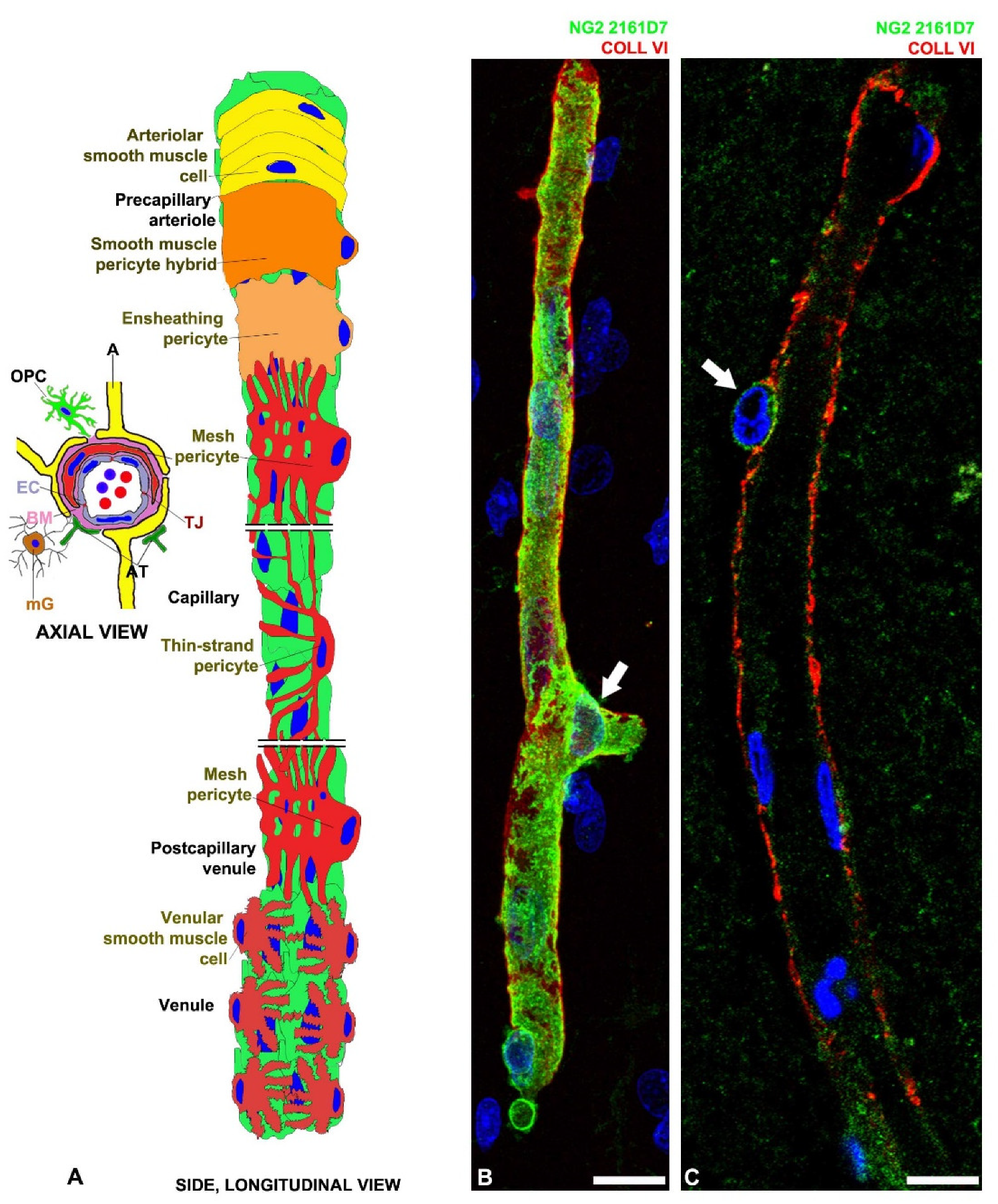
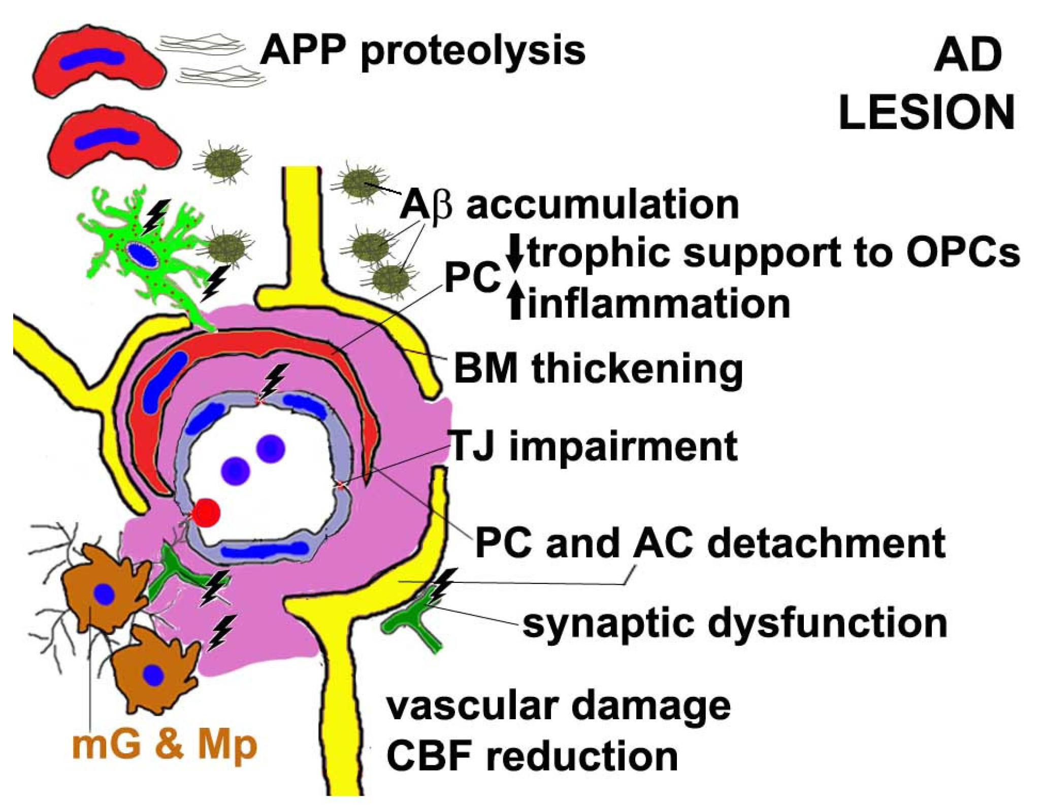
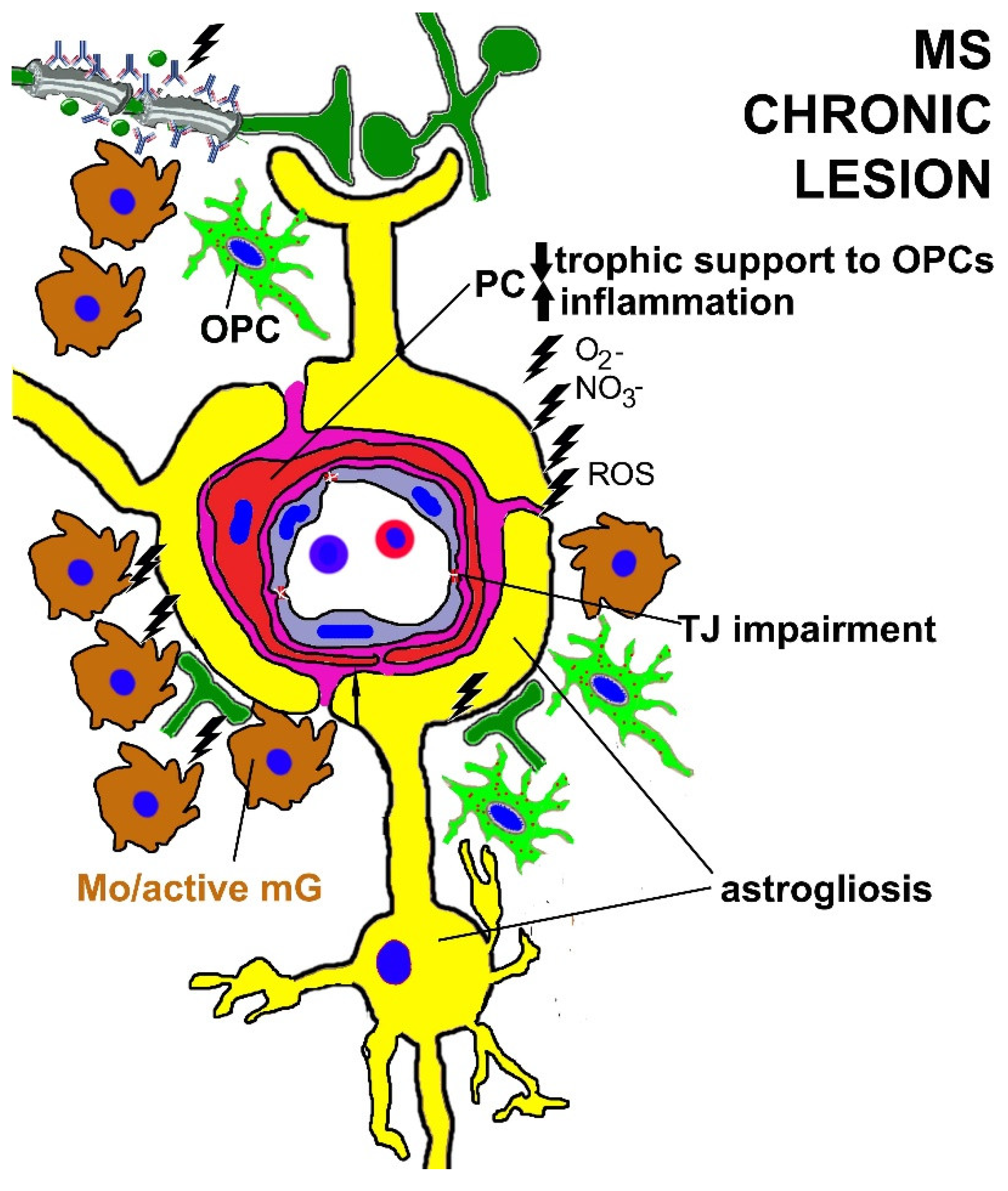
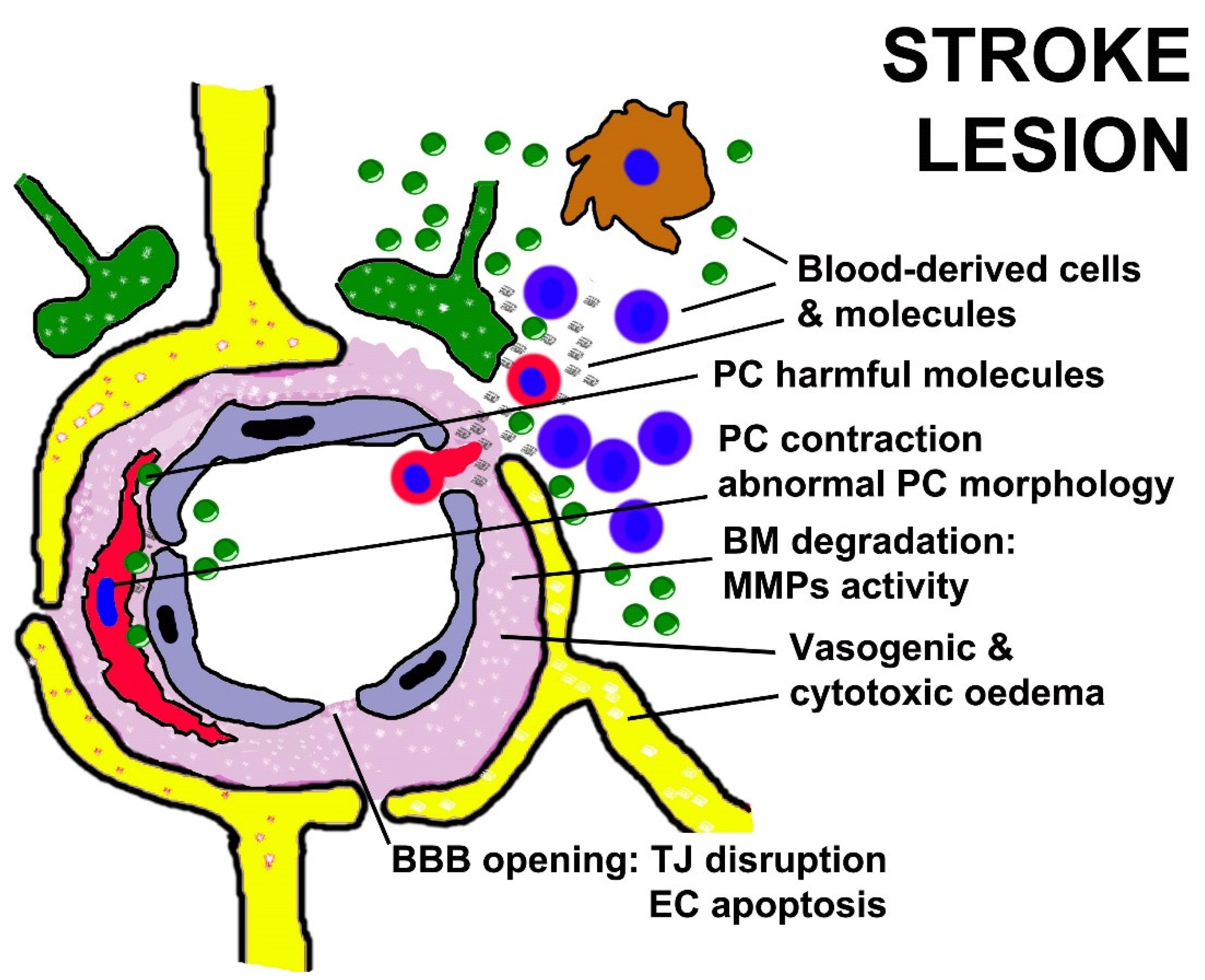
Table 1.
Best known markers of CNS PCs and their dynamic expression during development and adulthood.
Table 1.
Best known markers of CNS PCs and their dynamic expression during development and adulthood.
| PC Marker | Development | Healthy Adulthood |
|---|---|---|
| Neuron-glial antigen 2 (NG2) | [19,63] | Only activated PCs of CNS [64,65] |
| Platelet derived growth factor receptor beta (PDGFRβ) | [66,67] | [68] |
| Alanyl aminopeptidase (CD13) | [69] | [70,71] |
| Melanoma Cell Adhesion Molecule (CD146) | [71,72] | [73] |
| Endosialin (CD248) | [66,74] | Not expressed |
| Fluoro-Nissl dye NeuroTrace 500/525 | [75] | [76] |
Table 2.
Summary of PC dysfunction during clinical and experimental CNS conditions.
| Disease | PC Dysfunction(s) in the Clinical Condition | PC Dysfunction(s) in Models |
|---|---|---|
| Autism spectrum disorder | Increased density [182]; genetic risk after PC gene modifications [370] | Putative NVU dysfunction [371] |
| Prenatal life hypoxia | Reduced PC coverage [189,190]; increased angiogenesis and bFGF [372] | |
| White matter degeneration | [373] | Reduced PC coverage [154] |
| Altered neuronal circuitry | [120] | Ablation of PC induce poor interaction between glia and neural precursors [184,374]. |
| AD | Hypoperfusion, PC degeneration and BBB breakdown [117,162,193,194,206,207,209,375]; PC inability to support oligodendrogenesis [233] | Decreased density of PDGFRβ+ PCs in 5xFAD mice [211] and CD13+ PCs in pericyte-deficient APPsw/0; Pdgfrβ+/− [213]; dysfunction and apoptosis of desmin+ PCs in APPswe/PS1 mice [225] |
| MS | PC inability to support oligodendrogenesis [232]; increased density of PDGFRβ+ CD146+ PC in active MS lesions [239]; increased angiogenesis [255,256,257] | PC inability to support oligodendrogenesis in different models [69,144,145,149,150,154,232,244,253]; PDGFRβ+ PC triggers inflammation [245,246,247,248,249,250,251,376,377,378]; increased angiogenesis [258,259,261,262,263,264,265] |
| Stroke | Microvascular PC contractions aggravate ischemia [121]; PCs release harmful molecules [297] | In different models, PCs release harmful molecules [184,225,248,254,352]; decreased PC density, EC apoptosis, and BBB disruption [347,348,350,351]; abnormal PC morphology [354] |
| NeuroCOVID-19 | SARS-CoV-2 infects ACE2+ PCs [356,363,365] | PC vasoconstriction [368,369] |
Publisher’s Note: MDPI stays neutral with regard to jurisdictional claims in published maps and institutional affiliations. |
© 2022 by the authors. Licensee MDPI, Basel, Switzerland. This article is an open access article distributed under the terms and conditions of the Creative Commons Attribution (CC BY) license (https://creativecommons.org/licenses/by/4.0/).
Share and Cite
MDPI and ACS Style
Girolamo, F.; Errede, M.; Bizzoca, A.; Virgintino, D.; Ribatti, D. Central Nervous System Pericytes Contribute to Health and Disease. Cells 2022, 11, 1707. https://0-doi-org.brum.beds.ac.uk/10.3390/cells11101707
AMA Style
Girolamo F, Errede M, Bizzoca A, Virgintino D, Ribatti D. Central Nervous System Pericytes Contribute to Health and Disease. Cells. 2022; 11(10):1707. https://0-doi-org.brum.beds.ac.uk/10.3390/cells11101707
Chicago/Turabian StyleGirolamo, Francesco, Mariella Errede, Antonella Bizzoca, Daniela Virgintino, and Domenico Ribatti. 2022. "Central Nervous System Pericytes Contribute to Health and Disease" Cells 11, no. 10: 1707. https://0-doi-org.brum.beds.ac.uk/10.3390/cells11101707
Note that from the first issue of 2016, this journal uses article numbers instead of page numbers. See further details here.