Next Issue
Volume 11, March-1
Previous Issue
Volume 11, February-1


Cells, Volume 11, Issue 4 (February-2 2022) – 167 articles

Cover Story (view full-size image): 
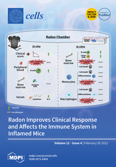
A therapy with the noble gas radon can be used in the treatment of painful, chronic inflammatory diseases as patients report long-lasting analgesic effects. So far, there is a lack in understanding the underlying molecular mechanisms and involved cell types. Next to the modulation of the immune system, an involvement of the antioxidative system is assumed. We utilized the controlled environment of a radon chamber in an in vivo as well as an ex vivo approach. We revealed that radon exposure significantly improves clinical disease progression and significantly increases B cell numbers and IL-5 levels, while no alterations in the antioxidative system were found in K/BxN serum-induced arthritic mice. We thus conclude that radon is able to modulate the immune system and, alongside with other, yet to be identified, parameters, improves painful inflammatory diseases. View this paper
  • Issues are regarded as officially published after their release is announced to the table of contents alert mailing list.
  • You may sign up for e-mail alerts to receive table of contents of newly released issues.
  • PDF is the official format for papers published in both, html and pdf forms. To view the papers in pdf format, click on the "PDF Full-text" link, and use the free Adobe Reader to open them.
Order results
Result details
Section
Select all
Export citation of selected articles as:
22 pages, 4043 KiB  
Article
Divalent Metal Ions Boost Effect of Nucleic Acids Delivered by Cell-Penetrating Peptides
by Maria Maloverjan, Kärt Padari, Aare Abroi, Ana Rebane and Margus Pooga
Cells 2022, 11(4), 756; https://0-doi-org.brum.beds.ac.uk/10.3390/cells11040756 - 21 Feb 2022
Cited by 2 | Viewed by 2337
Abstract
Cell-penetrating peptides (CPPs) are promising tools for the transfection of various substances, including nucleic acids, into cells. The aim of the current work was to search for novel safe and effective approaches for enhancing transfection efficiency of nanoparticles formed from CPP and splice-correcting [...] Read more.
Cell-penetrating peptides (CPPs) are promising tools for the transfection of various substances, including nucleic acids, into cells. The aim of the current work was to search for novel safe and effective approaches for enhancing transfection efficiency of nanoparticles formed from CPP and splice-correcting oligonucleotide (SCO) without increasing the concentration of peptide. We analyzed the effect of inclusion of calcium and magnesium ions into nanoparticles on CPP-mediated transfection in cell culture. We also studied the mechanism of such transfection as well as its efficiency, applicability in case of different cell lines, nucleic acid types and peptides, and possible limitations. We discovered a strong positive effect of these ions on transfection efficiency of SCO, that translated to enhanced synthesis of functional reporter protein. We observed significant changes in intracellular distribution and trafficking of nanoparticles formed by the addition of the ions, without increasing cytotoxicity. We propose a novel strategy for preparing CPP-oligonucleotide nanoparticles with enhanced efficiency and, thus, higher therapeutic potential. Our discovery may be translated to primary cell cultures and, possibly, in vivo studies, with the aim of increasing CPP-mediated transfection efficiency and the likelihood of using CPPs in clinics. Full article
Show Figures

Graphical abstract

22 pages, 2965 KiB  
Article
Digital Holographic Microscopy for Label-Free Detection of Leukocyte Alternations Associated with Perioperative Inflammation after Cardiac Surgery
by David Rene Steike, Michael Hessler, Eberhard Korsching, Florian Lehmann, Christina Schmidt, Christian Ertmer, Jürgen Schnekenburger, Hans Theodor Eich, Björn Kemper and Burkhard Greve
Cells 2022, 11(4), 755; https://0-doi-org.brum.beds.ac.uk/10.3390/cells11040755 - 21 Feb 2022
Cited by 11 | Viewed by 3077
Abstract
In a prospective observational pilot study on patients undergoing elective cardiac surgery with cardiopulmonary bypass, we evaluated label-free quantitative phase imaging (QPI) with digital holographic microscopy (DHM) to describe perioperative inflammation by changes in biophysical cell properties of lymphocytes and monocytes. Blood samples [...] Read more.
In a prospective observational pilot study on patients undergoing elective cardiac surgery with cardiopulmonary bypass, we evaluated label-free quantitative phase imaging (QPI) with digital holographic microscopy (DHM) to describe perioperative inflammation by changes in biophysical cell properties of lymphocytes and monocytes. Blood samples from 25 patients were investigated prior to cardiac surgery and postoperatively at day 1, 3 and 6. Biophysical and morphological cell parameters accessible with DHM, such as cell volume, refractive index, dry mass, and cell shape related form factor, were acquired and compared to common flow cytometric blood cell markers of inflammation and selected routine laboratory parameters. In all examined patients, cardiac surgery induced an acute inflammatory response as indicated by changes in routine laboratory parameters and flow cytometric cell markers. DHM results were associated with routine laboratory and flow cytometric data and correlated with complications in the postoperative course. In a subgroup analysis, patients were classified according to the inflammation related C-reactive protein (CRP) level, treatment with epinephrine and the occurrence of postoperative complications. Patients with regular courses, without epinephrine treatment and with low CRP values showed a postoperative lymphocyte volume increase. In contrast, the group of patients with increased CRP levels indicated an even further enlarged lymphocyte volume, while for the groups of epinephrine treated patients and patients with complicative courses, no postoperative lymphocyte volume changes were detected. In summary, the study demonstrates the capability of DHM to describe biophysical cell parameters of perioperative lymphocytes and monocytes changes in cardiac surgery patients. The pattern of correlations between biophysical DHM data and laboratory parameters, flow cytometric cell markers, and the postoperative course exemplify DHM as a promising diagnostic tool for a characterization of inflammatory processes and course of disease. Full article
(This article belongs to the Special Issue Current Trends in Quantitative Phase Imaging of Cells and Tissues)
Show Figures

Figure 1

15 pages, 16838 KiB  
Article
Basal Autophagy Is Necessary for A Pharmacologic PPARα Transactivation
by Eun Young Kim and Jae Man Lee
Cells 2022, 11(4), 754; https://0-doi-org.brum.beds.ac.uk/10.3390/cells11040754 - 21 Feb 2022
Cited by 1 | Viewed by 2070
Abstract
Autophagy is a conserved cellular process of catabolism leading to nutrient recycling upon starvation and maintaining tissue and energy homeostasis. Tissue-specific loss of core-autophagy-related genes often triggers diverse diseases, including cancer, neurodegeneration, inflammatory disease, metabolic disorder, and muscle disease. The nutrient-sensing nuclear receptors [...] Read more.
Autophagy is a conserved cellular process of catabolism leading to nutrient recycling upon starvation and maintaining tissue and energy homeostasis. Tissue-specific loss of core-autophagy-related genes often triggers diverse diseases, including cancer, neurodegeneration, inflammatory disease, metabolic disorder, and muscle disease. The nutrient-sensing nuclear receptors peroxisome proliferator-activated receptor α (PPARα) plays a key role in fasting-associated metabolisms such as autophagy, fatty acid oxidation, and ketogenesis. Here we show that autophagy defects impede the transactivation of PPARα. Liver-specific ablation of the Atg7 gene in mice showed reduced expression levels of PPARα target genes in response to its synthetic agonist ligands. Since NRF2, an antioxidant transcription factor, is activated in autophagy-deficient mice due to p62/SQSTM1 accumulation and its subsequent interaction with KEAP1, an E3 ubiquitin ligase. We hypothesize that the nuclear accumulation of NRF2 by autophagy defects blunts the transactivation of PPARα. Consistent with this idea, we find that NRF2 activation is sufficient to inhibit the pharmacologic transactivation of PPARα, which is dependent on the Nrf2 gene. These results reveal an unrecognized requirement of basal autophagy for the transactivation of PPARα by preventing NRF2 from a nuclear translocation and suggest a clinical significance of basal autophagy to expect a pharmacologic efficacy of synthetic PPARα ligands. Full article
Show Figures

Figure 1

14 pages, 4260 KiB  
Article
The HIFα-Stabilizing Drug Roxadustat Increases the Number of Renal Epo-Producing Sca-1+ Cells
by Aline Jatho, Anke Zieseniss, Katja Brechtel-Curth, Jia Guo, Kai Oliver Böker, Gabriela Salinas, Roland H. Wenger and Dörthe M. Katschinski
Cells 2022, 11(4), 753; https://0-doi-org.brum.beds.ac.uk/10.3390/cells11040753 - 21 Feb 2022
Cited by 4 | Viewed by 3265
Abstract
Inhibition of the prolyl-4-hydroxylase domain (PHD) enzymes, leading to the stabilization of hypoxia-inducible factor (HIF) α as well as to the stimulation of erythropoietin (Epo) synthesis, is the functional mechanism of the new anti-anemia drug roxadustat. Little is known about the effects of [...] Read more.
Inhibition of the prolyl-4-hydroxylase domain (PHD) enzymes, leading to the stabilization of hypoxia-inducible factor (HIF) α as well as to the stimulation of erythropoietin (Epo) synthesis, is the functional mechanism of the new anti-anemia drug roxadustat. Little is known about the effects of roxadustat on the Epo-producing cell pool. To gain further insights into the function of PHD inhibitors, we characterized the abundance of mesenchymal stem cell (MSC)-like cells after roxadustat treatment of mice. The number of Sca-1+ mesenchymal cells following roxadustat treatment increased exclusively in the kidneys. Isolated Sca-1+ cells demonstrated typical features of MSC-like cells, including adherence to tissue culture plates, trilineage differentiation potential, and expression of MSC markers. Kidney-derived Sca-1+ MSC-like cells were cultured for up to 21 days. Within the first few days in culture, cells stabilized HIF-1α and HIF-2α and temporarily increased Epo production upon incubation in hypoxia. In summary, we have identified a Sca-1+ MSC-like cell population that is involved in renal Epo production and might contribute to the strong anti-anemic effect of the PHD inhibitor roxadustat. Full article
(This article belongs to the Special Issue Gene Regulation by HIFs during Hypoxia 2022)
Show Figures

Figure 1

16 pages, 647 KiB  
Article
Circulating Serum MiRNA-8074 as a Novel Prognostic Biomarker for Multiple Myeloma
by Aneta Szudy-Szczyrek, Radosław Mlak, Michał Mielnik, Marcin Mazurek, Sylwia Chocholska, Martyna Podgajna, Michał Szczyrek, Iwona Homa-Mlak, Teresa Małecka-Massalska and Marek Hus
Cells 2022, 11(4), 752; https://0-doi-org.brum.beds.ac.uk/10.3390/cells11040752 - 21 Feb 2022
Cited by 4 | Viewed by 2319
Abstract
MiRNA-8074 is a molecule with the potential to regulate the expression of key genes related to the pathogenesis of multiple myeloma (MM), i.e., TP53, MYC, MAPK1, and KIAA. We analyzed the predictive and prognostic value of miRNA-8074 expression in [...] Read more.
MiRNA-8074 is a molecule with the potential to regulate the expression of key genes related to the pathogenesis of multiple myeloma (MM), i.e., TP53, MYC, MAPK1, and KIAA. We analyzed the predictive and prognostic value of miRNA-8074 expression in MM patients. In total, 105 newly diagnosed MM patients treated with thalidomide (n = 27), bortezomib (n = 41) and bortezomib with thalidomide (n = 37) were studied. For miRNA analysis, the column method and the Real-Time PCR technique with specific TaqMan Fast Advanced Master Mix and TaqMan probes were used. Factors that were associated with a significant reduction in progression-free survival (PFS) included: ECOG > 1, ISS stage III, low hemoglobin, thrombocytopenia, hypoalbuminemia, abnormal renal function, elevated creatinine, GFR < 60 mL/min/1.73 m2, elevated LDH, del(17p), t(11;14), the use of a single drug regimen (thalidomide or bortezomib) and high miRNA-8074 expression (HR = 2.01, 95% CI: 1.16–3.49; p = 0.0233). In addition to the known prognostic factors, such as ECOG > 1, Durie–Salmon stage III, diagnosis of light chain disease or non-secreting MM, renal failure, hypoalbuminemia, hypercalcemia, high β2-microglobulin, elevated LDH, and t(14;16), a high expression of miRNA-8074 was significantly associated with a higher risk of death (HR = 4.12, 95% CI: 2.20–7.70; p = 0.0009). In summary, miRNA-8074 may be a useful diagnostic tool to assess the prognosis in MM patients. Full article
(This article belongs to the Special Issue MicroRNAs in Cancer: Biomarkers, Functions and Therapies)
Show Figures

Figure 1

15 pages, 3782 KiB  
Article
Functional and Genetic Characterization of Porcine Beige Adipocytes
by Lilan Zhang, Silu Hu, Chunwei Cao, Chuanhe Chen, Jiali Liu, Yu Wang, Jianfeng Liu, Jianguo Zhao, Cong Tao and Yanfang Wang
Cells 2022, 11(4), 751; https://0-doi-org.brum.beds.ac.uk/10.3390/cells11040751 - 21 Feb 2022
Cited by 3 | Viewed by 2483
Abstract
Beige adipocytes are a distinct type of fat cells with a thermogenic activity that have gained substantial attention as an alternative cellular anti-obesity target in humans. These cells may provide an alternative strategy for the genetic selection of pigs with reduced fat deposition. [...] Read more.
Beige adipocytes are a distinct type of fat cells with a thermogenic activity that have gained substantial attention as an alternative cellular anti-obesity target in humans. These cells may provide an alternative strategy for the genetic selection of pigs with reduced fat deposition. Despite the presence of beige adipocytes in piglets, the molecular signatures of porcine beige adipocytes remain unclear. Here, white and beige adipocytes from Tibetan piglets were primarily cultured and differentiated. Compared to the white adipocytes, the beige adipocytes exhibited a stronger thermogenic capacity. RNA-sequencing-based genome-wide comparative analyses revealed distinct gene expression profiles for white and beige adipocytes. In addition, two genes, integrin alpha-2 (ITGA2) and calponin 1 (CNN1), which were specifically differentially expressed in porcine beige adipocytes, were further functionally characterized using a loss-of-function approach. Our data showed that both genes were involved in differentiation and thermogenesis of porcine beige adipocytes. Collectively, these data furthered our understanding of gene expression in porcine white and beige adipocytes. Elucidating the genetic basis of beige adipogenesis in pigs will pave the way for molecular design breeding in both pigs and large animal models of human diseases. Full article
Show Figures

Graphical abstract

10 pages, 3237 KiB  
Review
Free Fatty Acid Receptors (FFARs) in Adipose: Physiological Role and Therapeutic Outlook
by Saeed Al Mahri, Shuja Shafi Malik, Maria Al Ibrahim, Esraa Haji, Ghida Dairi and Sameer Mohammad
Cells 2022, 11(4), 750; https://0-doi-org.brum.beds.ac.uk/10.3390/cells11040750 - 21 Feb 2022
Cited by 28 | Viewed by 4950
Abstract
Fatty acids (FFAs) are important biological molecules that serve as a major energy source and are key components of biological membranes. In addition, FFAs play important roles in metabolic regulation and contribute to the development and progression of metabolic disorders like diabetes. Recent [...] Read more.
Fatty acids (FFAs) are important biological molecules that serve as a major energy source and are key components of biological membranes. In addition, FFAs play important roles in metabolic regulation and contribute to the development and progression of metabolic disorders like diabetes. Recent studies have shown that FFAs can act as important ligands of G-protein-coupled receptors (GPCRs) on the surface of cells and impact key physiological processes. Free fatty acid-activated receptors include FFAR1 (GPR40), FFAR2 (GPR43), FFAR3 (GPR41), and FFAR4 (GPR120). FFAR2 and FFAR3 are activated by short-chain fatty acids like acetate, propionate, and butyrate, whereas FFAR1 and FFAR4 are activated by medium- and long-chain fatty acids like palmitate, oleate, linoleate, and others. FFARs have attracted considerable attention over the last few years and have become attractive pharmacological targets in the treatment of type 2 diabetes and metabolic syndrome. Several lines of evidence point to their importance in the regulation of whole-body metabolic homeostasis including adipose metabolism. Here, we summarize our current understanding of the physiological functions of FFAR isoforms in adipose biology and explore the prospect of FFAR-based therapies to treat patients with obesity and Type 2 diabetes. Full article
(This article belongs to the Special Issue Free Fatty Acids and Pathogenesis of Diabetes Mellitus)
Show Figures

Graphical abstract

18 pages, 13626 KiB  
Article
CRISPR/Cas9 Mediated Knockout of Cyclooxygenase-2 Gene Inhibits Invasiveness in A2058 Melanoma Cells
by Cathleen Haase-Kohn, Markus Laube, Cornelius K. Donat, Birgit Belter and Jens Pietzsch
Cells 2022, 11(4), 749; https://0-doi-org.brum.beds.ac.uk/10.3390/cells11040749 - 21 Feb 2022
Cited by 7 | Viewed by 2703
Abstract
The inducible isoenzyme cyclooxygenase-2 (COX-2) is an important hub in cellular signaling, which contributes to tumor progression by modulating and enhancing a pro-inflammatory tumor microenvironment, tumor growth, apoptosis resistance, angiogenesis and metastasis. In order to understand the role of COX-2 expression in melanoma, [...] Read more.
The inducible isoenzyme cyclooxygenase-2 (COX-2) is an important hub in cellular signaling, which contributes to tumor progression by modulating and enhancing a pro-inflammatory tumor microenvironment, tumor growth, apoptosis resistance, angiogenesis and metastasis. In order to understand the role of COX-2 expression in melanoma, we investigated the functional knockout effect of COX-2 in A2058 human melanoma cells. COX-2 knockout was validated by Western blot and flow cytometry analysis. When comparing COX-2 knockout cells to controls, we observed significantly reduced invasion, colony and spheroid formation potential in cell monolayers and three-dimensional models in vitro, and significantly reduced tumor development in xenograft mouse models in vivo. Moreover, COX-2 knockout alters the metabolic activity of cells under normoxia and experimental hypoxia as demonstrated by using the radiotracers [18F]FDG and [18F]FMISO. Finally, a pilot protein array analysis in COX-2 knockout cells verified significantly altered downstream signaling pathways that can be linked to cellular and molecular mechanisms of cancer metastasis closely related to the enzyme. Given the complexity of the signaling pathways and the multifaceted role of COX-2, targeted suppression of COX-2 in melanoma cells, in combination with modulation of related signaling pathways, appears to be a promising therapeutic approach. Full article
(This article belongs to the Special Issue Cellular and Molecular Mechanisms of Cancer Metastasis)
Show Figures

Figure 1

17 pages, 7065 KiB  
Review
The Role of MeCP2 in Regulating Synaptic Plasticity in the Context of Stress and Depression
by Carla L. Sánchez-Lafuente, Lisa E. Kalynchuk, Hector J. Caruncho and Juan Ausió
Cells 2022, 11(4), 748; https://0-doi-org.brum.beds.ac.uk/10.3390/cells11040748 - 21 Feb 2022
Cited by 13 | Viewed by 4385
Abstract
Methyl-CpG-binding protein 2 (MeCP2) is a transcriptional regulator that is highly abundant in the brain. It binds to methylated genomic DNA to regulate a range of physiological functions implicated in neuronal development and adult synaptic plasticity. MeCP2 has mainly been studied for its [...] Read more.
Methyl-CpG-binding protein 2 (MeCP2) is a transcriptional regulator that is highly abundant in the brain. It binds to methylated genomic DNA to regulate a range of physiological functions implicated in neuronal development and adult synaptic plasticity. MeCP2 has mainly been studied for its role in neurodevelopmental disorders, but alterations in MeCP2 are also present in stress-related disorders such as major depression. Impairments in both stress regulation and synaptic plasticity are associated with depression, but the specific mechanisms underlying these changes have not been identified. Here, we review the interplay between stress, synaptic plasticity, and MeCP2. We focus our attention on the transcriptional regulation of important neuronal plasticity genes such as BDNF and reelin (RELN). Moreover, we provide evidence from recent studies showing a link between chronic stress-induced depressive symptoms and dysregulation of MeCP2 expression, underscoring the role of this protein in stress-related pathology. We conclude that MeCP2 is a promising target for the development of novel, more efficacious therapeutics for the treatment of stress-related disorders such as depression. Full article
(This article belongs to the Special Issue Transcriptional Regulatory Mechanisms in Health and Disease)
Show Figures

Graphical abstract

48 pages, 3107 KiB  
Review
Normal and Neoplastic Growth Suppression by the Extended Myc Network
by Edward V. Prochownik and Huabo Wang
Cells 2022, 11(4), 747; https://0-doi-org.brum.beds.ac.uk/10.3390/cells11040747 - 21 Feb 2022
Cited by 12 | Viewed by 3248
Abstract
Among the first discovered and most prominent cellular oncogenes is MYC, which encodes a bHLH-ZIP transcription factor (Myc) that both activates and suppresses numerous genes involved in proliferation, energy production, metabolism and translation. Myc belongs to a small group of bHLH-ZIP transcriptional regulators [...] Read more.
Among the first discovered and most prominent cellular oncogenes is MYC, which encodes a bHLH-ZIP transcription factor (Myc) that both activates and suppresses numerous genes involved in proliferation, energy production, metabolism and translation. Myc belongs to a small group of bHLH-ZIP transcriptional regulators (the Myc Network) that includes its obligate heterodimerization partner Max and six “Mxd proteins” (Mxd1–4, Mnt and Mga), each of which heterodimerizes with Max and largely opposes Myc’s functions. More recently, a second group of bHLH-ZIP proteins (the Mlx Network) has emerged that bears many parallels with the Myc Network. It is comprised of the Myc-like factors ChREBP and MondoA, which, in association with the Max-like member Mlx, regulate smaller and more functionally restricted repertoires of target genes, some of which are shared with Myc. Opposing ChREBP and MondoA are heterodimers comprised of Mlx and Mxd1, Mxd4 and Mnt, which also structurally and operationally link the two Networks. We discuss here the functions of these “Extended Myc Network” members, with particular emphasis on their roles in suppressing normal and neoplastic growth. These roles are complex due to the temporal- and tissue-restricted expression of Extended Myc Network proteins in normal cells, their regulation of both common and unique target genes and, in some cases, their functional redundancy. Full article
Show Figures

Figure 1

21 pages, 14170 KiB  
Article
Enhanced Autophagic Flux, Suppressed Apoptosis and Reduced Macrophage Infiltration by Dasatinib in Kidneys of Obese Mice
by Hassan Reda Hassan Elsayed, Randa El-Gamal, Mohammed R. Rabei, Mona G. Elhadidy, Shereen Hamed, Basma H. Othman, Mohamed Mahmoud Abdelraheem Elshaer, Mostafa Khaled Sedky, Ahmed Tarek Abd Elbaset Hassan and Mohammad El-Nablaway
Cells 2022, 11(4), 746; https://0-doi-org.brum.beds.ac.uk/10.3390/cells11040746 - 21 Feb 2022
Cited by 4 | Viewed by 2758
Abstract
Obesity causes renal changes (ORC), characterized by defective renal autophagy, lipogenesis, enhanced macrophage infiltration and apoptosis. We hypothesize that Dasatinib, a tyrosine kinase inhibitor, may ameliorate changes associated with obesity. We the mice with either Obesogenic diet (OD) or a standard basal diet. [...] Read more.
Obesity causes renal changes (ORC), characterized by defective renal autophagy, lipogenesis, enhanced macrophage infiltration and apoptosis. We hypothesize that Dasatinib, a tyrosine kinase inhibitor, may ameliorate changes associated with obesity. We the mice with either Obesogenic diet (OD) or a standard basal diet. After 12 weeks, the mice received either vehicle or Dasatinib 4 mg/kg/d for an additional four weeks. We examined serum creatinine, urea, lipid profile and renal cortical mRNA expression for lipogenesis marker SREBP1, inflammatory macrophage marker iNOS and fibrosis markers; TGFβ and PDGFA genes; immunohistochemical (IHC) staining for CD68; inflammatory macrophage marker and ASMA; fibrosis marker, LC3 and SQSTM1/P62; autophagy markers and western blotting (WB) for caspase-3; and, as an apoptosis marker, LC3II/I and SQSTM1/P62 in addition to staining for H&E, PAS, Sirius red and histopathological scoring. Dasatinib attenuated renal cortical mRNA expression for SREBP1, iNOS, PDGFA and TGFβ and IHC staining for CD68, ASMA and SQSTM1/P62 and WB for caspase-3 and SQSTM1/P62, while elevating LC3 expression. Moreover, Dasatinib ameliorated ORC; glomerulosclerosis, glomerular expansion, tubular dilatation, vacuolation and casts; inflammatory cellular infiltration; and fibrosis. Dasatinib is a promising therapy for ORC by correcting autophagy impairment, attenuating lipogenesis, apoptosis and macrophage infiltration by inducing antifibrotic activity. Full article
(This article belongs to the Special Issue Recent Advances in Development and Progression of Kidney Diseases)
Show Figures

Figure 1

19 pages, 3107 KiB  
Review
A Focused Review on Primary Graft Dysfunction after Clinical Lung Transplantation: A Multilevel Syndrome
by Jan Van Slambrouck, Dirk Van Raemdonck, Robin Vos, Cedric Vanluyten, Arno Vanstapel, Elena Prisciandaro, Lynn Willems, Michaela Orlitová, Janne Kaes, Xin Jin, Yanina Jansen, Geert M. Verleden, Arne P. Neyrinck, Bart M. Vanaudenaerde and Laurens J. Ceulemans
Cells 2022, 11(4), 745; https://0-doi-org.brum.beds.ac.uk/10.3390/cells11040745 - 21 Feb 2022
Cited by 7 | Viewed by 4253
Abstract
Primary graft dysfunction (PGD) is the clinical syndrome of acute lung injury after lung transplantation (LTx). However, PGD is an umbrella term that encompasses the ongoing pathophysiological and -biological mechanisms occurring in the lung grafts. Therefore, we aim to provide a focused review [...] Read more.
Primary graft dysfunction (PGD) is the clinical syndrome of acute lung injury after lung transplantation (LTx). However, PGD is an umbrella term that encompasses the ongoing pathophysiological and -biological mechanisms occurring in the lung grafts. Therefore, we aim to provide a focused review on the clinical, physiological, radiological, histological and cellular level of PGD. PGD is graded based on hypoxemia and chest X-ray (CXR) infiltrates. High-grade PGD is associated with inferior outcome after LTx. Lung edema is the main characteristic of PGD and alters pulmonary compliance, gas exchange and circulation. A conventional CXR provides a rough estimate of lung edema, while a chest computed tomography (CT) results in a more in-depth analysis. Macroscopically, interstitial and alveolar edema can be distinguished below the visceral lung surface. On the histological level, PGD correlates to a pattern of diffuse alveolar damage (DAD). At the cellular level, ischemia-reperfusion injury (IRI) is the main trigger for the disruption of the endothelial-epithelial alveolar barrier and inflammatory cascade. The multilevel approach integrating all PGD-related aspects results in a better understanding of acute lung failure after LTx, providing novel insights for future therapies. Full article
(This article belongs to the Special Issue Advances in Lung Transplantation)
Show Figures

Figure 1

20 pages, 6988 KiB  
Article
Protective Effects of Chaya against Mitochondrial and Synaptic Toxicities in the Type 2 Diabetes Mouse Model TallyHO
by Bhagavathi Ramasubramanian, Cameron Griffith, Madison Hanson, Lloyd E. Bunquin, Arubala P. Reddy, Vijay Hegde and P. Hemachandra Reddy
Cells 2022, 11(4), 744; https://0-doi-org.brum.beds.ac.uk/10.3390/cells11040744 - 21 Feb 2022
Cited by 2 | Viewed by 2148
Abstract
The purpose of our study is to determine the protective effects of the chaya leaf against mitochondrial abnormalities and synaptic damage in the Type 2 diabetes (T2D) mouse model, TallyHO (TH). The TH mouse is a naturally occurring polygenic mouse model of diabetes [...] Read more.
The purpose of our study is to determine the protective effects of the chaya leaf against mitochondrial abnormalities and synaptic damage in the Type 2 diabetes (T2D) mouse model, TallyHO (TH). The TH mouse is a naturally occurring polygenic mouse model of diabetes that mimics many characteristics of human Type 2 diabetes. Only male TH mice develop hyperglycemia and moderate obesity. Female mice display moderate obesity but do not manifest overt diabetes. In this study, we evaluated three groups of mice over a period of 11 weeks: (1) the experimental group of TH diabetic mice fed with chaya chow; (2) a diabetic control group of TH diabetic mice fed with regular chow; and (3) a non-diabetic control group of SWR/J mice fed with regular chow. Body mass and fasting blood glucose were assessed weekly. Brain and other peripheral tissues were collected. Using qRT-PCR and immunoblotting analyses, we measured the mRNA abundance and protein levels of mitochondrial biogenesis, mitochondrial dynamics, autophagy/mitophagy, and synaptic genes. Using immunofluorescence analysis, we measured the regional immunoreactivities of mitochondrial and synaptic proteins. Using biochemical methods, we assessed mitochondrial function. We found increased body mass and fasting glucose levels in the TH diabetic mice relative to the non-diabetic control SWRJ mice. In chaya chow-fed TH diabetic mice, we found significantly reduced body mass and fasting glucose levels. Mitochondrial fission genes were increased and fusion, biogenesis, autophagy/mitophagy, and synaptic genes were reduced in the TH mice; however, in the chaya chow-fed TH diabetic mice, mitochondrial fission genes were reduced and fusion, biogenesis, autophagy/mitophagy, and synaptic genes were increased. Mitochondrial function was defective in the diabetic TH mice; however, it was rescued in the chaya chow-fed TH mice. These observations strongly suggest that chaya chow reduces the diabetic properties, mitochondrial abnormalities, and synaptic pathology in diabetic, TH male mice. Our data strongly indicates that chaya can be used as natural supplemental diet for prediabetic and diabetic subjects and individuals with metabolic disorders. Full article
Show Figures

Figure 1

21 pages, 12172 KiB  
Review
Role of Dietary Supplements and Probiotics in Modulating Microbiota and Bone Health: The Gut-Bone Axis
by Alessandro de Sire, Roberto de Sire, Claudio Curci, Fabiana Castiglione and Walter Wahli
Cells 2022, 11(4), 743; https://0-doi-org.brum.beds.ac.uk/10.3390/cells11040743 - 21 Feb 2022
Cited by 38 | Viewed by 7486
Abstract
Osteoporosis is characterized by an alteration of bone microstructure with a decreased bone mineral density, leading to the incidence of fragility fractures. Around 200 million people are affected by osteoporosis, representing a major health burden worldwide. Several factors are involved in the pathogenesis [...] Read more.
Osteoporosis is characterized by an alteration of bone microstructure with a decreased bone mineral density, leading to the incidence of fragility fractures. Around 200 million people are affected by osteoporosis, representing a major health burden worldwide. Several factors are involved in the pathogenesis of osteoporosis. Today, altered intestinal homeostasis is being investigated as a potential additional risk factor for reduced bone health and, therefore, as a novel potential therapeutic target. The intestinal microflora influences osteoclasts’ activity by regulating the serum levels of IGF-1, while also acting on the intestinal absorption of calcium. It is therefore not surprising that gut dysbiosis impacts bone health. Microbiota alterations affect the OPG/RANKL pathway in osteoclasts, and are correlated with reduced bone strength and quality. In this context, it has been hypothesized that dietary supplements, prebiotics, and probiotics contribute to the intestinal microecological balance that is important for bone health. The aim of the present comprehensive review is to describe the state of the art on the role of dietary supplements and probiotics as therapeutic agents for bone health regulation and osteoporosis, through gut microbiota modulation. Full article
(This article belongs to the Special Issue Gut Microbiota in Nutrition and Health)
Show Figures

Figure 1

14 pages, 1741 KiB  
Article
Sustained Elevated Circulating Activin A Impairs Global Longitudinal Strain in Pregnant Rats: A Potential Mechanism for Preeclampsia-Related Cardiac Dysfunction
by Bhavisha A. Bakrania, Ana C. Palei, Umesh Bhattarai, Yingjie Chen, Joey P. Granger and Sajid Shahul
Cells 2022, 11(4), 742; https://0-doi-org.brum.beds.ac.uk/10.3390/cells11040742 - 21 Feb 2022
Cited by 1 | Viewed by 2212
Abstract
Mediators of cardiac injury in preeclampsia are not well understood. Preeclamptic women have decreased cardiac global longitudinal strain (GLS), a sensitive measure of systolic function that indicates fibrosis and tissue injury. GLS is worse in preeclampsia compared to gestational hypertension, despite comparable blood [...] Read more.
Mediators of cardiac injury in preeclampsia are not well understood. Preeclamptic women have decreased cardiac global longitudinal strain (GLS), a sensitive measure of systolic function that indicates fibrosis and tissue injury. GLS is worse in preeclampsia compared to gestational hypertension, despite comparable blood pressure, suggesting that placental factors may be involved. We previously showed that Activin A, a pro-fibrotic factor produced in excess by the placenta in preeclampsia, predicts impaired GLS postpartum. Here, we hypothesized that chronic excess levels of Activin A during pregnancy induces cardiac dysfunction. Rats were assigned to sham or activin A infusion (1.25–6 µg/day) on a gestational day (GD) 14 (n = 6–10/group). All animals underwent blood pressure measurement and comprehensive echocardiography followed by euthanasia and the collection of tissue samples on GD 19. Increased circulating activin A (sham: 0.59 ± 0.05 ng/mL, 6 µg/day: 2.8 ± 0.41 ng/mL, p < 0.01) was associated with impaired GLS (Sham: −22.1 ± 0.8%, 6 µg/day: −14.7 ± 1.14%, p < 0.01). Activin A infusion (6 µg/day) increased beta-myosin heavy chain expression in heart tissue, indicating cardiac injury. In summary, our findings indicate that increasing levels of activin A during pregnancy induces cardiac dysfunction and supports the concept that activin A may serve as a possible mediator of PE-induced cardiac dysfunction. Full article
(This article belongs to the Special Issue The Pathophysiology of Preeclampsia and Eclampsia)
Show Figures

Figure 1

23 pages, 1412 KiB  
Review
Hepatitis B Viral Protein HBx and the Molecular Mechanisms Modulating the Hallmarks of Hepatocellular Carcinoma: A Comprehensive Review
by Enakshi Sivasudhan, Neil Blake, Zhiliang Lu, Jia Meng and Rong Rong
Cells 2022, 11(4), 741; https://0-doi-org.brum.beds.ac.uk/10.3390/cells11040741 - 21 Feb 2022
Cited by 24 | Viewed by 7155
Abstract
With 296 million cases estimated worldwide, chronic hepatitis B virus (HBV) infection is the most common risk factor for hepatocellular carcinoma (HCC). HBV-encoded oncogene X protein (HBx), a key multifunctional regulatory protein, drives viral replication and interferes with several cellular signalling pathways that [...] Read more.
With 296 million cases estimated worldwide, chronic hepatitis B virus (HBV) infection is the most common risk factor for hepatocellular carcinoma (HCC). HBV-encoded oncogene X protein (HBx), a key multifunctional regulatory protein, drives viral replication and interferes with several cellular signalling pathways that drive virus-associated hepatocarcinogenesis. This review article provides a comprehensive overview of the role of HBx in modulating the various hallmarks of HCC by supporting tumour initiation, progression, invasion and metastasis. Understanding HBx-mediated dimensions of complexity in driving liver malignancies could provide the key to unlocking novel and repurposed combinatorial therapies to combat HCC. Full article
(This article belongs to the Special Issue Molecular Mechanisms of Hepatocellular Carcinoma)
Show Figures

Graphical abstract

14 pages, 1103 KiB  
Article
Towards Personalized Medicine in Myasthenia Gravis: Role of Circulating microRNAs miR-30e-5p, miR-150-5p and miR-21-5p
by Francesca Beretta, Yu-Fang Huang and Anna Rostedt Punga
Cells 2022, 11(4), 740; https://0-doi-org.brum.beds.ac.uk/10.3390/cells11040740 - 20 Feb 2022
Cited by 5 | Viewed by 2765
Abstract
Myasthenia gravis (MG) is an autoimmune neuromuscular disease characterized by fatigable skeletal muscle weakness with a fluctuating unpredictable course. One main concern in MG is the lack of objective biomarkers to guide individualized treatment decisions. Specific circulating serum microRNAs (miRNAs) miR-30e-5p, miR-150-5p and [...] Read more.
Myasthenia gravis (MG) is an autoimmune neuromuscular disease characterized by fatigable skeletal muscle weakness with a fluctuating unpredictable course. One main concern in MG is the lack of objective biomarkers to guide individualized treatment decisions. Specific circulating serum microRNAs (miRNAs) miR-30e-5p, miR-150-5p and miR-21-5p levels have been shown to correlate with clinical course in specific MG patient subgroups. The aim of our study was to better characterize these miRNAs, regardless of the MG subgroup, at an early stage from diagnosis and determine their sensitivity and specificity for MG diagnosis, as well as their predictive power for disease relapse. Serum levels of these miRNAs in 27 newly diagnosed MG patients were compared with 245 healthy individuals and 20 patients with non-MG neuroimmune diseases. Levels of miR-30e-5p and miR-150-5p significantly differed between MG patients and healthy controls; however, no difference was seen compared with patients affected by other neuroimmune diseases. High levels of miR-30e-5p predicted MG relapse (p = 0.049) with a hazard ratio of 2.81. In summary, miR-150-5p is highly sensitive but has low specificity for MG, while miR-30e-5p has the greatest potential as a predictive biomarker for the disease course in MG, regardless of subgroup. Full article
Show Figures

Graphical abstract

19 pages, 2624 KiB  
Review
Re-Discovery of Pyrimidine Salvage as Target in Cancer Therapy
by Melanie Walter and Patrick Herr
Cells 2022, 11(4), 739; https://0-doi-org.brum.beds.ac.uk/10.3390/cells11040739 - 20 Feb 2022
Cited by 20 | Viewed by 9047
Abstract
Nucleotides are synthesized through two distinct pathways: de novo synthesis and nucleoside salvage. Whereas the de novo pathway synthesizes nucleotides from amino acids and glucose, the salvage pathway recovers nucleosides or bases formed during DNA or RNA degradation. In contrast to high proliferating [...] Read more.
Nucleotides are synthesized through two distinct pathways: de novo synthesis and nucleoside salvage. Whereas the de novo pathway synthesizes nucleotides from amino acids and glucose, the salvage pathway recovers nucleosides or bases formed during DNA or RNA degradation. In contrast to high proliferating non-malignant cells, which are highly dependent on the de novo synthesis, cancer cells can switch to the nucleoside salvage pathways to maintain efficient DNA replication. Pyrimidine de novo synthesis remains the target of interest in cancer therapy and several inhibitors showed promising results in cancer cells and in vivo models. In the 1980s and 1990s, poor responses were however observed in clinical trials with several of the currently existing pyrimidine synthesis inhibitors. To overcome the observed limitations in clinical trials, targeting pyrimidine salvage alone or in combination with pyrimidine de novo inhibitors was suggested. Even though this approach showed initially promising results, it received fresh attention only recently. Here we discuss the re-discovery of targeting pyrimidine salvage pathways for DNA replication alone or in combination with inhibitors of pyrimidine de novo synthesis to overcome limitations of commonly used antimetabolites in various preclinical cancer models and clinical trials. We also highlight newly emerged targets in pyrimidine synthesis as well as pyrimidine salvage as a promising target in immunotherapy. Full article
(This article belongs to the Special Issue Exploiting DNA Damage Response (DDR) Inhibitors in Cancer Therapy)
Show Figures

Figure 1

26 pages, 4577 KiB  
Article
Potential Role of Intracranial Mast Cells in Neuroinflammation and Neuropathology Associated with Food Allergy
by Danielle L. Germundson and Kumi Nagamoto-Combs
Cells 2022, 11(4), 738; https://0-doi-org.brum.beds.ac.uk/10.3390/cells11040738 - 20 Feb 2022
Cited by 5 | Viewed by 4702
Abstract
Mast cells (MCs) are the major effector cells of allergic responses and reside throughout the body, including in the brain and meninges. Previously, we showed in a mouse model of subclinical cow’s milk allergy that brain MC numbers were elevated in sensitized mice. [...] Read more.
Mast cells (MCs) are the major effector cells of allergic responses and reside throughout the body, including in the brain and meninges. Previously, we showed in a mouse model of subclinical cow’s milk allergy that brain MC numbers were elevated in sensitized mice. However, the neurophysiological consequences of intracranial MC accumulation and activation are unclear. We hypothesized that centrally recruited MCs in sensitized mice could be activated by the allergen via the IgE/FcεRI mechanism and increase the blood–brain barrier (BBB) permeability to promote neuroinflammation. Furthermore, we suspected that repeated allergen exposure could sustain MC activation. To investigate our hypothesis, we sensitized C57BL6/J mice to a bovine whey allergen, β-lactoglobulin (BLG), and subsequently placed them on a whey-containing diet for two weeks. MC activity and associated changes in the brain were examined. BLG-sensitized mice showed mobility changes and depression-like behavior with significantly increased MC numbers and histamine levels in select brain regions. IgG extravasation and perivascular astrogliosis were also evident. Importantly, myelin staining revealed cortical demyelination in the BLG-sensitized mice, suggesting a potential neural substrate for their behavioral changes. Our findings support the ability of brain MCs to release histamine and other mediators to increase BBB permeability and facilitate neuroinflammatory responses in the brain. Full article
(This article belongs to the Collection Mast Cells in Health and Diseases)
Show Figures

Figure 1

32 pages, 45552 KiB  
Article
Aryl Hydrocarbon Receptor Activation by Benzo[a]pyrene Prevents Development of Septic Shock and Fatal Outcome in a Mouse Model of Systemic Salmonella enterica Infection
by Christiane Fueldner, Sina Riemschneider, Janine Haupt, Harald Jungnickel, Felix Schulze, Katharina Zoldan, Charlotte Esser, Sunna Hauschildt, Jens Knauer, Andreas Luch, Stefan Kalkhof and Jörg Lehmann
Cells 2022, 11(4), 737; https://0-doi-org.brum.beds.ac.uk/10.3390/cells11040737 - 20 Feb 2022
Cited by 5 | Viewed by 2831
Abstract
This study focused on immunomodulatory effects of aryl hydrocarbon receptor (AhR) activation through benzo[a]pyrene (BaP) during systemic bacterial infection. Using a well-established mouse model of systemic Salmonella enterica (S.E.) infection, we studied the influence of BaP on the cellular and humoral [...] Read more.
This study focused on immunomodulatory effects of aryl hydrocarbon receptor (AhR) activation through benzo[a]pyrene (BaP) during systemic bacterial infection. Using a well-established mouse model of systemic Salmonella enterica (S.E.) infection, we studied the influence of BaP on the cellular and humoral immune response and the outcome of disease. BaP exposure significantly reduced mortality, which is mainly caused by septic shock. Surprisingly, the bacterial burden in BaP-exposed surviving mice was significantly higher compared to non-exposed mice. During the early phase of infection (days 1–3 post-infection (p.i.)), the transcription of proinflammatory factors (i.e., IL-12, IFN-γ, TNF-α, IL-1β, IL-6, IL-18) was induced faster under BaP exposure. Moreover, BaP supported the activity of antigen-presenting cells (i.e., CD64 (FcγRI), MHC II, NO radicals, phagocytosis) at the site of infection. However, early in infection, the anti-inflammatory cytokines IL-10 and IL-22 were also locally and systemically upregulated in BaP-exposed S.E.-infected mice. BaP-exposure resulted in long-term persistence of salmonellae up to day 90 p.i., which was accompanied by significantly elevated S.E.-specific antibody responses (i.e., IgG1, IgG2c). In summary, these data suggest that BaP-induced AhR activation is capable of preventing a fatal outcome of systemic S.E. infection, but may result in long-term bacterial persistence, which, in turn, may support the development of chronic inflammation. Full article
Show Figures

Figure 1

18 pages, 3335 KiB  
Article
The Sonic Hedgehog Pathway Modulates Survival, Proliferation, and Differentiation of Neural Progenitor Cells under Inflammatory Stress In Vitro
by Mohamed Tail, Hao Zhang, Guoli Zheng, Maryam Hatami, Thomas Skutella, Andreas Unterberg, Klaus Zweckberger and Alexander Younsi
Cells 2022, 11(4), 736; https://0-doi-org.brum.beds.ac.uk/10.3390/cells11040736 - 20 Feb 2022
Cited by 6 | Viewed by 2593
Abstract
The Sonic Hedgehog protein (Shh) has been extensively researched since its discovery in 1980. Its crucial role in early neurogenesis and endogenous stem cells of mature brains, as well as its recently described neuroprotective features, implicate further important effects on neuronal homeostasis. Here, [...] Read more.
The Sonic Hedgehog protein (Shh) has been extensively researched since its discovery in 1980. Its crucial role in early neurogenesis and endogenous stem cells of mature brains, as well as its recently described neuroprotective features, implicate further important effects on neuronal homeostasis. Here, we investigate its potential role in the survival, proliferation, and differentiation of neural precursors cells (NPCs) under inflammatory stress as a potential adjunct for NPC-transplantation strategies in spinal cord injury (SCI) treatment. To this end, we simulated an inflammatory environment in vitro using lipopolysaccharide (LPS) and induced the Shh-pathway using recombinant Shh or blocked it using Cyclopamine, a potent Smo inhibitor. We found that Shh mediates the proliferation and neuronal differentiation potential of NPCs in vitro, even in an inflammatory stress environment mimicking the subacute phase after SCI. At the same time, our results indicate that a reduction of the Shh-pathway activation by blockage with Cyclopamine is associated with reduced NPC-survival, reduced neuronal differentiation and increased astroglial differentiation. Shh might thus, play a role in endogenous NPC-mediated neuroregeneration or even be a potent conjunct to NPC-based therapies in the inflammatory environment after SCI. Full article
(This article belongs to the Collection Role of Stem Cells in Spinal Cord Injuries)
Show Figures

Figure 1

22 pages, 4825 KiB  
Article
A Transcriptional Link between HER2, JAM-A and FOXA1 in Breast Cancer
by Rodrigo G. B. Cruz, Stephen F. Madden, Kieran Brennan and Ann M. Hopkins
Cells 2022, 11(4), 735; https://0-doi-org.brum.beds.ac.uk/10.3390/cells11040735 - 19 Feb 2022
Cited by 8 | Viewed by 2722
Abstract
Overexpression of the human epidermal growth factor receptor-2 (HER2) is associated with aggressive disease in breast and certain other cancers. At a cellular level, the adhesion protein Junctional Adhesion Molecule-A (JAM-A) has been reported to regulate the expression of HER3 via a transcriptional [...] Read more.
Overexpression of the human epidermal growth factor receptor-2 (HER2) is associated with aggressive disease in breast and certain other cancers. At a cellular level, the adhesion protein Junctional Adhesion Molecule-A (JAM-A) has been reported to regulate the expression of HER3 via a transcriptional pathway involving FOXA1. Since FOXA1 is also a suggested transcription factor for HER2, this study set out to determine if JAM-A regulates HER2 expression via a similar mechanism. An integrated tripartite approach was taken, involving cellular expression studies after targeted disruption of individual players in the putative pathway, in silico identification of relevant HER2 promoter regions and, finally, interrogation of cancer patient survival databases to deconstruct functionally important links between HER2, JAM-A and FOXA1 gene expression. The outcome of these investigations revealed a unidirectional pathway in which JAM-A expression transcriptionally regulates that of HER2 by influencing the binding of FOXA1 to a specific site in the HER2 gene promoter. Moreover, a correlation between JAM-A and HER2 gene expression was identified in 75% of a sample of 40 cancer types from The Cancer Genome Atlas, and coincident high mean mRNA expression of JAM-A, HER2 and FOXA1 was associated with poorer survival outcomes in HER2-positive (but not HER2-negative) patients with either breast or gastric tumors. These investigations provide the first evidence of a transcriptional pathway linking JAM-A, HER2 and FOXA1 in cancer settings, and support potential future pharmacological targeting of JAM-A as an upstream regulator of HER2. Full article
(This article belongs to the Special Issue Cell-Cell Interactions and Cell Adhesion Signaling in Disease States)
Show Figures

Figure 1

14 pages, 8236 KiB  
Article
Histone H3 Lysine 4 and 27 Trimethylation Landscape of Human Alzheimer’s Disease
by Giuseppe Persico, Francesca Casciaro, Stefano Amatori, Martina Rusin, Francesco Cantatore, Amalia Perna, Lavinia Alberi Auber, Mirco Fanelli and Marco Giorgio
Cells 2022, 11(4), 734; https://0-doi-org.brum.beds.ac.uk/10.3390/cells11040734 - 19 Feb 2022
Cited by 9 | Viewed by 2715
Abstract
Background: Epigenetic remodeling is emerging as a critical process for both the onset and progression of Alzheimer’s disease (AD), the most common form of neurodegenerative dementia. However, it is not clear to what extent the distribution of histone modifications is involved in AD. [...] Read more.
Background: Epigenetic remodeling is emerging as a critical process for both the onset and progression of Alzheimer’s disease (AD), the most common form of neurodegenerative dementia. However, it is not clear to what extent the distribution of histone modifications is involved in AD. Methods: To investigate histone H3 modifications in AD, we compared the genome-wide distributions of H3K4me3 and H3K27me3 in entorhinal cortices from severe sporadic AD patients and from age-matched healthy individuals of both sexes. Results: AD samples were characterized by typical average levels and distributions of the H3K4me3 and H3K27me3 signals. However, AD patients showed a lower H3K4me3 and higher H3K27me3 signal, particularly in males. Interestingly, the genomic sites found differentially trimethylated at the H3K4 between healthy and AD samples involve promoter regions of genes belonging to AD-related pathways such as glutamate receptor signaling. Conclusions: The signatures of H3K4me3 and H3K27me3 identified in AD patients validate the role of epigenetic chromatin remodeling in neurodegenerative disease and shed light on the genomic adaptive mechanisms involved in AD. Full article
(This article belongs to the Special Issue Epigenetic Mechanisms Underlying Ageing and Age-Related Diseases)
Show Figures

Figure 1

24 pages, 7499 KiB  
Article
Early Cellular, Molecular, Morphological and Behavioral Changes in the Humanized Amyloid-Beta-Knock-In Mouse Model of Late-Onset Alzheimer’s Disease
by Sudhir Kshirsagar, Rainier Vladlen Alvir, Ashly Hindle, Subodh Kumar, Murali Vijayan, Jangampalli Adi Pradeepkiran, Arubala P. Reddy, Bhagavathi Ramasubramanian and P. Hemachandra Reddy
Cells 2022, 11(4), 733; https://0-doi-org.brum.beds.ac.uk/10.3390/cells11040733 - 19 Feb 2022
Cited by 10 | Viewed by 4018
Abstract
The purpose of our study is to investigate early cellular, molecular, morphological and behavioral changes in humanized amyloid-beta-knock-in (hAbKI) mice. Using seven-month-old homozygous hAbKI mice, we studied behavioral phenotype parameters, including spatial learning and memory (Morris Water Maze), locomotor activity (open field), working [...] Read more.
The purpose of our study is to investigate early cellular, molecular, morphological and behavioral changes in humanized amyloid-beta-knock-in (hAbKI) mice. Using seven-month-old homozygous hAbKI mice, we studied behavioral phenotype parameters, including spatial learning and memory (Morris Water Maze), locomotor activity (open field), working memory (Y-maze) and motor coordination (rotarod); mRNA abundance, protein levels, soluble amyloid-beta 40 and 42 levels and regional immunoreactivities of key markers of mitochondrial dynamics, mitochondrial biogenesis, synaptic health, mitophagy and autophagy; mitochondrial function and using transmission electron microscopy & Golgi–Cox staining, we assessed mitochondrial morphology and dendritic spines. Our extensive behavioral analysis revealed that seven-month-old hAbKI mice showed impairments in motor coordination, reduced locomotor and exploration activities, impairments in working memory and spatial learning and memory. Our mRNA and protein analyses revealed the increased expression of mitochondrial-fission genes and reduced expression of mitochondrial-fusion, mitochondrial-biogenesis, synaptic, autophagy and mitophagy genes in seven-month-old hAbKI mice. An immunofluorescence analysis revealed altered immunoreactivities and agreed with the immunoblot results. Transmission-electron-microscopy data revealed increased mitochondrial fragmentation and reduced mitochondrial length in both hippocampal and cortical tissues of seven-month-old hAbKI mice and mitochondrial function defective. A Golgi–Cox-staining analysis revealed reduced dendritic spines in both cerebral cortices and hippocampi of hAbKI mice. Soluble amyloid-beta (1–40 and 1–42) were detected in three-month-old hAbKI mice and progressively increased in seven-month-old mice. These observations suggest that the human amyloid-beta peptide is sufficient to cause behavioral, mitochondrial, synaptic and ultrastructural changes in seven-month-old hAbKI mice. Our study findings also suggest that hAbKI mice might serve as a model for preclinical studies of preventive therapies. Full article
(This article belongs to the Collection Determinants of Neuronal Susceptibility to Mitochondrial Disease)
Show Figures

Figure 1

21 pages, 4288 KiB  
Article
Repeated Social Defeat Enhances CaCl2-Induced Abdominal Aortic Aneurysm Expansion by Inhibiting the Early Fibrotic Response via the MAPK-MKP-1 Pathway
by Hiroshi Kubota, Hiroyuki Yamada, Takeshi Sugimoto, Naotoshi Wada, Shinichiro Motoyama, Makoto Saburi, Daisuke Miyawaki, Noriyuki Wakana, Daisuke Kami, Takehiro Ogata, Masakazu Ibi and Satoaki Matoba
Cells 2022, 11(4), 732; https://0-doi-org.brum.beds.ac.uk/10.3390/cells11040732 - 19 Feb 2022
Viewed by 2195
Abstract
Depression is an independent risk factor for cardiovascular disease and is significantly associated with the prevalence of abdominal aortic aneurysm (AAA). We investigated the effect of repeated social defeat (RSD) on AAA development. Eight-week-old male wild-type mice were exposed to RSD by being [...] Read more.
Depression is an independent risk factor for cardiovascular disease and is significantly associated with the prevalence of abdominal aortic aneurysm (AAA). We investigated the effect of repeated social defeat (RSD) on AAA development. Eight-week-old male wild-type mice were exposed to RSD by being housed with larger CD-1 mice in a shared cage. They were subjected to vigorous physical contact. After the confirmation of depressive-like behavior, calcium chloride was applied to the infrarenal aorta of the mice. At one week, AAA development was comparable between the defeated and control mice, without any differences being observed in the accumulated macrophages or in the matrix metalloproteinase activity. At two weeks, the maximum diameter and circumference of the aneurysm were significantly increased in the defeated mice, and a significant decrease in periaortic fibrosis was also observed. Consistently, the phosphorylation of the extracellular signal-regulated kinase and the incorporation of 5-bromo-2′-deoxyuridine in the primarily cultured aortic vascular smooth muscle cells were significantly reduced in the defeated mice, which was accompanied by a substantial increase in mitogen-activated protein kinase phosphatase-1 (MKP-1). The MKP-1 mRNA and protein expression levels during AAA were much higher in the defeated mice than they were in the control mice. Our findings demonstrate that RSD enhances AAA development by suppressing periaortic fibrosis after an acute inflammatory response and imply novel mechanisms that are associated with depression-related AAA development. Full article
(This article belongs to the Section Cells of the Cardiovascular System)
Show Figures

Figure 1

12 pages, 653 KiB  
Article
Species-Specific Paternal Age Effects and Sperm Methylation Levels of Developmentally Important Genes
by Andreas Prell, Mustafa Orkun Sen, Ramya Potabattula, Laura Bernhardt, Marcus Dittrich, Thomas Hahn, Martin Schorsch, Federica Zacchini, Grazyna Ewa Ptak, Heiner Niemann and Thomas Haaf
Cells 2022, 11(4), 731; https://0-doi-org.brum.beds.ac.uk/10.3390/cells11040731 - 19 Feb 2022
Cited by 7 | Viewed by 2506
Abstract
A growing number of sperm methylome analyses have identified genomic loci that are susceptible to paternal age effects in a variety of mammalian species, including human, bovine, and mouse. However, there is little overlap between different data sets. Here, we studied whether or [...] Read more.
A growing number of sperm methylome analyses have identified genomic loci that are susceptible to paternal age effects in a variety of mammalian species, including human, bovine, and mouse. However, there is little overlap between different data sets. Here, we studied whether or not paternal age effects on the sperm epigenome have been conserved in mammalian evolution and compared methylation patterns of orthologous regulatory regions (mainly gene promoters) containing both conserved and non-conserved CpG sites in 94 human, 36 bovine, and 94 mouse sperm samples, using bisulfite pyrosequencing. We discovered three (NFKB2, RASGEF1C, and RPL6) age-related differentially methylated regions (ageDMRs) in humans, four (CHD7, HDAC11, PAK1, and PTK2B) in bovines, and three (Def6, Nrxn2, and Tbx19) in mice. Remarkably, the identified sperm ageDMRs were all species-specific. Most ageDMRs were in genomic regions with medium methylation levels and large methylation variation. Orthologous regions in species not showing this age effect were either hypermethylated (>80%) or hypomethylated (<20%). In humans and mice, ageDMRs lost methylation, whereas bovine ageDMRs gained methylation with age. Our results are in line with the hypothesis that sperm ageDMRs are in regions under epigenomic evolution and may be part of an epigenetic mechanism(s) for lineage-specific environmental adaptations and provide a solid basis for studies on downstream effects in the genes analyzed here. Full article
(This article belongs to the Special Issue Epigenetic Mechanisms Underlying Ageing and Age-Related Diseases)
Show Figures

Figure 1

11 pages, 1707 KiB  
Article
High Throughput Screening Targeting the Dengue NS3-NS5 Interface Identifies Antivirals against Dengue, Zika and West Nile Viruses
by Sundy N. Y. Yang, Belinda Maher, Chunxiao Wang, Kylie M. Wagstaff, Johanna E. Fraser and David A. Jans
Cells 2022, 11(4), 730; https://0-doi-org.brum.beds.ac.uk/10.3390/cells11040730 - 18 Feb 2022
Cited by 6 | Viewed by 2332
Abstract
Dengue virus (DENV) threatens almost 70% of the world’s population, with no effective therapeutic currently available and controversy surrounding the one approved vaccine. A key factor in dengue viral replication is the interaction between DENV nonstructural proteins (NS) 5 and 3 (NS3) in [...] Read more.
Dengue virus (DENV) threatens almost 70% of the world’s population, with no effective therapeutic currently available and controversy surrounding the one approved vaccine. A key factor in dengue viral replication is the interaction between DENV nonstructural proteins (NS) 5 and 3 (NS3) in the infected cell. Here, we perform a proof-of-principle high-throughput screen to identify compounds targeting the NS5-NS3 binding interface. We use a range of approaches to show for the first time that two small molecules–repurposed drugs I-OMe tyrphostin AG538 (I-OMe-AG238) and suramin hexasodium (SHS)–inhibit NS5-NS3 binding at low μM concentration through direct binding to NS5 that impacts thermostability. Importantly, both have strong antiviral activity at low μM concentrations against not only DENV-2, but also Zika virus (ZIKV) and West Nile virus (WNV). This work highlights the NS5-NS3 binding interface as a viable target for the development of anti-flaviviral therapeutics. Full article
(This article belongs to the Section Cell Signaling)
Show Figures

Figure 1

16 pages, 3671 KiB  
Article
The Modulatory Effects of DMF on Microglia in Aged Mice Are Sex-Specific
by Virginia Mela, Aline Sayd Gaban, Eoin O’Neill, Sibylle Bechet, Aífe Walsh and Marina A. Lynch
Cells 2022, 11(4), 729; https://0-doi-org.brum.beds.ac.uk/10.3390/cells11040729 - 18 Feb 2022
Cited by 9 | Viewed by 2960
Abstract
There is a striking sex-related difference in the prevalence of many neurodegenerative diseases, highlighting the need to consider whether treatments may exert sex-specific effects. A change in microglial activation state is a common feature of several neurodegenerative diseases and is considered to be [...] Read more.
There is a striking sex-related difference in the prevalence of many neurodegenerative diseases, highlighting the need to consider whether treatments may exert sex-specific effects. A change in microglial activation state is a common feature of several neurodegenerative diseases and is considered to be a key factor in driving the inflammation that characterizes these conditions. Among the changes that have been described is a switch in microglial metabolism towards glycolysis which is associated with production of inflammatory mediators and reduced function. Marked sex-related differences in microglial number, phenotype and function have been described in late embryonic and early postnatal life in rodents and some reports suggest that sexual dimorphism extends into adulthood and age and, in models of Alzheimer’s disease, the changes are more profound in microglia from female, compared with male, mice. Dimethyl fumarate (DMF) is a fumaric acid ester used in the treatment of psoriasis and relapsing remitting multiple sclerosis and, while its mechanism of action is unclear, it possesses anti-inflammatory and anti-oxidant properties and also impacts on cell metabolism. Here we treated 16–18-month-old female and male mice with DMF for 1 month and assessed its effect on microglia. The evidence indicates that it exerted sex-specific effects on microglial morphology and metabolism, reducing glycolysis only in microglia from female mice. The data suggest that this may result from its ability to inactivate glyceraldehyde-3-phosphate dehydrogenase (GAPDH). Full article
(This article belongs to the Special Issue Cellular and Molecular Mechanisms of Neurodegenerative Diseases)
Show Figures

Figure 1

16 pages, 1563 KiB  
Article
Epigenetic Dysregulation of the Homeobox A5 (HOXA5) Gene Associates with Subcutaneous Adipocyte Hypertrophy in Human Obesity
by Luca Parrillo, Rosa Spinelli, Mattia Costanzo, Pasqualina Florese, Serena Cabaro, Antonella Desiderio, Immacolata Prevenzano, Gregory Alexander Raciti, Ulf Smith, Claudia Miele, Pietro Formisano, Raffaele Napoli and Francesco Beguinot
Cells 2022, 11(4), 728; https://0-doi-org.brum.beds.ac.uk/10.3390/cells11040728 - 18 Feb 2022
Cited by 6 | Viewed by 2144
Abstract
Along with insulin resistance and increased risk of type 2 diabetes (T2D), lean first-degree relatives of T2D subjects (FDR) feature impaired adipogenesis in subcutaneous adipose tissue (SAT) and subcutaneous adipocyte hypertrophy well before diabetes onset. The molecular mechanisms linking these events have only [...] Read more.
Along with insulin resistance and increased risk of type 2 diabetes (T2D), lean first-degree relatives of T2D subjects (FDR) feature impaired adipogenesis in subcutaneous adipose tissue (SAT) and subcutaneous adipocyte hypertrophy well before diabetes onset. The molecular mechanisms linking these events have only partially been clarified. In the present report, we show that silencing of the transcription factor Homeobox A5 (HOXA5) in human preadipocytes impaired differentiation in mature adipose cells in vitro. The reduced adipogenesis was accompanied by inappropriate WNT-signaling activation. Importantly, in preadipocytes from FDR individuals, HOXA5 expression was attenuated, with hypermethylation of the HOXA5 promoter region found responsible for its downregulation, as revealed by luciferase assay. Both HOXA5 gene expression and DNA methylation were significantly correlated with SAT adipose cell hypertrophy in FDR, whose increased adipocyte size marks impaired adipogenesis. In preadipocytes from FDR, the low HOXA5 expression negatively correlated with enhanced transcription of the WNT signaling downstream genes NFATC1 and WNT2B. In silico evidence indicated that NFATC1 and WNT2B were directly controlled by HOXA5. The HOXA5 promoter region also was hypermethylated in peripheral blood leukocytes from these same FDR individuals, which was further revealed in peripheral blood leukocytes from an independent group of obese subjects. Thus, HOXA5 controlled adipogenesis in humans by suppressing WNT signaling. Altered DNA methylation of the HOXA5 promoter contributed to restricted adipogenesis in the SAT of lean subjects who were FDR of type 2 diabetics and in obese individuals. Full article
Show Figures

Figure 1

17 pages, 10493 KiB  
Article
Characterization of Collapsin Response Mediator Protein 2 in Colorectal Cancer Progression in Subjects with Diabetic Comorbidity
by Yih-Hsin Chang, Hui-Ju Yang, Huan-Wen Chen, Chiao-Wan Hsiao, Yi-Chen Hsieh, Yu-Wei Chan, Shu-Wen Chang, Wei-Lun Hwang, Wei-Shone Chen, Hou-Hsuan Cheng, Teh-Ying Chou, Fu-Pang Chang, Hsiang-Ling Ho, Fang-Yeh Chu, Yu-Li Lo, Chun-Jung Chen, Hui-Fang Tsai and Ming-Yuh Shiau
Cells 2022, 11(4), 727; https://0-doi-org.brum.beds.ac.uk/10.3390/cells11040727 - 18 Feb 2022
Cited by 2 | Viewed by 1949
Abstract
Background: Common demographic risk factors are identified in colorectal cancer (CRC) and type 2 diabetes mellitus (DM), nevertheless, the molecular link and mechanism for CRC-DM comorbidity remain elusive. Dysregulated glycogen synthase kinase-3 beta under metabolic imbalance is suggested to accelerate CRC pathogenesis/progression via [...] Read more.
Background: Common demographic risk factors are identified in colorectal cancer (CRC) and type 2 diabetes mellitus (DM), nevertheless, the molecular link and mechanism for CRC-DM comorbidity remain elusive. Dysregulated glycogen synthase kinase-3 beta under metabolic imbalance is suggested to accelerate CRC pathogenesis/progression via regulating collpasin response mediator protein-2 (CRMP2). Accordingly, roles of CRMP2 in CRC and CRC-DM patients were investigated for elucidating the molecular convergence of CRC and DM. Methods: CRMP2 profile in tumor tissues from CRC and CRC-DM patients was investigated to explore the link between CRC and DM etiology. Meanwhile, molecular mechanism of glucose to regulate CRMP2 profile and CRC characteristics was examined in vitro and in vivo. Results: CRMP2 was significantly lower in tumor lesions and associated with advanced tumor stage in CRC-DM patients. Physiological hyperglycemia suppressed CRMP2 expression/activity and augmented malignant characteristics of CRC cells. Hyperglycemia promotes actin de-polymerization, cytoskeleton flexibility and cell proliferation/metastasis by downregulating CRMP2 profile and thus contributes to CRC disease progression. Conclusions: This study uncovers molecular evidence to substantiate and elucidate the link between CRC and T2DM, as well as characterizing the roles of CRMP2 in CRC-DM. Accordingly, altered metabolic adaptations are promising targets for anti-diabetic and cancer strategies. Full article
(This article belongs to the Special Issue Epigenetic and Metabolic Regulation of Cancer)
Show Figures

Figure 1

Previous Issue
Next Issue
Back to TopTop