Review

Genetic Kidney Diseases (GKDs) Modeling Using Genome Editing Technologies

by
Fernando Gómez-García
1,2,
Raquel Martínez-Pulleiro
1,2,
Noa Carrera
1,
Catarina Allegue
2,* and
Miguel A. Garcia-Gonzalez
1,3,*
1
Laboratorio de Nefroloxía (No. 11), Grupo de Xenética e Bioloxía do Desenvolvemento das Enfermidades Renais, Instituto de Investigación Sanitaria de Santiago (IDIS), Complexo Hospitalario de Santiago de Compostela (CHUS), 15706 Santiago de Compostela, Spain
2
Grupo de Medicina Xenómica, Centro Singular de Investigación en Medicina Molecular y Enfermedades Crónicas (CiMUS), 15706 Santiago de Compostela, Spain
3
Fundación Pública Galega de Medicina Xenómica-SERGAS, Complexo Hospitalario de Santiago de Compostela (CHUS), 15706 Santiago de Compostela, Spain
*
Authors to whom correspondence should be addressed.
Submission received: 31 March 2022 / Revised: 29 April 2022 / Accepted: 4 May 2022 / Published: 6 May 2022

Abstract

:
Genetic kidney diseases (GKDs) are a group of rare diseases, affecting approximately about 60 to 80 per 100,000 individuals, for which there is currently no treatment that can cure them (in many cases). GKDs usually leads to early-onset chronic kidney disease, which results in patients having to undergo dialysis or kidney transplant. Here, we briefly describe genetic causes and phenotypic effects of six GKDs representative of different ranges of prevalence and renal involvement (ciliopathy, glomerulopathy, and tubulopathy). One of the shared characteristics of GKDs is that most of them are monogenic. This characteristic makes it possible to use site-specific nuclease systems to edit the genes that cause GKDs and generate in vitro and in vivo models that reflect the genetic abnormalities of GKDs. We describe and compare these site-specific nuclease systems (zinc finger nucleases (ZFNs), transcription activator-like effect nucleases (TALENs) and regularly clustered short palindromic repeat-associated protein (CRISPR-Cas9)) and review how these systems have allowed the generation of cellular and animal GKDs models and how they have contributed to shed light on many still unknown fields in GKDs. We also indicate the main obstacles limiting the application of these systems in a more efficient way. The information provided here will be useful to gain an accurate understanding of the technological advances in the field of genome editing for GKDs, as well as to serve as a guide for the selection of both the genome editing tool and the gene delivery method most suitable for the successful development of GKDs models.

1. Introduction

Rare kidney diseases comprise more than 150 different diseases, with a global prevalence of around 60 to 80 cases per 100,000 individuals, and most of them have a genetic etiology (Genetic kidney diseases) [1,2]. Genetic kidney diseases (GKDs) are one of the leading causes of early onset chronic kidney disease (CKD), that is, CKD manifesting before 25 years of age [3]. CKD involves reduced glomerular filtration rate and/or increased urinary albumin excretion. Complications of CKD include increased all-cause mortality, acute kidney injury and kidney-disease progression [4,5]. Furthermore, GKDs are the fifth most common cause of end-stage renal disease (ESRD) after diabetes, hypertension, glomerulonephritis, and pyelonephritis and accounts for at least 10–15% of adult kidney replacement therapy (KRT) cases [1,4]. However, thanks to the progress in KRT, GKDs patients rarely die when their disease progresses and can live for many years. Nevertheless, these patients often have compromised health with a poor quality of life [1].
Genomic analyzes to detect pathogenic variants that cause GKDs are extremely important. The development and implementation of genomic analysis techniques over the last two decades, including high-throughput next-generation sequencing (NGS), have led to the discovery of many GKDs-responsible genes. Currently, more than 600 genes are associated with GKDs [6,7]. The proteins encoded by these genes are expressed in different segments of the nephron: glomerulus, proximal tubule (PT), thick ascending limb (TAL), distal convoluted tubule (DCT), and collecting duct (CD) (Figure 1) [8].
In most cases, the presence of a mutation in one of these genes is sufficient to generate the disease, that is, most GKDs are monogenic (also termed “Mendelian”), with a strong genotype-phenotype correlation, a clear pattern of inheritance and where environmental factors have limited influence [9]. The opposite situation happens with complex or polygenic diseases, where multiple genetic variants (mostly common risk variants) at different locus and environmental factors contribute to the pathology, with a lack of simple patterns of inheritance. Common genetic variants have been shown to contribute to several kidney diseases, such as IgA nephropathy, membranous nephropathy, or nephrotic syndrome [9].
Since most GKDs are monogenic, site-specific nuclease systems could be used to edit the genes that cause these diseases. These systems include zinc finger nucleases (ZFNs), transcription activator-like effect nucleases (TALENs) and regularly clustered short palindromic repeat-associated protein (CRISPR-Cas9) which can generate nucleotide changes in the gene of interest. Therefore, using these genome editing technologies, it would be possible to modify the genes causing GKDs to study the phenotypic effects associated with such modifications [8]. This would be very useful, since there is currently a profound misunderstanding of the pathophysiology of these diseases. In addition, we will be able to answer questions about the impact of individual proteins on the development of kidney disease, the pathophysiological mechanisms that mark disease progression and possible therapeutic interventions [8].
In this review, we recapitulate and describe the different genome editing systems generally used in monogenic kidney diseases with different nephron segment deficiencies and various prevalence rates (high prevalence: autosomal dominant polycystic kidney disease and Alport syndrome; intermediate prevalence: autosomal recessive polycystic kidney disease and autosomal dominant tubulointerstitial kidney disease; and finally, low prevalence: Gitelman and Bartter syndromes). This will allow us to have updated information on actual and near future strategies for an exciting new era on GKDs research and treatment.

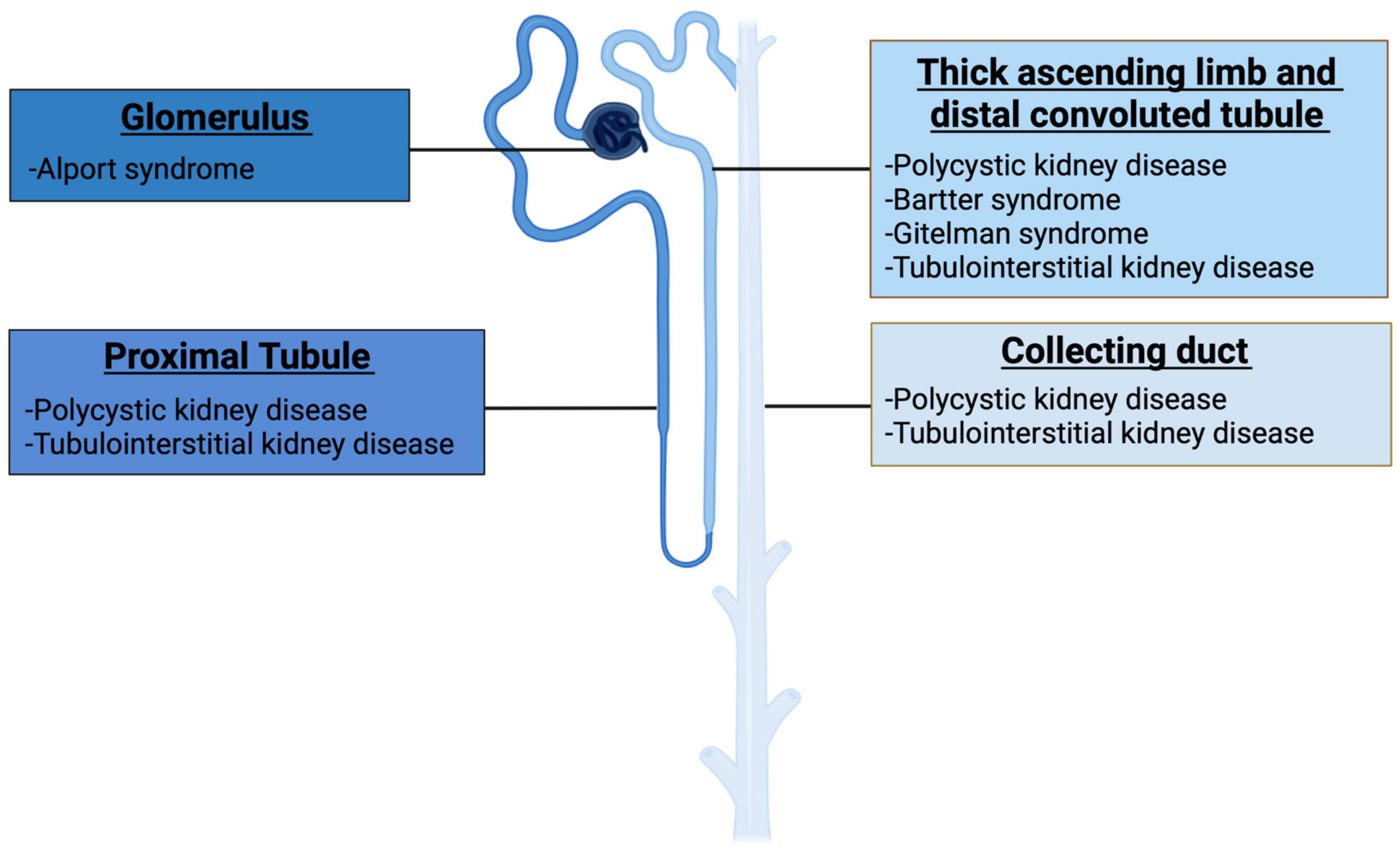
1.1. Genetic Kidney Diseases

As mentioned above, there are more than 600 genes linked to GKDs. These genes encode a wide variety of proteins including receptors, channels, enzymes, transcription factors, and structural components (Table 1) [10]. When the function of any of these proteins is compromised, vital processes such as water and electrolyte balance, blood pressure regulation, acid-base homeostasis, tissue oxygen supply, hormone and vitamin metabolism, innate and adaptive immunity, and central nervous and cognitive functions may be affected, resulting in various diseases [10].

1.1.1. Autosomal Dominant Polycystic Kidney Disease (ADPKD)

ADPKD is a monogenic disease caused mainly by mutations in PKD1 gene (accounts approximately for 85% of cases) which encodes polycystin-1 (PC1) and PKD2 gene (accounts approximately for 15% of cases) which encodes polcystin-2 (PC2) [12,13]. However, it can also be caused, in a minority percentage, by mutations in other genes such as GANAβ, DNAJB11, and IFT140 [14,15,16]. PC1 and PC2 proteins are involved in the differentiation and maintenance of renal tubular cells, and are predominantly, but not exclusively, localized in the primary cilium of these cells. Therefore, this kidney disease is classified as a ciliopathy [17,18,19]. ADPKD is the most common genetic kidney disease and affects ~1/400–1/1000 individuals [20]. It is the only GKD that is not a rare disease [1,2]. In approximately 90% of patients, the condition is inherited with a positive family history, while in 10% the disorder appears to be due to a de novo mutation [21].
The kidneys of patients with ADPKD are characterized by the formation of cysts that gradually grow in number and size. In early stage of the disease, the kidney contains few fluid-filled cysts and a large amount of well-preserved parenchyma [22]. In advanced ADPKD, marked kidney enlargement, vascular remodeling, interstitial fibrosis, and hepatic cysts are observed [23]. These pathological processes contribute to the development of ESRD, which is a severe renal disease requiring dialysis or kidney transplantation for patient survival. Particularly, 50% of ADPKD patients will reach ESRD before the age of 60 years, which puts ADPKD as a major cause of ESRD [24,25].

1.1.2. Autosomal Recessive Polycystic Kidney Disease (ARPKD)

ARPKD is a monogenic disease caused mainly by mutations in PKHD1 gene which encodes polyductin (also known as fibrocystin) [26,27]. However, it can also be caused, in a minority percentage, by mutations in DZIP1L gene [28]. Polyductin is involved in the terminal differentiation of the CD and biliary system and is localized in the primary cilium of renal epithelial cells, so this renal disease is also classified as a ciliopathy [17,29]. Even though ARPKD is less common than ADPKD (occurs in approximately 1 in 20,000 live births) [24], it is one of the common hereditary renal cystic diseases in infants [30].
The kidneys of patients with ARPKD are characterized by the presence of innumerable minute “microcysts” (1–2 mm) along the length of the DC, involving both the cortex and medulla and resulting in marked renal enlargement. Clinical features of ARPKD include ectasia of the DCs and hepatic bile ducts with associated renal and hepatic fibrosis [31]. Approximately 50% of patients with ARPKD present with this disease as neonates [32] and are born with very large kidneys [31]. These neonates suffer a 30% mortality rate due to respiratory and/or renal dysfunction [33]. For children who survive the perinatal period, they rarely survive beyond the age of 60 years [34].

1.1.3. Alport Syndrome (AS)

AS, also named type IV collagen nephropathy, is a monogenic disease caused by mutations in COL4A3, COL4A4, or COL4A5 genes which encode collagen chains α3, α4, and α5, respectively. These collagen chains are located, among others, in the glomerular basement membrane (GBM) [35], so this kidney disease is classified as a glomerulopathy [36]. AS is the second most common genetic kidney disease, after ADPKD [6]. Although its exact prevalence is unknown, a recent population-based study on the gnomAD cohort reports a frequency of 1/2320 individuals with a predicted pathogenic mutation in COL4A5, 1/106 individuals with a heterozygous predicted pathogenic mutation in COL4A3 or COL4A4, and 1/88,866 individuals with two heterozygous predicted pathogenic mutations in trans [37,38].
Individuals with pathogenic mutations in any of these genes can present with a wide phenotypic variability, ranging from isolated hematuria to renal failure, depending, among others, on the affected gene and if only one or the two copies of the gene are altered [39]. X-linked disease accounts for 70–75% of Alport patients [40,41,42]. Males with this condition are severely affected, with 90% probability of starting ESRD by the age of 40 years old, in addition to presenting with other extrarenal manifestations (e.g., hearing loss or ocular abnormalities) [43]. The phenotype of female carriers of heterozygous mutations in COL4A5 is highly variable, ranging from mild phenotypes with intermittent hematuria to severe cases [44]. Pathogenic mutations in the COL4A3 or COL4A4 genes cause the autosomal-recessive (AR) Alport syndrome, a similar phenotype to that of X-linked inheritance in males [45,46] or the “autosomal dominant” (AD) Alport syndrome, a less severe phenotype with persistent hematuria but without extrarenal manifestations. However, a portion of them may progress to ESRD [40,45,47,48].

1.1.4. Autosomal Dominant Tubulointerstitial Kidney Disease (ADTKD)

ADTKD is a monogenic disease caused by mutations in five different genes, including UMOD, MUC1, REN, HNF1β, and SEC61A1 which encode uromodulin, transmembrane epithelial mucin 1, renin, hepatocyte nuclear factor 1β and α1 subunit of the SEC61, respectively [49]. These proteins carry out renal and extrarenal functions and their malfunctioning generates tubular damage and interstitial fibrosis, so this kidney disease is classified as a tubulopathy [49,50]. Although the exact prevalence has not been determined, data suggest that ADTKD is one of the most common monogenic kidney diseases after ADPKD and AD, accounting for ~5% of monogenic disorders causing CKD [49,51].
ADTKD is characterized by a lack of specific clinical, biological, and pathological features [50]. For instance, affected individuals present with progressive CKD, normal or slightly elevated blood pressure, and normal or small kidney size [52]. In addition, the symptoms and organs affected may vary depending on the altered gene. For example, mutations in UMOD, MUC1, and REN mainly affect the kidney [53], whereas mutations in HNF1β and SEC61A1 also cause variable extrarenal manifestations [54,55].

1.1.5. Gitelman (GS) and Bartter (BS) Syndromes

GS and BS are monogenic diseases caused by mutations in different genes [56]. While GS is an autosomal-recessive disease produced by mutations in a single gene (SLC12A3) [57], BS can be caused by mutations in six distinct genes (SLC12A1, KCNJ1, CLCNKA, CLCNKB, BSND, and MAGED2) and be transmitted in three inheritance patterns (X-linked, autosomal-recessive, or autosomal-dominant) [58,59,60,61,62,63,64]. The proteins encode by these genes reside in different parts along the nephron, particularly in the DCT (associated with GS) and in the TAL (associated with BS), so these kidney diseases are classified as tubulopathies [56,65]. For GS, the estimated prevalence varies from 1 to 10 per 40,000 individuals [66], whereas for BS, the data are not well-defined (but an incidence of 1 per 1,000,000 is estimated) [67].
Although GS and BS have different etiologies, they share most of their clinical symptoms. Patients with GS or BS can present different symptoms as hypokalemia, hypochloremia, metabolic alkalosis, hyperreninemia, hyperaldosteronism or even CKD [65]. However, the main difference between the two is that patients with GS also present hypocalciuria. This is due to an increase in calcium (Ca2+) reabsorption to compensate for the loss of salts. This compensatory process does not occur in patients with BS [68].

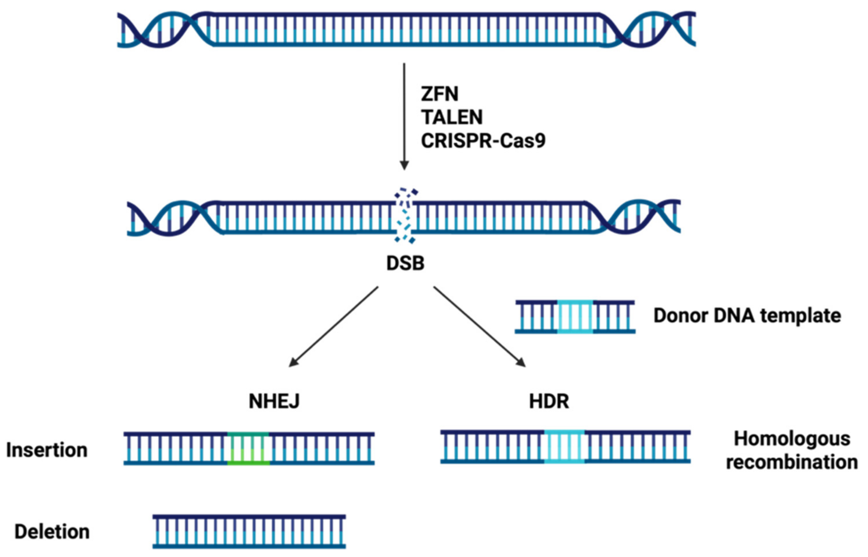
1.2. Site-Specific Nuclease Systems

Genome editing technologies have provided a sturdy tool for biomedical research with extraordinary versatility and feasibility. This technology can produce genome modifications, such as targeted mutagenesis or site-directed insertion/deletion/substitution at specific sites in the genome, using engineered nucleases. Genome editing relies on the production of site-specific double-strand DNA breaks (DSBs) and subsequent endogenous cellular repair through the error-prone nonhomologous end-joining (NHEJ) or the error-free homology-directed repair (HDR) pathway [69].
On the one hand, in NHEJ several enzymes are used to directly join the break ends of the DSBs without the need for a homologous repair template and can be carried out during any phase of the cell cycle in higher eukaryotes [70,71]. Due to its error-prone nature, NHEJ repair often leads to the insertion or deletion (InDels) of nucleotides and, thus, DNA sequence alterations at the targeted DSBs sites [69]. So NHEJ can be exploited to introduce frameshifts into the coding sequence of a gene (knock out) by a combination of two mechanisms: pre-mature truncation of the protein and nonsense-mediated decay of the mRNA transcript [71]. On the other hand, in HDR a homologous sequence serves as a template to repair the DSBs allowing an accurate repair and it occurs mainly during late S phase or G2 phase of the cell cycle [70,71]. Owing to its error-free nature, HDR can be utilized to insert or correct a specific mutation at the target loci (knock in) in the presence of an exogenously supplied donor oligo as a repair template by homologous recombination [69,71].
By taking advantage of the intrinsic DNA repair machinery of cellular organisms, tools that produce DSBs can be used to genome editing. The three major site-specific nuclease systems are: zinc finger nucleases (ZFNs), transcription activator-like effector nucleases (TALENs) and clustered regularly interspaced short palindromic repeats-CRISPR associated protein-9 (CRISPR-Cas9) (Figure 2).

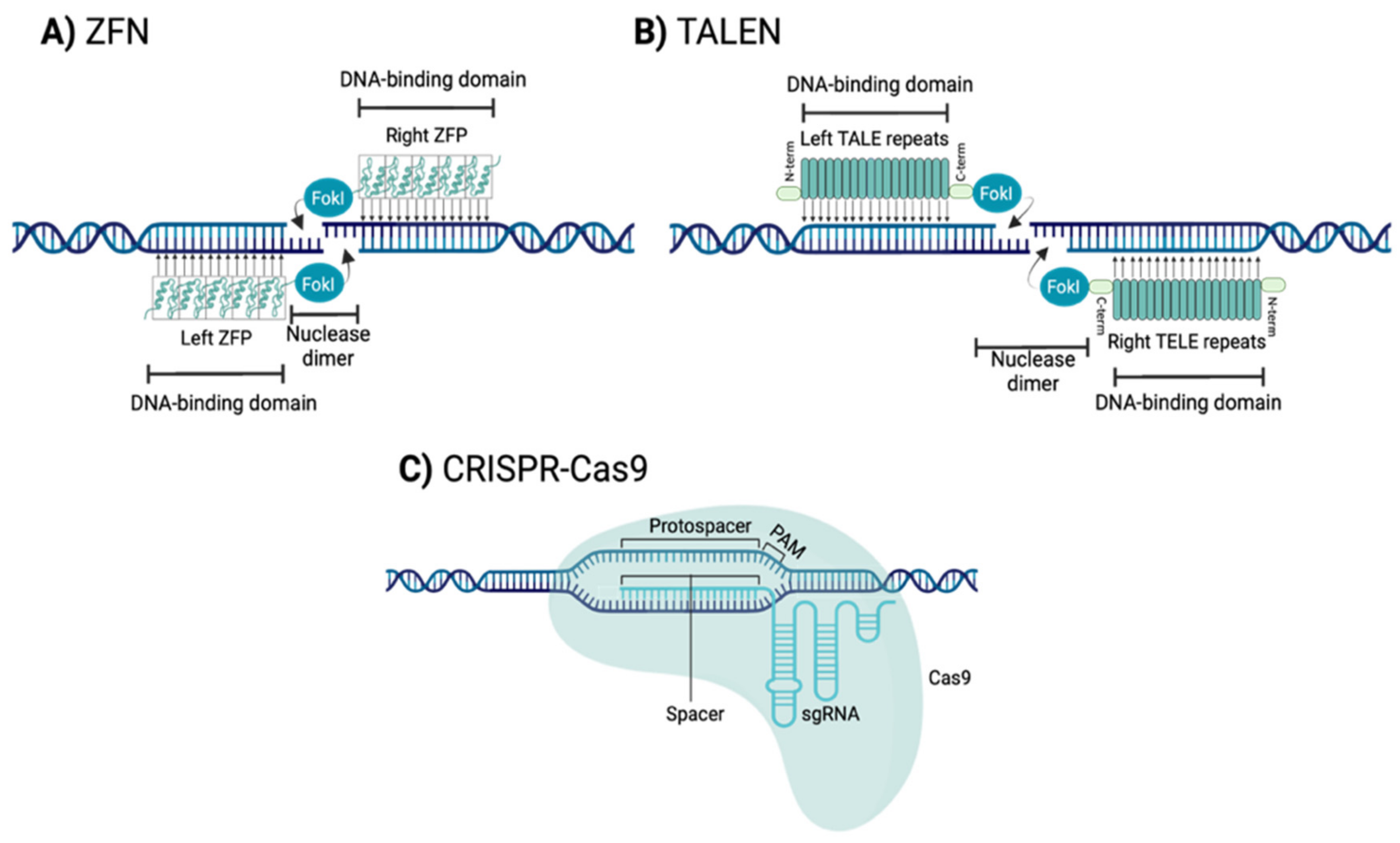
1.2.1. Zinc Finger Nucleases (ZFNs)

ZFNs are engineered nucleases composed of sequence-specific zinc finger DNA-binding domains and a non-specific DNA cleavage domain derived from the type II restriction endonuclease FokI (Figure 3A) [72]. The DNA-binding domain contains several linked zinc fingers (ZF) motifs, and each motif consists of approximately 30 amino acids in a conserved ββα secondary configuration [73]. Each individual ZF motif recognizes a 3-base pair (bp) specific DNA sequence and ZF motifs have been developed to recognize all of the 64 (4 x 4 x 4) possible nucleotide triplets [69,74]. This approach allows a specific tandem ZF motifs (ZFP) to potentially bind to a nucleotide sequence (typically with a length that is a multiple of 3, usually 9 bp to 18 bp) that is unique within a cell’s genome [71].
To induce a cleavage at a single and specific site in the genome two individuals ZFPs are required [75]. ZFPs must be designed as a pair able to recognize two sequences flanking the target site, one on the forward strand and the other on the reverse strand. Upon binding of the ZFPs, the pair of FokI domains dimerize and cleave the DNA at the target site, generating a DSB [76].

1.2.2. Transcription Activator-Like Effector Nucleases (TALENs)

TALENs are engineered nucleases composed of a transcription activator-like effector (TALE) DNA binding protein and a non-specific DNA endonuclease FokI (Figure 3B) [77]. TALE protein contains an N-terminal translocation signal, a C-terminal acidic transcription-activation domain, and a central DNA-binding domain [78]. The DNA-binding domain is composed of TALE repeats. Each repeat is 33 to 35 amino acids in length, including two relevant amino acids (termed the repeat-variable di-residue (RVD)) conferring specificity for one of the four DNA base pairs [78]. Therefore, each of the TALE repeats binds to a single nucleotide [69].
This direct relationship between TALE repeat and nucleotide recognition allows a specific tandem of TALE repeats to potentially bind to a single nucleotide sequence in a cell’s genome (typically 18-bp or even more) (typically 18 bp or even more) [69,75,79]. Like ZFNs, to induce a cleavage at a single and specific site in the genome two TALENs monomers are required to recognize flanking sequences of the target site. Upon binding of the TALENs on either side of the target site, the pair of FokI domains dimerize and cleave the DNA at the target site, generating a DSB [69,71].

1.2.3. Clustered Regularly Interspaced Short Palindromic Repeats-CRISPR Associated Protein 9 Nuclease (CRISPR-Cas9)

CRISPR-Cas9 are engineered systems composed of a Cas9 endonuclease, which is used to cleave the target sequence, and a single guide RNA (sgRNA), which defines the specificity of the Cas9 and guides Cas9 to the target DNA (Figure 3C) [80,81]. The sgRNA presents a sequence of 20 bp (spacer) nucleotides that are complementary to the target genomic sequence (protospacer) and a scaffold region that allows its association with Cas9. This association produces a conformational change in Cas9, which goes from a self-inhibited state to a functionally active state [82,83,84].
Activated Cas9 recognizes, in the target genome, a sequence of three nucleotides called protospacer adjacent motif (PAM). This PAM is different for each type of Cas9 (e.g., for Streptococcus pyogenes-SpCas9 it is (N)GG, where N is any nucleotide) [83,84]. Once the PAM sequence is recognized, the double strand dissociates. If the sgRNA spacer sequence is complementary to the target nucleotides adjacent to the PAM (protospacer), then the Cas9 cleaves the DNA at the target site, generating a DSB [82].
It should be noted that the CRISPR systems are continuously improving. New Cas9s have been discovered in other bacteria, such as Staphylococcus aureus-SaCas9 and Francisella novicida-FnCas9. In addition, new SpCas9s have been developed that offer improved efficiency and specificity, as well as being able to recognize different PAMs (e.g., SpCas9-NRRH, SpCas9-NRCH and SpCas9-NRTH, where H is A, C or T, and R is A or G). In addition, researchers can use catalytically dead Cas9 (dCas9), which cannot cut DNA, but does bind to specific DNA sequences to up- or down-regulate gene expression. New CRISPR–Cas-derived genomic editing agents have also been developed, such as Base Editing and Primer Editing, which allow changes to be introduced into DNA without the need to produce a DSB. Base editing consists of a Cas9 nickase (nCas9-an enzyme that cleaves only one of the DNA strands) linked to a deaminase enzyme (cytosine deaminase or adenine deaminase) that allows nucleotide transition in the target sequence. Prime editing consists of an nCas9 linked to a reverse transcriptase enzyme that allows nucleotide transition and transversion or insertion and deletion of small DNA fragments in the target sequence (reviewed in [85]).

1.2.4. Comparison of the Three Types of Genome Editing Technologies

Current gene editing techniques differ from one another in several ways such as target site recognition sequence, difficulty of delivery, nuclease size or off-target rate and have their respective pros and cons (summarized in Table 2).

Sequence Selection and Assembly Evaluation

ZFNs offer great versatility as they can be designed to target any sequence, nevertheless the specificity of individual zinc finger depends on two factor such as the context of the surrounding zinc fingers and DNA sequence [86]. Therefore, in practice, a suitable ZFN target sequence may not be found for a specific gene or chromosomal loci [69]. Furthermore, the assembly of ZFNs is complicated, involving laborious and time-consuming steps [74,92], and often assembled ZFNs fail to cleave target sites [86].
TALENs target selection is limited by the requirement that TALE binding sites should start with a thymidine residue (T), and it is sensitive to DNA methylation [87,93]. Not all TALENs are efficient to cleave target sites, and TALENs pairs must be experimentally validated [69]. The simple one-to-one specific recognition relationship between TALE repeats and the four nucleotides makes TALENs easier to design and assemble than ZFNs, but the assembly is still quite laborious and time-consuming [87]. One potential advantage over ZFNs is that the TALE repeat array can be easily extended to whatever length is desired [69]. Both ZFNs and TALENs rely on engineering proteins to specifically recognize target sequences and the complicated assembly process is the major hurdle preventing their wider application for genome editing [69].
In comparison, CRISPR-Cas9 target selection is limited by the presence of a PAM sequence downstream of the target sequence. Despite the PAM limitation, it is easy to find target sequences, for instance for the case of SpCas9 there is a PAM sequence (NGG) on average once every 8 bp. This simplicity is the main reason for the widespread adoption of the CRISPR-Cas9 systems and their application in numerous organisms [70].

Delivery Strategies

The major barrier for in vivo gene editing is the absence of safe and effective methods for local and systemic delivery of ZFNs, TALENs and CRISPR-Cas9 [94,95]. The delivery strategy must be able to specifically direct the genome editing complex to the target cell/tissue, and then this complex must be introduced into the cell, which requires crossing the plasma and nuclear membranes [96]. There are two strategies available to achieve this aim: viral and non-viral vectors (include physical and chemical methods) [95,96,97].
The most widely used method is viral vectors, in which nucleic acids coding for the protein complexes are packaged into viruses and delivered to the target cell. Nevertheless, viral vector cargo size is a limitation [96]. The typical size of a cDNA encoding a ZFN, a TALEN and a SpCas9 is approximately 1 kb, 3 kb and 4.2 kb in size, respectively. The CRISPR cDNA is somewhat larger than a TALEN monomer and much larger than a ZFN monomer (though both TALENs and ZFNs require dimerization, making their effective sizes larger) [69]. The size of the TALENs and Cas9 is a problem in the use of viral vectors that have a limited cargo size, such as adeno-associated virus (AAVs), with less than 5 kb [69,94,97]. However, to accommodate larger payloads, other viral vectors with larger capacities, such as adenoviral (AdVs) and lentiviral vectors (LVs), have also been investigated by various researchers for the gene-editing reagents delivery [88]. Nonetheless, these last two viral vectors have certain drawbacks such as immunogenicity and random integration, respectively [95], which would hinder their future application in gene therapy. Therefore, AAV is presented as the best option for its use in gene therapy in humans, as it does not develop immunogenicity.
Non-viral delivery methods have emerged as a viable alternative since they have the potential to solve the problem of cargo size and immunogenicity of viral vectors. However, the efficiency of delivery is slightly lower than that of viral vectors [98,99]. A major advantage of non-viral vectors is that they are artificially synthesized so that such important parameters as size, surface ligands, thermal properties, loading capacity, encapsulation efficiency and storage stability can be con-trolled [100,101]. What makes non-viral vectors even more promising is the rapid development of materials science applied to molecular biology, which will substantially facilitate the improvement of the efficiency and effectiveness of these non-viral vectors [99]. On the one hand, physical non-viral methods include microinjection, which involves injecting the genome editing complex directly into cells with a microscope and a needle; and electroporation, which applies pulses of electric current to stimulate the transient opening of pores in cell membranes, allowing delivery of the genome editing complex into cells [95]. On the other hand, non-viral chemical methods include lipid-based nanoparticles (LNPs), which involve encapsulation of negatively charged nucleic acids in positively charged liposomes for introduction into cells [102]; and cell penetrating peptides (CPPs), which are short peptides with an intrinsic ability to trans-locate across cell membranes, which is exploited to facilitate the delivery of a variety of cargoes into cells [103].

Specificity and Efficiency

An ideal gene editor would exhibit perfect specificity for the target sequence and cause no mutations to any other region of the genome. Unfortunately, ZFNs, TALENs and CRISPR-Cas9 rarely achieve such a high level of specificity and can generate mutations at the non-target locus (off-target activity) [104].
The specificity of ZFNs is determined by three major factors: the amino acid sequence of each ZF, the number of ZFs, and the interaction of the nuclease domain [89]. The specificity of TALENs is determined by RVDs, constituting a strikingly simple TALE–DNA recognition cipher [105,106]. Both ZFNs and TALENs tolerate a small number of positional mismatches, but TALENs exhibit better specificity than ZFNs [89]. The specificity of CRISPR-Cas9 is determined by the sgRNA (structure and composition) and PAM sequences [107,108,109]. CRISPR-Cas9 tolerates multiple consecutive mismatches and, compared to ZFNs and TALENs, CRISPR-Cas9 can exhibit higher off-target activity [89,110].
In addition to off-target activity, on-target effects must also be considered. This refers to large deletions, rearrangements or translations in the target sequence resulting from DSB repair induced by gene editing tools [111]. On-target effects are not dependent on the activity of nucleases but are associated with the functioning of DNA repair mechanisms in cells. To date, on-target effects cannot be predicted and may even be considered a bigger problem than off-target activity [112]. Therefore, on-target effects and off-target activity must be carefully monitored to avoid obtaining unintended gene modifications.
Efficiency is a difficult parameter to compare since, in many cases, it depends on the selected objective. In general, CRISPR-Cas9 (81%) has higher efficiency than ZFNs and TALENs, whereas TALENs (76%) usually exhibit higher efficiency than ZFNs (12%) [69,90].

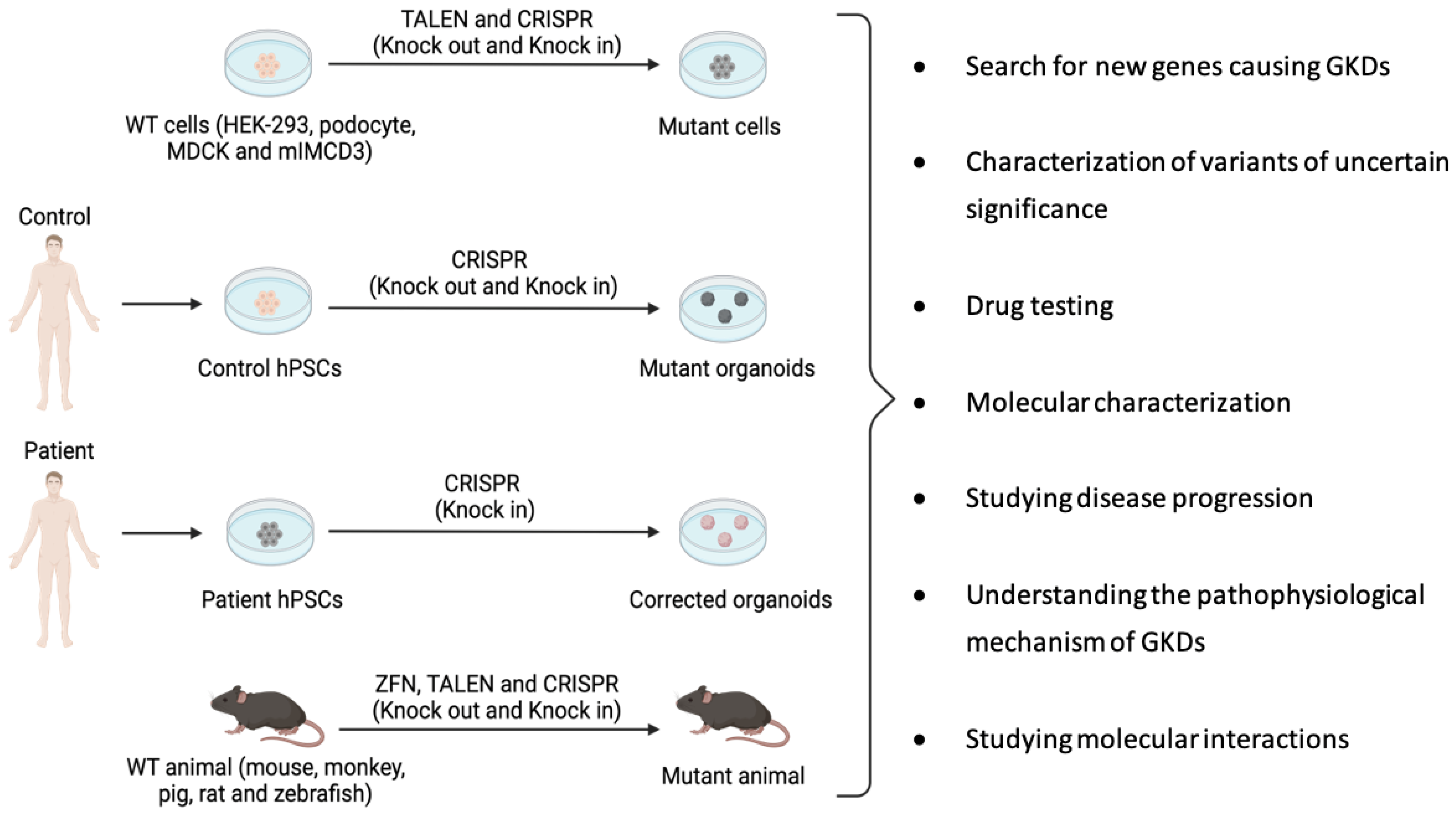
2. Site-Specific Nuclease Systems to Study Genetic Kidney Diseases

As previously mentioned, there is a profound lack of understanding of the pathophysiology and treatment of GKDs. The key to solving these problems is finding a suitable model (in vitro or in vivo) of these diseases for use in preclinical studies. Engineered nucleases (ZFNs, TALENs and CRISPR-Cas9) allow genome editing, which can be used to develop potentially useful models to study the progression and possible treatment of these human diseases. In addition, these nucleases enable find new genes involved in GKDs, since many of these genes remain unknown (Figure 4). The general approach is to knock out the gene responsible for the disease and to inspect the generated mutant models for phenotypic differences with isogenic controls (i.e., having a uniform genetic background) that were not modified by the nucleases [113].
There are numerous in vitro models to unravel the mechanisms of GKDs and perform drug screening tests with the aim of identifying a potential therapy for GKDs. In vitro models are available ranging from simple monolayer cultures of various renal cell lines to three-dimensional (3D) culture, and finally kidney organoids. The most common renal cell lines are Madin–Darby canine kidney (MDCK), inner medullary collecting duct (IMCD3) and human embryonic kidney cells (HEK293). In addition, human pluripotent stem cells (hPSCs) enable the production of organoids modeling human disease. Although these models clearly represent a very useful tool for understanding GKDs, it should be noted that due to the absence of their natural environment, these models are not representative of the physiology of the entire organism. Therefore, when interpreting the results of in vitro tests, it must be considered that these cell models lack important aspects, such as complex drug metabolization or tissue interactions. Hence, in vivo validation of results remains a mandatory step to understand the pathophysiological mechanisms of GKDs and drug development [114].
Just like in vitro models, a large number of in vivo models are available for the study of GKDs [115]. These models should recapitulate the phenotype observed in human pathology following the introduction of mutations in the orthologous gene corresponding to each type of GKDs [114]. In vivo models include invertebrate or lower vertebrate organisms, such as Xenopus or Danio rerio, useful for drug screening due to their fast generation time [116,117], as well as higher vertebrate organisms, especially mammals, which share greater genetic and physiological similarity with humans. Studies in the latter models better reflect human disease mechanisms and allow for more reliable observations, especially to determine treatment efficacy and adverse effects [114].
Although the field of GKDs is largely unknown, precision gene editing has been used in kidney research to achieve both gene knock-out and gene knock-in in a variety of model, from individual cells to animals complete (Figure 4; Table 3). These studies have generated new insights into the mechanisms of GKDs and have illustrated the potential of genome editing as a tool to enable progress in targeted gene therapy [118]. Below, gene editing tests carried out in different models for the study of GKDs are shown.

2.1. ADPKD Models Generated Using Genome Editing

ADPKD is the most common GKD and the most studied. There are several publications related to the application of genome editing for the study of ADPKD, most of them using CRISPR-Cas9 technology in organoids. The strategy was to knock-out PKD1 or PKD2 genes introducing InDels by NHEJ. However, studies have also been carried out using ZFNs and TALENs technologies in cells and animal models.

2.1.1. ADPKD Models Generated Using ZFNs

In 2015, Jin He et al., created a mini pig model by mono-allelic knockout of PKD1 gene by using ZFNs [119]. This model by 6 months of age macroscopically visible renal cysts appeared. These cysts grew in number and size and at 24 months these began to deform the normal kidney shape. Therefore, the PKD1 mono-allelic knock out is sufficient to trigger renal cystogenesis. No piglets PKD1/ were born, perhaps since it resulted in embryonic lethality as occurs in human [143] and in some mouse models [144]. In addition, this model has been used for drug testing. In 2019, Lian et al., researched the effect of single and combined treatments with 2Deoxy-D-glucose and metformin on ADPKD progression. They conclude that both drug treatments significantly inhibited cystogenesis in the ADPKD mini pig model, being the combined therapy the most effective [145].

2.1.2. ADPKD Models Generated Using TALENs

TALENs technology has been applied to study the role of PDE1A gene in the pathogenesis of ADPKD. PDE1 encodes phosphodiesterase 1 (PDE-1), the only PDE activated by calcium and the main enzyme degrading cAMP in the distal nephron and CD. Substantial evidence supports the hypothesis that disruption of polycystin function results in dysregulation of intracellular calcium dynamics, producing the inhibition of PDE-1 and causing an increase in intracellular cAMP, thus contributing to the development and the progression of ADPKD [146,147,148]. To validate this hypothesis, Hong Ye and colleagues in 2016 and Xiaofang Wang et al., in 2017, developed a Pde1a knockout (KO) mouse model using TALEN tool [128,129]. They conclude that Pde1a/ aggravates cystic formation on a Pkd2 mutant background. These results support an important role of PDE1A in the renal pathogenesis of ADPKD. In addition, this model may contribute to a better understanding of the mechanisms responsible for increased cAMP signaling in ADPKD and to the search for treatments capable of increasing PDE1 expression to delay cyst growth [128,129].
Furthermore, it is noteworthy the easily implementable workflow developed by Alexis Hofherr and coworkers in 2017 [120]. They applied TALEN and CRISPR systems for the rapid generation of targeted heterozygous and homozygous genomic sequence alterations in differentiated renal epithelial cells (MDCK and mIMCD3). In order to demonstrate the versatility of their genome editing approach, they established multiple novel cell lines for the study of ADPKD by introducing targeted mutations into Pkd1 and Pkd2, generating deletions of Pkd genes, rescuing polycystin expression, and developing cell lines incorporating multiple allelic features [120].

2.1.3. ADPKD Models Generated Using CRISPR-Cas9

CRISPR has been the most widely used tool to study ADPKD due to its cost-effectiveness. This tool has been mainly applied in organoids, with the aim of disease modeling and drug screening. Freedman and colleagues, in 2015, were the first to establish not only a protocol for differentiate hPSCs into kidney organoids (multicellular units containing podocytes, proximal tubules and distal tubules cells), but also the application of CRISPR in organoids to model GKDs [121]. They introduced frameshifts mutations by CRISPR-Cas9 in either PKD1 and PKD2 genes and established the first genetically edited models of disease in human kidney organoids. It is highly relevant that shortly after differentiation, organoids with PKD mutations formed cysts in vitro while no cysts were detected in control organoids with identical genetic backgrounds. In addition, using this protocol, the problem of variability among hPSCs from different patients to differentiate into kidney organoids disappears, as CRISPR generates series of PKD1/PKD2 mutant hPSCs that are otherwise isogenic. Nevertheless, one limitation of this study was that only 6% of CRISPR-PKD organoids developed cysts [121].
Two years later, Cruz et al., improved cystogenesis (~75% of CRISPR-PKD organoids developed cysts) by growing CRISPR-PKD kidney organoids in suspension culture [122]. Taking advantage of this high percentage of cyst formation, kidney organoids could be a perfect tool for understanding the pathogenesis of ADPKD and for drug screening.
In 2020, Shohei Kuraoka and colleagues generated CRISPR kidney organoids and demonstrated that cAMP signaling is required for in vitro cystogenesis in ureteric bud (UB) organoids [123]. On the one hand, using CRISPR-Cas9 technology, PKD1 gene was deleted in hPSCs and then PKD1+/ and PKD1/ clones were induced to differentiate into UB organoids. On the other hand, they generated UB organoids from hPSCs from patients with ADPKD who had a heterozygous missense mutation. Subsequently, both types of UB organoids were treated with forskolin (an activator of cAMP signaling). CRISPR-PKD1+/ and CRISPR-PKD1/ organoids exhibited cystogenesis upon forskolin treatment. In contrast, no cysts were formed in PKD1+/+ organoids after treatment. Similarly, PKD1+/ organoids from patients exhibit cystogenesis upon treatment, but no cysts were formed in PKD1+/+ organoids. Although cAMP stimulation by forskolin may not accurately mimic cyst formation signals in vivo, UB organoids could be used to study the balance between cAMP and PC1/PC2 mediated signals that cause cyst formation [123].
Recently, Yasaman Shamshirgaran and coworkers established a faster protocol for generating genetically engineered kidney organoids [124]. To this end, they set out two modifications: (1) using a doxycycline-inducible Cas9 expressing hPSC line instead of transfecting them with Cas9-plasmid; and (2) using KO pools instead of generating isogenic clonal lines. Applying these procedures, they generated PKD1 and PKD2 mutant hPSC, both of which exhibited an editing frequency of 80%. Then, both pools of hPSCs were differentiated into kidney organoids. They observed cyst formation in approximately 50% of the PKD KO pools organoids, while a practically negligible rate of cysts was observed in control organoids (less than 1%). Therefore, by using KO pools instead of generating isogenic clonal lines, it is possible to generate cystogenesis in kidney organoids, providing a platform for rapid target validation in the context of disease modeling [124].
As shown, applying both hPSCs differentiation and CRISPR tool, is opening new opportunities for the study the genetic basis of ADPKD and the evaluation of new therapeutic options. For instance, in 2018, Czerniecki and colleagues treated kidney organoids derived from CRISPR-PKD hPSCs with blebbistatin, a specific inhibitor of non-muscle myosin II [149]. This treatment induced a significant dose-dependent increase in cyst formation, which revealed an unexpected role for myosin in ADPKD. The result suggests that the myosin pathway and the regulation of actin-myosin activation may be implicate in cystogenesis [149].
CRISPR technology can also be used to discover potential new disease-causing genes. Mutations in PKD1 or PKD2 were the known causes of ADPKD, but there was a percentage of families affected by ADPKD that were genetically unresolved (GUR). In 2016, Binu Porath et al., carried out genetic studies in GUR ADPKD-affected families and suggested that mutations in GANAβ gene (encodes glucosidase II subunit α -GIIα) produce ADPKD [14]. To validate this hypothesis, they knocked out GANAβ gene in renal cortical tubular epithelial (RCTE) cells by CRISPR-Cas9. The results showed that GANAβ/ cells exhibited failed trafficking of PC1 and PC2 to primary cilia. However, a normal localization of PC1 was rescued by wild type (WT) GIIα, but not by the mutant. In GANAβ+/ a reduced mature PC1 was seen. Therefore, mutant GIIα cause maturation and localization defects of PC1 and PC2, and this may be related to cystogenesis [14].
Although kidney organoids are a very promising tool for the study of diseases, to date they have some limitations. Organoids lack vasculature so their growth is limited, they lack immune cells so they cannot be used to study processes that require inflammatory responses. Cellular composition varies significantly depending on the cell line and protocol used to generate organoids. Furthermore, when renal organoids self-assemble, tissues develop somewhat randomly, and hPSC-derived organoids show high variability. As existing culture methods cannot faithfully replicate renal conditions in vivo, the use of animal models is still necessary [150,151].
In 2019, Tomoyuki Tsukiyama and colleagues created cynomolgus monkeys with mutations in PKD1 gene by CRISPR-Cas9 [125]. The resulting PKD1+/ monkeys exhibited perinatal cyst formation in the distal tubules, possibly reflecting the initial pathology in humans. Many monkeys survived after cyst formation, and cysts progressed with age. However, no monkeys PKD1/ were born, which is consistent with the embryonic lethality observed in Pkd1-deficient mice, humans, and mini pigs [119,143,144]. Therefore, this model could elucidate the onset and progression of ADPKD, helping to establish new therapeutic strategies and discover new pharmacological treatments [125].
Masahito Watanabe et al., in 2022 applied CRISPR-Cas9 to generate heterozygous PKD1 mutant pigs [126]. PKD1+/ pigs had many symptomatic similarities to patients with ADPKD caused by heterozygous mutation of PKD1 gene. For instance, presence of macroscopic renal cysts during the neonatal stage, cystogenesis process, interstitial fibrosis, and asymptomatic period during the first half of life. Taking advantage of these similarities between both, PKD1+/ pigs can be used to study origin and development of this disease, check the effects of different treatment and test long-term treatments that cannot be carried out in rodent models [126].
It is important to note that although studies with cellular and animal ADPKD models have shed some light on this disease and have allowed potential treatments to be tested, such as Anti-TWEAK, which significantly slows disease progression, preserves renal function, and improves survival in an ADPKD mouse model. However, no definitive therapies are available [152]. Tolvaptan is the drug currently used in patients with ADPKD, it can slow the progression of renal cyst formation, but it is not capable of making them remit [153]. Therefore, more research of this disease is needed, and gene-edited cellular and animal models are essential.

2.2. ARPKD Models Generated Using Genome Editing

ARPKD is a less studied disease than ADPKD. Although, both are polycystic kidney disease and share their main renal symptom (cyst formation), ARPKD is much less prevalent than ADPKD due to its recessive nature. For the study of ARPKD, CRISPR technology has been the only gene editing tool that has been used.

ARPKD Models Generated Using CRISPR-Cas9

In 2018, Phillip Chumley and coworkers applied CRISPR technology in HEK-293 cell lines to generate diverse truncating mutations along multiple sites of PKHD1 gene [127]. Their objective was to determine whether polyductin deficiency produces alterations in energy metabolism and proliferation rate, as occurs with polycystin 1 deficiency [154]. The results indicated that the PKHD1/ clones did not show a significant proliferation rate compared to the WT clones, but an increase in metabolism was observed. In parallel, they performed the same procedures for PKD2 gene, but did not observe significant changes in the metabolism of PKD2/ clones. These results indicate that it is possible that the energy metabolism is altered in ARPKD, so this field could be studied to search for possible metabolism-related treatments [127].
The main application of CRISPR-Cas9 technology is to eliminate the expression of a certain gene through the formation of InDels. However, CRISPR can also be used to correct a specific mutation by introducing a donor oligo (WT sequence). In 2019, Low and colleagues applied the CRISPR-Cas9 gene editing tool in hPSCs from ARPKD patients to correct a specific mutation in PKHD1 gene [130]. After forskolin treatment, ARPKD kidney organoids exhibited drastic cystogenesis. In contrast, CRISPR-corrected ARPKD kidney organoids showed only marginal cyst formation, as did WT hPSCs-derived kidney organoids [130]. Although the application of CRISPR technology as a mutation correction tool requires further study, this publication demonstrates the great potential of CRISPR-knockin and its possible application as gene therapy.
Renal defects in ARPKD heterozygous patients are very mild and poorly understood since renal tissue is not available as no biopsies are performed in these patients. To extend the knowledge about the impact of heterozygous mutations in this autosomal recessive disorder, in 2019, Dan Shan and coworkers generated heterozygous mutant Pkhd1 mice by CRISPR [131]. Aged Pkhd1+/ females mice developed ectasia of PTs, but not of DCTs, or CDs. These results suggest that patients with heterozygous mutations in PKHD1 have a similar phenotype and develop only PT ectasia [131].
As occurs in ADPKD with GANAβ gene, in ARPKD there is also another gene that in a very small percentage can cause this disease. Mutations in PKHD1 gene are the known causes of ARPKD, but in 2016, Hao Lu and coworkers identified patients with homozygous mutations in DZIP1L gene, who had a phenotype similar to patients with homozygous mutations in PKHD1 (e.g., multiple small cysts) [28]. DZIP1L encodes cilium assembly protein DZIP1L, which through its interaction with septin2, participates in the maintenance of the periciliary diffusion barrier at the ciliary transition zone, allowing proper translocation of PC1/PC2. To validate that DZIP1L is a gene involved in the pathogenesis of ARPKD, they used CRISPR-Cas9 to develop a DZIP1L loss-of-function zebrafish model. They observed that zebrafish deficient for dzip1l had fewer motile cilia in the pronephric tubules. These results indicate that DZIP1L plays an important role in primary ciliogenesis and help to better understand the genetic heterogeneity underlying the pathogenesis of this disorder [28].
CRISPR-Cas9 technology can be applied to study the role of a specific gene in disease pathogenicity. For instance, the ionotropic P2X7 receptors are known to activate pannexin-1, a plasma channel capable of releasing ATP into the lumen. These receptors are thought to play a major role in cyst formation due to their high prevalence in the cyst wall (compared to healthy tissues) and the high ATP accumulation observed in cyst fluid [132,155]. To study mechanisms of P2X7 involvement in cystogenesis, Sergey N. Arkhipov et al., in 2019 generated a global KO of the P2rx7 gene in PCK rats (a model of ARPKD) by CRISPR. They concluded that PCK-P2rx7/ rats showed slower cyst growth (but not slower formation of new cysts) compared to PCK-P2rx7+/ and PCK-P2rx7+/+ rats [132]. These results support the involvement of ATP in cystic growth and highlight the role of P2X7 in the renal pathogenesis of ARPKD.

2.3. AS Models Generated Using Genome Editing

AS is the second most common genetic kidney disease and the second in which there are most publications related to genome editing. Nevertheless, as with ARPKD, CRISPR has been the only technology used to study this disease. The most widely used cellular model has been podocytes, which are the only cells that synthesize and secrete α3, α4 and α5 heterodimers in the glomerulus. These heterodimers are essential for GBM maintenance and mutations in the genes (COL4A3, COL4A4 and COL4A5) encoding these proteins generate AS [35,156].

AS Models Generated Using CRISPR-Cas9

During the progression of AS, the number of podocytes is substantially reduced due to apoptotic processes. However, the mutational mechanisms that trigger this podocyte death are unknown. To shed light on this field, in 2019, Hui-Di Zhang and colleagues generated Col4a3 KO mouse monoclonal podocytes using CRISPR [133]. They observed variations in the levels of endoplasmic reticulum (ER) stress-related proteins (increased expression of PERK, GRP94, CHOP, and phosphor-eIF2a) and in the levels of apoptosis-related proteins (increased expression of cleaved caspase 3 and decreased expression of Bcl-2) [133]. These results suggest that podocyte death could be triggered by dysregulation of both pathways due to COL4A3 deficiency.
The following year, Sergio Daga et al., corrected a specific mutation in COL4A3 and COL4A5 genes in podocyte from two AS patients by Cas9 and an oligo donor [134]. They used an innovative self-inactivating dual-plasmid system, one plasmid carried the donor DNA and the variant-specific sgRNA, while the other plasmid carried a self-cleaving SpCas9. Applying these procedures, they generated CRISPR-corrected COL4A3 and COL4A5 podocytes, which showed a homology-directed repair frequency close to 50%. Although podocytes are not renewable, these results demonstrate the ability of CRISPR to correct a mutation and it is likely that the application of this technology in the early stages of the disease, when podocytes are still present, would be effective in treating this disease [134].
Most of the mutations described in COL4A5 gene are missense mutation and most are described as likely pathogenic with uncertain significance [135,157]. To find out if the variant is responsible for the disease, CRISPR technology can be used to knock in the variant into the gene of interest and study how it changes the phenotype of the model. In 2021, Lei Sun et al., followed this strategy and introduced a missense mutation of the COL4A5 gene (observed in a patient) into immortalized human podocyte cell lines to confirm whether this variant could cause abnormal collagen alpha-5 (IV) chain expression and consequently cause AS [135]. They concluded that this variant significantly reduced α5 chain expression in podocytes. These results highlight the possibility of using CRISPR-knockin as a tool to confirm the pathogenicity of missense variants in vitro [135].
Regarding the use of genome engineering in mouse models, CRISPR technology has been used to develop a murine model harboring a nonsense mutation (observed in one patient) in the Col4a5 gene (Col4a5-R471X). In 2019, Kentarou Hashikam and coworkers generated male Col4a5 hemizygous mutant mice, which exhibited pathologies similar to those observed in AS patients [136]. Among these pathologies were increased levels of urinary albumin and blood urea nitrogen (BUN). These findings are indicative of impaired renal function. Since the mechanisms of progression are unknown and there is no effective treatment for AS, this animal model could be useful for studying the mechanisms involved in the development of AS and for preclinical trials to restore adequate renal function [136].
As in other diseases such as ADPKD with PDEA1, in AS there are also other genes that by themselves do not cause the disease, but the presence of mutations in these genes in patients with mutations in COL4 genes, aggravates the pathogenicity of the disease. An example would be the LAMB2 gene encoding for Laminin b2, which is a major component of the GBM. In 2018, Steven D. Funk and collogues generated mice with a specific mutation in Lamb2 gene (Lamb2-S80R) by CRISPR-knockin technology [137]. The results showed that the Lamb2S83R/S83R and Lamb2S83R/− mice did not exhibit a pathogenic phenotype. However, Lamb2S83R/+ significantly increased the rate of progression to kidney failure on a Col4a3/ background and increased proteinuria in females with a Col4a5+/ background. These data demonstrate how the phenotype associated with AS can vary due to mutations in other genes that also encode GBM components. Furthermore, this mouse model could help explain the wide range of phenotypes observed in AS patients, even in those who share the same COL4 mutation [137].

2.4. ADTKD Models Generated Using Genome Editing

ADTKD is the third most common monogenic kidney disease after exclusion of ADPKD and AS. This disease can be caused by mutations in five different genes; however, gene editing technologies have not been applied to all of these genes. ZFN and CRISPR have been the tools used to study this disease.

2.4.1. ADTKD Models Generated Using ZFNs

REN gene, which encodes for renin, is one of the causative genes for ADTKD. Renin is known to play an important role in renal function. To shed light on the importance of renin in the context of ADTKD in vivo, Carol Moreno et al., in 2018 used ZFN technology to knock out Ren gene in SS rats (rodent model of Salt-Sensitive hypertension) [138]. They observed that Ren/ SS rats had elevated BUN levels (indicative of poor renal function), abnormal renal morphology with cortical interstitial fibrosis and incorrect orientation of CDs and DCTs. These results highlight the importance of renin for proper renal morphology and function [138].

2.4.2. ADTKD Models Generated Using CRISPR-Cas9

As previously shown with other genes such as PKD1 and PKD2, the combination of kidney organoids and CRISPR technology is ideal for studying how deletion of a given gene influences renal development and disease [121,122,124]. In this sense, Aneta Przepiorski and coworkers in 2018 knocked out the HNF1β transcription factor gene in renal organoids by CRISPR-Cas9, which is known to be one of the genes responsible for ADTKD and plays an essential role in tubulogenesis [139]. HNF1β/ organoids showed reduced levels of markers associated with PT (LRP2) and TAL (UMOD, SLC12A1). These results indicate that KO of HNF1β prevents proper formation of the PT and TAL segments [139]. This is consistent with that described in the Hnf1b KO mouse [158]. However, cyst formation was not observed in HNF1β/ kidney organoids, even after several weeks in culture [159]. This indicates that, although organoids are useful for studying human kidney development, more research is needed for organoids to more accurately mimic the ADTKD phenotype.
As already mentioned, CRISPR technology can be used to discover new disease-causing genes. Until 2016, the known ADTKD-causing genes were REN, UMOD, and MUC1. In that year, Nikhita Ajit Bolar and colleagues identified two families with heterozygous missense variants in SEC61A1 gene and attempted to determine the pathogenicity of the missense variants in vivo [55]. To do so, they applied CRISPR technology to delete the human SEC61A1 ortholog, sec61al2, in zebrafish. Zebrafish showed defects of the pronephric tubules (consistent with the tubular atrophy observed in affected individuals), confirming that the SEC61 complex and its translocation function are required for normal renal development. These results highlight the role of SEC61A1 in the formation of the nephron and broaden the spectrum of ADTKD-causing genes [55].
UMOD gene, which encodes for uromodulin, is another ADTKD-causing gene. However, its role in disease progression is unknown. Studies suggest that it protects against uropathogenic bacteria, controls water transport in the kidney to concentrate urine [159,160] and is critical in preventing crystallization of calcium from the tubular filtrate [161]. In patients, mutant uromodulin has been shown to be poorly secreted and to accumulate in the ER of the distal renal epithelium. To further explore the signaling pathways triggered by misfolded uromodulin, Bryce G. Johnson et al., in 2017, developed a mouse model carrying a mutation in Umod gene (observed in patients) using CRISPR and an oligo donor [140]. Umod+/ mice exhibited renal failure at 24 weeks (elevated BUN and serum creatine levels), decreased body weight, renal fibrosis, reduced urinary uromodulin levels and accumulation of uromodulin in the ER, which activates unfolded protein response mechanisms and triggers ER stress-dependent cell death in renal tubule epithelial cells. This murine model recapitulates many features of ADTKD-UMOD patients and provides critical insight into signaling pathways altered in the pathogenesis of this disease. This makes it a valuable animal model for future studies [140].

2.5. GS and BS Models Generated Using Genome Editing

GS and BS are the least prevalent of the diseases discussed in this review. Moreover, they are those in which there are fewer publications related to the application of gene editing tools. On the one hand, no gene editing studies have been performed in GS. On the other hand, although BS is less prevalent than GS, two gene editing studies have been performed, one with ZFNs and the other with TALENs.

2.5.1. BS Models Generated Using ZFNs

KCNJ1 gene, which encodes for ATP-sensitive inward rectifier potassium channel 1, is one of the causative genes for BS. It mediates potassium recycling and facilitates sodium reabsorption in the TAL and potassium secretion in the cortical CD [162]. To investigate the effects of knocking out KCNJ1 gene on systemic and renal hemodynamics, in 2013, Xiaoyan Zhou and coworkers developed Kcnj1/ and Kcnj1+/ SS rats by ZFNs [141]. SS rats Kcnj1/ pups recapitulated many of the features of BS in humans such as severe volume depletion, increased BUN, hyperkalemia, and metabolic acidosis. Kcnj1+/ and Kcnj11+/+ SS rats exhibited the same phenotype when fed a low-salt diet, but when fed a higher-salt diet, Kcnj1+/ SS rats exhibited a reduced blood pressure (BP) and signs of protection from renal injury, compared with the Kcnj1+/+ littermates. These results are also observed in humans with KCNJ1 heterozygous mutation, but the mechanism of BP reduction by KCNJ1 disruption is not entirely understood. Nevertheless, since KCNJ1 plays a critical role in the TAL and in the cortical CD, it has been hypothesized that kidney has a central role in BP control. Future studies using this rat model would help to further understand the role of KCNJ1 in hypertension and renal injury [141].

2.5.2. BS Models Generated Using TALENs

Another gene that causes BS is CLCNKB, which codes for chloride channel protein ClC-Kb, and its function is to reabsorb chlorine in the TAL [60,163]. Chlorine reabsorption by ClC-Kb is thought to facilitate transepithelial salt reabsorption in the TAL [164]. Therefore, compromised ClC-Kb function could result in severe salt-losing nephropathy. To evaluate the reabsorptive and regulatory function of ClC-Kb in vivo, Alexandra Grill and colleagues, in 2016, used TALEN technology to generate Clcnk2 KO mouse lines (orthologous gene of CLCNKB in mice) [142]. ClC-K2-deficient mice showed a phenotype similar to that of BS patients, such as marked diuresis and low urinary concentrating ability, accompanied by a reduced salt-retaining capacity. These data suggest that ClC-K2 has an important role in TAL transepithelial salt transport and renal concentrating ability [142].

3. Main Challenges to Overcome

All this research demonstrates how extremely useful the application of site-specific nucleases systems in cellular and animal models can be in gaining more information about GKDs. Despite the promise, there are multiple obstacles to accurate an efficient in vivo genome editing in kidney. These challenges include: the ability of the vectors to effectively introduce the gene editing machinery into renal cells, and the immune response against the newly expressed proteins, and against the vector itself.
Evidence of the difficulty of performing renal gene editing in vivo is that in all of the publications cited above using animal models, gene editing was performed ex vivo and not in vivo. The strategies used were zygote injection (i.e., injection of the construct of interest into the pronucleus of fertilized eggs, and then these injected eggs are implanted into the oviduct of a pseudopregnant adoptive mother) or somatic cell nuclear transfer (i.e., transfer of the nucleus of a somatic cell into the cytoplasm of an enucleated egg, becoming the nucleus of the zygote, and then this zygote is implanted into the oviduct of a pseudopregnant adoptive mother).
Overcoming these challenges would not only facilitate the development of in vivo models of GKDs but would also open the door to the possible use of these technologies as a potential gene therapy against GKDs. There is evidence that the kidney has the capacity for plasticity. In 2021, Ke Dong et al., demonstrated that re-expression of Pkd1 or Pkd2 genes results in rapid reversal of ADPKD, as cystic cell proliferation and inflammation are reduced, kidneys regain their normal size, and renal function improves [165]. These promising results suggest that ADPKD, as well as other GKDs in which renal integrity and function are compromised, could be treated with gene therapy to correct the disease-causing mutation.

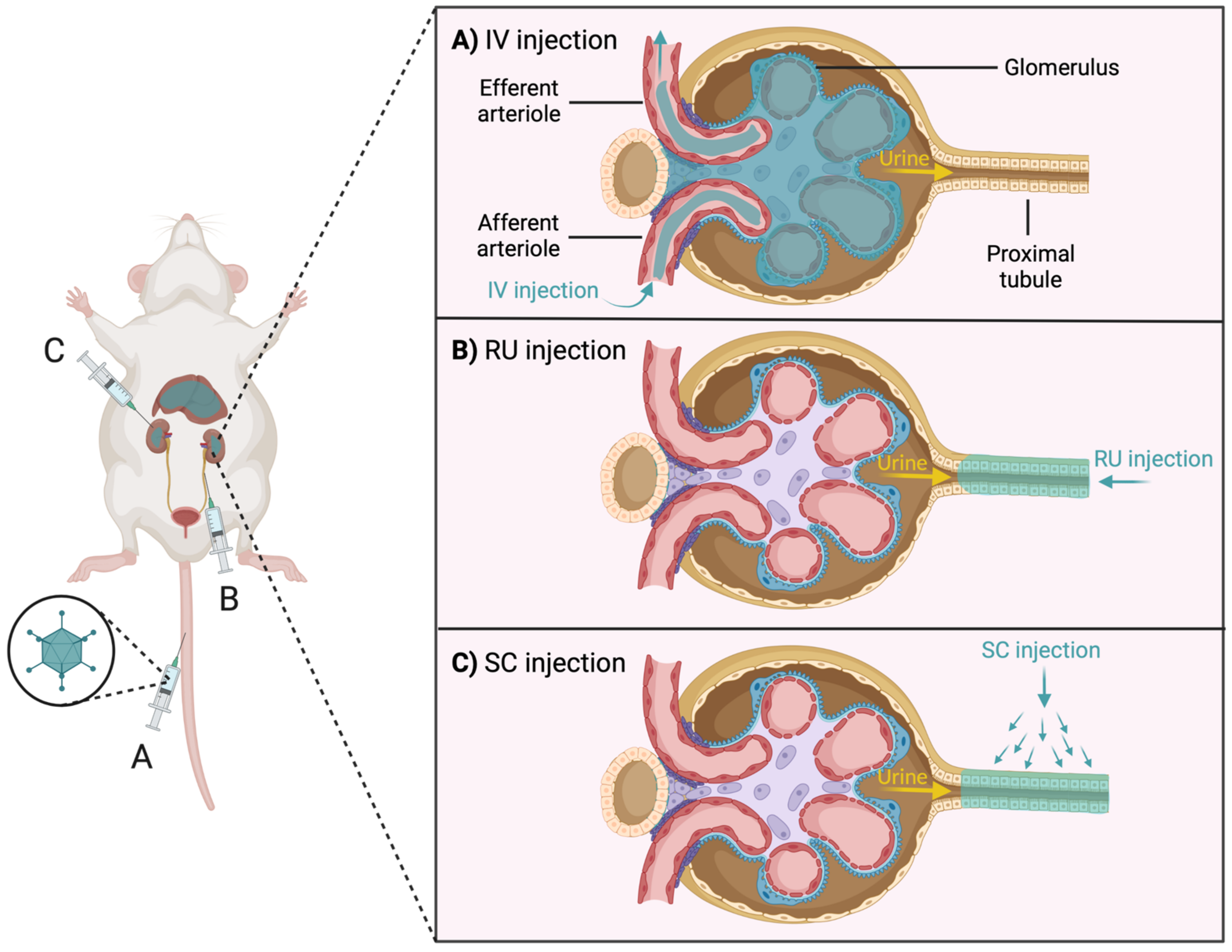
3.1. Difficulty of Gene Delivery to the Kidney

There are two main problems for efficient delivery of the gene editing machinery by vectors to renal cells: the access problem and the tropism problem. The access problem refers to whether the vectors can reach the target cells. The tropism problem considers whether, if the access problem is overcome, the vectors can interact with the target cells in the kidney [166].
The access problem is due, in part, to the stringent filtering functions intrinsic to the kidney. This filtration takes place in the glomerulus (specifically in the glomerular filtration membrane-GFM) of each nephron [166]. Blood, via the afferent arteriole, reaches the glomerulus, where molecules with a weight greater than 50 kDa and a size greater than 10 nm are excluded [166,167,168]. Blood is drained from the glomerulus through the efferent arteriole that branches forming the peritubular capillaries (PTCs), which extend along the PT and DCT, producing the exchange of substances. Finally, the PTCs flow into arcuate vein and the blood leaves the kidney through the renal vein [167].
Since most viral and non-viral vectors are larger than 10 nm in diameter and megadaltons in size, intravenous (IV) injection (Figure 5A) of vectors is not a useful strategy for the kidney, as they would not be able to cross the GFM [168]. It has been shown that after IV administration of AAV (the smallest viral vector, 25 nm), these were not able to cross the GFM and only glomerular cells were infected [166]. Therefore, IV injection of vectors seems to be relevant for the study of glomerulopathies, such as Alport syndrome, but not for other ERGs (e.g., tubulopathies) [166].
It is noteworthy that although these vectors cannot reach tubular cells by crossing the GFM, there is the possibility of reaching tubular cells through PTCs. It has been published that by injection of naked plasmid DNA into the renal vein, it is possible to transfer it to interstitial fibroblasts near PTCs, but not to tubular cells [169,170]. It has also been reported that by IV injection of nanoparticles (20 and 100 nm in size), these accumulated in the glomerulus and PTCs, but not in the tubules [171]. However, promising results show that after IV injection of mesoscale nanoparticles (~400 nm in size), they were internalized by the proximal tubule epithelial cells at basal side via passing through the peritubular capillaries [172]. Another study showed that, after IV injection of nanoparticles (140 nm iron oxide cubes and clusters, with PEG), they were able not only to penetrate the tubular cells but also to reach the lumen and be excreted via urine [173]. Nevertheless, further studies are needed to elucidate the mechanisms of translation and excretion of these nanoparticles that are larger than the GFM limit.
To avoid the GFM limitation there is the possibility of injecting the vectors directly into the kidney by two strategies: (1) retrograde ureteral (RU) injections (Figure 5B), avoiding the filtration effect of the glomerulus but with the limitation of going upstream against the natural flow of urine or (2) subcapsular (SC) injections (Figure 5C), both limitations are avoided, and the infection of the tubular cells is improved [166]. However, it has been observed that in each of the three injection strategies (IV, RU and SC), infection levels in other organs such as the liver were elevated, highlighting the problem of vector tropism [166,168,174,175].
The tropism problem greatly conditions efficient delivery to the kidney. Most vectors used for in vivo gene delivery have the liver or spleen as their first target organ, making it very difficult for the vector to reach the kidney [118]. For instance, after IV injection, the liver absorbs approximately 98% of the injected AdVs [176,177]. To solve the problem of vector tropism, vector modification may be a strategy to pursue. By adding certain peptides to the viral capsid, their specificity for the kidney can be increased, diverting their attraction to the liver [166]. In addition, off-target expression of the transgene can be restricted using kidney-specific promoters, such as Ksp-cadherin gene promoter [178]. However, delivery of the gene editing machinery outside the primary target, the kidney, may not pose substantial safety concerns. It would even be beneficial to some diseases such as ADPKD or ARPKD, in which the liver is also affected (liver cyst formation) [179].
In general, AdVs and AAVs are the most robust viral vectors for in vivo gene delivery. Several serotypes of these vectors exist, varying in target cell specificity and immunogenicity. The most used serotype of AdVs for renal research is AdV5, although this serotype is also largely taken up by the liver following IV injection [168]. Since adenoviruses are non-enveloped and can be retargeted by genetic or chemical modifications of their various capsid proteins [180,181], attempts have been made to increase the tropism of AdV5 for the kidney. For this purpose, peptides were introduced into the HI loop, which is a C-terminal region of the fiber protein (its function is to allow the adenovirus to bind to the target cell) [182,183,184]. Using this approach, it was possible to increase the infection of renal cells after IV injection [168].
In relation to AAVs, there is a wide variety of serotypes (e.g., AAV1, 2, 5, 6, 7, 8, 9). The serotypes with the best efficiencies for delivery in the kidney are 6 and 8 [166,185]. However, as with AdVs, a large leakage of AAVs to the liver is observed, even when injected directly into the kidney. A possible solution to this problem is the use of clamps (i.e., clamping of the renal artery, renal vein, and ureter to increase the time of physical exposure of AAVs in the injection fluid to kidney cells), which has markedly increased renal cell infection [166,174]. These results indicate that clamps may be a good way to increase and restrict infection to the kidney.
Although it has been shown here that there are different solutions to the problems posed for the delivery of genetic material into the kidney through the different types of existing vectors, there is still much room for improvement. Overcoming the delivery problem will certainly facilitate the development of more animal models to increase knowledge about GKDs.

3.2. Immune System against Gene Editing

As mentioned above, viral vectors are currently the most efficient and widely used platform for delivering gene editing machinery to target cells. In addition, many of the proteins used in gene editing are derived from bacteria (e.g., SpCas9). The fact that the components necessary to perform in vivo gene editing are foreign to the immune system may stimulate immune responses through three main pathways: innate immunity, humoral immunity, or cellular immunity [186]. This immune response further limits the efficacy of gene editing in renal cells.
Innate immunity uses pattern recognition receptors to recognize conserved features in microbes [187]. This raises the possibility that the viral vector may be recognized and eliminated [104]. Humoral immunity is mediated by antibodies, which can eliminate engineered nucleases or delivery vectors. It has been shown in studies in different animal models that administration by viral vectors of SpCas9 or SaCas9 results in the development of antibodies against these proteins [188,189,190]. Finally, cellular immunity is mediated by cytotoxic T cells, which can eliminate cells expressing the engineered nucleases. As with humoral immunity, studies in animal models have demonstrated the development of cellular immunity to Cas9 after delivery of SpCas9 and SaCas9 by viral vectors [188,189,190].
Therefore, for gene editing to be effective, it is important to determine and control the immune response against the delivery vector as well as against the engineered nucleases. A possible solution to reduce immunogenicity against these components is to identify the epitopes that cause immune stimulation, to modify them and thus reduce their immunogenicity [104]. Another possible solution is to replace the use of viral vectors with non-viral vectors. Noteworthy is the use of LNPs, which are a clinically approved drug delivery system [191]. LNPs present a matrix containing soluble lipophilic molecules, which gives them the advantage of interacting better with cells and thus facilitating greater cellular uptake of the transported drug [192]. Like non-viral vectors, it has the disadvantage that it accumulates naturally in the liver, so it is necessary to include modifications to ensure good uptake in other cell types [193]. Regarding the kidneys, the size and charge of LNPs considerably influences delivery efficiency (e.g., small LNPs penetrate more easily into the kidneys but are more easily eliminated) [194,195]. Although further research is needed to increase the efficiency and specificity of LNPs for the kidney, these systems have been successfully used in several studies for drug delivery to the kidney [196,197,198,199].

4. Conclusions

Gene editing systems based on site-specific nuclease are a versatile tool offering a multitude of applications for the study of genetic diseases. ZFNs, TALENs, and CRISPR-Cas9 have allowed the establishment of several in vitro and in vivo models that mimic the GKDs observed in patients, helping to improve the understanding of the pathophysiological mechanisms of GKDs, discover new disease-causing genes, resolve variants of uncertain significance, and drug testing. However, the application of these technologies as a possible treatment in the field of GKDs has not yet been successful due to the lack of knowledge about on-target and off-target effects and the difficulty of in vivo gene delivery into the kidney. The selection of an appropriate gene delivery method is crucial to optimize the efficacy of genome editing in the kidney. Currently, the most commonly used vectors are of viral origin and present problems of access and tropism to the kidney and may trigger an adverse immune response. It is worth trying to overcome these problems to facilitate and promote the successful establishment of more GKDs in vivo models that will contribute to generating new knowledge on disease development and progression. Furthermore, given the demonstrated plasticity capacity of the kidney, gene therapy based on gene editing tools could be a future option for treating Genetic kidney diseases.

Author Contributions

Writing—original draft preparation, F.G.-G.; writing—review and editing, F.G.-G., R.M.-P., N.C., C.A. and M.A.G.-G. All authors have read and agreed to the published version of the manuscript.

Funding

This work was funded by PI18/00378 from Instituto de Salud Carlos III under FIS/FEDER funds (to M.A.G.-G.), by IN607B-2016/020 from Axencia Galega de Innovación (to M.A.G.-G.), by ED431G 2019/02 (to C.A.) from the Xunta de Galicia, by RD21/0005/0020 from Redes de Investigación Cooperativa Orientadas a Resultados en Salud (to M.A.G.-G.) and by RD16/0009/0024 from Red de Investigación Renal (to M.A.G.-G.).

Institutional Review Board Statement

Not applicable.

Informed Consent Statement

Not applicable.

Acknowledgments

Figures were created with BioRender.com (accessed on 14 February 2022).

Conflicts of Interest

The authors declare no conflict of interest.

References

  1. Devuyst, O.; Knoers, N.V.A.M.; Remuzzi, G.; Schaefer, F. Rare inherited kidney diseases: Challenges, opportunities, and perspectives. Lancet 2014, 383, 1844–1859. [Google Scholar] [CrossRef] [Green Version]
  2. Aymé, S.; Bockenhauer, D.; Day, S.; Devuyst, O.; Guay-Woodford, L.M.; Ingelfinger, J.R.; Klein, J.B.; Knoers, N.V.; Perrone, R.D.; Roberts, J.; et al. Common Elements in Rare Kidney Diseases: Conclusions from a Kidney Disease: Improving Global Outcomes (KDIGO) Controversies Conference. Kidney Int. 2017, 92, 796–808. [Google Scholar] [CrossRef] [PubMed] [Green Version]
  3. Vivante, A.; Hildebrandt, F. Exploring the genetic basis of early-onset chronic kidney disease. Nat. Rev. Nephrol. 2016, 12, 133–146. [Google Scholar] [CrossRef] [PubMed] [Green Version]
  4. Torra, R.; Furlano, M.; Ortiz, A.; Ars, E. Genetic kidney diseases as an underrecognized cause of chronic kidney disease: The key role of international registry reports. Clin. Kidney J. 2021, 14, 1879–1885. [Google Scholar] [CrossRef] [PubMed]
  5. Jha, V.; Garcia-Garcia, G.; Iseki, K.; Li, Z.; Naicker, S.; Plattner, B.; Saran, R.; Wang, A.Y.M.; Yang, C.W. Chronic kidney disease: Global dimension and perspectives. Lancet 2013, 382, 260–272. [Google Scholar] [CrossRef]
  6. Groopman, E.E.; Marasa, M.; Cameron-Christie, S.; Petrovski, S.; Aggarwal, V.S.; Rasouly, H.M.; Li, Y.; Zhang, J.; Nestor, J.; Krithivasan, P.; et al. Diagnostic Utility of Exome Sequencing for Kidney Disease. N. Engl. J. Med. 2019, 380, 142–151. [Google Scholar] [CrossRef]
  7. Connaughton, D.M.; Kennedy, C.; Shril, S.; Mann, N.; Murray, S.L.; Williams, P.A.; Conlon, E.; Nakayama, M.; van der Ven, A.T.; Ityel, H.; et al. Monogenic causes of chronic kidney disease in adults. Kidney Int. 2019, 95, 914–928. [Google Scholar] [CrossRef]
  8. Hofmeister, A.F.; Kömhoff, M.; Weber, S.; Grgic, I. Disease modeling in genetic kidney diseases: Mice. Cell Tissue Res. 2017, 369, 159–170. [Google Scholar] [CrossRef]
  9. Köttgen, A.; Gall, E.C.-L.; Halbritter, J.; Kiryluk, K.; Mallett, A.J.; Parekh, R.S.; Rasouly, H.M.; Sampson, M.G.; Tin, A.; Antignac, C.; et al. Genetics in chronic kidney disease: Conclusions from a Kidney Disease: Improving Global Outcomes (KDIGO) Controversies Conference. Kidney Int. 2022. [Google Scholar] [CrossRef]
  10. van der Wijst, J.; Belge, H.; Bindels, R.J.M.; Devuyst, O. Learning Physiology From Inherited Kidney Disorders. Physiol. Rev. 2019, 99, 1575–1653. [Google Scholar] [CrossRef]
  11. UniProt. Available online: https://www.uniprot.org/ (accessed on 22 March 2022).
  12. Garcia-Gonzalez, M.A.; Jones, J.G.; Allen, S.K.; Palatucci, C.M.; Batish, S.D.; Seltzer, W.K.; Lan, Z.; Allen, E.; Qian, F.; Lens, X.M.; et al. Evaluating the clinical utility of a molecular genetic test for polycystic kidney disease. Mol. Genet. Metab. 2007, 92, 160–167. [Google Scholar] [CrossRef] [PubMed] [Green Version]
  13. Cordido, A.; Besada-Cerecedo, L.; García-González, M.A. The Genetic and Cellular Basis of Autosomal Dominant Polycystic Kidney Disease-A Primer for Clinicians. Front. Pediatr. 2017, 5, 279. [Google Scholar] [CrossRef] [PubMed]
  14. Porath, B.; Gainullin, V.G.; Gall, E.C.-L.; Dillinger, E.K.; Heyer, C.M.; Hopp, K.; Edwards, M.E.; Madsen, C.D.; Mauritz, S.R.; Banks, C.J.; et al. Mutations in GANAB, Encoding the Glucosidase IIα Subunit, Cause Autosomal-Dominant Polycystic Kidney and Liver Disease. Am. J. Hum. Genet. 2016, 98, 1193–1207. [Google Scholar] [CrossRef] [PubMed] [Green Version]
  15. Gall, E.C.-L.; Olson, R.J.; Besse, W.; Heyer, C.M.; Gainullin, V.G.; Smith, J.M.; Audrézet, M.P.; Hopp, K.; Porath, B.; Shi, B.; et al. Monoallelic Mutations to DNAJB11 Cause Atypical Autosomal-Dominant Polycystic Kidney Disease. Am. J. Hum. Genet. 2018, 102, 832–844. [Google Scholar] [CrossRef] [Green Version]
  16. Senum, S.R.; Li, Y.S.M.; Benson, K.A.; Joli, G.; Olinger, E.; Lavu, S.; Madsen, C.D.; Gregory, A.V.; Neatu, R.; Kline, T.L.; et al. Monoallelic IFT140 pathogenic variants are an important cause of the autosomal dominant polycystic kidney-spectrum phenotype. Am. J. Hum. Genet. 2022, 109, 136–156. [Google Scholar] [CrossRef]
  17. Hildebrandt, F.; Benzing, T.; Katsanis, N. Ciliopathies. N. Engl. J. Med. 2011, 364, 1533–1543. [Google Scholar] [CrossRef] [Green Version]
  18. Kim, E.; Walz, G. Sensitive cilia set up the kidney. Nat. Med. 2007, 13, 1409–1411. [Google Scholar] [CrossRef]
  19. Harris, P.C.; Torres, V.E. Genetic mechanisms and signaling pathways in autosomal dominant polycystic kidney disease. J. Clin. Investig. 2014, 124, 2315–2324. [Google Scholar] [CrossRef] [Green Version]
  20. Reiterová, J.; Štekrová, J.; Merta, M.; Kotlas, J.; Elišáková, V.; Lněnička, P.; Korabečná, M.; Kohoutová, M.; Tesař, V. Autosomal dominant polycystic kidney disease in a family with mosaicism and hypomorphic allele. BMC Nephrol. 2013, 14, 59. [Google Scholar] [CrossRef] [Green Version]
  21. Mehta, L.; Jim, B. Hereditary Renal Diseases. Semin. Nephrol. 2017, 37, 354–361. [Google Scholar] [CrossRef]
  22. Chebib, F.T.; Torres, V.E. Autosomal Dominant Polycystic Kidney Disease: Core Curriculum 2016. Am. J. Kidney Dis. 2016, 67, 792–810. [Google Scholar] [CrossRef] [PubMed] [Green Version]
  23. Garcia-Gonzalez, M.A.; Outeda, P.; Zhou, Q.; Zhou, F.; Menezes, L.F.; Qian, F.; Huso, D.L.; Germino, G.G.; Piontek, K.B.; Watnick, T. Pkd1 and Pkd2 are required for normal placental development. PLoS ONE 2010, 5, e12821. [Google Scholar] [CrossRef] [PubMed] [Green Version]
  24. Bergmann, C.; Guay-Woodford, L.M.; Harris, P.C.; Horie, S.; Peters, D.J.M.; Torres, V.E. Polycystic kidney disease. Nat. Rev. Dis. Prim. 2018, 4. [Google Scholar] [CrossRef]
  25. Parfrey, P.S.; Bear, J.C.; Morgan, J.; Cramer, B.C.; McManamon, P.J.; Gault, M.H.; Churchill, D.N.; Singh, M.; Hewitt, R.; Somlo, S.; et al. The diagnosis and prognosis of autosomal dominant polycystic kidney disease. N. Engl. J. Med. 1990, 323, 1085–1090. [Google Scholar] [CrossRef] [PubMed]
  26. Onuchic, L.F.; Furu, L.; Nagasawa, Y.; Hou, X.; Eggermann, T.; Ren, Z.; Bergmann, C.; Senderek, J.; Esquivel, E.; Zeltner, R.; et al. PKHD1, the polycystic kidney and hepatic disease 1 gene, encodes a novel large protein containing multiple immunoglobulin-like plexin-transcription-factor domains and parallel beta-helix 1 repeats. Am. J. Hum. Genet. 2002, 70, 1305–1317. [Google Scholar] [CrossRef] [PubMed] [Green Version]
  27. Cordido, A.; Vizoso-gonzalez, M.; Garcia-Gonzalez, M.A. Molecular Pathophysiology of Autosomal Recessive Polycystic Kidney Disease. Int. J. Mol. Sci. 2021, 22, 6523. [Google Scholar] [CrossRef] [PubMed]
  28. Lu, H.; Galeano, M.C.R.; Ott, E.; Kaeslin, G.; Kausalya, P.J.; Kramer, C.; Ortiz-Brüchle, N.; Hilger, N.; Metzis, V.; Hiersche, M.; et al. Mutations in DZIP1L, which encodes a ciliary-transition-zone protein, cause autosomal recessive polycystic kidney disease. Nat. Genet. 2017, 49, 1025–1034. [Google Scholar] [CrossRef] [PubMed]
  29. Kim, I.; Fu, Y.; Hui, K.; Moeckel, G.; Mai, W.; Li, C.; Liang, D.; Zhao, P.; Ma, J.; Chen, X.Z.; et al. Fibrocystin/polyductin modulates renal tubular formation by regulating polycystin-2 expression and function. J. Am. Soc. Nephrol. 2008, 19, 455–468. [Google Scholar] [CrossRef] [Green Version]
  30. Guay-Woodford, L.M.; Muecher, G.; Hopkins, S.D.; Avner, E.D.; Germino, G.G.; Guillot, A.P.; Herrin, J.; Holleman, R.; Irons, D.A.; Primack, W.; et al. The Severe Perinatal Form of Autosomal Recessive Polycystic Kidney Disease Maps to Chromosome 6p21.1-p12: Implications for Genetic Counseling. Am. J. Hum. Genet. 1995, 56, 1101. [Google Scholar]
  31. Zerres, K.; Rudnik-Schöneborn, S.; Steinkamm, C.; Becker, J.; Mücher, G. Autosomal recessive polycystic kidney disease. J. Mol. Med. 1998, 76, 303–309. [Google Scholar] [CrossRef]
  32. Capisonda, R.; Phan, V.; Traubuci, J.; Daneman, A.; Balfe, J.W.; Guay-Woodford, L.M. Autosomal recessive polycystic kidney disease: Outcomes from a single-center experience. Pediatr. Nephrol. 2003, 18, 119–126. [Google Scholar] [CrossRef] [PubMed]
  33. Guay-Woodford, L.M.; Desmond, R.A. Autosomal recessive polycystic kidney disease: The clinical experience in North America. Pediatrics 2003, 111, 1072–1080. [Google Scholar] [CrossRef] [PubMed] [Green Version]
  34. Fonck, C.; Chauveau, D.; Gagnadoux, M.F.; Pirson, Y.; Grünfeld, J.P. Autosomal recessive polycystic kidney disease in adulthood. Nephrol. Dial. Transplant. 2001, 16, 1648–1652. [Google Scholar] [CrossRef] [PubMed] [Green Version]
  35. Stokman, M.F.; Renkema, K.Y.; Giles, R.H.; Schaefer, F.; Knoers, N.V.A.M.; Van Eerde, A.M. The expanding phenotypic spectra of kidney diseases: Insights from genetic studies. Nat. Rev. Nephrol. 2016, 12, 472–483. [Google Scholar] [CrossRef] [PubMed]
  36. Pinto e Vairo, F.; Prochnow, C.; Kemppainen, J.L.; Lisi, E.C.; Steyermark, J.M.; Kruisselbrink, T.M.; Pichurin, P.N.; Dhamija, R.; Hager, M.M.; Albadri, S.; et al. Genomics Integration Into Nephrology Practice. Kidney Med. 2021, 3, 785–798. [Google Scholar] [CrossRef] [PubMed]
  37. Gibson, J.; Fieldhouse, R.; Chan, M.M.Y.; Sadeghi-Alavijeh, O.; Burnett, L.; Izzi, V.; Persikov, A.V.; Gale, D.P.; Storey, H.; Savige, J. Prevalence Estimates of Predicted Pathogenic COL4A3-COL4A5 Variants in a Population Sequencing Database and Their Implications for Alport Syndrome. J. Am. Soc. Nephrol. 2021, 32, 2273–2290. [Google Scholar] [CrossRef]
  38. gnomAD. Available online: https://gnomad.broadinstitute.org/ (accessed on 28 April 2022).
  39. Martínez-Pulleiro, R.; García-Murias, M.; Fidalgo-Díaz, M.; García-González, M.Á. Molecular Basis, Diagnostic Challenges and Therapeutic Approaches of Alport Syndrome: A Primer for Clinicians. Int. J. Mol. Sci. 2021, 22, 110663. [Google Scholar] [CrossRef]
  40. Fallerini, C.; Dosa, L.; Tita, R.; Del Prete, D.; Feriozzi, S.; Gai, G.; Clementi, M.; La Manna, A.; Miglietti, N.; Mancini, R.; et al. Unbiased next generation sequencing analysis confirms the existence of autosomal dominant Alport syndrome in a relevant fraction of cases. Clin. Genet. 2014, 86, 252–257. [Google Scholar] [CrossRef]
  41. Morinière, V.; Dahan, K.; Hilbert, P.; Lison, M.; Lebbah, S.; Topa, A.; Bole-Feysot, C.; Pruvost, S.; Nitschke, P.; Plaisier, E.; et al. Improving mutation screening in familial hematuric nephropathies through next generation sequencing. J. Am. Soc. Nephrol. 2014, 25, 2740–2751. [Google Scholar] [CrossRef] [Green Version]
  42. Yamamura, T.; Nozu, K.; Minamikawa, S.; Horinouchi, T.; Sakakibara, N.; Nagano, C.; Aoto, Y.; Ishiko, S.; Nakanishi, K.; Shima, Y.; et al. Comparison between conventional and comprehensive sequencing approaches for genetic diagnosis of Alport syndrome. Mol. Genet. Genom. Med. 2019, 7, e883. [Google Scholar] [CrossRef] [Green Version]
  43. Jais, J.P.; Knebelmann, B.; Giatras, I.; De Marchi, M.; Rizzoni, G.; Renieri, A.; Weber, M.; Gross, O.; Netzer, K.O.; Flinter, F.; et al. X-linked Alport syndrome: Natural history in 195 families and genotype- phenotype correlations in males. J. Am. Soc. Nephrol. 2000, 11, 649–657. [Google Scholar] [CrossRef] [PubMed]
  44. Savige, J.; Colville, D.; Rheault, M.; Gear, S.; Lennon, R.; Lagas, S.; Finlay, M.; Flinter, F. Alport Syndrome in Women and Girls. Clin. J. Am. Soc. Nephrol. 2016, 11, 1713–1720. [Google Scholar] [CrossRef] [PubMed] [Green Version]
  45. Storey, H.; Savige, J.; Sivakumar, V.; Abbs, S.; Flinter, F.A. COL4A3/COL4A4 Mutations and Features in Individuals with Autosomal Recessive Alport Syndrome. J. Am. Soc. Nephrol. 2013, 24, 1945–1954. [Google Scholar] [CrossRef] [PubMed] [Green Version]
  46. Pescucci, C.; Mari, F.; Longo, I.; Vogiatzi, P.; Caselli, R.; Scala, E.; Abaterusso, C.; Gusmano, R.; Seri, M.; Miglietti, N.; et al. Autosomal-dominant Alport syndrome: Natural history of a disease due to COL4A3 or COL4A4 gene. Kidney Int. 2004, 65, 1598–1603. [Google Scholar] [CrossRef] [PubMed] [Green Version]
  47. Fallerini, C.; Baldassarri, M.; Trevisson, E.; Morbidoni, V.; La Manna, A.; Lazzarin, R.; Pasini, A.; Barbano, G.; Pinciaroli, A.R.; Garosi, G.; et al. Alport syndrome: Impact of digenic inheritance in patients management. Clin. Genet. 2017, 92, 34–44. [Google Scholar] [CrossRef]
  48. Savige, J. Should We Diagnose Autosomal Dominant Alport Syndrome When There Is a Pathogenic Heterozygous COL4A3 or COL4A4 Variant? Kidney Int. Rep. 2018, 3, 1239–1241. [Google Scholar] [CrossRef] [Green Version]
  49. Devuyst, O.; Olinger, E.; Weber, S.; Eckardt, K.U.; Kmoch, S.; Rampoldi, L.; Bleyer, A.J. Autosomal dominant tubulointerstitial kidney disease. Nat. Rev. Dis. Prim. 2019, 5, 1–20. [Google Scholar] [CrossRef] [Green Version]
  50. Eckardt, K.U.; Alper, S.L.; Antignac, C.; Bleyer, A.J.; Chauveau, D.; Dahan, K.; Deltas, C.; Hosking, A.; Kmoch, S.; Rampoldi, L.; et al. Autosomal dominant tubulointerstitial kidney disease: Diagnosis, classification, and management—A KDIGO consensus report. Kidney Int. 2015, 88, 676–683. [Google Scholar] [CrossRef] [Green Version]
  51. Cormican, S.; Connaughton, D.M.; Kennedy, C.; Murray, S.; Živná, M.; Kmoch, S.; Fennelly, N.K.; O’Kelly, P.; Benson, K.A.; Conlon, E.T.; et al. Autosomal dominant tubulointerstitial kidney disease (ADTKD) in Ireland. Ren. Fail. 2019, 41, 832–841. [Google Scholar] [CrossRef] [Green Version]
  52. Bohle, A.; Mackensen-Haen, S.; Van Gise, H. Significance of tubulointerstitial changes in the renal cortex for the excretory function and concentration ability of the kidney: A morphometric contribution. Am. J. Nephrol. 1987, 7, 421–433. [Google Scholar] [CrossRef]
  53. Ayasreh, N.; Bullich, G.; Miquel, R.; Furlano, M.; Ruiz, P.; Lorente, L.; Valero, O.; García-González, M.A.; Arhda, N.; Garin, I.; et al. Autosomal Dominant Tubulointerstitial Kidney Disease: Clinical Presentation of Patients With ADTKD-UMOD and ADTKD-MUC1. Am. J. Kidney Dis. 2018, 72, 411–418. [Google Scholar] [CrossRef]
  54. Heidet, L.; Decramer, S.; Pawtowski, A.; Morinière, V.; Bandin, F.; Knebelmann, B.; Lebre, A.S.; Faguer, S.; Guigonis, V.; Antignac, C.; et al. Spectrum of HNF1B mutations in a large cohort of patients who harbor renal diseases. Clin. J. Am. Soc. Nephrol. 2010, 5, 1079–1090. [Google Scholar] [CrossRef] [PubMed] [Green Version]
  55. Bolar, N.A.; Golzio, C.; Živná, M.; Hayot, G.; Van Hemelrijk, C.; Schepers, D.; Vandeweyer, G.; Hoischen, A.; Huyghe, J.R.; Raes, A.; et al. Heterozygous Loss-of-Function SEC61A1 Mutations Cause Autosomal-Dominant Tubulo-Interstitial and Glomerulocystic Kidney Disease with Anemia. Am. J. Hum. Genet. 2016, 99, 174–187. [Google Scholar] [CrossRef] [PubMed] [Green Version]
  56. Downie, M.L.; Lopez Garcia, S.C.; Kleta, R.; Bockenhauer, D. Inherited Tubulopathies of the Kidney: Insights from Genetics. Clin. J. Am. Soc. Nephrol. 2021, 16, 620–630. [Google Scholar] [CrossRef] [PubMed] [Green Version]
  57. Simon, D.B.; Nelson-Williams, C.; Bia, M.J.; Ellison, D.; Karet, F.E.; Molina, A.M.; Vaara, I.; Iwata, F.; Cushner, H.M.; Koolen, M.; et al. Gitelman’s variant of Bartter’s syndrome, inherited hypokalaemic alkalosis, is caused by mutations in the thiazide-sensitive Na-Cl cotransporter. Nat. Genet. 1996, 12, 24–30. [Google Scholar] [CrossRef]
  58. Simon, D.B.; Karet, F.E.; Hamdan, J.M.; Di Pietro, A.; Sanjad, S.A.; Lifton, R.P. Bartter’s syndrome, hypokalaemic alkalosis with hypercalciuria, is caused by mutations in the Na-K-2Cl cotransporter NKCC2. Nat. Genet. 1996, 13, 183–188. [Google Scholar] [CrossRef]
  59. Simon, D.B.; Karet, F.E.; Rodriguez-Soriano, J.; Hamdan, J.H.; DiPietro, A.; Trachtman, H.; Sanjad, S.A.; Lifton, R.P. Genetic heterogeneity of Bartter’s syndrome revealed by mutations in the K+ channel, ROMK. Nat. Genet. 1996, 14, 152–156. [Google Scholar] [CrossRef]
  60. Simon, D.B.; Bindra, R.S.; Mansfield, T.A.; Nelson-Williams, C.; Mendonca, E.; Stone, R.; Schurman, S.; Nayir, A.; Alpay, H.; Bakkaloglu, A.; et al. Mutations in the chloride channel gene, CLCNKB, cause Bartter’s syndrome type III. Nat. Genet. 1997, 17, 171–178. [Google Scholar] [CrossRef]
  61. Birkenhäger, R.; Otto, E.; Schürmann, M.J.; Vollmer, M.; Ruf, E.M.; Maier-Lutz, I.; Beekmann, F.; Fekete, A.; Omran, H.; Feldmann, D.; et al. Mutation of BSND causes Bartter syndrome with sensorineural deafness and kidney failure. Nat. Genet. 2001, 29, 310–314. [Google Scholar] [CrossRef]
  62. Laghmani, K.; Beck, B.B.; Yang, S.-S.; Seaayfan, E.; Wenzel, A.; Reusch, B.; Vitzthum, H.; Priem, D.; Demaretz, S.; Bergmann, K.; et al. Polyhydramnios, Transient Antenatal Bartter’s Syndrome, and MAGED2 Mutations. N. Engl. J. Med. 2016, 374, 1853–1863. [Google Scholar] [CrossRef]
  63. Schlingmann, K.P.; Konrad, M.; Jeck, N.; Waldegger, P.; Reinalter, S.C.; Holder, M.; Seyberth, H.W.; Waldegger, S. Salt wasting and deafness resulting from mutations in two chloride channels. N. Engl. J. Med. 2004, 350, 1314–1319. [Google Scholar] [CrossRef] [PubMed]
  64. Bichet, D.G.; Fujiwara, T.M. Reabsorption of sodium chloride—Lessons from the chloride channels. N. Engl. J. Med. 2004, 350, 1281–1283. [Google Scholar] [CrossRef] [PubMed]
  65. Nuñez-Gonzalez, L.; Carrera, N.; Garcia-Gonzalez, M.A. Molecular Basis, Diagnostic Challenges and Therapeutic Approaches of Bartter and Gitelman Syndromes: A Primer for Clinicians. Int. J. Mol. Sci. 2021, 22, 11414. [Google Scholar] [CrossRef]
  66. Bao, M.; Cai, J.; Yang, X.; Ma, W. Genetic screening for Bartter syndrome and Gitelman syndrome pathogenic genes among individuals with hypertension and hypokalemia. Clin. Exp. Hypertens. 2019, 41, 381–388. [Google Scholar] [CrossRef] [PubMed]
  67. Orphanet: Bartter Syndrome. Available online: https://www.orpha.net/consor/cgi-bin/OC_Exp.php?Lng=GB&Expert=112 (accessed on 14 March 2022).
  68. Besouw, M.T.P.; Kleta, R.; Bockenhauer, D. Bartter and Gitelman syndromes: Questions of class. Pediatr. Nephrol. 2020, 35, 1815–1824. [Google Scholar] [CrossRef] [Green Version]
  69. Zhang, H.; Zhang, J.; Lang, Z.; Botella, J.R.; Zhu, J.K. Genome Editing—Principles and Applications for Functional Genomics Research and Crop Improvement. Crit. Rev. Plant Sci. 2017, 36, 291–309. [Google Scholar] [CrossRef]
  70. Puchta, H. The repair of double-strand breaks in plants: Mechanisms and consequences for genome evolution. J. Exp. Bot. 2005, 56, 1–14. [Google Scholar] [CrossRef] [Green Version]
  71. Gupta, R.M.; Musunuru, K. Expanding the genetic editing tool kit: ZFNs, TALENs, and CRISPR-Cas9. J. Clin. Investig. 2014, 124, 4154–4161. [Google Scholar] [CrossRef] [Green Version]
  72. Kim, Y.G.; Cha, J.; Chandrasegaran, S. Hybrid restriction enzymes: Zinc finger fusions to Fok I cleavage domain. Proc. Natl. Acad. Sci. USA 1996, 93, 1156–1160. [Google Scholar] [CrossRef] [Green Version]
  73. Beerli, R.R.; Barbas, C.F. Engineering polydactyl zinc-finger transcription factors. Nat. Biotechnol. 2002, 20, 135–141. [Google Scholar] [CrossRef]
  74. Carroll, D.; Morton, J.J.; Beumer, K.J.; Segal, D.J. Design, construction and in vitro testing of zinc finger nucleases. Nat. Protoc. 2006, 1, 1329–1341. [Google Scholar] [CrossRef] [PubMed]
  75. Peng, Y.; Clark, K.J.; Campbell, J.M.; Panetta, M.R.; Guo, Y.; Ekker, S.C. Making designer mutants in model organisms. Development 2014, 141, 4042–4054. [Google Scholar] [CrossRef] [PubMed] [Green Version]
  76. Urnov, F.D.; Rebar, E.J.; Holmes, M.C.; Zhang, H.S.; Gregory, P.D. Genome editing with engineered zinc finger nucleases. Nat. Rev. Genet. 2010, 11, 636–646. [Google Scholar] [CrossRef] [PubMed]
  77. Miller, J.C.; Tan, S.; Qiao, G.; Barlow, K.A.; Wang, J.; Xia, D.F.; Meng, X.; Paschon, D.E.; Leung, E.; Hinkley, S.J.; et al. A TALE nuclease architecture for efficient genome editing. Nat. Biotechnol. 2011, 29, 143–150. [Google Scholar] [CrossRef]
  78. Bogdanove, A.J.; Schornack, S.; Lahaye, T. TAL effectors: Finding plant genes for disease and defense. Curr. Opin. Plant Biol. 2010, 13, 394–401. [Google Scholar] [CrossRef]
  79. Guilinger, J.P.; Pattanayak, V.; Reyon, D.; Tsai, S.Q.; Sander, J.D.; Joung, J.K.; Liu, D.R. Broad specificity profiling of TALENs results in engineered nucleases with improved DNA-cleavage specificity. Nat. Methods 2014, 11, 429–435. [Google Scholar] [CrossRef] [Green Version]
  80. Cong, L.; Ran, F.A.; Cox, D.; Lin, S.; Barretto, R.; Habib, N.; Hsu, P.D.; Wu, X.; Jiang, W.; Marraffini, L.A.; et al. Multiplex genome engineering using CRISPR/Cas systems. Science 2013, 339, 819–823. [Google Scholar] [CrossRef] [Green Version]
  81. Mali, P.; Yang, L.; Esvelt, K.M.; Aach, J.; Guell, M.; DiCarlo, J.E.; Norville, J.E.; Church, G.M. RNA-guided human genome engineering via Cas9. Science 2013, 339, 823–826. [Google Scholar] [CrossRef] [Green Version]
  82. Szczelkun, M.D.; Tikhomirova, M.S.; Sinkunas, T.; Gasiunas, G.; Karvelis, T.; Pschera, P.; Siksnys, V.; Seidel, R. Direct observation of R-loop formation by single RNA-guided Cas9 and Cascade effector complexes. Proc. Natl. Acad. Sci. USA 2014, 111, 9798–9803. [Google Scholar] [CrossRef] [Green Version]
  83. Jinek, M.; Chylinski, K.; Fonfara, I.; Hauer, M.; Doudna, J.A.; Charpentier, E. A programmable dual-RNA-guided DNA endonuclease in adaptive bacterial immunity. Science 2012, 337, 816–821. [Google Scholar] [CrossRef]
  84. Jinek, M.; Jiang, F.; Taylor, D.W.; Sternberg, S.H.; Kaya, E.; Ma, E.; Anders, C.; Hauer, M.; Zhou, K.; Lin, S.; et al. Structures of Cas9 endonucleases reveal RNA-mediated conformational activation. Science 2014, 343. [Google Scholar] [CrossRef] [PubMed] [Green Version]
  85. Anzalone, A.V.; Koblan, L.W.; Liu, D.R. Genome editing with CRISPR-Cas nucleases, base editors, transposases and prime editors. Nat. Biotechnol. 2020, 38, 824–844. [Google Scholar] [CrossRef] [PubMed]
  86. Ramirez, C.L.; Foley, J.E.; Wright, D.A.; Müller-Lerch, F.; Rahman, S.H.; Cornu, T.I.; Winfrey, R.J.; Sander, J.D.; Fu, F.; Townsend, J.A.; et al. Unexpected failure rates for modular assembly of engineered zinc fingers. Nat. Methods 2008, 5, 374–375. [Google Scholar] [CrossRef] [PubMed]
  87. Cermak, T.; Doyle, E.L.; Christian, M.; Wang, L.; Zhang, Y.; Schmidt, C.; Baller, J.A.; Somia, N.V.; Bogdanove, A.J.; Voytas, D.F. Efficient design and assembly of custom TALEN and other TAL effector-based constructs for DNA targeting. Nucleic Acids Res. 2011, 39, e82. [Google Scholar] [CrossRef] [PubMed] [Green Version]
  88. Cheng, R.; Peng, J.; Yan, Y.; Cao, P.; Wang, J.; Qiu, C.; Tang, L.; Liu, D.; Tang, L.; Jin, J.; et al. Efficient gene editing in adult mouse livers via adenoviral delivery of CRISPR/Cas9. FEBS Lett. 2014, 588, 3954–3958. [Google Scholar] [CrossRef]
  89. Li, H.; Yang, Y.; Hong, W.; Huang, M.; Wu, M.; Zhao, X. Applications of genome editing technology in the targeted therapy of human diseases: Mechanisms, advances and prospects. Signal Transduct. Target. Ther. 2020, 5, 1–23. [Google Scholar] [CrossRef]
  90. Chen, L.; Tang, L.; Xiang, H.; Jin, L.; Li, Q.; Dong, Y.; Wang, W.; Zhang, G. Advances in genome editing technology and its promising application in evolutionary and ecological studies. Gigascience 2014, 3, 2047-217X. [Google Scholar] [CrossRef] [Green Version]
  91. Miyagi, A.; Lu, A.; Humphreys, B.D. Gene Editing: Powerful New Tools for Nephrology Research and Therapy. J. Am. Soc. Nephrol. 2016, 27, 2940–2947. [Google Scholar] [CrossRef] [Green Version]
  92. Gonzalez, B.; Schwimmer, L.J.; Fuller, R.P.; Ye, Y.; Asawapornmongkol, L.; Barbas, C.F. Modular system for the construction of zinc-finger libraries and proteins. Nat. Protoc. 2010, 5, 791–810. [Google Scholar] [CrossRef] [Green Version]
  93. Valton, J.; Dupuy, A.; Daboussi, F.; Thomas, S.; Maréchal, A.; Macmaster, R.; Melliand, K.; Juillerat, A.; Duchateau, P. Overcoming transcription activator-like effector (TALE) DNA binding domain sensitivity to cytosine methylation. J. Biol. Chem. 2012, 287, 38427–38432. [Google Scholar] [CrossRef] [Green Version]
  94. Ates, I.; Rathbone, T.; Stuart, C.; Bridges, P.H.; Cottle, R.N. Delivery approaches for therapeutic genome editing and challenges. Genes 2020, 11, 1113. [Google Scholar] [CrossRef] [PubMed]
  95. Yip, B.H. Recent advances in CRISPR/Cas9 delivery strategies. Biomolecules 2020, 10, 839. [Google Scholar] [CrossRef] [PubMed]
  96. Rui, Y.; Wilson, D.R.; Green, J.J. Non-Viral Delivery To Enable Genome Editing. Trends Biotechnol. 2019, 37, 281–293. [Google Scholar] [CrossRef] [PubMed]
  97. Wang, L.; Li, F.; Dang, L.; Liang, C.; Wang, C.; He, B.; Liu, J.; Li, D.; Wu, X.; Xu, X.; et al. In Vivo delivery systems for therapeutic genome editing. Int. J. Mol. Sci. 2016, 17, 626. [Google Scholar] [CrossRef] [PubMed] [Green Version]
  98. Faneca, H. Non-Viral Gene Delivery Systems. Pharmaceutics 2021, 13, 446. [Google Scholar] [CrossRef] [PubMed]
  99. Lan, T.; Que, H.; Luo, M.; Zhao, X.; Wei, X. Genome editing via non-viral delivery platforms: Current progress in personalized cancer therapy. Mol. Cancer 2022, 21, 1–15. [Google Scholar] [CrossRef]
  100. Wang, M.; Glass, Z.A.; Xu, Q. Non-viral delivery of genome-editing nucleases for gene therapy. Gene Ther. 2017, 24, 144–150. [Google Scholar] [CrossRef]
  101. Sakellari, G.I.; Zafeiri, I.; Batchelor, H.; Spyropoulos, F. Formulation design, production and characterisation of solid lipid nanoparticles (SLN) and nanostructured lipid carriers (NLC) for the encapsulation of a model hydrophobic active. Food Hydrocoll. Health 2021, 1, 100024. [Google Scholar] [CrossRef]
  102. Pensado, A.; Seijo, B.; Sanchez, A. Current strategies for DNA therapy based on lipid nanocarriers. Expert Opin. Drug Deliv. 2014, 11, 1721–1731. [Google Scholar] [CrossRef]
  103. Lim, S.; Koo, J.H.; Choi, J.M. Use of Cell-Penetrating Peptides in Dendritic Cell-Based Vaccination. Immune Netw. 2016, 16, 33–43. [Google Scholar] [CrossRef] [Green Version]
  104. Hirakawa, M.P.; Krishnakumar, R.; Timlin, J.A.; Carney, J.P.; Butler, K.S. Gene editing and CRISPR in the clinic: Current and future perspectives. Biosci. Rep. 2020, 40, BSR20200127. [Google Scholar] [CrossRef] [PubMed] [Green Version]
  105. Boch, J.; Scholze, H.; Schornack, S.; Landgraf, A.; Hahn, S.; Kay, S.; Lahaye, T.; Nickstadt, A.; Bonas, U. Breaking the code of DNA binding specificity of TAL-type III effectors. Science 2009, 326, 1509–1512. [Google Scholar] [CrossRef] [PubMed]
  106. Moscou, M.J.; Bogdanove, A.J. A simple cipher governs DNA recognition by TAL effectors. Science 2009, 326, 1501. [Google Scholar] [CrossRef]
  107. Zhang, X.H.; Tee, L.Y.; Wang, X.G.; Huang, Q.S.; Yang, S.H. Off-target effects in CRISPR/Cas9-mediated genome engineering. Mol. Ther. Nucleic Acids 2015, 4, e264. [Google Scholar] [CrossRef] [PubMed]
  108. Zetsche, B.; Volz, S.E.; Zhang, F. A split-Cas9 architecture for inducible genome editing and transcription modulation. Nat. Biotechnol. 2015, 33, 139–142. [Google Scholar] [CrossRef] [PubMed] [Green Version]
  109. Moon, S.B.; Kim, D.Y.; Ko, J.H.; Kim, J.S.; Kim, Y.S. Improving CRISPR Genome Editing by Engineering Guide RNAs. Trends Biotechnol. 2019, 37, 870–881. [Google Scholar] [CrossRef]
  110. Fu, Y.; Foden, J.A.; Khayter, C.; Maeder, M.L.; Reyon, D.; Joung, J.K.; Sander, J.D. High-frequency off-target mutagenesis induced by CRISPR-Cas nucleases in human cells. Nat. Biotechnol. 2013, 31, 822–826. [Google Scholar] [CrossRef] [Green Version]
  111. Kosicki, M.; Tomberg, K.; Bradley, A. Repair of double-strand breaks induced by CRISPR-Cas9 leads to large deletions and complex rearrangements. Nat. Biotechnol. 2018, 36, 765–771. [Google Scholar] [CrossRef]
  112. Korablev, A.; Lukyanchikova, V.; Serova, I.; Battulin, N. On-Target CRISPR/Cas9 Activity Can Cause Undesigned Large Deletion in Mouse Zygotes. Int. J. Mol. Sci. 2020, 21, 3604. [Google Scholar] [CrossRef]
  113. Cruz, N.M.; Freedman, B.S. CRISPR Gene Editing in the Kidney. Am. J. Kidney Dis. 2018, 71, 874–883. [Google Scholar] [CrossRef]
  114. Koslowski, S.; Latapy, C.; Auvray, P.; Blondel, M.; Meijer, L. An Overview of In Vivo and In Vitro Models for Autosomal Dominant Polycystic Kidney Disease: A Journey from 3D-Cysts to Mini-Pigs. Int. J. Mol. Sci. 2020, 21, 4537. [Google Scholar] [CrossRef] [PubMed]
  115. Ortiz, A.; Sanchez-Niño, M.D.; Izquierdo, M.C.; Martin-Cleary, C.; Garcia-Bermejo, L.; Moreno, J.A.; Ruiz-Ortega, M.; Draibe, J.; Cruzado, J.M.; Garcia-Gonzalez, M.A.; et al. Translational value of animal models of kidney failure. Eur. J. Pharmacol. 2015, 759, 205–220. [Google Scholar] [CrossRef] [PubMed]
  116. Pandey, U.B.; Nichols, C.D. Human disease models in Drosophila melanogaster and the role of the fly in therapeutic drug discovery. Pharmacol. Rev. 2011, 63, 411–436. [Google Scholar] [CrossRef] [PubMed] [Green Version]
  117. Strange, K. Drug Discovery in Fish, Flies, and Worms. ILAR J. 2016, 57, 133–143. [Google Scholar] [CrossRef]
  118. WareJoncas, Z.; Campbell, J.M.; Martínez-Gálvez, G.; Gendron, W.A.C.; Barry, M.A.; Harris, P.C.; Sussman, C.R.; Ekker, S.C. Precision Gene Editing and Applications in Nephrology. Nat. Rev. Nephrol. 2018, 14, 663. [Google Scholar] [CrossRef]
  119. He, J.; Li, Q.; Fang, S.; Guo, Y.; Liu, T.; Ye, J.; Yu, Z.; Zhang, R.; Zhao, Y.; Hu, X.; et al. PKD1 mono-allelic knockout is sufficient to trigger renal cystogenesis in a mini-pig model. Int. J. Biol. Sci. 2015, 11, 361–369. [Google Scholar] [CrossRef] [Green Version]
  120. Hofherr, A.; Busch, T.; Huber, N.; Nold, A.; Bohn, A.; Viau, A.; Bienaimé, F.; Kuehn, E.W.; Arnold, S.J.; Köttgen, M. Efficient genome editing of differentiated renal epithelial cells. Pflugers Arch. 2017, 469, 303–311. [Google Scholar] [CrossRef] [Green Version]
  121. Freedman, B.S.; Brooks, C.R.; Lam, A.Q.; Fu, H.; Morizane, R.; Agrawal, V.; Saad, A.F.; Li, M.K.; Hughes, M.R.; Werff, R.V.; et al. Modelling kidney disease with CRISPR-mutant kidney organoids derived from human pluripotent epiblast spheroids. Nat. Commun. 2015, 6, 1–13. [Google Scholar] [CrossRef] [Green Version]
  122. Cruz, N.M.; Song, X.; Czerniecki, S.M.; Gulieva, R.E.; Churchill, A.J.; Kim, Y.K.; Winston, K.; Tran, L.M.; Diaz, M.A.; Fu, H.; et al. Organoid cystogenesis reveals a critical role of microenvironment in human polycystic kidney disease. Nat. Mater. 2017, 16, 1112–1119. [Google Scholar] [CrossRef] [Green Version]
  123. Kuraoka, S.; Tanigawa, S.; Taguchi, A.; Hotta, A.; Nakazato, H.; Osafune, K.; Kobayashi, A.; Nishinakamura, R. PKD1-Dependent Renal Cystogenesis in Human Induced Pluripotent Stem Cell-Derived Ureteric Bud/Collecting Duct Organoids. J. Am. Soc. Nephrol. 2020, 31, 2355–2371. [Google Scholar] [CrossRef]
  124. Shamshirgaran, Y.; Jonebring, A.; Svensson, A.; Leefa, I.; Bohlooly, Y.M.; Firth, M.; Woollard, K.J.; Hofherr, A.; Rogers, I.M.; Hicks, R. Rapid target validation in a Cas9-inducible hiPSC derived kidney model. Sci. Rep. 2021, 11, 1–9. [Google Scholar] [CrossRef]
  125. Tsukiyama, T.; Kobayashi, K.; Nakaya, M.; Iwatani, C.; Seita, Y.; Tsuchiya, H.; Matsushita, J.; Kitajima, K.; Kawamoto, I.; Nakagawa, T.; et al. Monkeys mutant for PKD1 recapitulate human autosomal dominant polycystic kidney disease. Nat. Commun. 2019, 10, 1–15. [Google Scholar] [CrossRef] [PubMed] [Green Version]
  126. Watanabe, M.; Umeyama, K.; Nakano, K.; Matsunari, H.; Fukuda, T.; Matsumoto, K.; Tajiri, S.; Yamanaka, S.; Hasegawa, K.; Okamoto, K.; et al. Generation of heterozygous PKD1 mutant pigs exhibiting early-onset renal cyst formation. Lab. Investig. 2022, 102, 560–569. [Google Scholar] [CrossRef] [PubMed]
  127. Chumley, P.; Zhou, J.; Mrug, S.; Chacko, B.; Parant, J.M.; Challa, A.K.; Wilson, L.S.; Berryhill, T.F.; Barnes, S.; Kesterson, R.A.; et al. Truncating PKHD1 and PKD2 mutations alter energy metabolism. Am. J. Physiol. Renal Physiol. 2019, 316, F414–F425. [Google Scholar] [CrossRef] [PubMed]
  128. Ye, H.; Wang, X.; Sussman, C.R.; Hopp, K.; Irazabal, M.V.; Bakeberg, J.L.; LaRiviere, W.B.; Manganiello, V.C.; Vorhees, C.V.; Zhao, H.; et al. Modulation of Polycystic Kidney Disease Severity by Phosphodiesterase 1 and 3 Subfamilies. J. Am. Soc. Nephrol. 2016, 27, 1312–1320. [Google Scholar] [CrossRef] [Green Version]
  129. Wang, X.; Yamada, S.; Lariviere, W.B.; Ye, H.; Bakeberg, J.L.; Irazabal, M.V.; Chebib, F.T.; Van Deursen, J.; Harris, P.C.; Sussman, C.R.; et al. Generation and phenotypic characterization of Pde1a mutant mice. PLoS ONE 2017, 12, e0181087. [Google Scholar] [CrossRef]
  130. Low, J.H.; Li, P.; Chew, E.G.Y.; Zhou, B.; Suzuki, K.; Zhang, T.; Lian, M.M.; Liu, M.; Aizawa, E.; Esteban, C.R.; et al. Generation of Human PSC-Derived Kidney Organoids with Patterned Nephron Segments and a De Novo Vascular Network. Cell Stem Cell 2019, 25, 373–387.e9. [Google Scholar] [CrossRef]
  131. Shan, D.; Rezonzew, G.; Mullen, S.; Roye, R.; Zhou, J.; Chumley, P.; Revell, D.Z.; Challa, A.; Kim, H.; Lockhart, M.E.; et al. Heterozygous Pkhd1 C642* mice develop cystic liver disease and proximal tubule ectasia that mimics radiographic signs of medullary sponge kidney. Am. J. Physiol. Renal Physiol. 2019, 316, F463–F472. [Google Scholar] [CrossRef]
  132. Arkhipov, S.N.; Potter, D.L.; Geurts, A.M.; Pavlov, T.S. Knockout of P2rx7 purinergic receptor attenuates cyst growth in a rat model of ARPKD. Am. J. Physiol. Renal Physiol. 2019, 317, F1649–F1655. [Google Scholar] [CrossRef]
  133. Zhang, H.D.; Huang, J.N.; Liu, Y.Z.; Ren, H.; Xie, J.Y.; Chen, N. Endoplasmic reticulum stress and proteasome pathway involvement in human podocyte injury with a truncated COL4A3 mutation. Chin. Med. J. 2019, 132, 1823–1832. [Google Scholar] [CrossRef]
  134. Daga, S.; Donati, F.; Capitani, K.; Croci, S.; Tita, R.; Giliberti, A.; Valentino, F.; Benetti, E.; Fallerini, C.; Niccheri, F.; et al. New frontiers to cure Alport syndrome: COL4A3 and COL4A5 gene editing in podocyte-lineage cells. Eur. J. Hum. Genet. 2020, 28, 480–490. [Google Scholar] [CrossRef] [PubMed] [Green Version]
  135. Kuang, X.; Sun, L.; Wu, Y.; Huang, W. A novel missense mutation of COL4A5 gene alter collagen IV α5 chain to cause X-linked Alport syndrome in a Chinese family. Transl. Pediatr. 2020, 9, 587–595. [Google Scholar] [CrossRef] [PubMed]
  136. Hashikami, K.; Asahina, M.; Nozu, K.; Iijima, K.; Nagata, M.; Takeyama, M. Establishment of X-linked Alport syndrome model mice with a Col4a5 R471X mutation. Biochem. Biophys. Rep. 2018, 17, 81–86. [Google Scholar] [CrossRef] [PubMed]
  137. Funk, S.D.; Bayer, R.H.; Malone, A.F.; McKee, K.K.; Yurchenco, P.D.; Miner, J.H. Pathogenicity of a human laminin β2 mutation revealed in models of alport syndrome. J. Am. Soc. Nephrol. 2018, 29, 949–960. [Google Scholar] [CrossRef] [Green Version]
  138. Moreno, C.; Hoffman, M.; Stodola, T.J.; Didier, D.N.; Lazar, J.; Geurts, A.M.; North, P.E.; Jacob, H.J.; Greene, A.S. Creation and characterization of a renin knockout rat. Hypertension 2011, 57, 614–619. [Google Scholar] [CrossRef] [Green Version]
  139. Przepiorski, A.; Sander, V.; Tran, T.; Hollywood, J.A.; Sorrenson, B.; Shih, J.H.; Wolvetang, E.J.; McMahon, A.P.; Holm, T.M.; Davidson, A.J. A Simple Bioreactor-Based Method to Generate Kidney Organoids from Pluripotent Stem Cells. Stem Cell Rep. 2018, 11, 470–484. [Google Scholar] [CrossRef] [Green Version]
  140. Johnson, B.G.; Dang, L.T.; Marsh, G.; Roach, A.M.; Levine, Z.G.; Monti, A.; Reyon, D.; Feigenbaum, L.; Duffield, J.S. Uromodulin p.Cys147Trp mutation drives kidney disease by activating ER stress and apoptosis. J. Clin. Investig. 2017, 127, 3954–3969. [Google Scholar] [CrossRef]
  141. Zhou, X.; Zhang, Z.; Shin, M.K.; Horwitz, S.B.; Levorse, J.M.; Zhu, L.; Sharif-Rodriguez, W.; Streltsov, D.Y.; Dajee, M.; Hernandez, M.; et al. Heterozygous disruption of renal outer medullary potassium channel in rats is associated with reduced blood pressure. Hypertension 2013, 62, 288–294. [Google Scholar] [CrossRef] [Green Version]
  142. Grill, A.; Schießl, I.M.; Gess, B.; Fremter, K.; Hammer, A.; Castrop, H. Salt-losing nephropathy in mice with a null mutation of the Clcnk2 gene. Acta Physiol. 2016, 218, 198–211. [Google Scholar] [CrossRef]
  143. Hopp, K.; Ward, C.J.; Hommerding, C.J.; Nasr, S.H.; Tuan, H.F.; Gainullin, V.G.; Rossetti, S.; Torres, V.E.; Harris, P.C. Functional polycystin-1 dosage governs autosomal dominant polycystic kidney disease severity. J. Clin. Investig. 2012, 122, 4257–4273. [Google Scholar] [CrossRef] [Green Version]
  144. Lu, W.; Peissel, B.; Babakhanlou, H.; Pavlova, A.; Geng, L.; Fan, X.; Larson, C.; Brent, G.; Zhou, J. Perinatal lethality with kidney and pancreas defects in mice with a targetted Pkd1 mutation. Nat. Genet. 1997, 17, 179–181. [Google Scholar] [CrossRef] [PubMed]
  145. Lian, X.; Wu, X.; Li, Z.; Zhang, Y.; Song, K.; Cai, G.; Li, Q.; Lin, S.; Chen, X.; Bai, X.Y. The combination of metformin and 2-deoxyglucose significantly inhibits cyst formation in miniature pigs with polycystic kidney disease. Br. J. Pharmacol. 2019, 176, 711–724. [Google Scholar] [CrossRef] [Green Version]
  146. Torres, V.E.; Harris, P.C. Strategies targeting cAMP signaling in the treatment of polycystic kidney disease. J. Am. Soc. Nephrol. 2014, 25, 18–32. [Google Scholar] [CrossRef] [PubMed]
  147. Chebib, F.T.; Sussman, C.R.; Wang, X.; Harris, P.C.; Torres, V.E. Vasopressin and disruption of calcium signalling in polycystic kidney disease. Nat. Rev. Nephrol. 2015, 11, 451–464. [Google Scholar] [CrossRef] [PubMed] [Green Version]
  148. Gattone, V.H.; Wang, X.; Harris, P.C.; Torres, V.E. Inhibition of renal cystic disease development and progression by a vasopressin V2 receptor antagonist. Nat. Med. 2003, 9, 1323–1326. [Google Scholar] [CrossRef]
  149. Czerniecki, S.M.; Cruz, N.M.; Harder, J.L.; Menon, R.; Annis, J.; Otto, E.A.; Gulieva, R.E.; Islas, L.V.; Kim, Y.K.; Tran, L.M.; et al. High-Throughput Screening Enhances Kidney Organoid Differentiation from Human Pluripotent Stem Cells and Enables Automated Multidimensional Phenotyping. Cell Stem Cell 2018, 22, 929–940.e4. [Google Scholar] [CrossRef] [Green Version]
  150. Romero-Guevara, R.; Ioannides, A.; Xinaris, C. Kidney Organoids as Disease Models: Strengths, Weaknesses and Perspectives. Front. Physiol. 2020, 11, 1384. [Google Scholar] [CrossRef]
  151. Wu, H.; Uchimura, K.; Donnelly, E.L.; Kirita, Y.; Morris, S.A.; Humphreys, B.D. Comparative Analysis and Refinement of Human PSC-Derived Kidney Organoid Differentiation with Single-Cell Transcriptomics. Cell Stem Cell 2018, 23, 869–881.e8. [Google Scholar] [CrossRef] [Green Version]
  152. Cordido, A.; Nunez-Gonzalez, L.; Martinez-Moreno, J.M.; Lamas-Gonzalez, O.; Rodriguez-Osorio, L.; Perez-Gomez, M.V.; Martin-Sanchez, D.; Outeda, P.; Chiaravalli, M.; Watnick, T.; et al. TWEAK Signaling Pathway Blockade Slows Cyst Growth and Disease Progression in Autosomal Dominant Polycystic Kidney Disease. J. Am. Soc. Nephrol. 2021, 32, 1913–1932. [Google Scholar] [CrossRef]
  153. Torres, V.E.; Chapman, A.B.; Devuyst, O.; Gansevoort, R.T.; Grantham, J.J.; Higashihara, E.; Perrone, R.D.; Krasa, H.B.; Ouyang, J.; Czerwiec, F.S. Tolvaptan in patients with autosomal dominant polycystic kidney disease. N. Engl. J. Med. 2012, 367, 2407–2418. [Google Scholar] [CrossRef] [Green Version]
  154. Menezes, L.F.; Zhou, F.; Patterson, A.D.; Piontek, K.B.; Krausz, K.W.; Gonzalez, F.J.; Germino, G.G. Network analysis of a Pkd1-mouse model of autosomal dominant polycystic kidney disease identifies HNF4α as a disease modifier. PLoS Genet. 2012, 8, e1003053. [Google Scholar] [CrossRef] [PubMed] [Green Version]
  155. Palygin, O.; Ilatovskaya, D.V.; Levchenko, V.; Klemens, C.A.; Dissanayake, L.; Williams, A.M.; Pavlov, T.S.; Staruschenko, A. Characterization of purinergic receptor expression in ARPKD cystic epithelia. Purinergic Signal. 2018, 14, 485–497. [Google Scholar] [CrossRef] [PubMed]
  156. Kruegel, J.; Rubel, D.; Gross, O. Alport syndrome—Insights from basic and clinical research. Nat. Rev. Nephrol. 2013, 9, 170–178. [Google Scholar] [CrossRef] [PubMed]
  157. Hertz, J.M.; Thomassen, M.; Storey, H.; Flinter, F. Clinical utility gene card for: Alport syndrome—Update 2014. Eur. J. Hum. Genet. 2015, 23, 713. [Google Scholar] [CrossRef] [Green Version]
  158. Massa, F.; Garbay, S.; Bouvier, R.; Sugitani, Y.; Noda, T.; Gubler, M.C.; Heidet, L.; Pontoglio, M.; Fischer, E. Hepatocyte nuclear factor 1β controls nephron tubular development. Development 2013, 140, 886–896. [Google Scholar] [CrossRef] [Green Version]
  159. Bates, J.M.; Raffi, H.M.; Prasadan, K.; Mascarenhas, R.; Laszik, Z.; Maeda, N.; Hultgren, S.J.; Kumar, S. Tamm-Horsfall protein knockout mice are more prone to urinary tract infection: Rapid communication. Kidney Int. 2004, 65, 791–797. [Google Scholar] [CrossRef] [Green Version]
  160. Renigunta, A.; Renigunta, V.; Saritas, T.; Decher, N.; Mutig, K.; Waldegger, S. Tamm-Horsfall glycoprotein interacts with renal outer medullary potassium channel ROMK2 and regulates its function. J. Biol. Chem. 2011, 286, 2224–2235. [Google Scholar] [CrossRef] [Green Version]
  161. Mo, L.; Liaw, L.; Evan, A.P.; Sommer, A.J.; Lieske, J.C.; Wu, X.R. Renal calcinosis and stone formation in mice lacking osteopontin, Tamm-Horsfall protein, or both. Am. J. Physiol. Renal Physiol. 2007, 293, F1935–F1943. [Google Scholar] [CrossRef]
  162. Welling, P.A.; Ho, K. A comprehensive guide to the ROMK potassium channel: Form and function in health and disease. Am. J. Physiol. Renal Physiol. 2009, 297, F849–F863. [Google Scholar] [CrossRef] [Green Version]
  163. Bartter, F.C.; Pronove, P.; Gill, J.R.; Maccardle, R.C. Hyperplasia of the juxtaglomerular complex with hyperaldosteronism and hypokalemic alkalosis. A new syndrome. Am. J. Med. 1962, 33, 811–828. [Google Scholar] [CrossRef]
  164. Greger, R. Ion transport mechanisms in thick ascending limb of Henle’s loop of mammalian nephron. Physiol. Rev. 1985, 65, 760–797. [Google Scholar] [CrossRef] [PubMed]
  165. Dong, K.; Zhang, C.; Tian, X.; Coman, D.; Hyder, F.; Ma, M.; Somlo, S. Renal plasticity revealed through reversal of polycystic kidney disease in mice. Nat. Genet. 2021, 53, 1649–1663. [Google Scholar] [CrossRef] [PubMed]
  166. Rubin, J.D.; Nguyen, T.V.; Allen, K.L.; Ayasoufi, K.; Barry, M.A. Comparison of Gene Delivery to the Kidney by Adenovirus, Adeno-Associated Virus, and Lentiviral Vectors After Intravenous and Direct Kidney Injections. Hum. Gene Ther. 2019, 30, 1559–1571. [Google Scholar] [CrossRef] [PubMed]
  167. Kida, Y. Peritubular Capillary Rarefaction: An Underappreciated Regulator of CKD Progression. Int. J. Mol. Sci. 2020, 21, 8255. [Google Scholar] [CrossRef] [PubMed]
  168. Rubin, J.D.; Barry, M.A. Improving Molecular Therapy in the Kidney. Mol. Diagn. Ther. 2020, 24, 375–396. [Google Scholar] [CrossRef] [PubMed]
  169. Maruyama, H.; Higuchi, N.; Nishikawa, Y.; Hirahara, H.; Iino, N.; Kameda, S.; Kawachi, H.; Yaoita, E.; Gejyo, F.; Miyazaki, J.I. Kidney-targeted naked DNA transfer by retrograde renal vein injection in rats. Hum. Gene Ther. 2002, 13, 455–468. [Google Scholar] [CrossRef] [PubMed] [Green Version]
  170. Kameda, S.; Maruyama, H.; Higuchi, N.; Iino, N.; Nakamura, G.; Miyazaki, J.; Gejyo, F. Kidney-targeted naked DNA transfer by retrograde injection into the renal vein in mice. Biochem. Biophys. Res. Commun. 2004, 314, 390–395. [Google Scholar] [CrossRef]
  171. Liu, G.W.; Pippin, J.W.; Eng, D.G.; Lv, S.; Shankland, S.J.; Pun, S.H. Nanoparticles exhibit greater accumulation in kidney glomeruli during experimental glomerular kidney disease. Physiol. Rep. 2020, 8, e14545. [Google Scholar] [CrossRef]
  172. Williams, R.M.; Shah, J.; Ng, B.D.; Minton, D.R.; Gudas, L.J.; Park, C.Y.; Heller, D.A. Mesoscale nanoparticles selectively target the renal proximal tubule epithelium. Nano Lett. 2015, 15, 2358–2364. [Google Scholar] [CrossRef] [Green Version]
  173. Naumenko, V.; Nikitin, A.; Kapitanova, K.; Melnikov, P.; Vodopyanov, S.; Garanina, A.; Valikhov, M.; Ilyasov, A.; Vishnevskiy, D.; Markov, A.; et al. Intravital microscopy reveals a novel mechanism of nanoparticles excretion in kidney. J. Control. Release 2019, 307, 368–378. [Google Scholar] [CrossRef]
  174. Rocca, C.J.; Ur, S.N.; Harrison, F.; Cherqui, S. rAAV9 combined with renal vein injection is optimal for kidney-targeted gene delivery: Conclusion of a comparative study. Gene Ther. 2014, 21, 618–628. [Google Scholar] [CrossRef] [PubMed] [Green Version]
  175. Shen, X.; Xu, Y.; Bai, Z.; Ma, D.; Niu, Q.; Meng, J.; Fan, S.; Zhang, L.; Hao, Z.; Zhang, X.; et al. Transparenchymal Renal Pelvis Injection of Recombinant Adeno-Associated Virus Serotype 9 Vectors Is a Practical Approach for Gene Delivery in the Kidney. Hum. Gene Ther. Methods 2018, 29, 251–258. [Google Scholar] [CrossRef] [PubMed]
  176. Lieber, A.; He, C.Y.; Meuse, L.; Schowalter, D.; Kirillova, I.; Winther, B.; Kay, M.A. The role of Kupffer cell activation and viral gene expression in early liver toxicity after infusion of recombinant adenovirus vectors. J. Virol. 1997, 71, 8798–8807. [Google Scholar] [CrossRef] [PubMed] [Green Version]
  177. Worgall, S.; Wolff, G.; Falck-Pedersen, E.; Crystal, R.G. Innate immune mechanisms dominate elimination of adenoviral vectors following in vivo administration. Hum. Gene Ther. 1997, 8, 37–44. [Google Scholar] [CrossRef] [PubMed]
  178. Shao, X.; Johnson, J.E.; Richardson, J.A.; Hiesberger, T.; Igarashi, P. A minimal Ksp-cadherin promoter linked to a green fluorescent protein reporter gene exhibits tissue-specific expression in the developing kidney and genitourinary tract. J. Am. Soc. Nephrol. 2002, 13, 1824–1836. [Google Scholar] [CrossRef] [PubMed] [Green Version]
  179. Watnick, T.J.; Torres, V.E.; Gandolph, M.A.; Qian, F.; Onuchic, L.F.; Klinger, K.W.; Landes, G.; Germino, G.G. Somatic mutation in individual liver cysts supports a two-hit model of cystogenesis in autosomal dominant polycystic kidney disease. Mol. Cell 1998, 2, 247–251. [Google Scholar] [CrossRef]
  180. Doronin, K.; Shashkova, E.V.; May, S.M.; Hofherr, S.E.; Barry, M.A. Chemical modification with high molecular weight polyethylene glycol reduces transduction of hepatocytes and increases efficacy of intravenously delivered oncolytic adenovirus. Hum. Gene Ther. 2009, 20, 975–988. [Google Scholar] [CrossRef]
  181. Barry, M.A.; Weaver, E.A.; Chen, C.Y. Mining the adenovirus “virome” for systemic oncolytics. Curr. Pharm. Biotechnol. 2012, 13, 1804–1808. [Google Scholar] [CrossRef]
  182. Krasnykh, V.; Dmitriev, I.; Mikheeva, G.; Miller, C.R.; Belousova, N.; Curiel, D.T. Characterization of an adenovirus vector containing a heterologous peptide epitope in the HI loop of the fiber knob. J. Virol. 1998, 72, 1844–1852. [Google Scholar] [CrossRef] [Green Version]
  183. Dmitriev, I.; Krasnykh, V.; Miller, C.R.; Wang, M.; Kashentseva, E.; Mikheeva, G.; Belousova, N.; Curiel, D.T. An adenovirus vector with genetically modified fibers demonstrates expanded tropism via utilization of a coxsackievirus and adenovirus receptor-independent cell entry mechanism. J. Virol. 1998, 72, 9706–9713. [Google Scholar] [CrossRef] [Green Version]
  184. Wickham, T.J.; Tzeng, E.; Shears, L.L.; Roelvink, P.W.; Li, Y.; Lee, G.M.; Brough, D.E.; Lizonova, A.; Kovesdi, I. Increased in vitro and in vivo gene transfer by adenovirus vectors containing chimeric fiber proteins. J. Virol. 1997, 71, 8221–8229. [Google Scholar] [CrossRef] [PubMed] [Green Version]
  185. Saito, S.; Ohno, S.I.; Harada, Y.; Oikawa, K.; Fujita, K.; Mineo, S.; Gondo, A.; Kanno, Y.; Kuroda, M. rAAV6-mediated miR-29b delivery suppresses renal fibrosis. Clin. Exp. Nephrol. 2019, 23, 1345–1356. [Google Scholar] [CrossRef] [PubMed]
  186. Chew, W.L. Immunity to CRISPR Cas9 and Cas12a therapeutics. Wiley Interdiscip. Rev. Syst. Biol. Med. 2018, 10, e1408. [Google Scholar] [CrossRef] [PubMed]
  187. Barbalat, R.; Ewald, S.E.; Mouchess, M.L.; Barton, G.M. Nucleic acid recognition by the innate immune system. Annu. Rev. Immunol. 2011, 29, 185–214. [Google Scholar] [CrossRef] [PubMed]
  188. McCullough, K.T.; Boye, S.L.; Fajardo, D.; Calabro, K.; Peterson, J.J.; Strang, C.E.; Chakraborty, D.; Gloskowski, S.; Haskett, S.; Samuelsson, S.; et al. Somatic Gene Editing of GUCY2D by AAV-CRISPR/Cas9 Alters Retinal Structure and Function in Mouse and Macaque. Hum. Gene Ther. 2019, 30, 571–589. [Google Scholar] [CrossRef] [PubMed]
  189. Nelson, C.E.; Wu, Y.; Gemberling, M.P.; Oliver, M.L.; Waller, M.A.; Bohning, J.D.; Robinson-Hamm, J.N.; Bulaklak, K.; Rivera, R.M.C.; Collier, J.H.; et al. Long-term evaluation of AAV-CRISPR genome editing for Duchenne muscular dystrophy. Nat. Med. 2019, 25, 427–432. [Google Scholar] [CrossRef]
  190. Chew, W.L.; Tabebordbar, M.; Cheng, J.K.W.; Mali, P.; Wu, E.Y.; Ng, A.H.M.; Zhu, K.; Wagers, A.J.; Church, G.M. A multifunctional AAV-CRISPR-Cas9 and its host response. Nat. Methods 2016, 13, 868–874. [Google Scholar] [CrossRef] [Green Version]
  191. Rosenblum, D.; Gutkin, A.; Kedmi, R.; Ramishetti, S.; Veiga, N.; Jacobi, A.M.; Schubert, M.S.; Friedmann-Morvinski, D.; Cohen, Z.R.; Behlke, M.A.; et al. CRISPR-Cas9 genome editing using targeted lipid nanoparticles for cancer therapy. Sci. Adv. 2020, 6, eabc9450. [Google Scholar] [CrossRef]
  192. Xue, H.; Guo, P.; Wen, W.-C.; Wong, H. Lipid-Based Nanocarriers for RNA Delivery. Curr. Pharm. Des. 2015, 21, 3140–3147. [Google Scholar] [CrossRef]
  193. Dammes, N.; Goldsmith, M.; Ramishetti, S.; Dearling, J.L.J.; Veiga, N.; Packard, A.B.; Peer, D. Conformation-sensitive targeting of lipid nanoparticles for RNA therapeutics. Nat. Nanotechnol. 2021, 16, 1030–1038. [Google Scholar] [CrossRef]
  194. Ma, Y.; Cai, F.; Li, Y.; Chen, J.; Han, F.; Lin, W. A review of the application of nanoparticles in the diagnosis and treatment of chronic kidney disease. Bioact. Mater. 2020, 5, 732–743. [Google Scholar] [CrossRef] [PubMed]
  195. Huang, Y.; Wang, J.; Jiang, K.; Chung, E.J. Improving kidney targeting: The influence of nanoparticle physicochemical properties on kidney interactions. J. Control. Release 2021, 334, 127–137. [Google Scholar] [CrossRef] [PubMed]
  196. Hu, J.B.; Song, G.L.; Liu, D.; Li, S.J.; Wu, J.H.; Kang, X.Q.; Qi, J.; Jin, F.Y.; Wang, X.J.; Xu, X.L.; et al. Sialic acid-modified solid lipid nanoparticles as vascular endothelium-targeting carriers for ischemia-reperfusion-induced acute renal injury. Drug Deliv. 2017, 24, 1856–1867. [Google Scholar] [CrossRef] [Green Version]
  197. Liu, H.; Zhang, H.; Yin, N.; Zhang, Y.; Gou, J.; Yin, T.; He, H.; Ding, H.; Zhang, Y.; Tang, X. Sialic acid-modified dexamethasone lipid calcium phosphate gel core nanoparticles for target treatment of kidney injury. Biomater. Sci. 2020, 8, 3871–3884. [Google Scholar] [CrossRef] [PubMed]
  198. Van Alem, C.M.A.; Boonstra, M.; Prins, J.; Bezhaeva, T.; Van Essen, M.F.; Ruben, J.M.; Vahrmeijer, A.L.; Van Der Veer, E.P.; De Fijter, J.W.; Reinders, M.E.; et al. Local delivery of liposomal prednisolone leads to an anti-inflammatory profile in renal ischaemia-reperfusion injury in the rat. Nephrol. Dial. Transplant 2018, 33, 44–53. [Google Scholar] [CrossRef] [PubMed]
  199. He, Z.; Tang, H.; You, X.; Huang, K.; Dhinakar, A.; Kang, Y.; Yu, Q.; Wu, J. BAPTA-AM Nanoparticle for the Curing of Acute Kidney Injury Induced by Ischemia/Reperfusion. J. Biomed. Nanotechnol. 2018, 14, 868–883. [Google Scholar] [CrossRef]
Figure 1. Representation of the nephron and selection of Genetic kidney diseases. Diseases are arranged and categorized according to the predominant localization and manifestation of the defect within the nephron.
Cells 11 01571 g001
Figure 2. Cellular repair mechanisms of double-strand DNA breaks (DSBs). Site-specific nuclease systems carry out precise and efficient genome modification by producing a DSB, which would be corrected by nonhomologous end-joining (NHEJ) or homology-directed repair (HDR) mechanisms.
Cells 11 01571 g002
Figure 3. Site-specific nuclease systems. (A) Zinc-finger nucleases (ZFNs) recognize DNA target sequence using specific tandems of three base pair recognition motifs (ZFP). Paired ZFNs bind to the opposite strands to dimerize FokI, producing a DSB at the desired site; (B) transcription activator-like effector nucleases (TALENs) recognize the specific DNA sequence through TALE repeats that include repeat-variable di-residues (RVDs). TALE proteins contain an N-terminal region, a central domain of repeats and a C-terminal region. Paired TALENs bind to the opposite strands to dimerize FokI, producing a DSB at the desired site; (C) CRISPR-Cas9 recognizes the specific DNA sequence using a single guide RNA (sgRNA) and the Cas9 protein recognizes the protospacer adjacent motif (PAM) and cleaves the DNA at the target site, generating a DSB.
Cells 11 01571 g003
Figure 4. Workflow for disease modeling using genome editing technologies in different models. The different gene editing tools used in each of the possible models (cells, organoids, or animals) are represented, indicating their implication in the study of GKDs.
Cells 11 01571 g004
Figure 5. Expected target for infection in the context of the organism and the renal corpuscle after intravenous and direct kidney injection of typical viral vectors. Injection of viral vectors by any of the three strategies would infect renal cells, but mainly hepatocytes (left); (A) intravenous (IV) injection of viral vectors would cause only glomerular cells to be infected or recirculate into the bloodstream, due to the strict filtering functions of the glomerulus; (B) retrograde ureteral (RU) injection of viral vectors would avoid the filtering effects of the glomerulus but would be faced with going upstream against the natural flow of urine, although it would theoretically allow the vectors access to the tubules; (C) subcapsular (SC) injection of viral vectors would bypass the glomerulus and urinary flow and allow the vectors access to the tubules. Transduction is depicted with tissues and cells shaded in blue. Adenoviral vector (AdV).
Cells 11 01571 g005
Table 1. List and classification of Genetic kidney diseases reviewed in the text.
DiseaseEstimated Incidende (per 100,000 Population)GeneProteinFunction
Autosomal dominant polycystic kidney disease (CL)~100 individualsPKD1
PKD2
GANAβ
DNAJB11
IFT140
Polycystin-1
Polycystin-2
Glucosidase II subunit α
DnaJ homolog subfamily B member 11
Intraflagellar transport protein 140 homolog
Calcium ion transmembrane transport
Alpha-glucosidase activity
Misfolded protein binding
Cilium assembly
Autosomal recessive polycystic kidney disease (CL)~5 individualsPKHD1
DZIP1L
Polyductin
Cilium assembly protein DZIP1L
Cell-cell adhesion
Metal ion binding
Alport syndrome (GL)~50 individualsCOL4A3
COL4A4
COL4A5
Collagen alpha-3 (IV) chain
Collagen alpha-4 (IV) chain
Collagen alpha-5 (IV) chain
Extracellular matrix organization
Autosomal dominant tubulointerstitial kidney disease (TL)N/AUMOD
MUC1
REN
HNF1B
SEC61A1
Uromodulin
Mucin-1
Renin
Hepatocyte nuclear factor 1β
α1 subunit of the SEC61
Cellular defense response
DNA damage response
Cell maturation
Transcription factor
Endoplasmatic reticulum organization
Gitelman syndrome (TL)~2 individualsSLC12A3Solute carrier family 12 member 3Ion transport
Bartter syndrome (TL)<1 individualSLC12A1
KCNJ1
CLCNKA
CLCNKB
BSND
MAGED2
Solute carrier family 12 member 1
ATP-sensitive inward rectifier potassium channel 1
Chloride channel protein ClC-Ka
Chloride channel protein ClC-Kb
Barttin
Melanoma-associated antigen D2
Ion transport
Potassium ion transport
Chloride transport
Chloride transport
Chloride transport
Sodium ion absorption
Abbreviations: CL: ciliopathy; GL: glomerulopathy; TL: tubulopathy [11]. N/A: not available.
Table 2. Comparison of ZFNs, TALENs and CRISPR-Cas9.
ZFNsTALENsCRISPR-Cas9References
Recognition siteZinc finger motifRVD region of tandem TALE repeatSingle-strand guide RNA[72,77,80]
EndonucleaseFokI nucleaseFokI nucleaseCas9 nuclease[72,77,80]
Target sequence size9–18 bp per ZFP monomer14–20 bp per TALE monomer20 bp guide sequence[71,79,83]
Targeting limitationsDifficult to target non-G-rich sites5ʹ targeted base must be a T for each TALEN monomerTargeted site must precede a PAM sequence[69,86,87]
Delivery (in vivo)AAVs, LVs, AdVsLVs, AdVsAAVs, LVs[69,88]
SpecificityTolerating a small number of positional mismatchesTolerating a small number of positional mismatchesTolerating multiple consecutive mismatches[89]
Efficiency~12%~76%~81%[69,90]
Cost (Single experiment)$5000$500$30[91]
Overall evaluationGoodVery goodExcellent
Abbreviations: ZFNs, zinc finger nucleases; TALENs, transcription activator-like effector nucleases; CRISPR-Cas9, clustered regularly interspaced short palindromic repeats-CRISPR associated protein-9 nuclease; AAVs, adeno-associated virus; LVs, lentiviral vectors; AdVs, adenoviral vectors.
Table 3. Use of site-specific nuclease systems in Genetic kidney diseases research.
GeneToolModelKey FindingRefs.
Autosomal dominant polycystic kidney disease
PKD1ZFNMini pigHeterozygous PKD1 mini pigs develop cysts.[119]
TALEN and CRISPRMDCK and mIMCD3Protocol for creating knockout cell lines[120]
CRISPRKidney organoidsKnockout of PKD1 causes cysts[121]
CRISPRKidney organoidsGrowing kidney organoids in suspension culture enhances cystogenesis[122]
CRISPRUB organoidscAMP signaling is involved in cystogenesis[123]
CRISPRKidney organoidsBy using knockout pools it is possible to generate cystogenesis[124]
CRISPRMonkeyHeterozygous PKD1 monkeys show cystogenesis perinatally[125]
CRISPRPigHeterozygous PKD1 pigs develop many pathological conditions similar to ADPKD patients[126]
PKD2TALENMDCK and mIMCD3Protocol to creation knockout cell lines[120]
CRISPRKidney organoidsKnockout of PKD2 causes cysts[121]
CRISPRKidney organoidsGrowing organoids in suspension culture enhances cystogenesis[122]
CRISPRKidney organoidsBy using knockout pools it is possible to generate cystogenesis[124]
CRISPRHEK-293Knockout of PKD2 does not alter energy metabolism[127]
Pde1aTALENMouseKnockout of Pde1a aggravates cystogenesis[128]
TALENMouseKnockout of Pde1a aggravates cystogenesis[129]
GANAβCRISPRRCTEKnockout of GANAβ causes PC1 and PC2 maturation and localization defects[14]
Autosomal recessive polycystic kidney disease
PKHD1CRISPRHEK-293Knockout of PKHD1 alters energy metabolism[127]
CRISPRKidney organoidsCRISPR-knockin as a method to correct pathogenic variants.[130]
CRISPRMouseHeterozygous Pkhd1 develop proximal tubule ectasia[131]
dzip1lCRISPRZebrafishDZIP1L is involved in the formation of primary cilia[28]
P2rx7CRISPRMouseP2X7 contributes to cyst growth by increasing pannexin-1-dependent ATP release into the lumen[132]
Alport Syndrome
COL4A3CRISPRMouse podocytesKnockout of Col4a causes endoplasmic reticulum stress and apoptosis[133]
CRISPRHuman podocytesInnovative protocol for COL4A3 correction by HDR[134]
COL4A5CRISPRHuman podocytesInnovative protocol for COL4A5 correction by HDR[134]
CRISPRHuman podocytesCRISPR-knockin as a method for confirming the pathogenicity of missense variants[135]
CRISPRMouseHeterozygous Col4a5 male mice develop many pathological conditions similar to AS patients[136]
Lamb2CRISPRMouseHeterozygous mutations in a gene encoding GBM components aggravate AS phenotype[137]
Autosomal dominant tubulointerstitial kidney disease
RenZFNSS ratKnockout of Ren causes poor renal function[138]
HNF1BCRISPRKidney organoidsKnockout of HNF1B prevents proper formation of certain components of the nephron[139]
sec61al2CRISPRZebrafishMutations in sec61al2 causes pronephric tubules defects[55]
UmodCRISPRMouseHeterozygous Umod mice develop many pathological conditions similar to ADTKD-UMOD patient[140]
Gitelman and Bartter syndromes
Kcnj1ZFNSS ratKnockout of Kcnj1 protects against salt-induced hypertension and renal injury[141]
Clcnk2TALENMouseClC-K2-deficient mice develop many pathological conditions similar to BS patient[142]
Schematic representation of publications related to the application of gene editing tools in GKDs research, grouped by type of disease.
Publisher’s Note: MDPI stays neutral with regard to jurisdictional claims in published maps and institutional affiliations.

