Next Issue
Volume 9, January
Previous Issue
Volume 8, November


Cells, Volume 8, Issue 12 (December 2019) – 189 articles

Cover Story (view full-size image): 
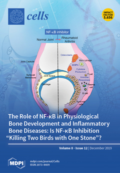
Rheumatoid arthritis (RA) is a chronic inflammatory disease that causes progressive joint destruction over time. RA is characterized by extensive synovitis, cartilage erosion, and bone destruction by excessive immune and inflammatory responses. Synovial cells and immune cells produce inflammatory cytokines, such as IL-1, IL-6, and TNFα, and matrix metalloproteases (MMPs). Since NF-ĸB is a transcription factor that regulates the expression of inflammatory cytokines, including TNF-α and IL-6, and serves as a mediator for RANK signaling, selective inhibition of the classical NF-ĸB pathway appears to be a target for bone destruction of RA. NF-ĸB inhibitors, such as decoy oligonucleotides, NEMO-biding domain (NBD) peptide, TAT-IĸBα-super repressor, or IKKβ inhibitor, suppress bone destruction by suppressing local inflammation and RANK signaling. View this paper.
  • Issues are regarded as officially published after their release is announced to the table of contents alert mailing list.
  • You may sign up for e-mail alerts to receive table of contents of newly released issues.
  • PDF is the official format for papers published in both, html and pdf forms. To view the papers in pdf format, click on the "PDF Full-text" link, and use the free Adobe Reader to open them.
Order results
Result details
Section
Select all
Export citation of selected articles as:
15 pages, 1425 KiB  
Article
The Phenotype and Secretory Activity of Adipose-Derived Mesenchymal Stem Cells (ASCs) of Patients with Rheumatic Diseases
by Ewa Kuca-Warnawin, Urszula Skalska, Iwona Janicka, Urszula Musiałowicz, Krzysztof Bonek, Piotr Głuszko, Piotr Szczęsny, Marzena Olesińska and Ewa Kontny
Cells 2019, 8(12), 1659; https://0-doi-org.brum.beds.ac.uk/10.3390/cells8121659 - 17 Dec 2019
Cited by 20 | Viewed by 3481
Abstract
Mesenchymal stem/stromal cells (MSCs) have immunosuppressive and regenerative properties. Adipose tissue is an alternative source of MSCs, named adipose-derived mesenchymal stem cells (ASCs). Because the biology of ASCs in rheumatic diseases (RD) is poorly understood, we performed a basic characterization of RD/ASCs. The [...] Read more.
Mesenchymal stem/stromal cells (MSCs) have immunosuppressive and regenerative properties. Adipose tissue is an alternative source of MSCs, named adipose-derived mesenchymal stem cells (ASCs). Because the biology of ASCs in rheumatic diseases (RD) is poorly understood, we performed a basic characterization of RD/ASCs. The phenotype and expression of adhesion molecules (intracellular adhesion molecule (ICAM)-1 and vascular cell adhesion molecule (VCAM)-1) on commercially available healthy donors (HD), ASC lines (n = 5) and on ASCs isolated from patients with systemic lupus erythematosus (SLE, n = 16), systemic sclerosis (SSc, n = 17) and ankylosing spondylitis (AS, n = 16) were analyzed by flow cytometry. The secretion of immunomodulatory factors by untreated and cytokine-treated ASCs was measured by ELISA. RD/ASCs have reduced basal levels of CD90 and ICAM-1 expression, correlated with interleukin (IL)-6 and transforming growth factor (TGF)-β1 release, respectively. Compared with HD/ASCs, untreated and tumour necrosis factor (TNF) + interferon (IFN)-γ (TI)-treated RD/ASCs produced similar amounts of prostaglandin E2 (PGE2), IL-6, leukemia inhibiting factor (LIF), and TGF-β1, more IL-1Ra, soluble human leukocyte antigen G (sHLA-G) and tumor necrosis factor-inducible gene (TSG)-6, but less kynurenines and galectin-3. Basal secretion of galectin-3 was inversely correlated with the patient’s erythrocyte sedimentation rate (ESR) value. IFN-α and IL-23 slightly raised galectin-3 release from SLE/ASCs and AS/ASCs, respectively. TGF-β1 up-regulated PGE2 secretion by SSc/ASCs. In conclusion, RD/ASCs are characterized by low basal levels of CD90 and ICAM-1 expression, upregulated secretion of IL-1Ra, TSG-6 and sHLA-G, but impaired release of kynurenines and galectin-3. These abnormalities may modify biological activities of RD/ASCs. Full article
(This article belongs to the Special Issue Adipose-Derived Stromal/Stem Cells)
Show Figures

Figure 1

22 pages, 4203 KiB  
Article
CD47-SIRPα Signaling Induces Epithelial-Mesenchymal Transition and Cancer Stemness and Links to a Poor Prognosis in Patients with Oral Squamous Cell Carcinoma
by Shin Pai, Oluwaseun Adebayo Bamodu, Yen-Kuang Lin, Chun-Shu Lin, Pei-Yi Chu, Ming-Hsien Chien, Liang-Shun Wang, Michael Hsiao, Chi-Tai Yeh and Jo-Ting Tsai
Cells 2019, 8(12), 1658; https://0-doi-org.brum.beds.ac.uk/10.3390/cells8121658 - 17 Dec 2019
Cited by 46 | Viewed by 5651
Abstract
Background: Oral squamous cell carcinoma (OSCC), with high mortality rates, is one of the most diagnosed head and neck cancers. Epithelial-to-mesenchymal transition (EMT) and the generation of cancer stem cells (CSCs) are two keys for therapy-resistance, relapse, and distant metastasis. Accumulating evidence indicates [...] Read more.
Background: Oral squamous cell carcinoma (OSCC), with high mortality rates, is one of the most diagnosed head and neck cancers. Epithelial-to-mesenchymal transition (EMT) and the generation of cancer stem cells (CSCs) are two keys for therapy-resistance, relapse, and distant metastasis. Accumulating evidence indicates that aberrantly expressed cluster of differentiation (CD)47 is associated with cell-death evasion and metastasis; however, the role of CD47 in the generation of CSCs in OSCC is not clear. Methods: We investigated the functional roles of CD47 in OSCC cell lines SAS, TW2.6, HSC-3, and FaDu using the bioinformatics approach, immunoblotting, immunofluorescence staining, and assays for cellular migration, invasion, colony, and orosphere formation, as well as radiosensitivity. Results: We demonstrated increased expression of CD47 in OSCC patients was associated with an estimated poorly survival disadvantage (p = 0.0391) and positively correlated with the expression of pluripotency factors. Silencing CD47 significantly suppressed cell viability and orosphere formation, accompanied by a downregulated expression of CD133, SRY-Box transcription factor 2 (SOX2), octamer-binding transcription factor 4 (OCT4), and c-Myc. In addition, CD47-silenced OSCC cells showed reduced EMT, migration, and clonogenicity reflected by increased E-cadherin and decreased vimentin, Slug, Snail, and N-cadherin expression. Conclusion: Of therapeutic relevance, CD47 knockdown enhanced the anti-OSCC effect of radiotherapy. Collectively, we showed an increased CD47 expression promoted the generation of CSCs and malignant OSCC phenotypes. Silencing CD47, in combination with radiation, could provide an alternative and improved therapeutic efficacy for OSCC patients. Full article
Show Figures

Graphical abstract

16 pages, 1287 KiB  
Review
Salinity Stress Responses and Adaptation Mechanisms in Eukaryotic Green Microalgae
by Prateek Shetty, Margaret Mukami Gitau and Gergely Maróti
Cells 2019, 8(12), 1657; https://0-doi-org.brum.beds.ac.uk/10.3390/cells8121657 - 17 Dec 2019
Cited by 156 | Viewed by 10517
Abstract
High salinity is a challenging environmental stress for organisms to overcome. Unicellular photosynthetic microalgae are especially vulnerable as they have to grapple not only with ionic imbalance and osmotic stress but also with the generated reactive oxygen species (ROS) interfering with photosynthesis. This [...] Read more.
High salinity is a challenging environmental stress for organisms to overcome. Unicellular photosynthetic microalgae are especially vulnerable as they have to grapple not only with ionic imbalance and osmotic stress but also with the generated reactive oxygen species (ROS) interfering with photosynthesis. This review attempts to compare and contrast mechanisms that algae, particularly the eukaryotic Chlamydomonas microalgae, exhibit in order to immediately respond to harsh conditions caused by high salinity. The review also collates adaptation mechanisms of freshwater algae strains under persistent high salt conditions. Understanding both short-term and long-term algal responses to high salinity is integral to further fundamental research in algal biology and biotechnology. Full article
(This article belongs to the Special Issue Chlamydomonas Cell Biology)
Show Figures

Figure 1

30 pages, 1943 KiB  
Review
Epigenetic Control of Autophagy in Cancer Cells: A Key Process for Cancer-Related Phenotypes
by Paul Peixoto, Céline Grandvallet, Jean-Paul Feugeas, Michaël Guittaut and Eric Hervouet
Cells 2019, 8(12), 1656; https://0-doi-org.brum.beds.ac.uk/10.3390/cells8121656 - 17 Dec 2019
Cited by 34 | Viewed by 4120
Abstract
Although autophagy is a well-known and extensively described cell pathway, numerous studies have been recently interested in studying the importance of its regulation at different molecular levels, including the translational and post-translational levels. Therefore, this review focuses on the links between autophagy and [...] Read more.
Although autophagy is a well-known and extensively described cell pathway, numerous studies have been recently interested in studying the importance of its regulation at different molecular levels, including the translational and post-translational levels. Therefore, this review focuses on the links between autophagy and epigenetics in cancer and summarizes the. following: (i) how ATG genes are regulated by epigenetics, including DNA methylation and post-translational histone modifications; (ii) how epidrugs are able to modulate autophagy in cancer and to alter cancer-related phenotypes (proliferation, migration, invasion, tumorigenesis, etc.) and; (iii) how epigenetic enzymes can also regulate autophagy at the protein level. One noteable observation was that researchers most often reported conclusions about the regulation of the autophagy flux, following the use of epidrugs, based only on the analysis of LC3B-II form in treated cells. However, it is now widely accepted that an increase in LC3B-II form could be the consequence of an induction of the autophagy flux, as well as a block in the autophagosome-lysosome fusion. Therefore, in our review, all the published results describing a link between epidrugs and autophagy were systematically reanalyzed to determine whether autophagy flux was indeed increased, or inhibited, following the use of these potentially new interesting treatments targeting the autophagy process. Altogether, these recent data strongly support the idea that the determination of autophagy status could be crucial for future anticancer therapies. Indeed, the use of a combination of epidrugs and autophagy inhibitors could be beneficial for some cancer patients, whereas, in other cases, an increase of autophagy, which is frequently observed following the use of epidrugs, could lead to increased autophagy cell death. Full article
Show Figures

Figure 1

11 pages, 804 KiB  
Perspective
Syntaxin 16’s Newly Deciphered Roles in Autophagy
by Bor Luen Tang
Cells 2019, 8(12), 1655; https://0-doi-org.brum.beds.ac.uk/10.3390/cells8121655 - 17 Dec 2019
Cited by 12 | Viewed by 3666
Abstract
Syntaxin 16, a Qa-SNARE (soluble N-ethylmaleimide-sensitive factor activating protein receptor), is involved in a number of membrane-trafficking activities, particularly transport processes at the trans-Golgi network (TGN). Recent works have now implicated syntaxin 16 in the autophagy process. In fact, syntaxin 16 appears [...] Read more.
Syntaxin 16, a Qa-SNARE (soluble N-ethylmaleimide-sensitive factor activating protein receptor), is involved in a number of membrane-trafficking activities, particularly transport processes at the trans-Golgi network (TGN). Recent works have now implicated syntaxin 16 in the autophagy process. In fact, syntaxin 16 appears to have dual roles, firstly in facilitating the transport of ATG9a-containing vesicles to growing autophagosomes, and secondly in autolysosome formation. The former involves a putative SNARE complex between syntaxin 16, VAMP7 and SNAP-47. The latter occurs via syntaxin 16’s recruitment by Atg8/LC3/GABARAP family proteins to autophagosomes and endo-lysosomes, where syntaxin 16 may act in a manner that bears functional redundancy with the canonical autophagosome Qa-SNARE syntaxin 17. Here, I discuss these recent findings and speculate on the mechanistic aspects of syntaxin 16’s newly found role in autophagy. Full article
Show Figures

Figure 1

21 pages, 4529 KiB  
Article
Early Stroke Induces Long-Term Impairment of Adult Neurogenesis Accompanied by Hippocampal-Mediated Cognitive Decline
by Carolin Kathner-Schaffert, Lina Karapetow, Madlen Günther, Max Rudolph, Mahmoud Dahab, Eileen Baum, Thomas Lehmann, Otto W. Witte, Christoph Redecker, Christian W. Schmeer and Silke Keiner
Cells 2019, 8(12), 1654; https://0-doi-org.brum.beds.ac.uk/10.3390/cells8121654 - 17 Dec 2019
Cited by 23 | Viewed by 3927
Abstract
Stroke increases neurogenesis in the adult dentate gyrus in the short term, however, long-term effects at the cellular and functional level are poorly understood. Here we evaluated the impact of an early stroke lesion on neurogenesis and cognitive function of the aging brain. [...] Read more.
Stroke increases neurogenesis in the adult dentate gyrus in the short term, however, long-term effects at the cellular and functional level are poorly understood. Here we evaluated the impact of an early stroke lesion on neurogenesis and cognitive function of the aging brain. We hypothesized that a stroke disturbs dentate neurogenesis during aging correlate with impaired flexible learning. To address this issue a stroke was induced in 3-month-old C57Bl/6 mice by a middle cerebral artery occlusion (MCAO). To verify long-term changes of adult neurogenesis the thymidine analogue BrdU (5-Bromo-2′-deoxyuridine) was administrated at different time points during aging. One and half months after BrdU injections learning and memory performance were assessed with a modified version of the Morris water maze (MWM) that includes the re-learning paradigm, as well as hippocampus-dependent and -independent search strategies. After MWM performance mice were transcardially perfused. To further evaluate in detail the stroke-mediated changes on stem- and progenitor cells as well as endogenous proliferation nestin-green-fluorescent protein (GFP) mice were used. Adult nestin-GFP mice received a retroviral vector injection in the hippocampus to evaluate changes in the neuronal morphology. At an age of 20 month the nestin-GFP mice were transcardially perfused after MWM performance and BrdU application 1.5 months later. The early stroke lesion significantly decreased neurogenesis in 7.5- and 9-month-old animals and also endogenous proliferation in the latter group. Furthermore, immature doublecortin (DCX)-positive neurons were reduced in 20-month-old nestin-GFP mice after lesion. All MCAO groups showed an impaired performance in the MWM and mostly relied on hippocampal-independent search strategies. These findings indicate that an early ischemic insult leads to a dramatical decline of neurogenesis during aging that correlates with a premature development of hippocampal-dependent deficits. Our study supports the notion that an early stroke might lead to long-term cognitive deficits as observed in human patients after lesion. Full article
(This article belongs to the Special Issue Aging and Regeneration)
Show Figures

Figure 1

20 pages, 876 KiB  
Review
Cell-Free microRNAs as Potential Oral Cancer Biomarkers: From Diagnosis to Therapy
by Óscar Rapado-González, Rafael López-López, José Luis López-Cedrún, Gabriel Triana-Martínez, Laura Muinelo-Romay and María Mercedes Suárez-Cunqueiro
Cells 2019, 8(12), 1653; https://0-doi-org.brum.beds.ac.uk/10.3390/cells8121653 - 17 Dec 2019
Cited by 29 | Viewed by 5122
Abstract
Oral cavity cancer is the most frequent malignancy of the head and neck. Unfortunately, despite educational interventions for prevention and early diagnosis, oral cancer patients are often diagnosed in advanced stages associated with poor prognosis and life expectancy. Therefore, there is an urgent [...] Read more.
Oral cavity cancer is the most frequent malignancy of the head and neck. Unfortunately, despite educational interventions for prevention and early diagnosis, oral cancer patients are often diagnosed in advanced stages associated with poor prognosis and life expectancy. Therefore, there is an urgent need to find noninvasive biomarkers to improve early detection of this tumor. Liquid biopsy has emerged as a valuable tool in medical oncology which provides new horizons for improving clinical decision making. Notably, cell-free microRNAs (miRNAs), a class of short non-coding RNAs, are emerging as novel noninvasive cancer biomarkers. Here, we provide an overview of the potential clinical application of cell-free miRNAs as diagnostic, prognostic, and therapeutic biomarkers in oral cancer. Full article
(This article belongs to the Special Issue Cancer Related microRNAs)
Show Figures

Figure 1

13 pages, 1386 KiB  
Article
Cigarette Smoke Condensate Exposure Changes RNA Content of Extracellular Vesicles Released from Small Airway Epithelial Cells
by Tiziana Corsello, Andrzej S. Kudlicki, Roberto P. Garofalo and Antonella Casola
Cells 2019, 8(12), 1652; https://0-doi-org.brum.beds.ac.uk/10.3390/cells8121652 - 17 Dec 2019
Cited by 24 | Viewed by 3917
Abstract
Exposure to environmental tobacco smoke (ETS) is a known risk factor for the development of chronic lung diseases, cancer, and the exacerbation of viral infections. Extracellular vesicles (EVs) have been identified as novel mediators of cell–cell communication through the release of biological content. [...] Read more.
Exposure to environmental tobacco smoke (ETS) is a known risk factor for the development of chronic lung diseases, cancer, and the exacerbation of viral infections. Extracellular vesicles (EVs) have been identified as novel mediators of cell–cell communication through the release of biological content. Few studies have investigated the composition/function of EVs derived from human airway epithelial cells (AECs) exposed to cigarette smoke condensate (CSC), as surrogates for ETS. Using novel high-throughput technologies, we identified a diverse range of small noncoding RNAs (sncRNAs), including microRNA (miRNAs), Piwi-interacting RNA (piRNAs), and transfer RNA (tRNAs) in EVs from control and CSC-treated SAE cells. CSC treatment resulted in significant changes in the EV content of miRNAs. A total of 289 miRNAs were identified, with five being significantly upregulated and three downregulated in CSC EVs. A total of 62 piRNAs were also detected in our EV preparations, with five significantly downregulated and two upregulated in CSC EVs. We used TargetScan and Gene Ontology (GO) analysis to predict the biological targets of hsa-miR-3913-5p, the most represented miRNA in CSC EVs. Understanding fingerprint molecules in EVs will increase our knowledge of the relationship between ETS exposure and lung disease, and might identify potential molecular targets for future treatments. Full article
(This article belongs to the Section Intracellular and Plasma Membranes)
Show Figures

Graphical abstract

13 pages, 1783 KiB  
Review
Heart Failure with Reduced Ejection Fraction (HFrEF) and Preserved Ejection Fraction (HFpEF): The Diagnostic Value of Circulating MicroRNAs
by Yei-Tsung Chen, Lee Lee Wong, Oi Wah Liew and Arthur Mark Richards
Cells 2019, 8(12), 1651; https://0-doi-org.brum.beds.ac.uk/10.3390/cells8121651 - 16 Dec 2019
Cited by 38 | Viewed by 5710
Abstract
Circulating microRNAs offer attractive potential as epigenetic disease biomarkers by virtue of their biological stability and ready accessibility in liquid biopsies. Numerous clinical cohort studies have revealed unique microRNA profiles in different disease settings, suggesting utility as markers with diagnostic and prognostic applications. [...] Read more.
Circulating microRNAs offer attractive potential as epigenetic disease biomarkers by virtue of their biological stability and ready accessibility in liquid biopsies. Numerous clinical cohort studies have revealed unique microRNA profiles in different disease settings, suggesting utility as markers with diagnostic and prognostic applications. Given the complex network of microRNA functions in modulating gene expression and post-transcriptional modifications, the circulating microRNA landscape in disease may reflect pathophysiological status, providing valuable information for delineating distinct subtypes and/or stages of complex diseases. Heart failure (HF) is an increasingly significant global health challenge, imposing major economic liability and health care burden due to high hospitalization, morbidity, and mortality rates. Although HF is defined as a syndrome characterized by symptoms and findings on physical examination, it may be further differentiated based on left ventricular ejection fraction (LVEF) and categorized as HF with reduced ejection fraction (HFrEF) and HF with preserved ejection fraction (HFpEF). The presenting clinical syndromes in HFpEF and HFrEF are similar but mortality differs, being somewhat lower in HFpEF than in HFrEF. However, while HFrEF is responsive to an array of therapies, none has been shown to improve survival in HFpEF. Herein, we review recent HF cohort studies focusing on the distinct microRNA profiles associated with HF subtypes to reveal new insights to underlying mechanisms and explore the possibility of exploiting these differences for diagnostic/prognostic applications. Full article
(This article belongs to the Collection Regulatory Functions of microRNAs)
Show Figures

Figure 1

20 pages, 5997 KiB  
Article
Self-Assembling Scaffolds Supported Long-Term Growth of Human Primed Embryonic Stem Cells and Upregulated Core and Naïve Pluripotent Markers
by Christina McKee, Christina Brown and G. Rasul Chaudhry
Cells 2019, 8(12), 1650; https://0-doi-org.brum.beds.ac.uk/10.3390/cells8121650 - 16 Dec 2019
Cited by 8 | Viewed by 4993
Abstract
The maintenance and expansion of human embryonic stem cells (ESCs) in two-dimensional (2-D) culture is technically challenging, requiring routine manipulation and passaging. We developed three-dimensional (3-D) scaffolds to mimic the in vivo microenvironment for stem cell proliferation. The scaffolds were made of two [...] Read more.
The maintenance and expansion of human embryonic stem cells (ESCs) in two-dimensional (2-D) culture is technically challenging, requiring routine manipulation and passaging. We developed three-dimensional (3-D) scaffolds to mimic the in vivo microenvironment for stem cell proliferation. The scaffolds were made of two 8-arm polyethylene glycol (PEG) polymers functionalized with thiol (PEG-8-SH) and acrylate (PEG-8-Acr) end groups, which self-assembled via a Michael addition reaction. When primed ESCs (H9 cells) were mixed with PEG polymers, they were encapsulated and grew for an extended period, while maintaining their viability, self-renewal, and differentiation potential both in vitro and in vivo. Three-dimensional (3-D) self-assembling scaffold-grown cells displayed an upregulation of core pluripotency genes, OCT4, NANOG, and SOX2. In addition, the expression of primed markers decreased, while the expression of naïve markers substantially increased. Interestingly, the expression of mechanosensitive genes, YAP and TAZ, was also upregulated. YAP inhibition by Verteporfin abrogated the increased expression of YAP/TAZ as well as core and naïve pluripotent markers. Evidently, the 3-D culture conditions induced the upregulation of makers associated with a naïve state of pluripotency in the primed cells. Overall, our 3-D culture system supported the expansion of a homogenous population of ESCs and should be helpful in advancing their use for cell therapy and regenerative medicine. Full article
(This article belongs to the Special Issue 3D Stem Cell Culture)
Show Figures

Figure 1

24 pages, 4496 KiB  
Article
Phosphatidylinositol Monophosphates Regulate Optimal Vav1 Signaling Output
by Sonia Rodríguez-Fdez, Carmen Citterio, L. Francisco Lorenzo-Martín, Jesús Baltanás-Copado, Clara Llorente-González, Senena Corbalán-García, Miguel Vicente-Manzanares and Xosé R. Bustelo
Cells 2019, 8(12), 1649; https://0-doi-org.brum.beds.ac.uk/10.3390/cells8121649 - 16 Dec 2019
Cited by 7 | Viewed by 4287
Abstract
Phosphatidylinositol–5 phosphate (PI5P) and other mono-phosphoinositides (mono-PIs) play second messenger roles in both physiological and pathological conditions. Despite this, their intracellular targets and mechanisms of action remain poorly characterized. Here, we show that Vav1, a protein that exhibits both Rac1 GDP/GTP exchange and [...] Read more.
Phosphatidylinositol–5 phosphate (PI5P) and other mono-phosphoinositides (mono-PIs) play second messenger roles in both physiological and pathological conditions. Despite this, their intracellular targets and mechanisms of action remain poorly characterized. Here, we show that Vav1, a protein that exhibits both Rac1 GDP/GTP exchange and adaptor activities, is positively modulated by PI5P and, possibly, other mono-PIs. Unlike other phospholipid–protein complexes, the affinity and specificity of the Vav1–lipid interaction entail a new structural solution that involves the synergistic action of the Vav1 C1 domain and an adjacent polybasic tail. This new regulatory layer, which is not conserved in the Vav family paralogs, favors the engagement of optimal Vav1 signaling outputs in lymphocytes. Full article
(This article belongs to the Special Issue GTPase Pathways in Health and Diseases)
Show Figures

Graphical abstract

12 pages, 507 KiB  
Review
Small Particles, Big Effects: The Interplay Between Exosomes and Dendritic Cells in Antitumor Immunity and Immunotherapy
by Bruno Deltreggia Benites, Marisa Claudia Alvarez and Sara Teresinha Olalla Saad
Cells 2019, 8(12), 1648; https://0-doi-org.brum.beds.ac.uk/10.3390/cells8121648 - 16 Dec 2019
Cited by 18 | Viewed by 3691
Abstract
Dendritic cells play a fundamental role in the antitumor immunity cycle, and the loss of their antigen-presenting function is a recognized mechanism of tumor evasion. We have recently demonstrated the effect of exosomes extracted from serum of patients with acute myeloid leukemia as [...] Read more.
Dendritic cells play a fundamental role in the antitumor immunity cycle, and the loss of their antigen-presenting function is a recognized mechanism of tumor evasion. We have recently demonstrated the effect of exosomes extracted from serum of patients with acute myeloid leukemia as important inducers of dendritic cell immunotolerance, and several other works have recently demonstrated the effects of these nanoparticles on immunity to other tumor types as well. The aim of this review was to highlight the recent findings on the effects of tumor exosomes on dendritic cell functions, the mechanisms by which they can lead to tumor evasion, and their manipulation as a possible strategy in cancer treatment. Full article
Show Figures

Figure 1

16 pages, 480 KiB  
Review
Myeloid-Derived Suppressor Cells: Major Figures that Shape the Immunosuppressive and Angiogenic Network in Cancer
by Eleni-Kyriaki Vetsika, Aristeidis Koukos and Athanasios Kotsakis
Cells 2019, 8(12), 1647; https://0-doi-org.brum.beds.ac.uk/10.3390/cells8121647 - 15 Dec 2019
Cited by 79 | Viewed by 5343
Abstract
Myeloid-derived suppressor cells (MDSCs) constitute a vast population of immature myeloid cells implicated in various conditions. Most notably, their role in cancer is of great complexity. They exert immunosuppressive functions like hampering cancer immunity mediated by T lymphocytes and natural killer cells, while [...] Read more.
Myeloid-derived suppressor cells (MDSCs) constitute a vast population of immature myeloid cells implicated in various conditions. Most notably, their role in cancer is of great complexity. They exert immunosuppressive functions like hampering cancer immunity mediated by T lymphocytes and natural killer cells, while simultaneously they can recruit T regulatory cells to further promote immunosuppression, thus shielding tumor cells against the immune defenses. In addition, they were shown to support tumor invasion and metastasis by inducing vascularization. Yet again, in order to exert their angiogenic activities, they do have at their disposal a variety of occasionally overlapping mechanisms, mainly driven by VEGF/JAK/STAT signaling. In this concept, they have risen to be a rather attractive target for therapies, including depletion or maturation, so as to overcome cancer immunity and suppress angiogenic activity. Even though, many studies have been conducted to better understand these cells, there is much to be done yet. This article hopes to shed some light on the paradoxal complexity of these cells, while elucidating some of the key features of MDSCs in relation to immunosuppression and, most importantly, to the vascularization processes, along with current therapeutic options in cancer, in relation to MDSC depletion. Full article
Show Figures

Figure 1

16 pages, 4410 KiB  
Article
Copresentation of BMP-6 and RGD Ligands Enhances Cell Adhesion and BMP-Mediated Signaling
by Francesca Posa, Anna Luise Grab, Volker Martin, Dirk Hose, Anja Seckinger, Giorgio Mori, Slobodan Vukicevic and Elisabetta Ada Cavalcanti-Adam
Cells 2019, 8(12), 1646; https://0-doi-org.brum.beds.ac.uk/10.3390/cells8121646 - 15 Dec 2019
Cited by 11 | Viewed by 3655
Abstract
We report on the covalent immobilization of bone morphogenetic protein 6 (BMP-6) and its co-presentation with integrin ligands on a nanopatterned platform to study cell adhesion and signaling responses which regulate the transdifferentiation of myoblasts into osteogenic cells. To immobilize BMP-6, the heterobifunctional [...] Read more.
We report on the covalent immobilization of bone morphogenetic protein 6 (BMP-6) and its co-presentation with integrin ligands on a nanopatterned platform to study cell adhesion and signaling responses which regulate the transdifferentiation of myoblasts into osteogenic cells. To immobilize BMP-6, the heterobifunctional linker MU-NHS is coupled to amine residues of the growth factor; this prevents its internalization while ensuring that its biological activity is maintained. Additionally, to allow cells to adhere to such platform and study signaling events arising from the contact to the surface, we used click-chemistry to immobilize cyclic-RGD carrying an azido group reacting with PEG-alkyne spacers via copper-catalyzed 1,3-dipolar cycloaddition. We show that the copresentation of BMP-6 and RGD favors focal adhesion formation and promotes Smad 1/5/8 phosphorylation. When presented in low amounts, BMP-6 added to culture media of cells adhering to the RGD ligands is less effective than BMP-6 immobilized on the surfaces in inducing Smad complex activation and in inhibiting myotube formation. Our results suggest that a local control of ligand density and cell signaling is crucial for modulating cell response. Full article
(This article belongs to the Special Issue Mechanotransduction in Control of Cell Fate and Function)
Show Figures

Figure 1

12 pages, 3001 KiB  
Article
Temporal Dynamics of VEGFA-Induced VEGFR2/FAK Co-Localization Depend on SHB
by Ilkka Pietilä, Djenolan Van Mourik, Andreas Tamelander, Vitezslav Kriz, Lena Claesson-Welsh, Anders Tengholm and Michael Welsh
Cells 2019, 8(12), 1645; https://0-doi-org.brum.beds.ac.uk/10.3390/cells8121645 - 15 Dec 2019
Cited by 12 | Viewed by 3570
Abstract
Focal adhesion kinase (FAK) is essential for vascular endothelial growth factor-A (VEGFA)/VEGF receptor-2 (VEGFR2)-stimulated angiogenesis and vascular permeability. We have previously noted that presence of the Src homology-2 domain adapter protein B (SHB) is of relevance for VEGFA-stimulated angiogenesis in a FAK-dependent manner. [...] Read more.
Focal adhesion kinase (FAK) is essential for vascular endothelial growth factor-A (VEGFA)/VEGF receptor-2 (VEGFR2)-stimulated angiogenesis and vascular permeability. We have previously noted that presence of the Src homology-2 domain adapter protein B (SHB) is of relevance for VEGFA-stimulated angiogenesis in a FAK-dependent manner. The current study was conducted in order address the temporal dynamics of co-localization between these components in HEK293 and primary lung endothelial cells (EC) by total internal reflection fluorescence microscopy (TIRF). An early (<2.5 min) VEGFA-induced increase in VEGFR2 co-localization with SHB was dependent on tyrosine 1175 in VEGFR2. VEGFA also enhanced SHB co-localization with FAK. FAK co-localization with VEGFR2 was dependent on SHB since it was significantly lower in SHB deficient EC after VEGFA addition. Absence of SHB also resulted in a gradual decline of VEGFR2 co-localization with FAK under basal (prior to VEGFA addition) conditions. A similar basal response was observed with expression of the Y1175F-VEGFR2 mutant in wild type EC. The distribution of focal adhesions in SHB-deficient EC was altered with a primarily perinuclear location. These live cell data implicate SHB as a key component regulating FAK activity in response to VEGFA/VEGFR2. Full article
(This article belongs to the Special Issue Vascular Signalling)
Show Figures

Figure 1

17 pages, 3104 KiB  
Article
Genetically Engineered Lung Cancer Cells for Analyzing Epithelial–Mesenchymal Transition
by Michał Kiełbus, Jakub Czapiński, Joanna Kałafut, Justyna Woś, Andrzej Stepulak and Adolfo Rivero-Müller
Cells 2019, 8(12), 1644; https://0-doi-org.brum.beds.ac.uk/10.3390/cells8121644 - 15 Dec 2019
Cited by 6 | Viewed by 3584
Abstract
Cell plasticity, defined as the ability to undergo phenotypical transformation in a reversible manner, is a physiological process that also exerts important roles in disease progression. Two forms of cellular plasticity are epithelial–mesenchymal transition (EMT) and its inverse process, mesenchymal–epithelial transition (MET). These [...] Read more.
Cell plasticity, defined as the ability to undergo phenotypical transformation in a reversible manner, is a physiological process that also exerts important roles in disease progression. Two forms of cellular plasticity are epithelial–mesenchymal transition (EMT) and its inverse process, mesenchymal–epithelial transition (MET). These processes have been correlated to the poor outcome of different types of neoplasias as well as drug resistance development. Since EMT/MET are transitional processes, we generated and validated a reporter cell line. Specifically, a far-red fluorescent protein was knocked-in in-frame with the mesenchymal gene marker VIMENTIN (VIM) in H2170 lung cancer cells. The vimentin reporter cells (VRCs) are a reliable model for studying EMT and MET showing cellular plasticity upon a series of stimulations. These cells are a robust platform to dissect the molecular mechanisms of these processes, and for drug discovery in vitro and in vivo in the future. Full article
(This article belongs to the Section Cellular Pathology)
Show Figures

Graphical abstract

16 pages, 5730 KiB  
Article
STK3/4 Expression Is Regulated in Uterine Endometrial Cells during the Estrous Cycle
by Sohyeon Moon, Ok-Hee Lee, Sujin Lee, Jihyun Lee, Haeun Park, Miseon Park, Eun Mi Chang, Keun-Hong Park and Youngsok Choi
Cells 2019, 8(12), 1643; https://0-doi-org.brum.beds.ac.uk/10.3390/cells8121643 - 15 Dec 2019
Cited by 13 | Viewed by 3503
Abstract
The uterus is dynamically regulated in response to various signaling triggered by hormones during the estrous cycle. The Hippo signaling pathway is known as an important signaling for regulating cellular processes during development by balancing between cell growth and apoptosis. Serine/threonine protein kinase [...] Read more.
The uterus is dynamically regulated in response to various signaling triggered by hormones during the estrous cycle. The Hippo signaling pathway is known as an important signaling for regulating cellular processes during development by balancing between cell growth and apoptosis. Serine/threonine protein kinase 3/4 (STK3/4) is a key component of the Hippo signaling network. However, the regulation of STK3/4-Hippo signaling in the uterus is little known. In this study, we investigated the regulation and expression of STK3/4 in the uterine endometrium during the estrous cycle. STK3/4 expression was dynamically regulated in the uterus during the estrous cycle. STK3/4 protein expression was gradually increased from the diestrus stage and reached the highest in the estrus stage. STK3/4 was exclusively localized in the luminal and glandular epithelial cells of the uterus, and phosphorylated STK3/4 was also increased at the estrus stage. Moreover, the increase of STK3/4 expression in uteri was induced by administration of estradiol, but not by progesterone injection in ovariectomized mice. Pretreatment with an estrogen receptor antagonist ICI 182,780 reduced estrogen-induced STK3/4 expression and its phosphorylation. The estrogen-induced STK3/4 expression was related to the increase in phosphorylation of downstream targets including LATS1/2 and YAP. These findings suggest that STK3/4-Hippo signaling acts a novel signaling pathway in the uterine epithelium and STK3/4-Hippo is one of key molecules for connecting between the estrogen downstream signaling pathway and the Hippo signaling pathway leading to regulate dynamic uterine epithelium during the estrous cycle. Full article
Show Figures

Figure 1

19 pages, 5538 KiB  
Review
The Microbiome–Estrogen Connection and Breast Cancer Risk
by Sheetal Parida and Dipali Sharma
Cells 2019, 8(12), 1642; https://0-doi-org.brum.beds.ac.uk/10.3390/cells8121642 - 15 Dec 2019
Cited by 109 | Viewed by 22325
Abstract
The microbiome is undoubtedly the second genome of the human body and has diverse roles in health and disease. However, translational progress is limited due to the vastness of the microbiome, which accounts for over 3.3 million genes, whose functions are still unclear. [...] Read more.
The microbiome is undoubtedly the second genome of the human body and has diverse roles in health and disease. However, translational progress is limited due to the vastness of the microbiome, which accounts for over 3.3 million genes, whose functions are still unclear. Numerous studies in the past decade have demonstrated how microbiome impacts various organ-specific cancers by altering the energy balance of the body, increasing adiposity, synthesizing genotoxins and small signaling molecules, and priming and regulating immune response and metabolism of indigestible dietary components, xenobiotics, and pharmaceuticals. In relation to breast cancer, one of the most prominent roles of the human microbiome is the regulation of steroid hormone metabolism since endogenous estrogens are the most important risk factor in breast cancer development especially in postmenopausal women. Intestinal microbes encode enzymes capable of deconjugating conjugated estrogen metabolites marked for excretion, pushing them back into the enterohepatic circulation in a biologically active form. In addition, the intestinal microbes also break down otherwise indigestible dietary polyphenols to synthesize estrogen-like compounds or estrogen mimics that exhibit varied estrogenic potency. The present account discusses the potential role of gastrointestinal microbiome in breast cancer development by mediating metabolism of steroid hormones and synthesis of biologically active estrogen mimics. Full article
Show Figures

Figure 1

12 pages, 2584 KiB  
Communication
Critical Impact of Human Amniotic Membrane Tension on Mitochondrial Function and Cell Viability In Vitro
by Laura Poženel, Andrea Lindenmair, Katy Schmidt, Andrey V. Kozlov, Johannes Grillari, Susanne Wolbank, Asmita Banerjee and Adelheid Weidinger
Cells 2019, 8(12), 1641; https://0-doi-org.brum.beds.ac.uk/10.3390/cells8121641 - 15 Dec 2019
Cited by 7 | Viewed by 2947
Abstract
Amniotic cells show exciting stem cell features, which has led to the idea of using living cells of human amniotic membranes (hAMs) in toto for clinical applications. However, under common cell culture conditions, viability of amniotic cells decreases rapidly, whereby reasons for this [...] Read more.
Amniotic cells show exciting stem cell features, which has led to the idea of using living cells of human amniotic membranes (hAMs) in toto for clinical applications. However, under common cell culture conditions, viability of amniotic cells decreases rapidly, whereby reasons for this decrease are unknown so far. Recently, it has been suggested that loss of tissue tension in vivo leads to apoptosis. Therefore, the aim of this study was to investigate the effect of tissue distention on the viability of amniotic cells in vitro. Thereby, particular focus was put on vital mitochondria-linked parameters, such as respiration and ATP synthesis. Biopsies of hAMs were incubated for 7–21 days either non-distended or distended. We observed increased B-cell lymphoma 2-associated X protein (BAX)/B-cell lymphoma (BCL)-2 ratios in non-distended hAMs at day seven, followed by increased caspase 3 expression at day 14, and, consequently, loss of viability at day 21. In contrast, under distention, caspase 3 expression increased only slightly, and mitochondrial function and cellular viability were largely maintained. Our data suggest that a mechano-sensing pathway may control viability of hAM cells by triggering mitochondria-mediated apoptosis upon loss of tension in vitro. Further studies are required to elucidate the underlying molecular mechanisms between tissue distention and viability of hAM cells. Full article
(This article belongs to the Section Mitochondria)
Show Figures

Figure 1

21 pages, 5535 KiB  
Article
The Effect of Fibroblast Growth Factor 15 Signaling in Non-Steatotic and Steatotic Liver Transplantation from Cardiocirculatory Death
by Cindy G. Avalos-de León, Mónica B. Jiménez-Castro, María Eugenia Cornide-Petronio, José Gulfo, Floriana Rotondo, Jordi Gracia-Sancho, Araní Casillas-Ramírez and Carmen Peralta
Cells 2019, 8(12), 1640; https://0-doi-org.brum.beds.ac.uk/10.3390/cells8121640 - 14 Dec 2019
Cited by 5 | Viewed by 3722
Abstract
We elucidate the relevance of fibroblast growth factor 15 (FGF15) in liver transplantation (LT) using rats with both steatotic and non-steatotic organs from donors after cardiocirculatory death (DCD). Compared to LT from non-DCDs, the induction of cardiocirculatory death (CD) increases hepatic damage, proliferation, [...] Read more.
We elucidate the relevance of fibroblast growth factor 15 (FGF15) in liver transplantation (LT) using rats with both steatotic and non-steatotic organs from donors after cardiocirculatory death (DCD). Compared to LT from non-DCDs, the induction of cardiocirculatory death (CD) increases hepatic damage, proliferation, and intestinal and circulatory FGF15. This is associated with high levels of FGF15, bilirubin and bile acids (BAs), and overexpression of the enzyme involved in the alternative BA synthesis pathway, CYP27A1, in non-steatotic livers. Furthermore, CD activates the proliferative pathway, Hippo/YAP, in these types of liver. Blocking FGF15 action in LT from DCDs does not affect CYP27A1 but causes an overexpression of CYP7A, an enzyme from the classic BA synthesis pathway, and this is related to further accumulation of BAs and exacerbated damage. FGF15 inhibition also impairs proliferation without changing Hippo/YAP. In spite of worse damage, steatosis prevents a proliferative response in livers from DCDs. In steatotic grafts, CD does not modify CYP7A1, CYP27A1, BA, or the Hippo/YAP pathway, and FGF15 is not involved in damage or proliferation. Thus, endogenous FGF15 protects against BA accumulation and damage and promotes regeneration independently of the Hippo/YAP pathway, in non-steatotic LT from DCDs. Herein we show a minor role of FGF15 in steatotic LT from DCDs. Full article
Show Figures

Figure 1

17 pages, 5229 KiB  
Article
Knock Down of Plakophillin 2 Dysregulates Adhesion Pathway through Upregulation of miR200b and Alters the Mechanical Properties in Cardiac Cells
by Luca Puzzi, Daniele Borin, Priyatansh Gurha, Raffaella Lombardi, Valentina Martinelli, Marek Weiss, Laura Andolfi, Marco Lazzarino, Luisa Mestroni, Ali J. Marian and Orfeo Sbaizero
Cells 2019, 8(12), 1639; https://0-doi-org.brum.beds.ac.uk/10.3390/cells8121639 - 14 Dec 2019
Cited by 16 | Viewed by 3717
Abstract
Background: Mutations in genes encoding intercalated disk/desmosome proteins, such as plakophilin 2 (PKP2), cause arrhythmogenic cardiomyopathy (ACM). Desmosomes are responsible for myocyte–myocyte attachment and maintaining mechanical integrity of the myocardium. Methods: We knocked down Pkp2 in HL-1 mouse atrial cardiomyocytes (HL-1Pkp2-shRNA) [...] Read more.
Background: Mutations in genes encoding intercalated disk/desmosome proteins, such as plakophilin 2 (PKP2), cause arrhythmogenic cardiomyopathy (ACM). Desmosomes are responsible for myocyte–myocyte attachment and maintaining mechanical integrity of the myocardium. Methods: We knocked down Pkp2 in HL-1 mouse atrial cardiomyocytes (HL-1Pkp2-shRNA) and characterized their biomechanical properties. Gene expression was analyzed by RNA-Sequencing, microarray, and qPCR. Immunofluorescence was used to detect changes in cytoskeleton and focal adhesion. Antagomirs were used to knock down expression of selected microRNA (miR) in the rescue experiments. Results: Knockdown of Pkp2 was associated with decreased cardiomyocyte stiffness and work of detachment, and increased plasticity index. Altered mechanical properties were associated with impaired actin cytoskeleton in HL-1Pkp2-shRNA cells. Analysis of differentially expressed genes identified focal adhesion and actin cytoskeleton amongst the most dysregulated pathways, and miR200 family (a, b, and 429) as the most upregulated miRs in HL-1Pkp2-shRNA cells. Knockdown of miR-200b but not miR-200a, miR-429, by sequence-specific shRNAs partially rescued integrin-α1 (Itga1) levels, actin organization, cell adhesion (on collagen), and stiffness. Conclusions: PKP2 deficiency alters cardiomyocytes adhesion through a mechanism that involves upregulation of miR-200b and suppression of Itga1 expression. These findings provide new insights into the molecular basis of altered mechanosensing in ACM. Full article
(This article belongs to the Section Cellular Pathology)
Show Figures

Graphical abstract

16 pages, 1566 KiB  
Review
Non-Coding RNAs in IGF-1R Signaling Regulation: The Underlying Pathophysiological Link between Diabetes and Cancer
by Baoqing Chen, Junyan Li, Dongmei Chi, Iman Sahnoune, Steliana Calin, Leonard Girnita and George A. Calin
Cells 2019, 8(12), 1638; https://0-doi-org.brum.beds.ac.uk/10.3390/cells8121638 - 14 Dec 2019
Cited by 28 | Viewed by 5139
Abstract
The intricate molecular network shared between diabetes mellitus (DM) and cancer has been broadly understood. DM has been associated with several hormone-dependent malignancies, including breast, pancreatic, and colorectal cancer (CRC). Insulin resistance, hyperglycemia, and inflammation are the main pathophysiological mechanisms linking DM to [...] Read more.
The intricate molecular network shared between diabetes mellitus (DM) and cancer has been broadly understood. DM has been associated with several hormone-dependent malignancies, including breast, pancreatic, and colorectal cancer (CRC). Insulin resistance, hyperglycemia, and inflammation are the main pathophysiological mechanisms linking DM to cancer. Non-coding RNAs (ncRNAs), particularly microRNAs (miRNAs) and long non-coding RNAs (lncRNAs), are widely appreciated as pervasive regulators of gene expression, governing the evolution of metabolic disorders, including DM and cancer. The ways ncRNAs affect the development of DM complicated with cancer have only started to be revealed in recent years. Insulin-like growth factor 1 receptor (IGF-1R) signaling is a master regulator of pathophysiological processes directing DM and cancer. In this review, we briefly summarize a number of well-known miRNAs and lncRNAs that regulate the IGF-1R in DM and cancer, respectively, and further discuss the potential underlying molecular pathogenesis of this disease association. Full article
Show Figures

Figure 1

28 pages, 3478 KiB  
Article
Identification and Validation Model for Informative Liquid Biopsy-Based microRNA Biomarkers: Insights from Germ Cell Tumor In Vitro, In Vivo and Patient-Derived Data
by João Lobo, Ad J.M. Gillis, Annette van den Berg, Lambert C.J. Dorssers, Gafanzer Belge, Klaus-Peter Dieckmann, Henk P. Roest, Luc J.W. van der Laan, Jourik Gietema, Robert J. Hamilton, Carmen Jerónimo, Rui Henrique, Daniela Salvatori and Leendert H.J. Looijenga
Cells 2019, 8(12), 1637; https://0-doi-org.brum.beds.ac.uk/10.3390/cells8121637 - 14 Dec 2019
Cited by 70 | Viewed by 5468
Abstract
Liquid biopsy-based biomarkers, such as microRNAs, represent valuable tools for patient management, but often do not make it to integration in the clinic. We aim to explore issues impeding this transition, in the setting of germ cell tumors, for which novel biomarkers are [...] Read more.
Liquid biopsy-based biomarkers, such as microRNAs, represent valuable tools for patient management, but often do not make it to integration in the clinic. We aim to explore issues impeding this transition, in the setting of germ cell tumors, for which novel biomarkers are needed. We describe a model for identifying and validating clinically relevant microRNAs for germ cell tumor patients, using both in vitro, in vivo (mouse model) and patient-derived data. Initial wide screening of candidate microRNAs is performed, followed by targeted profiling of potentially relevant biomarkers. We demonstrate the relevance of appropriate (negative) controls, experimental conditions (proliferation), and issues related to sample origin (serum, plasma, cerebral spinal fluid) and pre-analytical variables (hemolysis, contaminants, temperature), all of which could interfere with liquid biopsy-based studies and their conclusions. Finally, we show the value of our identification model in a specific scenario, contradicting the presumed role of miR-375 as marker of teratoma histology in liquid biopsy setting. Our findings indicate other putative microRNAs (miR-885-5p, miR-448 and miR-197-3p) fulfilling this clinical need. The identification model is informative to identify the best candidate microRNAs to pursue in a clinical setting. Full article
(This article belongs to the Special Issue microRNA as Biomarker)
Show Figures

Graphical abstract

16 pages, 1499 KiB  
Review
The Role of NF-κB in Physiological Bone Development and Inflammatory Bone Diseases: Is NF-κB Inhibition “Killing Two Birds with One Stone”?
by Eijiro Jimi, Nana Takakura, Fumitaka Hiura, Ichiro Nakamura and Shizu Hirata-Tsuchiya
Cells 2019, 8(12), 1636; https://0-doi-org.brum.beds.ac.uk/10.3390/cells8121636 - 14 Dec 2019
Cited by 81 | Viewed by 7276
Abstract
Nuclear factor-κB (NF-κB) is a transcription factor that regulates the expression of various genes involved in inflammation and the immune response. The activation of NF-κB occurs via two pathways: inflammatory cytokines, such as TNF-α and IL-1β, activate the “classical pathway”, and cytokines involved [...] Read more.
Nuclear factor-κB (NF-κB) is a transcription factor that regulates the expression of various genes involved in inflammation and the immune response. The activation of NF-κB occurs via two pathways: inflammatory cytokines, such as TNF-α and IL-1β, activate the “classical pathway”, and cytokines involved in lymph node formation, such as CD40L, activate the “alternative pathway”. NF-κB1 (p50) and NF-κB2 (p52) double-knockout mice exhibited severe osteopetrosis due to the total lack of osteoclasts, suggesting that NF-κB activation is required for osteoclast differentiation. These results indicate that NF-κB may be a therapeutic target for inflammatory bone diseases, such as rheumatoid arthritis and periodontal disease. On the other hand, mice that express the dominant negative form of IκB kinase (IKK)-β specifically in osteoblasts exhibited increased bone mass, but there was no change in osteoclast numbers. Therefore, inhibition of NF-κB is thought to promote bone formation. Taken together, the inhibition of NF-κB leads to “killing two birds with one stone”: it suppresses bone resorption and promotes bone formation. This review describes the role of NF-κB in physiological bone metabolism, pathologic bone destruction, and bone regeneration. Full article
(This article belongs to the Special Issue NF-kappa B in Inflammation and Immunity)
Show Figures

Figure 1

13 pages, 1165 KiB  
Article
Improving the Quantification of DNA Sequences Using Evolutionary Information Based on Deep Learning
by Hilal Tayara and Kil To Chong
Cells 2019, 8(12), 1635; https://0-doi-org.brum.beds.ac.uk/10.3390/cells8121635 - 14 Dec 2019
Cited by 18 | Viewed by 3699
Abstract
It is known that over 98% of the human genome is non-coding, and 93% of disease associated variants are located in these regions. Therefore, understanding the function of these regions is important. However, this task is challenging as most of these regions are [...] Read more.
It is known that over 98% of the human genome is non-coding, and 93% of disease associated variants are located in these regions. Therefore, understanding the function of these regions is important. However, this task is challenging as most of these regions are not well understood in terms of their functions. In this paper, we introduce a novel computational model based on deep neural networks, called DQDNN, for quantifying the function of non-coding DNA regions. This model combines convolution layers for capturing regularity motifs at multiple scales and recurrent layers for capturing long term dependencies between the captured motifs. In addition, we show that integrating evolutionary information with raw genomic sequences improves the performance of the predictor significantly. The proposed model outperforms the state-of-the-art ones using raw genomics sequences only and also by integrating evolutionary information with raw genomics sequences. More specifically, the proposed model improves 96.9% and 98% of the targets in terms of area under the receiver operating characteristic curve and the precision-recall curve, respectively. In addition, the proposed model improved the prioritization of functional variants of expression quantitative trait loci (eQTLs) compared with the state-of-the-art models. Full article
(This article belongs to the Special Issue Biocomputing and Synthetic Biology in Cells)
Show Figures

Figure 1

22 pages, 2362 KiB  
Review
The Butterfly Effect of RNA Alterations on Transcriptomic Equilibrium
by Ng Desi and Yvonne Tay
Cells 2019, 8(12), 1634; https://0-doi-org.brum.beds.ac.uk/10.3390/cells8121634 - 13 Dec 2019
Cited by 8 | Viewed by 4530
Abstract
Post-transcriptional regulation plays a key role in modulating gene expression, and the perturbation of transcriptomic equilibrium has been shown to drive the development of multiple diseases including cancer. Recent studies have revealed the existence of multiple post-transcriptional processes that coordinatively regulate the expression [...] Read more.
Post-transcriptional regulation plays a key role in modulating gene expression, and the perturbation of transcriptomic equilibrium has been shown to drive the development of multiple diseases including cancer. Recent studies have revealed the existence of multiple post-transcriptional processes that coordinatively regulate the expression and function of each RNA transcript. In this review, we summarize the latest research describing various mechanisms by which small alterations in RNA processing or function can potentially reshape the transcriptomic landscape, and the impact that this may have on cancer development. Full article
(This article belongs to the Special Issue Cancer Related microRNAs)
Show Figures

Figure 1

15 pages, 690 KiB  
Review
Shaping of Innate Immune Response by Fatty Acid Metabolite Palmitate
by Hong-Tai Tzeng, I-Tsu Chyuan and Wei-Yu Chen
Cells 2019, 8(12), 1633; https://0-doi-org.brum.beds.ac.uk/10.3390/cells8121633 - 13 Dec 2019
Cited by 30 | Viewed by 7739
Abstract
Innate immune cells monitor invading pathogens and pose the first-line inflammatory response to coordinate with adaptive immunity for infection removal. Innate immunity also plays pivotal roles in injury-induced tissue remodeling and the maintenance of tissue homeostasis in physiological and pathological conditions. Lipid metabolites [...] Read more.
Innate immune cells monitor invading pathogens and pose the first-line inflammatory response to coordinate with adaptive immunity for infection removal. Innate immunity also plays pivotal roles in injury-induced tissue remodeling and the maintenance of tissue homeostasis in physiological and pathological conditions. Lipid metabolites are emerging as the key players in the regulation of innate immune responses, and recent work has highlighted the importance of the lipid metabolite palmitate as an essential component in this regulation. Palmitate modulates innate immunity not only by regulating the activation of pattern recognition receptors in local innate immune cells, but also via coordinating immunological activity in inflammatory tissues. Moreover, protein palmitoylation controls various cellular physiological processes. Herein, we review the updated evidence that palmitate catabolism contributes to innate immune cell-mediated inflammatory processes that result in immunometabolic disorders. Full article
(This article belongs to the Special Issue Metabolic Reprogramming in Innate Immune Cell Fate and Function)
Show Figures

Figure 1

12 pages, 902 KiB  
Review
CD38 Expression by Myeloma Cells and Its Role in the Context of Bone Marrow Microenvironment: Modulation by Therapeutic Agents
by Federica Costa, Benedetta Dalla Palma and Nicola Giuliani
Cells 2019, 8(12), 1632; https://0-doi-org.brum.beds.ac.uk/10.3390/cells8121632 - 13 Dec 2019
Cited by 26 | Viewed by 6563
Abstract
In the last decades CD38 has emerged as an attractive target for multiple myeloma (MM). CD38 is a novel multifunctional glycoprotein that acts as a receptor, adhesion molecule interacting with CD31 and as an ectoenzyme. As an ectoenzyme, CD38 functions as a metabolic [...] Read more.
In the last decades CD38 has emerged as an attractive target for multiple myeloma (MM). CD38 is a novel multifunctional glycoprotein that acts as a receptor, adhesion molecule interacting with CD31 and as an ectoenzyme. As an ectoenzyme, CD38 functions as a metabolic sensor catalyzing the extracellular conversion of NAD+ to the immunosuppressive factor adenosine (ADO). Other ectoenzymes, CD73 and CD203a, together with CD38, are also involved in the alternative axis of extracellular production of ADO, bypassing the canonical pathway mediated by CD39. CD38 is ubiquitously expressed in the bone marrow microenvironment; however, only MM cells display a very high surface density, which lead to the development of several anti-CD38 monoclonal antibodies (mAbs). The efficacy of anti-CD38 mAbs depends from the presence of CD38 on the surface of MM and immune-microenvironment cells. Interestingly, it has been reported that several drugs like lenalidomide, panobinostat, the all-trans retinoic acid and the DNA methyltransferase inhibitors may increase the expression of CD38. Hence, the possibility to modulate CD38 by increasing its expression on MM cells is the pre-requisite to potentiate the clinical efficacy of the anti-CD38 mAbs and to design clinical trials with the combination of anti-CD38 mAbs and these drugs. Full article
Show Figures

Figure 1

32 pages, 4699 KiB  
Article
Post-ER Stress Biogenesis of Golgi Is Governed by Giantin
by Cole P. Frisbie, Alexander Y. Lushnikov, Alexey V. Krasnoslobodtsev, Jean-Jack M. Riethoven, Jennifer L. Clarke, Elena I. Stepchenkova and Armen Petrosyan
Cells 2019, 8(12), 1631; https://0-doi-org.brum.beds.ac.uk/10.3390/cells8121631 - 13 Dec 2019
Cited by 7 | Viewed by 6692
Abstract
Background: The Golgi apparatus undergoes disorganization in response to stress, but it is able to restore compact and perinuclear structure under recovery. This self-organization mechanism is significant for cellular homeostasis, but remains mostly elusive, as does the role of giantin, the largest Golgi [...] Read more.
Background: The Golgi apparatus undergoes disorganization in response to stress, but it is able to restore compact and perinuclear structure under recovery. This self-organization mechanism is significant for cellular homeostasis, but remains mostly elusive, as does the role of giantin, the largest Golgi matrix dimeric protein. Methods: In HeLa and different prostate cancer cells, we used the model of cellular stress induced by Brefeldin A (BFA). The conformational structure of giantin was assessed by proximity ligation assay and atomic force microscopy. The post-BFA distribution of Golgi resident enzymes was examined by 3D SIM high-resolution microscopy. Results: We detected that giantin is rather flexible than an extended coiled-coil dimer and BFA-induced Golgi disassembly was associated with giantin monomerization. A fusion of the nascent Golgi membranes after BFA washout is forced by giantin re-dimerization via disulfide bond in its luminal domain and assisted by Rab6a GTPase. GM130-GRASP65-dependent enzymes are able to reach the nascent Golgi membranes, while giantin-sensitive enzymes appeared at the Golgi after its complete recovery via direct interaction of their cytoplasmic tail with N-terminus of giantin. Conclusion: Post-stress recovery of Golgi is conducted by giantin dimer and Golgi proteins refill membranes according to their docking affiliation rather than their intra-Golgi location. Full article
(This article belongs to the Section Intracellular and Plasma Membranes)
Show Figures

Figure 1

16 pages, 2741 KiB  
Article
Adenosine A1-A2A Receptor-Receptor Interaction: Contribution to Guanosine-Mediated Effects
by Débora Lanznaster, Caio M. Massari, Vendula Marková, Tereza Šimková, Romain Duroux, Kenneth A. Jacobson, Víctor Fernández-Dueñas, Carla I. Tasca and Francisco Ciruela
Cells 2019, 8(12), 1630; https://0-doi-org.brum.beds.ac.uk/10.3390/cells8121630 - 13 Dec 2019
Cited by 25 | Viewed by 3468
Abstract
Guanosine, a guanine-based purine nucleoside, has been described as a neuromodulator that exerts neuroprotective effects in animal and cellular ischemia models. However, guanosine’s exact mechanism of action and molecular targets have not yet been identified. Here, we aimed to elucidate a role of [...] Read more.
Guanosine, a guanine-based purine nucleoside, has been described as a neuromodulator that exerts neuroprotective effects in animal and cellular ischemia models. However, guanosine’s exact mechanism of action and molecular targets have not yet been identified. Here, we aimed to elucidate a role of adenosine receptors (ARs) in mediating guanosine effects. We investigated the neuroprotective effects of guanosine in hippocampal slices from A2AR-deficient mice (A2AR−/−) subjected to oxygen/glucose deprivation (OGD). Next, we assessed guanosine binding at ARs taking advantage of a fluorescent-selective A2AR antagonist (MRS7396) which could engage in a bioluminescence resonance energy transfer (BRET) process with NanoLuc-tagged A2AR. Next, we evaluated functional AR activation by determining cAMP and calcium accumulation. Finally, we assessed the impact of A1R and A2AR co-expression in guanosine-mediated impedance responses in living cells. Guanosine prevented the reduction of cellular viability and increased reactive oxygen species generation induced by OGD in hippocampal slices from wild-type, but not from A2AR−/− mice. Notably, while guanosine was not able to modify MRS7396 binding to A2AR-expressing cells, a partial blockade was observed in cells co-expressing A1R and A2AR. The relevance of the A1R and A2AR interaction in guanosine effects was further substantiated by means of functional assays (i.e., cAMP and calcium determinations), since guanosine only blocked A2AR agonist-mediated effects in doubly expressing A1R and A2AR cells. Interestingly, while guanosine did not affect A1R/A2AR heteromer formation, it reduced A2AR agonist-mediated cell impedance responses. Our results indicate that guanosine-induced effects may require both A1R and A2AR co-expression, thus identifying a molecular substrate that may allow fine tuning of guanosine-mediated responses. Full article
(This article belongs to the Special Issue Adenosine Receptors: From Cell Biology to Human Diseases)
Show Figures

Figure 1

Previous Issue
Next Issue
Back to TopTop