Article

Comparative Analysis of the Complete Chloroplast Genome Sequences of Three Closely Related East-Asian Wild Roses (Rosa sect. Synstylae; Rosaceae)

Department of Biological Sciences, Sungkyunkwan University, Suwon 16419, Korea
*
Author to whom correspondence should be addressed.
Submission received: 4 November 2018 / Revised: 18 December 2018 / Accepted: 27 December 2018 / Published: 3 January 2019
(This article belongs to the Section Population and Evolutionary Genetics and Genomics)

Abstract

:
Species belonging to Rosa section Synstylae (Rosaceae) are mainly distributed in East Asia, and represent recently diverged lineages within the genus. Over decades, inferring phylogenetic relationships within section Synstylae have been exceptional challenges, due to short branch lengths and low support values. Of approximately 36 species in the section Synstylae, Rosa multiflora, Rosa luciae and Rosa maximowicziana are widely distributed in the Sino-Japanese floristic region. In this study, we assembled chloroplast genomes of these three species to compare the genomic features within section Synstylae, and to compare with other infrageneric groups. We found that three Rosa sect. Synstylae species had lost infA genes with pseudogenization, and they were almost identical to each other. Two protein-coding gene regions (ndhF and ycf1) and five non-coding regions (5’matK-trnK, psbI-trnS-trnG, rps16-trnG, rpoB-trnC, and rps4-trnT) were identified as being highly informative markers. Within three section Synstylae chloroplast genomes, 85 simple sequence repeat (SSR) motifs were detected, of which at least 13 motifs were identified to be effective markers. The phylogenetic relationships of R. multiflora, R. luciae and R. maximowicziana could not be resolved, even with chloroplast genome-wide data. This study reveals the chloroplast genomic data of Rosa sect. Synstylae, and it provides valuable markers for DNA barcoding and phylogenetic analyses for further studies.

1. Introduction

The genus Rosa L. (Rosaceae) comprises approximately 125–200 species, distributed through the temperate and subtropical regions of the Northern hemisphere [1,2,3]. In the conventional taxonomy, genus Rosa is divided into four subgenera (Hulthemia, Rosa, Platyrhodon, and Hesperhodos), and the subgenus Rosa is further divided into 10 sections (Pimpinellifoliae, Gallicanae, Caninae, Carolinae, Rosa, Synstylae, Chinenses [syn. Indicae], Banksianae, Laevigatae, and Bracteatae) [1,4,5]. The Rosa section Synstylae DC. comprises approximately 36 species, which are well-circumscribed with the distinctive synapomorphic characteristic of connate styles into an exserted column [1,5], while the phylogenetic consectionality with section Chinenses has been suggested, from several previous studies [2,5,6,7,8]. Rosa sect. Synstylae species are mostly distributed in East Asia, of which Rosa multiflora Thunb. and Rosa luciae Franch. & Rochebr. ex Crép (syn. Rosa wichurana Crép) are the most well-known species, as ones of the wild ancestors of cultivated roses [9]. Rosa sect. Synstylae species are known to be recently diverged, based on poor resolutions and low support values from many previous molecular phylogenetic studies [2,6,7,8,10,11]. Even the most recent attempt to reconstruct the phylogeny of section Synstylae did not resolve the phylogenetic relationships with several phylogenetic problems, such as species delimitation failure, poor resolutions, and low support values, and inconsistency among gene trees [5]. Another evidence supporting the close species relationships in section Synstylae is incomplete reproductive barriers among congeneric species [12,13]. Indeed, the phylogenetic reconstruction of genus Rosa has been notoriously difficult, due to low sequence divergence level and hybridization [7].
Chloroplasts are the photosynthetic organelles in plant cells, and the chloroplast genomes contain various essential genes for carbon fixation and metabolite synthesis. Most chloroplast genomes range from 120 to 170 kb in size, and have the quadripartite structure, in which two inverted repeats (IRs) are separated by a large single-copy region (LSC) and a small single-copy region (SSC) [14]. Their genomic structure, gene content, gene order, and base composition are known to be highly conserved, especially in IR regions. For decades, various chloroplast genome regions have been identified as useful molecular markers in systematic and population genetic studies [15,16]. Whole-chloroplast genome sequencing, with the advent of high-throughput next-generation sequencing technique, now generates massive sequence data, helping to overcome the previously unresolved relationships. It also provides genomic information (e.g., structure, gene order, and content), mutations in which imply the presence of the critical events in the evolutionary histories.
In the Eastern Sino-Japanese floristic region, Chang et al. [17] suggested three major florae: the Amur flora, the Liaoning–Shandong flora, and the Southern Chinese–Japanese flora. Three representative species of Rosa sect. Synstylae in this study, i.e., R. multiflora, R. luciae, and Rosa maximowicziana Regel, are widely distributed in the Eastern Sino-Japanese floristic region, and each species represents different floristic elements. Rosa multiflora is found in the Liaoning–Shandong flora, while R. luciae and R. maximowicziana occur in the Southern Chinese flora and the Amur flora, respectively (Figure 1). Though their distributions are to some extent, parapatric (between R. maximowicziana and R. multiflora/R. luciae) or to overlap a large portion (between R. multiflora and R. luciae), these species are distinct in the ecological, morphological, or phenological characteristics (Table 1). While they have the evident differences in these characteristics, their species relationships or delimitations were not clear with molecular data in previous studies, suggesting short evolutionary history on them [2,5,6,7,10,11].
In previous studies, only several chloroplast region data were used to infer the phylogeny of Rosa sect. Synstylae [5,6,7,8,10]. In this study, whole chloroplast genome sequences of three Rosa sect. Synstylae species (R. multiflora, R. luciae, and R. maximowicziana) were sequenced to (1) understand the chloroplast genomic features of Rosa sect. Synstylae; (2) evaluate interspecific divergence among the chloroplast genomes of Rosa sect. Synstylae species; and (3) infer the phylogenetic relationship based on the chloroplast genomic data. In addition to the intra-sectional comparison chloroplast genomes of Rosa sect. Synstylae, the intra-generic comparison including chloroplast genomes of species in the different infrageneric group was conducted to obtain further insights on chloroplast genomes in genus Rosa. This study will provide the information of effective chloroplast markers to identify Rosa species, and the insight on directions of further systematic studies on Rosa sect. Synstylae. Moreover, it will be one of the good model study on the chloroplast genome evolution of very recently diverged species, which are geographically, ecologically, morphologically, or phenologically distinct.

2. Materials and Methods

2.1. Taxon Sampling and Genome Sequencing

Fresh young leaf samples were collected from wild individuals of three Rosa sect. Synstylae species (Rosa multiflora, R. luciae and R. maximowicziana; Table 2). Voucher specimens for three Rosa accessions were deposited in Ha Eun Herbarium, Sungkyunkwan University (SKK). Total genomic DNA was extracted using Exgene Plant SV mini kit (Geneall, Seoul, Korea) and sequenced using Illumina HiSeq 4000 platform (Illumina, San Diego, CA, USA) at Macrogen (Seoul, Korea), generating paired-end sequence reads.

2.2. Chloroplast Genome Assembly and Annotation

The paired-end reads of each species were assembled de novo into the draft chloroplast genome sequence contigs using Velvet 1.2.10 [18]. Assembled draft contigs were imported into Geneious R 10.2.3 (Biomatters Ltd., Auckland, New Zealand) to develop the circular chloroplast genome sequence [19]. Several ambiguous regions (e.g., simple sequence repeat regions) and the junctions between IRs and LSC/SSC regions were confirmed with Sanger sequencing by Polymerase Chane Reaction (PCR) amplification.
The assembled chloroplast genome sequences were annotated using Geneious. Protein coding genes and ribosomal RNA (rRNA) genes were primarily identified using DOGMA (Dual Organellar GenoMe Annotator) [20], and transfer RNA (tRNA) genes were identified using tRNAscan-SE 2.0 [21]. Further annotations were identified by comparing chloroplast genomes with the well-defined Nicotiana tabacum chloroplast genome (Z00044) [22] and Rosa roxburghii chloroplast genome (NC_032038) [23] from GenBank as references. Finally, all gene annotations were verified and accurately adjusted via BLASTN and BLASTX searches. Three Rosa chloroplast gene maps were drawn with OGDRAW (OrganellarGenomeDRAW) [24]. The annotated chloroplast genomes of three Rosa species were submitted to GenBank (Table 2).

2.3. Chloroplast Genome Comparative Analyses

The complete chloroplast genomes of R. multiflora, R. luciae, and R. maximowicziana from this study and chloroplast genomes of two other Rosa species from other infrageneric groups in GenBank, Rosa roxburghii (subgenus Platyrhodon; NC_032038) [23] and Rosa odorata var. gigantea (subgenus Rosa section Chinenses; KF753637) [25], were compared using mVISTA [26,27] with LAGAN alignment program [28]. To compare the chloroplast genomes both within section Synstylae and among infrageneric groups, R. multiflora chloroplast genome was used as a reference genome in this analysis. Major variations in gene contents or features of Rosa chloroplast genomes were manually identified with Geneious [19]. For the accurate genome comparison, gene annotations of NC_032038 and KF753637 were checked again with BLASTN, BLASTX, and tRNAscan-SE [21].
DNA polymorphisms analysis was performed using DnaSP (DNA Sequence Polymorphism) v6 [29] to calculate the nucleotide diversity (Pi), and to detect highly variable sites among Rosa chloroplast genomes. Chloroplast genome sequences were aligned using Geneious Alignment, and the alignment was manually adjusted by using Geneious [19]. Nucleotide diversities were calculated within section Synstylae and within genus Rosa. In DNA polymorphism analysis, the window length was set to 800 bp, and the step size was set to 200 bp.

2.4. Analysis of Simple Sequence Repeats

The simple sequence repeats (SSRs) of 1-6 bp repeats in three chloroplast genomes of sect. Synstylae were identified using MISA-web (MIcroSAtellite identification tool-web) [30,31]. The minimum repeat number thresholds were set to 10 for mononucleotide repeats, five for dinucleotide repeats, four for trinucleotide repeats, and three for tetranucleotide, pentanucleotide, or hexanucleotide repeats. The monomeric repeat regions with the repeat number of nine were also counted as SSRs if the same region in other species had 10 or greater.

2.5. Phylogenetic Analysis

In the phylogenetic reconstruction of Rosa species, five species including three from this study (i.e., R. roxburghii, R. odorata var. gigantea, R. multiflora, R. luciae, and R. maximowicziana) were included. The chloroplast genome of Fragaria vesca ssp. vesca (NC_015206) [32] and Dasiphora fruticosa (NC_036423) [33] from GenBank were used as the outgroups, because the genus Fragaria and Dasiphora are suggested to be ones of the closely related genera to Rosa [34]. The whole chloroplast genome sequences were aligned using Geneious Alignment and the alignment was manually adjusted in Geneious [19].
Maximum likelihood (ML) and Bayesian inference (BI) analyses were conducted to reconstruct the phylogenetic relationships. Tree reconstruction and the bootstrap for ML analysis were performed in IQ-TREE v1.6.1 [35]. The substitution model was selected using ModelFinder [36] in IQ-TREE. ModelFinder selected the three substitution-type models with unequal base frequency and the gamma rate heterogeneity (K3Pu+F+G4) as the best-fitting substitution model, based on Bayesian information criterion (BIC). The phylogenetic tree was reconstructed with IQ-TREE and the branch supports for the tree were estimated with 3,000 bootstrap replicates using UFBoot (Ultrafast Boostrap Approximation) [35,37]. Based on the model test result from ModelFinder, the general time-reversible model with the gamma rate heterogeneity (GTR+G4) was selected as the best-fitting model for Bayesian inference. MrBayes 3.2.6 [38] conducted the Bayesian inference of the Rosa chloroplast genomic phylogeny. In Markov chain Monte Carlo (MCMC), a couple of four chains were run for 2,000,000 generations (2 × 2,000,000 generations). The trees were sampled in every 100 generations, and the first 25% of sampling were discarded as burn-in trees. Remaining trees were used to construct a 50% majority-rule consensus tree.

3. Results

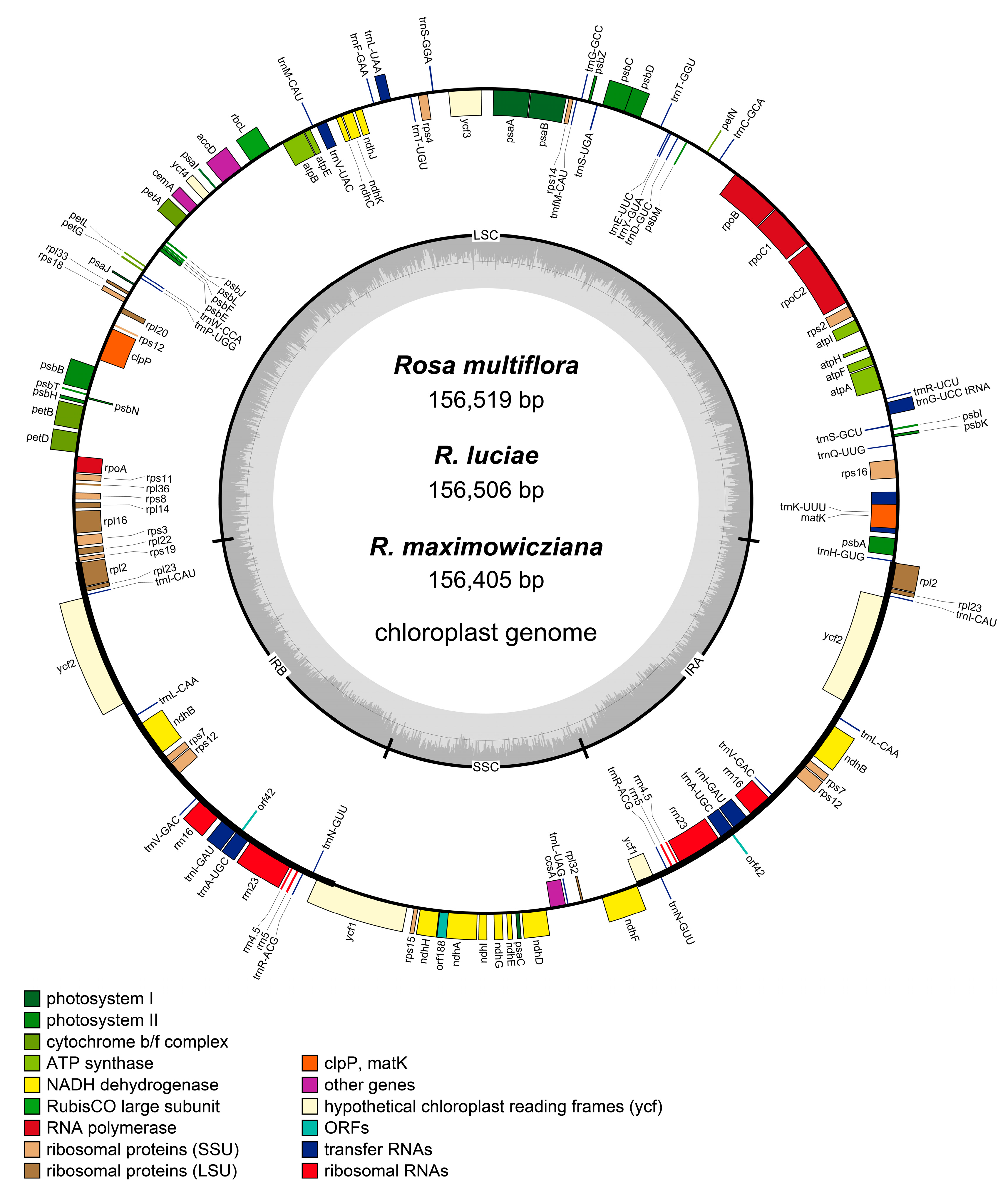
3.1. General Chloroplast Genome Features

The total genome sequence lengths of three Rosa sect. Synstylae species (R. multiflora, R. luciae, and R. maximowicziana) were from 156,405 bp (R. maximowicziana) to 156,519 bp (R. multiflora), and the depth of coverage for chloroplast genome contigs were from 214× (R. multiflora) to 1418× (R. maximowicziana). Each chloroplast genomes of Rosa sect. Synstylae species had a typical quadripartite structure, in which a large single-copy region (LSC; 85,529–85,643 bp) and a small single-copy region (SSC; 18,759–18,760 bp) were separated by two inverted repeats (IRs; 26,058 bp) (Figure 2). The gene numbers, gene orders, and GC contents were identical in chloroplast genomes of three Rosa sect. Synstylae species (Table 3). One hundred and fourteen unique genes were identified in each chloroplast genome of three Rosa sect. Synstylae species, including 80 protein-coding genes, 30 tRNA genes and four rRNA genes (Table 3). Among the unique genes, 15 genes contained one intron, and two genes contained two introns (Table S1).
Comparing to the previously identified chloroplast genomes of two Rosa species in other infrageneric groups, R. roxburghii (subgenus Platyrhodon) and R. odorata var. gigantea (subgenus Rosa section Chinenses), three Rosa sect. Synstylae chloroplast genomes (hereafter, the Synstylae chloroplast genomes) from this study had smaller genome sizes and fewer genes, whereas the GC contents were identical among five species (Table 3).

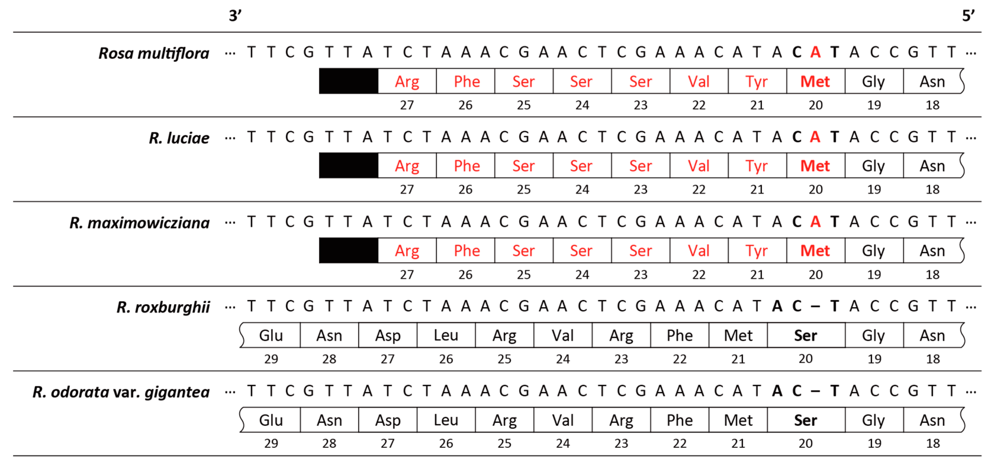
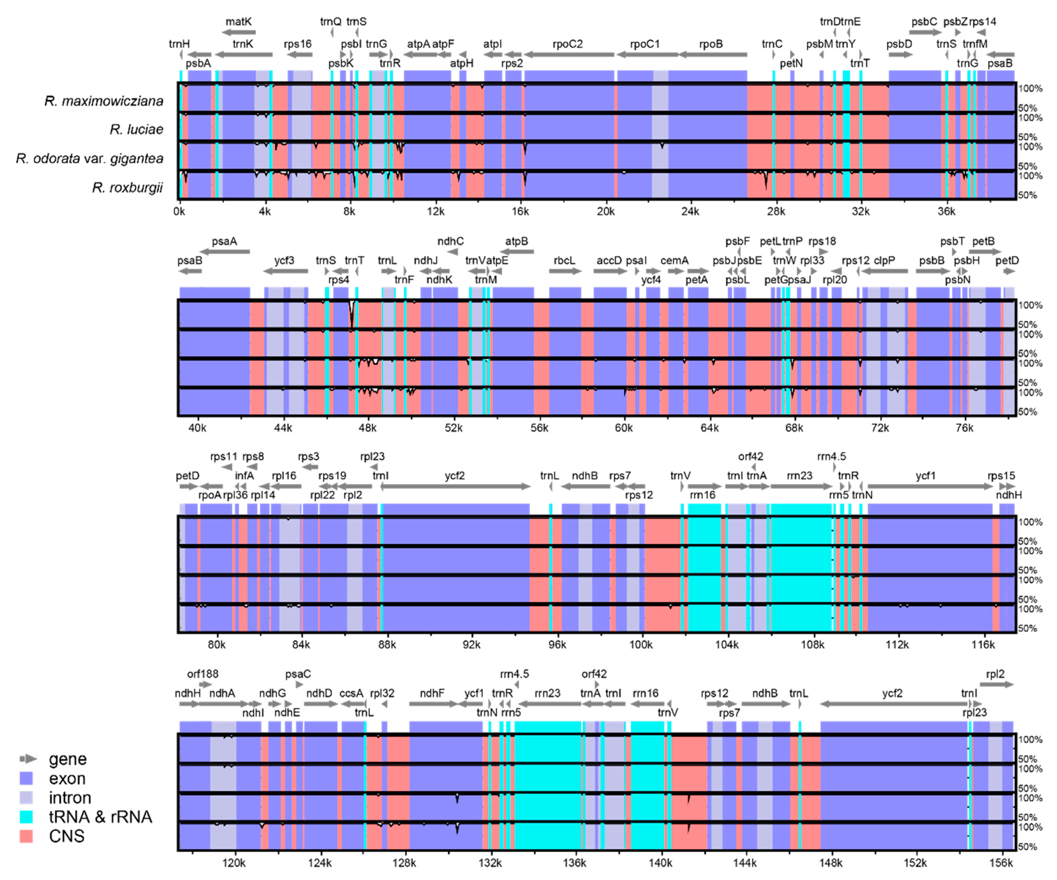
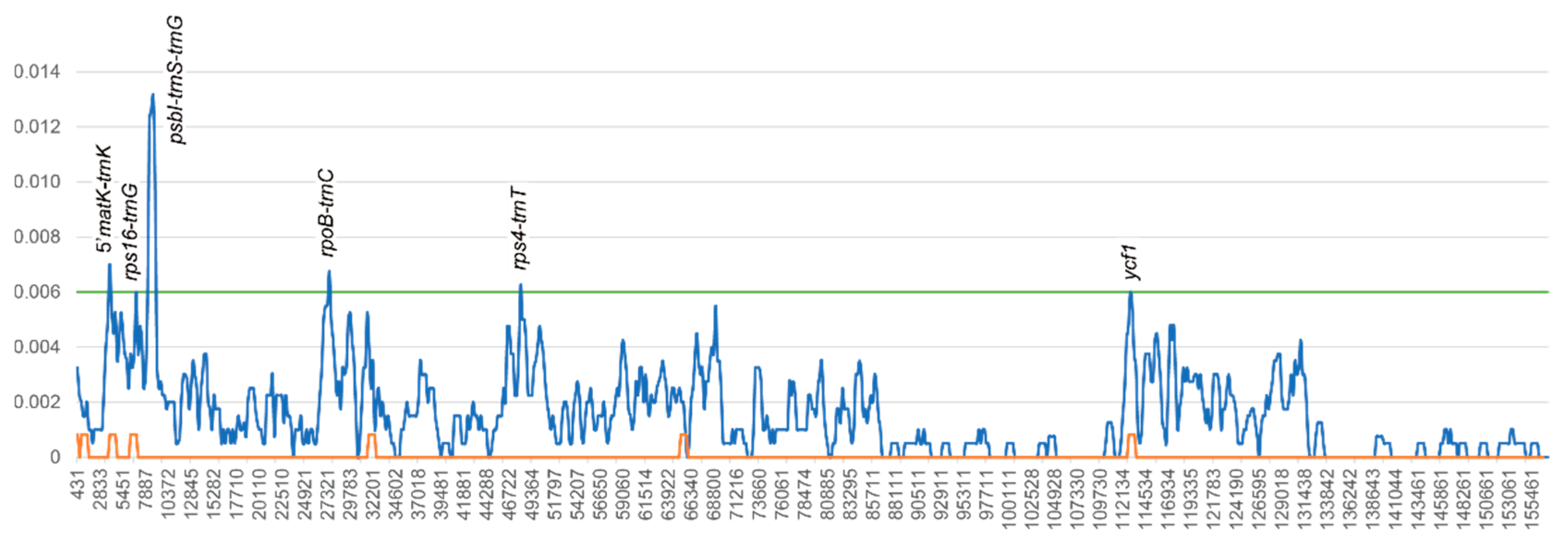
3.2. Interspecific Comparative Analyses

The interspecific comparison of the gene contents of five Rosa chloroplast genomes indicated that infA genes of the Synstylae chloroplast genomes was pseudogenized by a frameshift mutation (Figure 3). According to the original gene annotations, there are two more differences on gene contents among the Synstylae, R. roxburghii and R. odorata var. gigantea chloroplast genomes. First, trnG(GCC) gene was missing in R. roxburghii chloroplast genome. The other difference was that chloroplast genomes of R. roxburghii or R. odorata var. gigantea contained additional trnP(GGG) gene in the region where trnP(UGG) gene already exists. We conducted additional BLASTN and tRNAscan-SE searches on these genomes to verify whether these differences are evident. In BLASTN and tRNAscan-SE searches; however, trnG(GCC) gene was also found in R. roxburghii chloroplast genome. In the other difference, tRNAscan-SE only identified trnP(UGG) gene, not trnP(GGG) gene. Given these findings, the only difference in gene contents among the Rosa chloroplast genomes was that the Synstylae chloroplast genomes lost their infA genes by pseudogenization, while R. roxburghii and R. odorata var. gigantea chloroplast genomes contained infA genes.
Another remarkable difference was the mutations on the 3’ region of ndhF genes. In Rosa species, two kinds of mutations were found in the 3’ regions of ndhF genes. The mutation of T-insertion on the 3’ region of ndhF gene of R. maximowicziana caused the protein to terminate one amino acid early (746 amino acid residues), compared to the other Rosa species (747 amino acid residues). The other mutation was a point mutation on ndhF gene of R. roxburghii, altering phenylalanine 746 (Phe746) to leucine 746 (Leu746) (Figure 4).
In the overall interspecific chloroplast genome comparison, there was no large differences between R. multiflora chloroplast genome and each of the other Rosa chloroplast genome (Figure 5). Especially, within section Synstylae, the chloroplast genomes of R. luciae and R. maximowicziana were almost identical to the one of R. multiflora, showing very few differences. One distinct difference between the chloroplast genomes of R. maximowicziana and R. multiflora, however, was that the R. maximowicziana chloroplast genome has a 107 bp indel in the rps4-trnT(UGU) intergenic region. R. roxburghii and R. odorata var. gigantea had more variable sites compared to R. luciae and R. maximowicziana.
To estimate the genetic distance among Rosa chloroplast genomes, nucleotide diversity (Pi) was calculated using DnaSP. The average Pi value for five Rosa chloroplast genomes was 0.00154, and the average Pi value for the Synstylae chloroplast genomes was 0.00003. IR regions showed lower nucleotide diversity than single copy regions. While a comparison of three Synstylae chloroplast genomes was not informative, due to very few variations, a comparison of five Rosa chloroplast genome identified several variation hotspots (Figure 6). Six regions showed higher Pi values than other regions (Pi > 0.006). The hotspot with the highest Pi value was the psbI-trnS-trnG region (Pi = 0.01313), followed by four intergenic regions (5’matK-trnK, rps16-trnG, rpoB-trnC and rps4-trnT), and one gene region (ycf1).

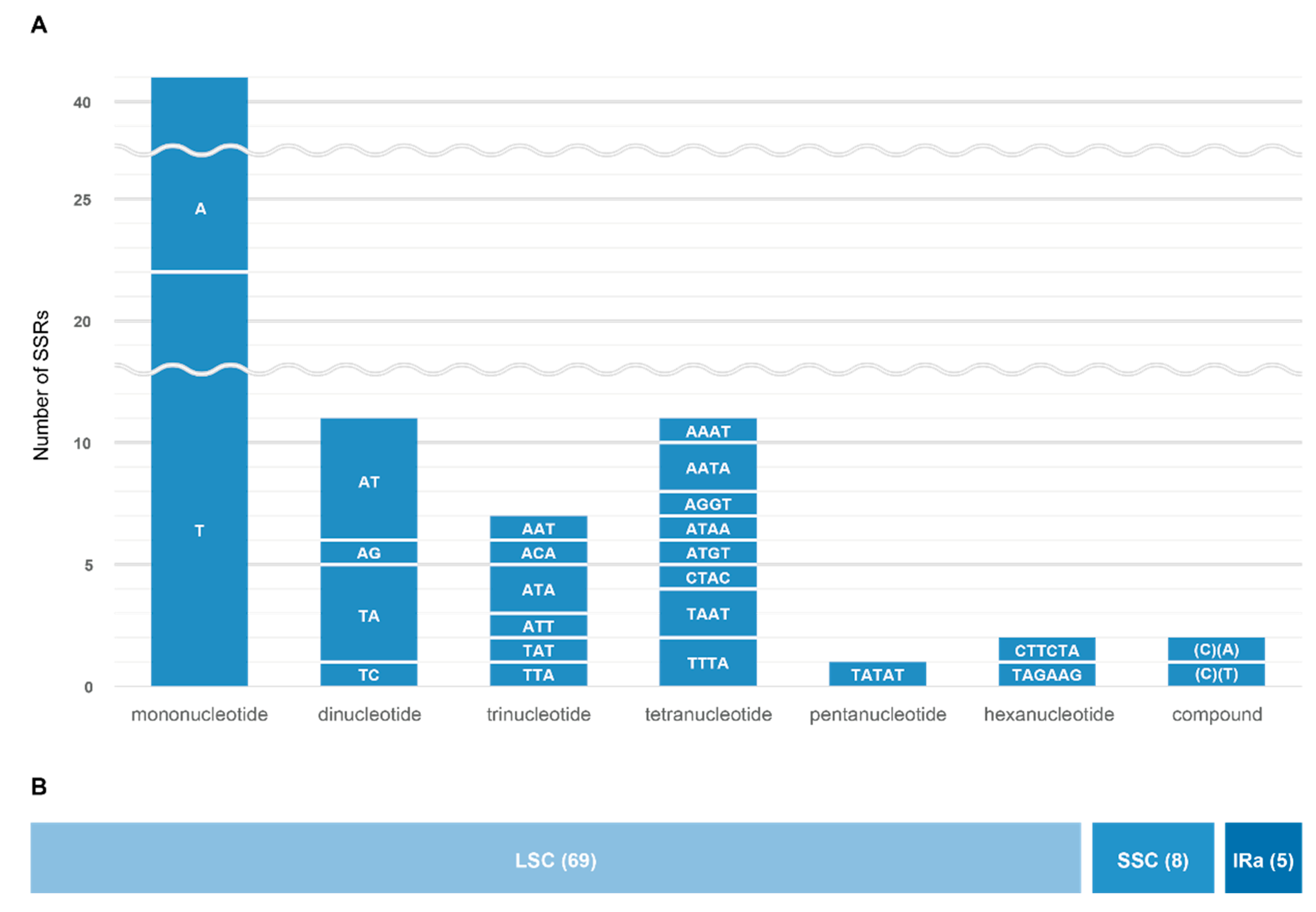
3.3. Simple Sequence Repeats

MISA identified 85 SSRs in the Synstylae chloroplast genomes, including two compound repeat regions (Figure 7 and Table S2). Except for two compound repeats, 51 mononucleotide, 11 dinucleotide, seven trinucleotide, 11 tetranucleotide, one pentanucleotide, and two hexanucleotide repeats were identified. Both compound repeats were compounds of two monomeric repeat regions. Most SSR regions were identified in single-copy regions, including 69 SSRs in the LSC region and eight SSRs in the SSC region, whereas five SSRs were identified in each IR region (Figure 7). Seventy-five SSRs had the repeat motifs of A/T or A/T combinations. Sixty-five SSRs were located in intergenic regions, 11 SSRs in protein-coding regions, nine SSRs in gene intron regions, and two SSRs in rDNA (Table S2).

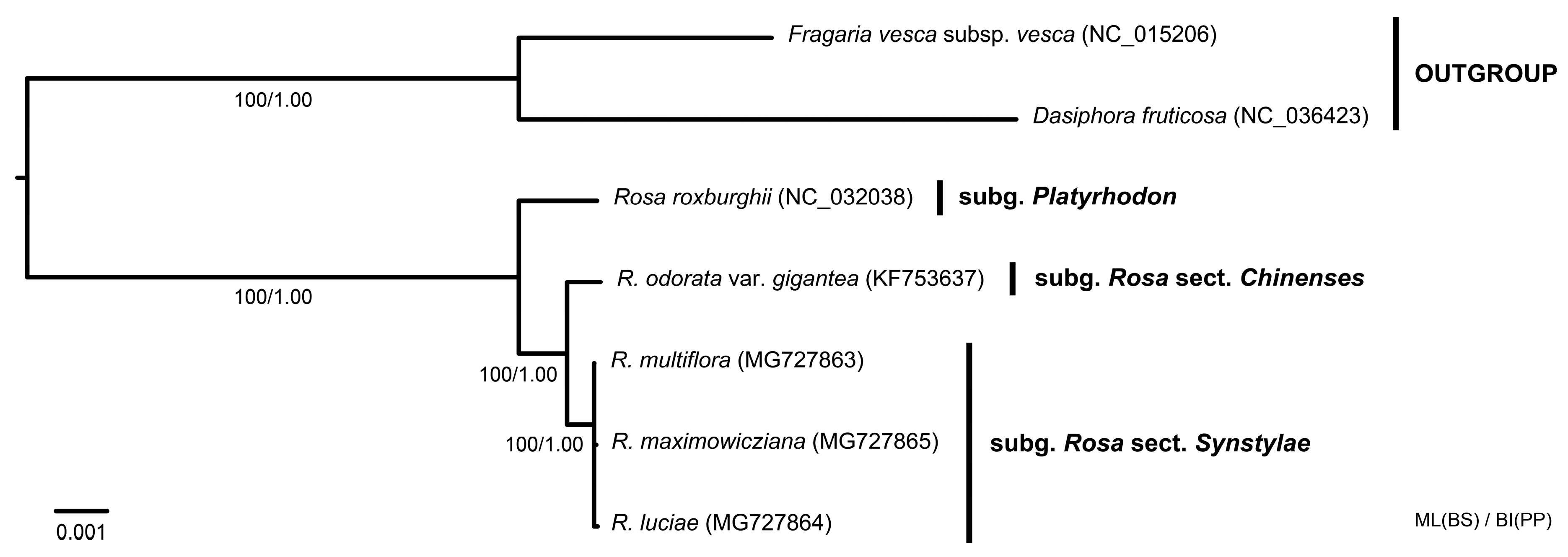
3.4. Phylogenetic Analyses

Maximum Likelihood and BI analyses showed identical tree topologies with high support values (Figure 8). Rosa sect. Synstylae species were monophyletic, strongly supported with a bootstrap value (BS) of 100% in ML, and a posterior probability (PP) of 1.00 in BI. In the chloroplast genome phylogeny, Rosa odorata var. gigantea was inferred to have a closer relationship with three Rosa sect. Synstylae species than R. roxburghii (100% BS and 1.00 PP support). Within the section Synstylae, however, no phylogenetic relationship was inferred, due to short branch lengths and poor support values.

4. Discussion

Three chloroplast genomes of Rosa sect. Synstylae (Rosa multiflora, R. luciae and R. maximowicziana) were de novo assembled for the first time in this study. The Synstylae chloroplast genomes were marginally different at best. Not surprisingly, more differences were found between the Synstylae chloroplast genomes, and two other chloroplast genomes of R. roxburghii (subg. Platyrhodon) and R. odorata var. gigantea (subg. Rosa sect. Chinenses). The major difference was that the Synstylae chloroplast genomes had lost their infA gene by pseudogenization (Figure 3). Independent infA gene loss is known to be quite common in angiosperm groups [39], but it is remarkable that there was a difference in infA gene loss within a genus. Within Synstylae species, chloroplast genomes were identical to each other in genome structures, gene contents, and GC contents. They also showed extremely low diversity (Pi = 0.00003), which has been one of major hurdles for the phylogenetic reconstruction in genus Rosa [7].
Because the Synstylae chloroplast genomes had only few variations and no genomic structural differences, several conservative protein-coding regions could be significant candidates for the chloroplast barcoding regions. The ndhF gene could be proposed to be one of those, as a frameshift mutation on the 3’ end of ndhF gene was identified in R. maximowicziana, which caused early termination of translation. This region may be able to be used in the higher taxon, genus Rosa, as a missense mutation occurred in the 3’ ndhF of R. roxburghii. Another candidate is ycf1 gene (tentatively suggested to be tic214 [40]), because a conversion of the 543th amino acid (phenylalanine to tyrosine) was identified in R. maximowicziana. In the generic level, this region was also suggested as a variation hotspot (Figure 6), showing numerous conservative and missense mutations within genus Rosa. These two regions (i.e., ndhF and ycf1 genes) are expected to be highly useful genetic markers in genus Rosa, confirming that these regions have been suggested as good markers for DNA barcoding and phylogenetic analyses in Rosaceae [34,41,42,43]. Especially, the ndhF gene is known to have a higher substitution rate, and its 3’ region was suggested to be phylogenetically useful, because it shows a high indel frequency, a high AT content and a low homoplasy rate [44]. Indeed, in Rubus subg. Idaeobatus, which is in the same subfamily with genus Rosa (subfamily Rosoideae), the 3’ regions of ndhF have shown a wide range of indel variations, resulting in frameshift mutations and alterations on the ndhF transcription termination [41]. Moreover, they may provide evolutionary insights on genus Rosa, given that they are the protein-coding genes. Because the Synstylae chloroplast genomes are highly conserved among species, chloroplast SSRs may provide valuable information for genotyping in the intra- and/or inter-specific level. For SSR genotyping in Synstylae species, 13 mononucleotide SSR regions were polymorphic among the species in this study (except for a deletion in the rps4-trnT region of R. maximowicziana). Among those, two regions (psbH-rpoA and rpl14-rps3 intergenic regions) could differentiate each accession of the Synstylae chloroplast genomes (Table S1).
Rosa sect. Synstylae species have been inferred to be very recently diverged, suggesting that R. multiflora and R. luciae have diverged from their common ancestor ca. 1.6 MYA, based on the chloroplast data [7]. Though R. maximowicziana showed two distinctive variations in its chloroplast genome (i.e., the frameshift mutation on the 3’ end of ndhF gene and a 107-bp indel in rps4-trnT(UGU) intergenic region), the Synstylae chloroplast genomes were almost identical, showing 0.00003 Pi value among three different genomes (Figure 4, Figure 5 and Figure 6). The comparative and phylogenetic analyses in this study suggested much closer relationships among three Synstylae species with lower diversities and shorter branch lengths than previous studies suggested [2,5,7]. The tree topology of sect. Synstylae revealed a polytomy, due to the extremely short branch lengths among three species, which suggest marginal phylogenetic differences among them (Figure 8). Given that these species are distinct in the distributions and the ecological, morphological, and phenological characteristics, the incongruence between those characters and the molecular phylogeny may imply the unique evolutionary histories of three Synstylae species. The close phylogenetic relationships of three Synstylae species may suggest that the evolutionary time was not sufficient for their chloroplast genomes to be distinctly diverged, and/or that they had interspecific genetic introgressions among the Synstylae species. Indeed, the incomplete lineage sorting and genetic introgression have been suggested in phylogenetic analyses with chloroplast data of Synstylae species [5]. If genetic introgression is occurring in the Synstylae clade, chloroplast capture events may occur, which would obscure inferring the species phylogeny. For more phylogenetic resolutions and systematic implications, additional phylogenetic analyses with nuclear markers would be required, because a nuclear genome phylogeny can be different from a chloroplast genome phylogeny [45]. Because the previous studies with various chloroplast markers suggested nonmonophyly of Rosa multiflora and R. luciae [5,7,8,10], these species might have more than one distinct haplotype. In this study, however, all Synstylae species samples were collected in the Korean Peninsula, while they are distributed in the wide range of the Eastern Sino-Japanese floristic region. This sampling bias could affect the chloroplast genome comparative analyses. To verify the existence of various distinct chloroplast haplotypes of R. multiflora and R. luciae, further phylogeographic and/or population genetic studies, including many accessions from representative sites in the Eastern Sino-Japanese floristic region will shed new light on the evolution of the Synstylae species. If there are various distinct chloroplast haplotypes in the section Synstylae, and if the haplotype differentiations are found along the geographic gradients, but not along the species boundaries, it is more plausible that the genetic introgressions have occurred, rather than that the chloroplast genome divergence was not sufficient. Several studies found the haplotype sharing in some taxa in which species are closely related, but defined by ecological or morphological differences [46,47,48]. Given that the marginal genome-wide differences in the Synstylae species in this study, these species also might share the chloroplast genomes beyond the species boundaries. Therefore, Rosa sect. Synstylae, including R. multiflora, R. luciae, and R. maximowicziana, can be another model taxonomic group for studying the chloroplast genome evolution of closely related species, which are defined by ecological or morphological differences, but shared the plastomes by the different species.

Supplementary Materials

The following are available online at https://0-www-mdpi-com.brum.beds.ac.uk/2073-4425/10/1/23/s1, Table S1: List of genes in chloroplast genomes of three Rosa sect. Synstylae species, Table S2: List of simple sequence repeat (SSR) regions in chloroplast genomes of three Rosa sect. Synstylae species.

Author Contributions

S.-C.K. and J.-H.J. conceived and designed the experiments; J.-H.J. performed the experiments and analyzed the data; J.-H.J. wrote the draft of manuscript and S.-C.K. subsequently edited.

Acknowledgments

This study was supported by Global Ph.D. Fellowship (GPF; NRF-2017H1A2A1045787) from National Research Foundation of Korea (NRF) to J.-H.J. We give special thanks to Young-woo Han for providing the collection information of Rosa maximowicziana.

Conflicts of Interest

The authors declare no conflict of interest. The founding sponsors had no role in the design of the study; in the collection, analyses, or interpretation of data; in the writing of the manuscript, and in the decision to publish the results.

References

  1. Rehder, A. Manual of Cultivated Trees and Shrubs; The Macmillan Company: New York, NY, USA, 1940; pp. 426–451. [Google Scholar]
  2. Wissemann, V.; Ritz, C.M. The genus Rosa (Rosoideae, Rosaceae) revisited: Molecular analysis of nrITS-1 and atpB-rbcL intergenic spacer (IGS) versus conventional taxonomy. Bot. J. Linn. Soc. 2005, 147, 275–290. [Google Scholar] [CrossRef]
  3. Christenhusz, M.J.M.; Fay, M.F.; Chase, M.W. Plants of the World: An Illustrated Encyclopedia of Vascular Plants; University of Chicago Press: Chicago, IL, USA, 2017; pp. 262–267. [Google Scholar]
  4. Rehder, A. Bibliography of Cultivated Trees and Shrubs Hardy in the Cooler Temperate Regions of the Northern Hemisphere; Arnold Arboretum of Harvard University: Jamaica Plain, MA, USA, 1949; pp. 296–317. [Google Scholar]
  5. Zhu, Z.-M.; Gao, X.-F.; Fougère-Danezan, M. Phylogeny of Rosa sections Chinenses and Synstylae (Rosaceae) based on chloroplast and nuclear markers. Mol. Phylogenetics Evol. 2015, 87, 50–64. [Google Scholar] [CrossRef] [PubMed]
  6. Wu, S.; Ueda, Y.; He, H.; Nishihara, S.; Matsumoto, S. Phylogenetic analysis of Japanese Rosa species using matK sequences. Breed. Sci. 2000, 50, 275–281. [Google Scholar] [CrossRef]
  7. Fougère-Danezan, M.; Joly, S.; Bruneau, A.; Gao, X.-F.; Zhang, L.-B. Phylogeny and biogeography of wild roses with specific attention to polyploids. Ann. Bot. 2014, 115, 275–291. [Google Scholar] [CrossRef] [Green Version]
  8. Liu, C.; Wang, G.; Wang, H.; Xia, T.; Zhang, S.; Wang, Q.; Fang, Y. Phylogenetic relationships in the genus Rosa revisited based on rpl16, trnL-F, and atpB-rbcL sequences. HortScience 2015, 50, 1618–1624. [Google Scholar]
  9. Debener, T.; Linde, M. Exploring complex ornamental genomes: The rose as a model plant. Crit. Rev. Plant Sci. 2009, 28, 267–280. [Google Scholar] [CrossRef]
  10. Bruneau, A.; Starr, J.R.; Joly, S. Phylogenetic relationships in the genus Rosa: New evidence from chloroplast DNA sequences and an appraisal of current knowledge. Syst. Bot. 2007, 32, 366–378. [Google Scholar] [CrossRef]
  11. Matsumoto, S.; Kouchi, M.; Fukui, H.; Ueda, Y. Phylogenetic analyses of the subgenus Eurosa using the its nrDNA sequence. In Proceedings of the XXV International Horticultural Congress, Part 11: Application of Biotechnology and Molecular Biology and Breeding—Gene Expression and Molecular Breeding, Genome Analysis, Brussels, Belgium, 2–7 August 1998; pp. 193–202. [Google Scholar]
  12. Ohba, H.; Akiyama, S.; Mikanagi, Y. Interspecific hybrids between Japanese species in Rosa section Synstylae (Rosaceae). J. Jap. Bot. 2007, 82, 45–53. [Google Scholar]
  13. Jeon, J.-H.; Maki, M.; Kim, S.-C. Inferring phylogenetic relationships among recently diverged East Asian species of roses (Rosa section Synstylae; Rosaceae). Unpublished work.
  14. Ruhlman, T.A.; Jansen, R.K. The plastid genomes of flowering plants. In Chloroplast Biotechnology: Methods and Protocols; Maliga, P., Ed.; Humana Press: Totowa, NJ, USA, 2014; pp. 3–38. [Google Scholar]
  15. Shaw, J.; Lickey, E.B.; Beck, J.T.; Farmer, S.B.; Liu, W.; Miller, J.; Siripun, K.C.; Winder, C.T.; Schilling, E.E.; Small, R.L. The tortoise and the hare II: Relative utility of 21 noncoding chloroplast DNA sequences for phylogenetic analysis. Am. J. Bot. 2005, 92, 142–166. [Google Scholar] [CrossRef]
  16. Shaw, J.; Lickey, E.B.; Schilling, E.E.; Small, R.L. Comparison of whole chloroplast genome sequences to choose noncoding regions for phylogenetic studies in angiosperms: The tortoise and the hare III. Am. J. Bot. 2007, 94, 275–288. [Google Scholar] [CrossRef]
  17. Chang, C.S.; Kim, H.; Chang, G.S. Illustrated Encyclopedia of Fauna & Flora of Korea, Vol. 43, Woody Plants; Ministry of Education: Seoul, Korea, 2011; p. 510.
  18. Zerbino, D.; Birney, E. Velvet: Algorithms for de novo short read assembly using de Bruijn graphs. Genome Res. 2008, 18, 821–829. [Google Scholar] [CrossRef] [PubMed]
  19. Kearse, M.; Moir, R.; Wilson, A.; Stones-Havas, S.; Cheung, M.; Sturrock, S.; Buxton, S.; Cooper, A.; Markowitz, S.; Duran, C.; et al. Geneious basic: An integrated and extendable desktop software platform for the organization and analysis of sequence data. Bioinformatics 2012, 28, 1647–1649. [Google Scholar] [CrossRef] [PubMed]
  20. Wyman, S.K.; Jansen, R.K.; Boore, J.L. Automatic annotation of organellar genomes with DOGMA. Bioinformatics 2004, 20, 3252–3255. [Google Scholar] [CrossRef] [PubMed] [Green Version]
  21. Lowe, T.M.; Chan, P.P. tRNAscan-SE on-line: Integrating search and context for analysis of transfer RNA genes. Nucleic Acids Res. 2016, 44, W54–W57. [Google Scholar] [CrossRef]
  22. Shinozaki, K.; Ohme, M.; Tanaka, M.; Wakasugi, T.; Hayashida, N.; Matsubayashi, T.; Zaita, N.; Chunwongse, J.; Obokata, J.; Yamaguchi-Shinozaki, K.; et al. The complete nucleotide sequence of the tobacco chloroplast genome: Its gene organization and expression. EMBO J. 1986, 5, 2043–2049. [Google Scholar] [CrossRef] [PubMed]
  23. Wang, Q.; Hu, H.; An, J.; Bai, G.; Ren, Q.; Liu, J. Complete chloroplast genome sequence of Rosa roxburghii and its phylogenetic analysis. Mitochondrial DNA Part B 2018, 3, 149–150. [Google Scholar] [CrossRef]
  24. Lohse, M.; Drechsel, O.; Kahlau, S.; Bock, R. OrganellarGenomeDRAW—A suite of tools for generating physical maps of plastid and mitochondrial genomes and visualizing expression data sets. Nucleic Acids Res. 2013, 41, W575–W581. [Google Scholar] [CrossRef]
  25. Yang, J.B.; Li, D.Z.; Li, H.T. Highly effective sequencing whole chloroplast genomes of angiosperms by nine novel universal primer pairs. Mol. Ecol. Resour. 2014, 14, 1024–1031. [Google Scholar] [CrossRef]
  26. Frazer, K.A.; Pachter, L.; Poliakov, A.; Rubin, E.M.; Dubchak, I. VISTA: Computational tools for comparative genomics. Nucleic Acids Res. 2004, 32, W273–W279. [Google Scholar] [CrossRef]
  27. Mayor, C.; Brudno, M.; Schwartz, J.R.; Poliakov, A.; Rubin, E.M.; Frazer, K.A.; Pachter, L.S.; Dubchak, I. VISTA: Visualizing global DNA sequence alignments of arbitrary length. Bioinformatics 2000, 16, 1046–1047. [Google Scholar] [CrossRef]
  28. Brudno, M.; Do, C.B.; Cooper, G.M.; Kim, M.F.; Davydov, E.; Green, E.D.; Sidow, A.; Batzoglou, S.; Program, N.C.S. LAGAN and multi-LAGAN: Efficient tools for large-scale multiple alignment of genomic DNA. Genome Res. 2003, 13, 721–731. [Google Scholar] [CrossRef] [PubMed]
  29. Rozas, J.; Ferrer-Mata, A.; Sánchez-DelBarrio, J.C.; Guirao-Rico, S.; Librado, P.; Ramos-Onsins, S.E.; Sánchez-Gracia, A. DnaSP 6: DNA sequence polymorphism analysis of large data sets. Mol. Biol. Evol. 2017, 34, 3299–3302. [Google Scholar] [CrossRef] [PubMed]
  30. Beier, S.; Thiel, T.; Münch, T.; Scholz, U.; Mascher, M. MISA-web: A web server for microsatellite prediction. Bioinformatics 2017, 33, 2583–2585. [Google Scholar] [CrossRef] [PubMed]
  31. Thiel, T.; Michalek, W.; Varshney, R.; Graner, A. Exploiting EST databases for the development and characterization of gene-derived SSR-markers in barley (Hordeum vulgare L.). Theor. Appl. Genet. 2003, 106, 411–422. [Google Scholar] [CrossRef] [PubMed]
  32. Shulaev, V.; Sargent, D.J.; Crowhurst, R.N.; Mockler, T.C.; Folkerts, O.; Delcher, A.L.; Jaiswal, P.; Mockaitis, K.; Liston, A.; Mane, S.P. The genome of woodland strawberry (Fragaria vesca). Nat. Genet. 2011, 43, 109–116. [Google Scholar] [CrossRef] [PubMed]
  33. Zhao, Y.; Lu, D.; Han, R.; Wang, L.; Qin, P. The complete chloroplast genome sequence of the shrubby cinquefoil Dasiphora fruticosa (Rosales: Rosaceae). Conserv. Genet. Resour. 2018, 10, 675–678. [Google Scholar] [CrossRef]
  34. Potter, D.; Eriksson, T.; Evans, R.C.; Oh, S.; Smedmark, J.E.E.; Morgan, D.R.; Kerr, M.; Robertson, K.R.; Arsenault, M.; Dickinson, T.A.; et al. Phylogeny and classification of Rosaceae. Plant Syst. Evol. 2007, 266, 5–43. [Google Scholar] [CrossRef]
  35. Nguyen, L.-T.; Schmidt, H.A.; von Haeseler, A.; Minh, B.Q. IQ-TREE: A fast and effective stochastic algorithm for estimating maximum-likelihood phylogenies. Mol. Biol. Evol. 2015, 32, 268–274. [Google Scholar] [CrossRef]
  36. Kalyaanamoorthy, S.; Minh, B.Q.; Wong, T.K.F.; von Haeseler, A.; Jermiin, L.S. ModelFinder: Fast model selection for accurate phylogenetic estimates. Nat. Methods 2017, 14, 587–589. [Google Scholar] [CrossRef]
  37. Minh, B.Q.; Nguyen, M.A.T.; von Haeseler, A. Ultrafast approximation for phylogenetic bootstrap. Mol. Biol. Evol. 2013, 30, 1188–1195. [Google Scholar] [CrossRef]
  38. Ronquist, F.; Teslenko, M.; Van Der Mark, P.; Ayres, D.L.; Darling, A.; Höhna, S.; Larget, B.; Liu, L.; Suchard, M.A.; Huelsenbeck, J.P. MrBayes 3.2: Efficient Bayesian phylogenetic inference and model choice across a large model space. Syst. Biol. 2012, 61, 539–542. [Google Scholar] [CrossRef] [PubMed]
  39. Millen, R.S.; Olmstead, R.G.; Adams, K.L.; Palmer, J.D.; Lao, N.T.; Heggie, L.; Kavanagh, T.A.; Hibberd, J.M.; Gray, J.C.; Morden, C.W.; et al. Many parallel losses of infA from chloroplast DNA during angiosperm evolution with multiple independent transfers to the nucleus. Plant Cell 2001, 13, 645–658. [Google Scholar] [CrossRef] [PubMed]
  40. Kikuchi, S.; Bédard, J.; Hirano, M.; Hirabayashi, Y.; Oishi, M.; Imai, M.; Takase, M.; Ide, T.; Nakai, M. Uncovering the protein translocon at the chloroplast inner envelope membrane. Science 2013, 339, 571–574. [Google Scholar] [CrossRef] [PubMed]
  41. Howarth, D.G.; Gardner, D.E.; Morden, C.W. Phylogeny of Rubus subgenus Idaeobatus (Rosaceae) and its implications toward colonization of the Hawaiian islands. Syst. Bot. 1997, 22, 433–441. [Google Scholar] [CrossRef]
  42. Dong, W.; Xu, C.; Li, C.; Sun, J.; Zuo, Y.; Shi, S.; Cheng, T.; Guo, J.; Zhou, S. Ycf1, the most promising plastid DNA barcode of land plants. Sci. Rep. 2015, 5, 8348. [Google Scholar] [CrossRef] [PubMed]
  43. Wang, S.; Shi, C.; Gao, L.-Z. Plastid genome sequence of a wild woody oil species, Prinsepia utilis, provides insights into evolutionary and mutational patterns of Rosaceae chloroplast genomes. PLoS ONE 2013, 8, e73946. [Google Scholar] [CrossRef] [PubMed]
  44. Kim, K.J.; Jansen, R.K. Ndhf sequence evolution and the major clades in the sunflower family. Proc. Natl. Acad. Sci. USA 1995, 92, 10379. [Google Scholar] [CrossRef] [PubMed]
  45. Wendel, J.F.; Doyle, J.J. Phylogenetic incongruence: Window into genome history and molecular evolution. In Molecular Systematics of Plants II; Springer: Berlin, Germany, 1998; pp. 265–296. [Google Scholar]
  46. Zhang, R.; Hipp, A.L.; Gailing, O. Sharing of chloroplast haplotypes among red oak species suggests interspecific gene flow between neighboring populations. Botany 2015, 93, 691–700. [Google Scholar] [CrossRef] [Green Version]
  47. Nevill, P.G.; Després, T.; Bayly, M.J.; Bossinger, G.; Ades, P.K. Shared phylogeographic patterns and widespread chloroplast haplotype sharing in Eucalyptus species with different ecological tolerances. Tree Genet. Genom. 2014, 10, 1079–1092. [Google Scholar] [CrossRef]
  48. Shepherd, L.D.; de Lange, P.J.; Perrie, L.R.; Heenan, P.B. Chloroplast phylogeography of New Zealand Sophora trees (Fabaceae): Extensive hybridization and widespread last glacial maximum survival. J. Biogeogr. 2017, 44, 1640–1651. [Google Scholar] [CrossRef]
Figure 1. The schematic distribution of three Rosa sect. Synstylae species. (A) Rosa multiflora in the Liaoning–Shandong flora; (B) Rosa luciae in the Southern Chinese–Japanese flora; (C) Rosa maximowicziana in the Amur flora. CN: China; KP: Korean Peninsula; PK: Primorsky Krai, Russia; JA: Japanese Archipelago.
Genes 10 00023 g001
Figure 2. Gene maps of three Rosa sect. Synstylae species: R. multiflora, R. luciae and R. maximowicziana. The outer circle shows the genes at each locus, and inverted repeat regions are indicated with thicker lines. Genes on the outside of the outer circle are transcribed in a counterclockwise direction, while genes on the inside of the outer circle are transcribed in a clockwise direction. The inner circle indicates the range of the large single-copy region (LSC), the small single-copy region (SSC), and two inverted repeats (IRs), and also shows a GC content graph of the genome. In the GC content graph, the dark gray lines indicate GC content, while light gray lines indicate the AT content at each locus.
Genes 10 00023 g002
Figure 3. Pseudogenization of infA genes in the Synstylae chloroplast genomes. In each lane, aligned DNA sequence data are shown on the top. Hyphens indicate indels in the sequences. Boxed ribbons show the amino acid sequences coded from the DNA sequences, and black closed boxes indicate the terminal codons. Numbers under the boxes indicates the amino acid residue numbers. The mutated residues are bolded, and mutations are in red.
Genes 10 00023 g003
Figure 4. The 3’ region of ndhF genes of five Rosa species and two other closely related species (Fragaria vesca ssp. vesca (GenBank accession: NC_015206; [32]) and Dasiphora fruticosa (GenBank accession: NC_036423; [33]). In each lane, aligned DNA sequence data are shown on the top. Hyphens indicate indels in the sequences. Boxed ribbons show the amino acid sequences that are coded from the DNA sequences, and black closed boxes indicate the terminal codons. Numbers under the boxes indicate the amino acid residue numbers. The mutated residues are bolded, and mutations are in red.
Genes 10 00023 g004
Figure 5. Visualized alignment of the Rosa chloroplast genome sequences with annotations, using mVISTA. Each horizontal lane shows the graph for the sequence pairwise identity with a R. multiflora chloroplast genome sequence. The x-axis represents the base sequence of the alignment and the y-axis represents the pairwise percent identity within 50–100%. Grey arrows represent the genes and their orientations. Blue boxes represent exon regions and light gray boxes represent intron regions; light blue boxes represent RNA-coding gene regions; red boxes represent non-coding sequence (CNS) regions.
Genes 10 00023 g005
Figure 6. Nucleotide diversity graphs of Rosa chloroplast genomes. The x-axis represents the base sequence of the alignment, and the y-axis represents Pi value. The blue line indicates the nucleotide diversity of five Rosa chloroplast genomes; the orange line indicates the nucleotide diversity of the Synstylae chloroplast genomes; and the green line indicates the threshold for variation hotspots (Pi threshold = 0.006). Each variation hotspot for Rosa chloroplast genomes is annotated on the graph.
Genes 10 00023 g006
Figure 7. Information of simple sequence repeats (SSRs) of the Synstylae chloroplast genomes. (A) SSR types and the number of different SSR types. Each repeat motif is indicated in the middle of each bar. (B) The proportion of SSR location in the Synstylae chloroplast genomes. The locations and SSR numbers were indicated in the middle of bars.
Genes 10 00023 g007
Figure 8. Phylogenetic tree of five Rosa chloroplast genomes inferred from maximum likelihood (ML) and Bayesian Inference (BI) based on whole chloroplast genome data. Bootstrap values (BS) from ML analysis and posterior probabilities (PP) from BI were annotated at each branch.
Genes 10 00023 g008
Table 1. The ecological, morphological, and phenological characteristics of three species of Rosa sect. Synstylae.
CharacteristicsRosa multifloraRosa luciaeRosa maximowicziana
HabitatForest edgeSea shore/Open spaceThicket/Near the water
Growth habitErectProstrateDecumbent
LeavesNot leatheryLeatheryNot leathery
StipulesPectinateSerrateSerrate
PricklesSparseSparseDense
StylesGlabrousPubescentGlabrous
FloweringApril–MayJune–JulyMay–June
Table 2. Accession information for three Rosa species.
SpeciesLocalityVoucher NumberGenBank Accession
R. multifloraNamyangju, Gyeonggi, KoreaJJH258MG727863
R. luciaeSeoguipo, Jeju, KoreaSGRW001MG727864
R. maximowiczianaHwaseong, Gyeonggi, KoreaCBRX001MG727865
Table 3. The general characteristics of five Rosa chloroplast genomes.
CharacteristicsR. multifloraR. luciaeR. maximowiczianaR. roxburghiiR. odorata var. gigantea
GenBank accession numberMG727863MG727864MG727865NC_032038KF753637
ReferenceThis studyThis studyThis studyWang et al., 2018 [23]Yang et al., 2014 [25]
Total cpDNA size (bp)156,519156,506156,405156,749156,634
LSC size (bp)85,64385,63185,52985,85285,767
SSC size (bp)18,76018,75918,76018,79118,761
IR size (bp)26,05826,05826,05826,05326,053
Number of different genes114114114115116 (115 *)
Number of different protein-coding genes8080808181
Number of different tRNA genes3030303031 (30 *)
Number of different rRNA genes44444
GC content (%)37.237.237.237.237.2
GC content of LSC (%)35.235.235.235.235.2
GC content of SSC (%)31.331.331.331.331.3
GC content of IR (%)42.742.742.742.742.7
cpDNA: chloroplast DNA; LSC: large single-copy region; SSC: small single-copy region; IR: inverted repeat; * The number of the genes after additional verification.

