Article

Exome Sequencing Identifies a Novel FBN1 Variant in a Pakistani Family with Marfan Syndrome That Includes Left Ventricle Diastolic Dysfunction

1
Department of Biotechnology, Faculty of Chemical and Life Sciences, Abdul Wali Khan University, Mardan 23200, Pakistan
2
Centre for Endocrinology, William Harvey Research Institute, Charterhouse Square Campus, Barts and The London School of Medicine and Dentistry, Queen Mary University of London, London EC1M 6BQ, UK
3
Center for Statistical Genetics, Gertrude H. Sergievsky Center, Department of Neurology, Columbia University Medical Center, New York, NY 10032, USA
4
Department of Zoology, College of Science, King Saud University, Riyadh I1451, Saudi Arabia
5
Taub Institute for Alzheimer’s Disease and the Aging Brain, Columbia University Medical Center, New York, NY 10032, USA
*
Authors to whom correspondence should be addressed.
Submission received: 2 August 2021 / Revised: 12 September 2021 / Accepted: 18 September 2021 / Published: 28 November 2021
(This article belongs to the Special Issue Cardiovascular Disease Risk Factors, Genetics, and Prevention)

Abstract

:
Introduction: Cardiomyopathies are diseases of the heart muscle and are important causes of heart failure. Dilated cardiomyopathy (DCM) is a common form of cardiomyopathy that can be acquired, syndromic or non-syndromic. The current study was conducted to explore the genetic defects in a Pakistani family with cardiac disease and features of Marfan’s syndrome (MFS). Methods: A family with left ventricle (LV) diastolic dysfunction and MFS phenotype was assessed in Pakistan. The clinical information and blood samples from the patients were collected after physical, cardiovascular, and ophthalmologic examinations. An affected individual (proband) was subjected to whole-exome sequencing (WES). The findings were further validated through Sanger sequencing in the family. Results: Through WES and sanger validation, we identified a novel variant NM_000138.4; c.1402A>G in the Fibrillin-1 (FBN1) gene that segregates with LV diastolic dysfunction and MFS. Furthermore, bioinformatic evaluation suggested that the novel variant is deleterious and disease-causing. Conclusions: This study identified for the first time a novel FBN1 variant in a family with LV diastolic dysfunction and MFS in Pakistan.

1. Introduction

Cardiomyopathies are a heterogeneous group of diseases that mostly result in progressive heart failure with significant morbidity and mortality. They represent an important and complex group of heart muscle disorders with heterogeneous phenotypic expression and multiple etiologies [1]. There are different causes of cardiomyopathies, most of them are genetic. These diseases are either part of a generalized systemic disorder or confined to the heart [2]. Dilated cardiomyopathy (DCM) is categorized into syndromic, non-syndromic and acquired types. Non-syndromic DCM is defined as DCM without other systemic involvement, while syndromic DCM is a multisystemic disorder [3], including those with mitochondrial myopathies, lysosomal storage disorders, and neuromuscular involvement [4]. Marfan syndrome (MFS) which involves the connective tissue and cardiovascular manifestations includes aortic dilation and ventricular arrhythmia [5].
Dilated cardiomyopathy is one of the major causes of heart failure and heart transplantation worldwide [6]. It is a myocardial disease involving dilation of the left ventricle, resulting in systolic dysfunction, heart failure, arrhythmia, and sudden cardiac death (SCD) [7]. DCM shows an autosomal-dominant inheritance pattern in ~90% of the cases and is X-linked or autosomal recessive in ~10% of cases, with variable penetrance and expressivity [8]. Furthermore, it is a genetically heterogeneous disease having the involvement of >100 genes [7,9]. More than 50 genes encoding the sarcomeric proteins, nuclear envelope, cytoskeleton, ion channels, sarcolemma and intercellular junctions have been associated with DCM [10]. Titin (TTN) is one of the important DCM genes, with truncating variants reported in about 25% of the familial form of DCM [11], while many other genes, such as LMNA, MYH7, MYBPC3, TNNT2, MYH6, and SCN5A, have been observed at lower frequencies [12].
MFS is a common autosomal-dominant inherited disorder of the fibrous connective tissues, involving prominent manifestations in many organ systems such as cardiovascular, skin, skeletal, ocular, pulmonary, and brain (dura mater abnormalities). MFS has a prevalence of 2 to 3 in 10,000 individuals [13,14]. Impaired left ventricular (LV) function in MFS occurs due to significant valvular heart disease [15,16]. The available data on left ventricular diastolic dysfunction in MFS are scarce [17,18]. MFS is mainly caused by heterozygous variants in the FBN1 gene (OMIM #134797). FBN1 encodes a 350 kDa glycoprotein member Fibrillin-1 of the Fibrillin family, which is a major constituent of 10–12 nm extracellular microfibrils that comprise the cardiac connective tissue scaffold and thus maintain the stability and assembly of the microfibrils [19,20]. The variants reported in the FBN1 gene are responsible for 70–80% of MFS in general and ~90% of autosomal-dominant MFS cases [21,22]. More than 1800 FBN1 variants have been associated with MFS, which are either predominantly inherited in an autosomal dominant fashion or are de novo in one-third of the patients [13].
To our knowledge, MFS with LV diastolic dysfunction has not been explored at the genetic level in the Pakistani population. Thus, we investigated if novel genes/variants can be involved in the genetic background of these cardiac diseases. Therefore, to test this hypothesis, we investigated a clinically well-characterized Pakistani family with LV diastolic dysfunction and skeletal features of MFS. A DNA sample from the proband of the family underwent WES and segregation of the identified variant was tested using Sanger sequencing. Furthermore, the identified variant was investigated for its effect on protein structure and function using in silico and bioinformatic tools. We identified a novel variant (NM_000138.4; c.1402A>G) in the FBN1 gene with possible deleterious and damaging effects. This variant may indicate the etiologies of LV diastolic dysfunction and MFS in Pakistani patients.

2. Methodology

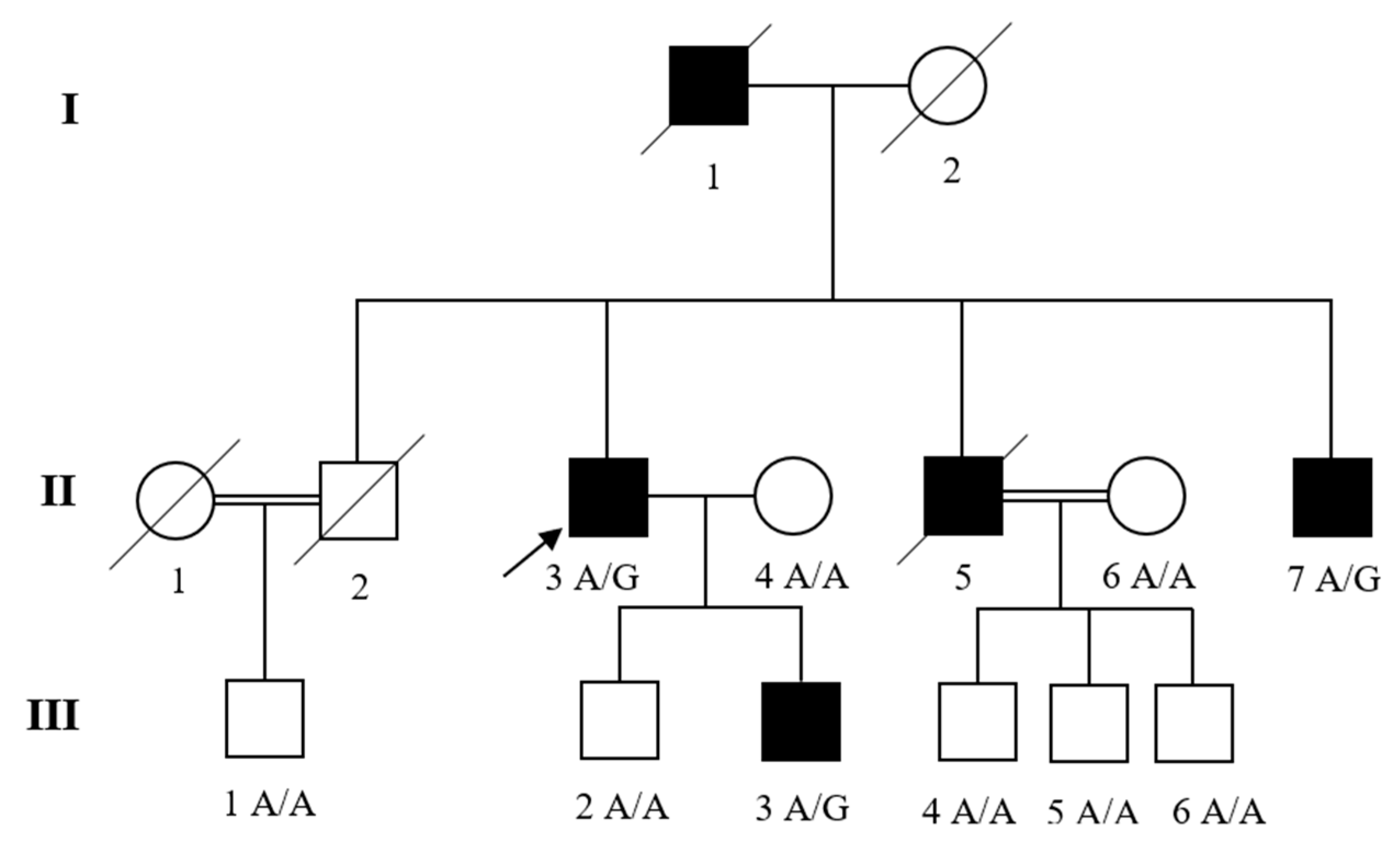
2.1. Family Recruitment and Clinical Diagnosis

A family (Figure 1) with LV diastolic dysfunction and MFS was recruited and selected for this study. The family was visited and interviewed for the clinical data and family history. Peripheral blood samples were collected from both affected and unaffected family members after obtaining written informed consent from every individual. The study was approved by the Institutional Review Boards (IRBs) of Abdul Wali Khan University Mardan, Mardan Medical Complex (MMC), Pakistan (IRB-AWKUM-2306) and Columbia University, USA (IRB-AAAS5864) in accordance with the declaration of Helsinki.
The affected family members were clinically evaluated and diagnosed by certified clinicians and cardiologists based on their family history, symptoms and medical records. The patients underwent detailed physical, cardiovascular, ophthalmologic, and skeletal system evaluations. They were examined by M-Mode and two-dimensional echocardiography, Doppler ultrasound and chest radiographs.

2.2. DNA Isolation and Whole Exome Sequencing

Genomic DNA was isolated via a GeneAll® Exgene™ Blood SV mini-Kit (GeneAll Biotechnology Co., Ltd., Seoul, Korea) according to the manufacturer’s protocol. The DNA sample was fragmented using Covaris technology. The DNA fragments were amplified by ligation-mediated PCR (LM-PCR) and were hybridized to exome array for enrichment. After washing the non-hybridized fragments, the captured products were circularized. DNA Nanoballs (DNBs) were produced using the rolling circle amplification (RCA) technique. The resulting qualified captured library was loaded onto the BGISEQ platform.
The Agilent SureSelect Human All Exon V6 kit (Agilent Technologies, Inc., Santa Clara, CA, USA) was used for exome capture and preparation of exome libraries with a target region of 60.33Mb using DNA sample of the proband from the family. Exome sequencing was performed on the DNBseq™ platform, a BGISEQ-500 instrument (BGI Tech Solutions Hongkong Co., Ltd., Hong Kong, China) with 100 bp paired-end reads and mean sequencing depth of 74.81X on target regions.

2.3. Variant’s Annotation and Filtering

The WES reads were aligned to the human reference genome (GRCh38/hg38) by the Burrows–Wheeler Aligner software (BWAv0.7.15) [23,24,25]. PCR duplicate reads were removed with Picard tools. Local realignment around insertions/deletions (InDels), base quality score recalibration and variant calling were performed using the Genome Analysis Toolkit (GATK) [26,27].
ANNOVAR (v2019Oct24) was used for variant annotation [28]. Variants were filtered based on their frequency of occurrence in various databases (Genome Aggregation Database (gnomAD), Greater Middle East (GME), Kaviar Genomic Variant (kaviar_20150923), and Brazilian genomic variants (ABraOM)). Variants with a minor allele frequency (MAF) of <0.0005 were retained [29,30,31,32] and synonymous variants were excluded. Furthermore, various bioinformatics tools (CADD, SIFT, Polyphen-2, Mutation Taster, and Mutation Assessor included in dbNSFPv3.5 and dbscSNV1.1) were applied to predict the deleterious effect of the identified variant on protein structure and function (to predict the functional impact of the candidate variant). The conservation was examined using GERP++, PhyloP and PhastCons [33,34]. The databases including dbSNP, OMIM, ClinVar, HGMD, Genetics Home Reference (GHR) website, and Varsome were screened to determine whether the variant had any associated phenotypes.

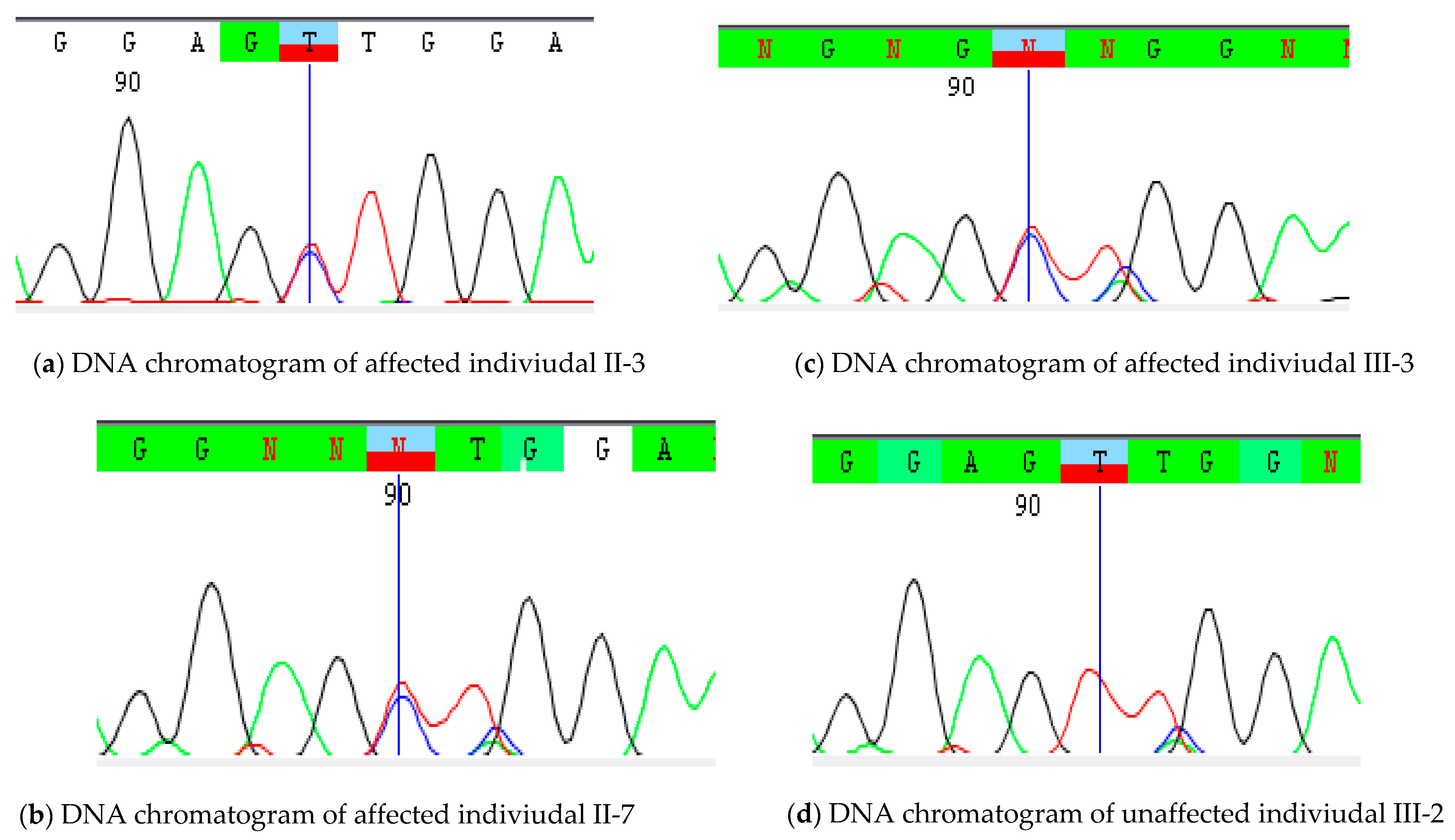
2.4. Sanger Validation

The available DNA samples in the pedigree underwent Sanger sequencing to validate candidate variant obtained from WES and confirm its segregation with phenotype in the pedigree. Forward and reverse primers (FBN1-F-ACAGAATTACAACAGACCCTTGG; FBN1-R- GCCTTGCAAGCTCTGTAACC; Product Size: 521bp) were designed using Primer3 software [35]. The amplified products were purified by ExoSAP-IT (USB Corp., Cleveland, OH, USA) and were sequenced using the BigDye Terminator v3.1 Cycle Sequencing Kit followed by capillary electrophoresis on an ABI 3730 DNA Analyzer (Applied Biosystems Inc., Foster City, CA USA). The DNA sequences were then aligned to the reference genome sequence using the CodonCode Aligner v7.1.2 (CodonCode Corp., Centerville, MA, USA).

3. Results

3.1. Clinical Manifestations

The affected individuals of this family were observed and diagnosed with cardiac complications. They had common features of chest pain, shortness of breath, syncope, dizziness, and fatigue. It was a three-generation pedigree with three affected individuals II-3, II-7, and III-3 (Figure 1). The ages of onset were 58, 53, 42 and 16 years, respectively. The proband II-3 was a 63-year-old man, diagnosed with LV diastolic dysfunction, mitral valve regurgitation, tall thin stature, and long arms and legs. His parents were non-consanguineous and there was no family history of MFS. The proband’s father (I-1) died at the age of 80 years due to heart failure. Echocardiography (M-Mode/two-dimensional) of patients II-3, II-7, and III-3 confirmed LV diastolic dysfunction, normal-size cardiac chambers and normal LV ejection fraction. Mitral valve regurgitation or mitral insufficiency was also observed in patients II-3, and II-7. Patient II-5 died at the age of 57 years; however, his medical history was obtained from the clinical records. The physical examination of affected individuals II-3, II-7, and III-3 revealed skeletal features including tall thin stature, and long arms and legs. No other abnormalities were present in these patients. The clinical manifestations of the Family are given in Table 1.

3.2. Genetic Analysis

WES was carried out for the proband (II-3) of the family. The average sequencing depth in the target region was 74.81X. In addition, 99.62% of the target bases had 1X coverage and 98.05% of target bases had 10X coverage. The raw reads, clean reads, and mean sequencing depth (%) on the target region obtained from the selected individual were 89620140, 89445598, and 86.50, respectively. Furthermore, the variants were identified and filtered in the family.

3.3. Variant’s Validation and Segregation

The identified variant was subjected to Sanger validation. Thus, a novel variant [NM_000138.4; c.1402A>G: p.(T468A)] was identified in FBN1 gene and was tested for segregation. Through Sanger sequencing, we found that family members II-7 and III-3 had the FBN1 variant segregated with MFS in the Family and was classified as likely pathogenic according to American College of Medical Genetics (ACMG) guidelines. The DNA sequence chromatograms of the FBN1 variant (NM_000138.4; c.1402A>G) are shown in Figure 2.

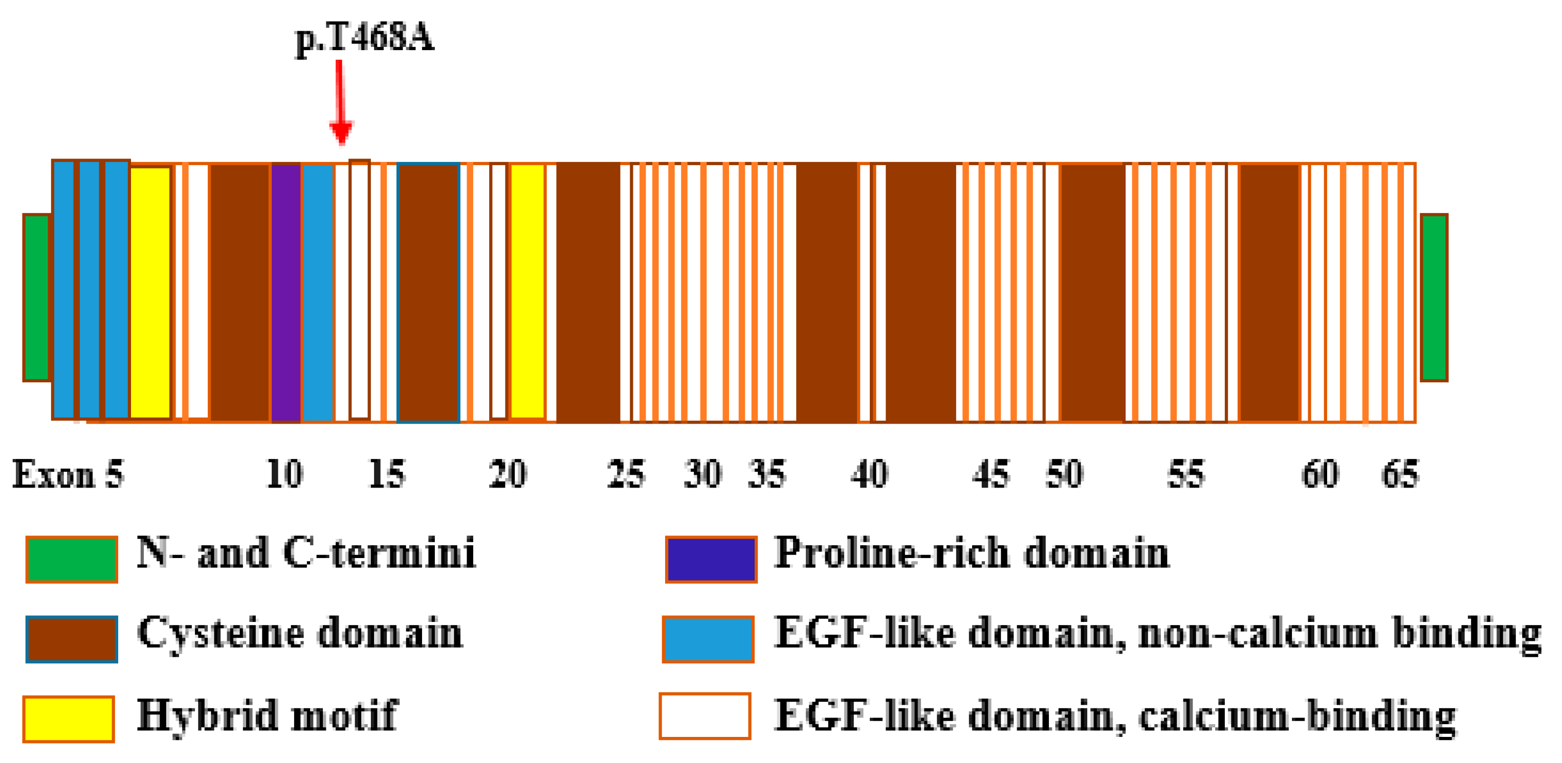
3.4. Bioinformatic Analysis

The missense variant p.(T468A) in FBN1 gene was designated to be damaging, deleterious and disease-causing by PROVEAN (Protein Variation Effect Analyzer), SIFT (Sorting Intolerant From Tolerant), FATHMM (Functional Analysis Through Hidden Markov Models), MutationTaster, and MutationAssessor. This variant is located in a highly conserved region of the calcium-binding epidermal growth factor-like domain 3 (cbEGF-like domain 3) of FBN1 protein and is predicted to affect protein function (Figure 3).

4. Discussion

In this study, a family with LV diastolic dysfunction and MFS was evaluated, as shown in Figure 1. The affected individuals II-3, II-5, II-7 and III-3 displayed tall thin stature with long arms and legs, consistent with MFS, LV diastolic dysfunction, dyspnea, chest pain, fatigue, and syncope. Furthermore, no other abnormalities had been noticed in any of the affected family members. The proband from the family was subjected to WES analysis, and the variant observed was tested for segregation in other family members. Through WES data analysis we identified a novel variant (NM_000138.4; c.1402A>G) in the FBN1 gene which segregated with MFS and LV diastolic dysfunction in the Family. The variant found in this study was analyzed using various online bioinformatic tools, such as PROVEAN, SIFT, FATHMM, MutationTaster, and MutationAssessor. Bioinformatic analysis suggested that the variant has deleterious and damaging effects. The FBN1 variant replaces highly conserved threonine residue with alanine (NP_000129.3, p.T468A) in the calcium-binding epidermal growth factor (cbEGF)-like domain 3, and thus may affect the protein structure by disturbing the microfilament arrangement (Figure 3). The FBN1 protein comprises 47 epidermal growth factor-like (EGF-like) domains, 7 TGFβ-binding (TB) domains, 2 hybrid (Hyb) domains, N- and C-terminal domains. Each EGF-like domain consists of six highly conserved cysteines linked by three disulfide bonds (C1-C3, C2-C4, and C5-C6) [36]. The disruption of these bonds and the loss or addition of cysteine residue results in domain misfolding [37].
This variant was predicted to be deleterious by PROVEAN, SIFT, FATHMM, Mutation Taster, and MutationAssessor, with a CADD score of 20.2. Previously, many FBN1 variants responsible for cardiovascular complications in MFS were identified [38]. In ClinVar, there are >2000 pathogenic or likely pathogenic FBN1 variants. FBN1 variants result in the loss of fibrillin-rich microfibrils in the cardiac extracellular matrix, and finally, weakness of the connective tissue. This results in the excessive activation of transforming growth factor-β (TGF-β), thus inducing phenotypic consequences in MFS [39,40,41]. Variants in exons 12–15 encoding cbEGF-like domains are responsible for a mild MFS phenotype (ectopia lentis) with late cardiovascular involvement [42]. However, a novel FBN1 mutation previously found in a large Pakistani family with ocular and skeletal system defects had no cardiovascular features of MFS [43]. That said, a large number of variants in the FBN1 gene have been associated with MFS, but the role of these variants in LV diastolic dysfunction has not been determined.
FBN1 is expressed in many human tissues, including cartilage, tendon, cornea, zonules, and the cardiovascular system [44]. According to the Kyoto Encyclopedia of Genes and Genomes (KEGG) pathway annotation, the FBN1 protein is involved in the TGF-β signaling pathway (hsa04350). Besides their structural role, fibrillin microfibrils also contribute to tissue homeostasis through their interaction with cell surface receptors integrins and through interaction with growth factors, such as TGF-β and bone morphogenetic proteins (BMPs) [45,46]. Increased amounts of active TGF-β due to reductions in fibrillin have been reported to be implicated in MFS pathogenesis in mice with fibrillin1 deficiencies. Lung emphysema, vascular complications and bone overgrowth are the most distinctive features of mice with reduced expressions of the FBN1 gene, and in MFS patients with heterozygous FBN1 variants [47].
In conclusion, we report a novel variant (NM_000138.4; c.1402A>G) in FBN1 gene associated with LV diastolic dysfunction and MFS in a Pakistani family. Our findings support the role of this variant in the genetic pathology of these cardiac diseases in the Pakistani population. Further investigation is needed to confirm the contribution of defects in the FBN1 gene to these disease phenotypes and to elucidate the effects of this gene variant on function of the gene.

5. Web Resources

ANNOVAR, http://annovar.openbioinformatics.org/ (17 January 2020).
Burrows–Wheeler Aligner, http://bio-bwa.sourceforge.net/ (30 November 2018).
Combined Annotation-Dependent Depletion (CADD), https://cadd.gs.washington.edu/ (17 January 2020).
Exome Aggregation Consortium (ExAC), http://exac.broadinstitute.org/ (17 January 2020).
gnomAD, http://gnomad.broadinstitute.org/ (17 January 2020).
Genome Analysis Toolkit (GATK), https://software.broadinstitute.org/gatk/ (30 November 2018).
Genomic Evolutionary Rate Profiling (GERP), http://mendel.stanford.edu/SidowLab/downloads/gerp/ (17 January 2020).
MutationTaster, http://www.mutationtaster.org/ (17 January 2020).
Online Mendelian Inheritance of Man (OMIM), https://www.omim.org/ (17 January 2020).
PhastCons and PhyloP, http://compgen.cshl.edu/phast/ (17 January 2020).
Picard Tools, https://broadinstitute.github.io/picard/ (30 November 2018).

Author Contributions

N.F. analyzed the data and participated in manuscript writing. N.F., Q.K. and Y.A. located the family, and collected blood samples and the clinical data. I.S. and N.F. performed bioinformatic analysis, N.F., A.A. and L.M.N.S. performed the laboratory experiments. L.A.M. provided funds for exome sequencing. S.M.L. supervised the work at the Columbia University in New York. F.J. being supervisor at AWKUM Pakistan helped in samples collection, project designing, writing, and editing the manuscript. I.S. and S.M.L. edited the manuscript. H.A.E.-S. helped in manuscript preparation and editing. All authors have read and agreed to the published version of the manuscript.

Funding

This research was funded by L.A.M, from Barts Charity (grants MGU0438 & MGU0583) and the Medical Research Council (MRC; Project Grant MR/K020455/1) and the APC was funded by the Researchers Supporting Project Number (RSP-2021/19), King Saud University, Riyadh, Saudi Arabia.

Institutional Review Board Statement

The study was conducted according to the guidelines of the Declaration of Helsinki, and approved by the Institutional Review Board (or Ethics Committee) of Abdul Wali Khan University Mardan, Mardan Medical Complex (MMC), Pakistan (IRB-AWKUM-2306) approval date (25 July 2019) and Columbia University, USA (IRB-AAAS5864) approval date (21 August 2019).

Informed Consent Statement

Written informed consent was obtained from all subjects involved in the study.

Data Availability Statement

The data produced in this research is presented in this manuscript and will be publicly available.

Acknowledgments

We are thankful to the family members for their participation in this study. The authors would like to extend their sincere appreciation to the Researchers Supporting Project Number (RSP-2021/19), King Saud University, Riyadh, Saudi Arabia.

Conflicts of Interest

The authors have no conflict of interest to declare.

References

  1. Wexler, R.; Elton, T.; Pleister, A.; Feldman, D. Cardiomyopathy: An overview. Am. Fam. Physician 2009, 79, 778. [Google Scholar]
  2. Maron, B.J.; Towbin, J.A.; Thiene, G.; Antzelevitch, C.; Corrado, D.; Arnett, D.; Moss, A.J.; Seidman, C.E.; Young, J.B. Contemporary definitions and classification of the cardiomyopathies: An American Heart Association scientific statement from the council on clinical cardiology, heart failure and transplantation committee; quality of care and outcomes research and functional genomics and translational biology interdisciplinary working groups; and council on epidemiology and prevention. Circulation 2006, 113, 1807–1816. [Google Scholar]
  3. Hershberger, R.E.; Jordan, E. Dilated Cardiomyopathy Overview. Genereviews® [Internet]; 2021. Available online: https://0-www-ncbi-nlm-nih-gov.brum.beds.ac.uk/books/NBK1309/pdf/Bookshelf_NBK1309.pdf (accessed on 2 August 2021).
  4. Rosenbaum, A.N.; Agre, K.E.; Pereira, N.L. Genetics of dilated cardiomyopathy: Practical implications for heart failure management. Nat. Rev. Cardiol. 2020, 17, 286–297. [Google Scholar] [CrossRef] [PubMed]
  5. Isekame, Y.; Gati, S.; Aragon-Martin, J.A.; Bastiaenen, R.; Seshasai, S.R.K.; Child, A. Cardiovascular management of adults with Marfan syndrome. Eur. Cardiol. Rev. 2016, 11, 102. [Google Scholar] [CrossRef]
  6. Tayal, U.; Prasad, S.; Cook, S.A. Genetics and genomics of dilated cardiomyopathy and systolic heart failure. Genome Med. 2017, 9, 1–14. [Google Scholar] [CrossRef] [PubMed] [Green Version]
  7. Weintraub, R.G.; Semsarian, C.; Macdonald, P. Dilated cardiomyopathy. Lancet 2017, 390, 400–414. [Google Scholar] [CrossRef]
  8. Hershberger, R.E.; Morales, A.; Siegfried, J.D. Clinical and genetic issues in dilated cardiomyopathy: A review for genetics professionals. Genet. Med. 2010, 12, 655–667. [Google Scholar] [CrossRef] [PubMed] [Green Version]
  9. Piran, S.; Liu, P.; Morales, A.; Hershberger, R.E. Where genome meets phenome: Rationale for integrating genetic and protein biomarkers in the diagnosis and management of dilated cardiomyopathy and heart failure. J. Am. Coll. Cardiol. 2012, 60, 283–289. [Google Scholar] [CrossRef] [PubMed] [Green Version]
  10. Merlo, M.; Cannata, A.; Gobbo, M.; Stolfo, D.; Elliott, P.M.; Sinagra, G. Evolving concepts in dilated cardiomyopathy. Eur. J. Heart Fail. 2018, 20, 228–239. [Google Scholar] [CrossRef] [Green Version]
  11. Herman, D.S.; Lam, L.; Taylor, M.R.; Wang, L.; Teekakirikul, P.; Christodoulou, D.; Conner, L.; DePalma, S.R.; McDonough, B.; Sparks, E. Truncations of titin causing dilated cardiomyopathy. N. Engl. J. Med. 2012, 366, 619–628. [Google Scholar] [CrossRef] [Green Version]
  12. Hershberger, R.E.; Hedges, D.J.; Morales, A. Dilated cardiomyopathy: The complexity of a diverse genetic architecture. Nat. Rev. Cardiol. 2013, 10, 531–547. [Google Scholar] [CrossRef] [PubMed]
  13. Judge, D.P.; Dietz, H.C. Marfan’s syndrome. Lancet 2005, 366, 1965–1976. [Google Scholar] [CrossRef]
  14. Verstraeten, A.; Alaerts, M.; Van Laer, L.; Loeys, B. Marfan syndrome and related disorders: 25 years of gene discovery. Hum. Mutat. 2016, 37, 524–531. [Google Scholar] [CrossRef] [PubMed]
  15. Meijboom, L.J.; Timmermans, J.; Van Tintelen, J.P.; Nollen, G.J.; De Backer, J.; Van Den Berg, M.P.; Boers, G.H.; Mulder, B.J. Evaluation of left ventricular dimensions and function in Marfan’s syndrome without significant valvular regurgitation. Am. J. Cardiol. 2005, 95, 795–797. [Google Scholar] [CrossRef] [PubMed]
  16. Chatrath, R.; Beauchesne, L.M.; Connolly, H.M.; Michels, V.V.; Driscoll, D.J. Left ventricular function in the Marfan syndrome without significant valvular regurgitation. Am. J. Cardiol. 2003, 91, 914–916. [Google Scholar] [CrossRef]
  17. Savolainen, A.; Nisula, L.; Keto, P.; Hekali, P.; Viitasalo, M.; Kaitila, L.; Kupari, M. Left ventricular function in children with the Marfan syndrome. Eur. Heart J. 1994, 15, 625–630. [Google Scholar] [CrossRef]
  18. Porciani, M.C.; Giurlani, L.; Chelucci, A.; Pepe, G.; Giusti, B.; Brunelli, T.; Attanasio, M.; Martinucci, P.; Fattori, R.; Abbatea, R. Diastolic subclinical primary alterations in Marfan syndrome and Marfan-related disorders. Clin. Cardiol. Int. Index. Peer-Rev. J. Adv. Treat. Cardiovasc. Dis. 2002, 25, 416–420. [Google Scholar] [CrossRef] [PubMed]
  19. Sakai, L.Y.; Keene, D.R.; Renard, M.; De Backer, J. FBN1: The disease-causing gene for Marfan syndrome and other genetic disorders. Gene 2016, 591, 279–291. [Google Scholar] [CrossRef] [PubMed]
  20. Rosenbloom, J.; Abrams, W.; Mecham, R. Extracellular matrix 4: The elastic fiber. FASEB J. 1993, 7, 1208–1218. [Google Scholar] [CrossRef] [Green Version]
  21. Stheneur, C.; Collod-Béroud, G.; Faivre, L.; Buyck, J.F.; Gouya, L.; Le Parc, J.-M.; Moura, B.; Muti, C.; Grandchamp, B.; Sultan, G. Identification of the minimal combination of clinical features in probands for efficient mutation detection in the FBN1 gene. Eur. J. Hum. Genet. 2009, 17, 1121–1128. [Google Scholar] [CrossRef]
  22. Xiao, Y.; Liu, X.; Guo, X.; Liu, L.; Jiang, L.; Wang, Q.; Gong, B. A novel FBN1 mutation causes autosomal dominant Marfan syndrome. Mol. Med. Rep. 2017, 16, 7321–7328. [Google Scholar] [CrossRef] [Green Version]
  23. Li, H.; Durbin, R. Fast and accurate short read alignment with Burrows–Wheeler transform. Bioinformatics 2009, 25, 1754–1760. [Google Scholar] [CrossRef] [PubMed] [Green Version]
  24. Li, H.; Durbin, R. Fast and accurate long-read alignment with Burrows–Wheeler transform. Bioinformatics 2010, 26, 589–595. [Google Scholar] [CrossRef] [PubMed] [Green Version]
  25. McKenna, A.; Hanna, M.; Banks, E.; Sivachenko, A.; Cibulskis, K.; Kernytsky, A.; Garimella, K.; Altshuler, D.; Gabriel, S.; Daly, M. The Genome Analysis Toolkit: A MapReduce framework for analyzing next-generation DNA sequencing data. Genome Res. 2010, 20, 1297–1303. [Google Scholar] [CrossRef] [PubMed] [Green Version]
  26. DePristo, M.A.; Banks, E.; Poplin, R.; Garimella, K.V.; Maguire, J.R.; Hartl, C.; Philippakis, A.A.; Del Angel, G.; Rivas, M.A.; Hanna, M. A framework for variation discovery and genotyping using next-generation DNA sequencing data. Nat. Genet. 2011, 43, 491–498. [Google Scholar] [CrossRef]
  27. Wang, K.; Li, M.; Hakonarson, H. ANNOVAR: Functional annotation of genetic variants from high-throughput sequencing data. Nucleic Acids Res. 2010, 38, e164. [Google Scholar] [CrossRef] [PubMed]
  28. Fu, W.; O’connor, T.D.; Jun, G.; Kang, H.M.; Abecasis, G.; Leal, S.M.; Gabriel, S.; Rieder, M.J.; Altshuler, D.; Shendure, J. Analysis of 6515 exomes reveals the recent origin of most human protein-coding variants. Nature 2013, 493, 216–220. [Google Scholar] [CrossRef] [PubMed] [Green Version]
  29. Consortium, G.P. A global reference for human genetic variation. Nature 2015, 526, 68. [Google Scholar] [CrossRef] [Green Version]
  30. Lek, M.; Karczewski, K.J.; Minikel, E.V.; Samocha, K.E.; Banks, E.; Fennell, T.; O’Donnell-Luria, A.H.; Ware, J.S.; Hill, A.J.; Cummings, B.B. Analysis of protein-coding genetic variation in 60,706 humans. Nature 2016, 536, 285–291. [Google Scholar] [CrossRef] [PubMed] [Green Version]
  31. Scott, E.M.; Halees, A.; Itan, Y.; Spencer, E.G.; He, Y.; Azab, M.A.; Gabriel, S.B.; Belkadi, A.; Boisson, B.; Abel, L. Characterization of Greater Middle Eastern genetic variation for enhanced disease gene discovery. Nat. Genet. 2016, 48, 1071–1076. [Google Scholar] [CrossRef] [PubMed]
  32. Jian, X.; Boerwinkle, E.; Liu, X. In silico prediction of splice-altering single nucleotide variants in the human genome. Nucleic Acids Res. 2014, 42, 13534–13544. [Google Scholar] [CrossRef] [PubMed] [Green Version]
  33. Liu, X.; Wu, C.; Li, C.; Boerwinkle, E. dbNSFP v3.0: A one-stop database of functional predictions and annotations for human nonsynonymous and splice-site SNVs. Hum. Mutat. 2016, 37, 235–241. [Google Scholar] [CrossRef] [PubMed] [Green Version]
  34. Koressaar, T.; Remm, M. Enhancements and modifications of primer design program Primer3. Bioinformatics 2007, 23, 1289–1291. [Google Scholar] [CrossRef] [PubMed] [Green Version]
  35. Dietz, H.C.; Pyeritz, R.E. Mutations in the human gene for fibrillin-1 (FBN1) in the Marfan syndrome and related disorders. Hum. Mol. Genet. 1995, 4, 1799–1809. [Google Scholar] [CrossRef] [PubMed]
  36. Whiteman, P.; Handford, P.A. Defective secretion of recombinant fragments of fibrillin-1: Implications of protein misfolding for the pathogenesis of Marfan syndrome and related disorders. Hum. Mol. Genet. 2003, 12, 727–737. [Google Scholar] [CrossRef] [PubMed] [Green Version]
  37. Yan, Y.; Chen, J.; Gong, L.; Zhang, Y.; Yuan, M.; Cui, B.; Wang, Y. Novel FBN1 mutations are responsible for cardiovascular manifestations of Marfan syndrome. Mol. Biol. Rep. 2016, 43, 1227–1232. [Google Scholar]
  38. Pepe, G.; Giusti, B.; Sticchi, E.; Abbate, R.; Gensini, G.F.; Nistri, S. Marfan syndrome: Current perspectives. Appl. Clin. Genet. 2016, 9, 55. [Google Scholar] [CrossRef] [Green Version]
  39. Vollbrandt, T.; Tiedemann, K.; El-Hallous, E.; Lin, G.; Brinckmann, J.; John, H.; Bätge, B.; Notbohm, H.; Reinhardt, D.P. Consequences of cysteine mutations in calcium-binding epidermal growth factor modules of fibrillin-1. J. Biol. Chem. 2004, 279, 32924–32931. [Google Scholar] [CrossRef] [Green Version]
  40. Ramirez, F.; Dietz, H.C. Marfan syndrome: From molecular pathogenesis to clinical treatment. Curr. Opin. Genet. Dev. 2007, 17, 252–258. [Google Scholar] [CrossRef]
  41. Pepe, G.; Lapini, I.; Evangelisti, L.; Attanasio, M.; Giusti, B.; Lucarini, L.; Fattori, R.; Pellicanò, G.; Scrivanti, M.; Porciani, M.C. Is ectopia lentis in some cases a mild phenotypic expression of Marfan syndrome? Need for a long-term follow-up. Mol. Vis. 2007, 13, 7. [Google Scholar]
  42. Micheal, S.; Khan, M.I.; Akhtar, F.; Weiss, M.M.; Islam, F.; Ali, M.; Qamar, R.; Maugeri, A.; den Hollander, A.I. Identification of a novel FBN1 gene mutation in a large Pakistani family with Marfan syndrome. Mol. Vis. 2012, 18, 1918. [Google Scholar] [PubMed]
  43. Dureau, P. Pathophysiology of zonular diseases. Curr. Opin. Ophthalmol. 2008, 19, 27–30. [Google Scholar] [CrossRef] [PubMed]
  44. Bax, D.V.; Bernard, S.E.; Lomas, A.; Morgan, A.; Humphries, J.; Shuttleworth, C.A.; Humphries, M.J.; Kielty, C.M. Cell adhesion to fibrillin-1 molecules and microfibrils is mediated by α5β1 and αvβ3 integrins. J. Biol. Chem. 2003, 278, 34605–34616. [Google Scholar] [CrossRef] [PubMed] [Green Version]
  45. Jovanović, J.; Takagi, J.; Choulier, L.; Abrescia, N.G.; Stuart, D.I.; van der Merwe, P.A.; Mardon, H.J.; Handford, P.A. αvβ6 is a novel receptor for human fibrillin-1: Comparative studies of molecular determinants underlying integrin-RGD affinity and specificity. J. Biol. Chem. 2007, 282, 6743–6751. [Google Scholar] [CrossRef] [PubMed] [Green Version]
  46. Neptune, E.R.; Frischmeyer, P.A.; Arking, D.E.; Myers, L.; Bunton, T.E.; Gayraud, B.; Ramirez, F.; Sakai, L.Y.; Dietz, H.C. Dysregulation of TGF-β activation contributes to pathogenesis in Marfan syndrome. Nat. Genet. 2003, 33, 407–411. [Google Scholar] [CrossRef] [PubMed]
  47. Gayraud, B.; Keene, D.R.; Sakai, L.Y.; Ramirez, F. New insights into the assembly of extracellular microfibrils from the analysis of the fibrillin 1 mutation in the tight skin mouse. J. Cell Biol. 2000, 150, 667–680. [Google Scholar] [CrossRef] [Green Version]
Figure 1. Pedigree of the family. Males and females are denoted by squares and circles, respectively. Filled symbols represent affected individuals and unfilled symbols unaffected family members. Crossed symbols indicate the family member is deceased. The arrow shows the proband. The proband (II-3) in the Family underwent exome sequencing while other members in this family underwent Sanger sequencing. The genotypes are shown for the FBN1 variant [c.1402A>G: p.(T468A)].
Genes 12 01915 g001
Figure 2. DNA sequence chromatograms of FBN1 variant (NM_000138.4; c.1402A>G) obtained after Sanger sequencing. (a) DNA chromatogram of affected indiviudal II-3; (b) DNA chromatogram of affected indiviudal II-7; (c) DNA chromatogram of affected indiviudal III-3; (d) DNA chromatogram of unaffected indiviudal III-2.
Genes 12 01915 g002
Figure 3. Schematic illustration of FBN1 (Fibrillin-1), a large glycoprotein (350 kDa, 2871 amino acids) with multiple functional domains. The figure shows that the variant p.(T468A) is located in cbEGF-like domain 3 of the protein [22].
Genes 12 01915 g003
Table 1. The echocardiographic parameters of the Family.
PatientsII-3II-5II-7III-3
Age (years)63574716
Age at the time of onset58534216
GenderMaleMaleMaleMale
Echocardiographic parameters
LVEDD (mm)46505342
LVEF (%)65715960
FS (%)35403236
MV E (m/sec)0.86 ± 1.60.74 ± 1.40.83 ± 0.20.72 ± 1.9
MV A (m/sec)0.56 ± 0.130.51 ± 0.110.47 ± 0.180.49 ± 0.15
E/A ratio1.6 ± 0.51.8 ± 0.51.5 ± 0.61.6 ± 0.3
DTE (msec)199 ± 32171.6 ± 41.5174.3 ± 63.2165 ± 37.1
IVRT (msec)73 ± 1368 ± 1572.4 ± 1070.2 ± 14.1
Mitral valve regurgitation+1+1+1
Skeletal system
Height (cm)185187178173.58
Arm Span (cm)196.1198.22190.46184
Arm span to height ratio1.061.061.071.06
LVEDD, left ventricular end-diastolic diameter; LVEF, left ventricular ejection fraction; FS, fractional shortening; MV E and MV A, mitral inflow velocities; DTE, deceleration time of mitral E wave; IVRT, isovolumic relaxation time.
Publisher’s Note: MDPI stays neutral with regard to jurisdictional claims in published maps and institutional affiliations.

