Article

Production of Membrane Vesicles in Listeria monocytogenes Cultured with or without Sub-Inhibitory Concentrations of Antibiotics and Their Innate Immune Responses In Vitro

1
Department of Microbiology, School of Medicine, Kyungpook National University, Daegu 41944, Korea
2
Department of Applied Mathematics, College of Science and Technology, Korea University, Sejong 30019, Korea
3
Institute of Science and Technology, College of Science and Technology, Korea University, Sejong 30019, Korea
*
Authors to whom correspondence should be addressed.
Submission received: 3 February 2021 / Revised: 10 March 2021 / Accepted: 11 March 2021 / Published: 13 March 2021
(This article belongs to the Special Issue Microbial Extracellular Vesicles)

Abstract

:
Listeriosis is a food-borne illness caused by Listeria monocytogenes. Ampicillin (AMP) alone or in combination with gentamicin (GEN) is the first-line treatment option. Membrane vesicle (MV) production in L. monocytogenes under antibiotic stress conditions and pathologic roles of these MVs in hosts have not been reported yet. Thus, the aim of this study was to investigate the production of MVs in L. monocytogenes cultured with sub-minimum inhibitory concentrations (MICs) of AMP, GEN, or trimethoprim/sulfamethoxazole (SXT) and determine pathologic effects of these MVs in colon epithelial Caco-2 cells. L. monocytogenes cultured in tryptic soy broth with 1/2 MIC of AMP, GEN, or SXT produced 6.0, 2.9, or 1.5 times more MV particles, respectively, than bacteria cultured without antibiotics. MVs from L. monocytogenes cultured with AMP (MVAMP), GEN (MVGEN), or SXT (MVSXT) were more cytotoxic to Caco-2 cell than MVs obtained from cultivation without antibiotics (MVTSB). MVAMP induced more expression of tumor necrosis factor (TNF)-α gene than MVTSB, MVGEN and MVSXT, whereas MVTSB induced more expression of interleukin (IL)- and IL-8 genes than other MVs. Expression of pro-inflammatory cytokine genes by L. monocytogenes MVs was significantly inhibited by proteinase K treatment of MVs. In conclusion, antibiotic stress can trigger the biogenesis of MVs in L. monocytogenes and MVs produced by L. monocytogenes exposed to sub-MIC of AMP can induce strong pro-inflammatory responses by expressing TNF-α gene in host cells, which may contribute to the pathology of listeriosis.

1. Introduction

L. monocytogenes is a ubiquitous Gram-positive, non-spore-forming, facultative intracellular bacterium responsible for human listeriosis with high mortality in risk group, including pregnant women, the elderly, newborns, and immunocompromised individuals [1,2]. Effective antimicrobial therapy is essential to treat listeriosis. β-lactam antibiotics such as ampicillin (AMP), penicillin, and amoxicillin have been used for the treatment of listeriosis as the first-line option [3]. These antibiotics can penetrate into host cells where L. monocytogenes resides and bind to penicillin-binding protein 3 (involved in peptidoglycan biosynthesis) expressed by L. monocytogenes [4]. The antimicrobial dose is important when treating L. monocytogenes infections because penicillin exhibits bacteriostatic effects within cells only when cells are exposed to a high concentration. Depending on clinical conditions of infected patients, aminoglycoside, classically gentamicin (GEN), has been used in combination with ampicillin [5,6]. Trimethoprim/sulfamethoxazole (SXT) can also be used to treat listeriosis as a second-line option [7,8].
Bacterial extracellular vesicles (EVs) were first reported in Escherichia coli in the 1960s [9,10]. Since then, the production and biological roles of EVs have been reported in a variety of bacterial species. Secreted OMVs derived from Gram-negative bacteria are responsible for host cell cytotoxicity, the production of biofilms, transfer of antibiotic resistant genes, and modulation of innate immune responses in hosts [11,12]. Therefore, OMVs play an important role in the pathogenesis of Gram-negative bacterial pathogens. Production of membrane vesicles (MVs) and their biological roles in Gram-positive bacteria such as Staphylococcus aureus [13], Bacillus spp. [14], Clostridium spp. [15], and L. monocytogenes [16] have also been characterized. However, biological roles of Gram-positive bacteria-derived MVs involved in host–pathogen interaction as possible facilitators are less understood compared to those of Gram-negative bacteria-derived OMVs. In previous studies [16], we have demonstrated that L. monocytogenes can produce MVs during in vitro culture and that transcription factor σB plays a pivotal role in the biogenesis of MVs. Moreover, L. monocytogenes can produce more MVs under harsh conditions such as carbon starvation or salt stress conditions than under normal culture conditions [16,17]. Pathogenic bacteria, including L. monocytogenes, may encounter antibiotic stress conditions during infection. Gram-positive bacteria such as Streptococcus pyogenes and S. aureus can produce MVs actively under sub-inhibitory concentrations of β-lactam antibiotics [18,19]. In this circumstance, we postulate that sub-inhibitory concentrations of antibiotic stress may stimulate the production of MVs in L. monocytogenes and that these MVs may also contribute to pathogenesis with different aspects to host cells from MVs produced under conditions without antibiotics. Therefore, the aim of this study was to investigate the production of MVs in L. monocytogenes cultured with sub-minimum inhibitory concentrations (MICs) of AMP, GEN, or SXT and determine the ability of these MVs to induce cytotoxicity and innate immune responses in colon epithelial Caco-2 cells in vitro.

2. Materials and Methods

2.1. Bacterial Strain and Cell Culture

Wild-type L. monocytogenes 10403S strain (serotype 1/2a) was used in this study [16]. Bacteria were maintained on tryptic soy broth (TSB) (BD Science, Franklin Lakes, NJ, USA) at 37 °C. Human colon epithelial Caco-2 cells were obtained from the Korean Cell Line Bank (Seoul, Korea) and grown in Dulbecco’s modified Eagle medium (HyClone, Logan, UT, USA) supplemented with 10% fetal bovine serum (HyClone), 2 mM L-glutamine, 1000 U/mL penicillin G, and 50 μg/mL streptomycin at 37 °C in a humidified atmosphere with 5% CO2. Confluent cells were harvested and seeded into 24- or 96-well microplates for cell viability assays or 24-well plates for cytokine gene expression assays.

2.2. Antimicrobial Susceptibility Testing

MICs of AMP, GEN, and SXT (trimethoprim:sulfamethoxazole, 1:5) were determined by microdilution methods according to the Clinical Laboratory Standards Institute (CLSI) [20]. S. aureus ATCC 29213 was used as a quality control strain.

2.3. Isolation of MVs

MVs produced by L. monocytogenes were isolated from bacterial culture supernatants as previously described [16]. Briefly, L. monocytogenes was cultured in 500 mL of TSB at 37 °C with shaking (180 rpm). Bacteria were cultured in 500 mL of TSB supplemented with 1/2 MIC of AMP (0.25 µg/mL), GEN (0.25 µg/mL), or SXT (0.25/1.25 µg/mL) at 37 °C with shaking to isolate MVs from L. monocytogenes under antibiotic stress conditions. Bacteria were cultured to late exponential phase at OD600 of 1.4. Cultured bacterial cells were then centrifuged at 6000× g for 15 min at 4 °C. Culture supernatants were filtered using a QuixStand Benchtop System (GE Healthcare, Amersham, UK) with a 0.2 µm hollow fiber membrane (GE Healthcare) and concentrated using a QuixStand Benchtop System with a 500 kDa hollow fiber membrane (GE Healthcare). MV samples were collected by ultracentrifugation at 150,000× g for 3 h at 4 °C and washed in phosphate-buffered saline (PBS) followed by another ultracentrifugation. MV pellets were resuspended in PBS. Purified MVs were streaked onto blood agar plates to check for sterility and then stored at −80 °C until use.

2.4. Nano Particle Tracking Analysis (NTA)

MV size and concentration were measured using a NanoSight NS500 instrument with a 488 nm laser module and sCMOS camera module (Malvern Instruments, Worcestershire, UK). The small volume of MV samples obtained from ultracentrifugation was serially diluted in MilliQ water and then we preliminarily checked the numbers of MV particles in the diluted samples. After calculation of dilution factor to obtain approximately 8~9 × 10⁸ particles/mL, MV samples were diluted in MilliQ water to such concentration and then NTA was performed to determine MV size and number of particles. NTA measurement yielded 50~100 particles per frame. Samples were loaded into the sample chamber. Videos were then recorded for 30 s three times. Captured data were analyzed using NTA 3.1 software build 3.1.46. All measurements were performed in triplicate at room temperature.

2.5. Cell Viability Assay

The viability of Caco-2 cells treated with L. monocytogenes MVs was measured using 3-[4,5-dimethylthiazol-2-yl]-2,5 diphenyltetrazolium bromide (MTT) assay (Abcam, Cambridge, UK). Live and dead bacteria were prepared to measure their cytotoxicities to Caco-2 cells. L. monocytogenes was cultured in TSB (OD600 of 1) to prepare live bacteria. Dead bacteria were prepared by fixing L. monocytogenes cells with 10% formalin for 20 h and then washed with PBS five times by centrifugation at 13,000 rpm for 10 min. Bacterial viability was checked on blood agar plates. Briefly, Caco-2 cells were seeded into 24- or 96-well microplates and then treated with different concentrations (1~20 μg/mL) of MVs for 24 h. Absorbance was measured at 570 nm after treatment with MTT reagent for 2 h. The cell viability assay was performed in three independent experiments.

2.6. Expression of Pro-Inflammatory Cytokine Genes Determined by Quantitative Real-Time Polymerase Chain Reaction (qPCR)

Expression levels of genes encoding glyceraldehyde 3-phosphate dehydrogenase (GAPDH), interleukin (IL)-, IL-8, and tumor necrosis factor (TNF)-α were assessed by qPCR as previously described [21]. Caco-2 cells (2.0 × 10⁵ cells/mL) were treated with 1, 2, or 5 μg/mL of L. monocytogenes MVs for 4 h. Total RNAs were extracted using an RNeasy Mini Kit (Qiagen, Valencia, CA, USA). cDNAs were then synthesized from 1 μg of total RNAs using oligo dT primers and M-MLV reverse transcriptase (Fermentas) in a total reaction volume of 40 μL. Gene expression was quantified using TOPrealTM qPCR 2X PreMIX (SYBR Green with high ROX) (Enzynomics) on an ABIPRISM 7500 Real-Time System (Applied Biosystems, Foster City, CA, USA). Fold changes in gene expression were calculated using the comparative Ct method. Expression levels of genes were normalized to GAPDH expression levels. Each experiment was performed in triplicate.

2.7. Treatment of L. monocytogenes MVs with Proteinase K and Lysozyme

Purified L. monocytogenes MVs were treated with 0.1 mg/mL proteinase K (Biofact, Daejeon, Korea) at 50 °C for 3 h to degrade MV proteins or 1 mg/mL lysozyme (Sigma-Aldrich, St. Louis, MO, USA) at 37 °C for 30 min to degrade MV peptidoglycans.

2.8. Statistical Analysis

Data were analyzed using R 3.3.4 (https://www.r-project.org/, accessed on 15 November 2020). Expression levels of genes were analyzed using a one-way analysis of variance (ANOVA) with Dunnett’s post hoc analysis. Student’s t-test was used to determine statistical significance of cell viability and cytokine gene expression between intact MVs and proteinase K- or lysozyme-treated MVs. Differences were considered statistically significant at p < 0.05.

3. Results

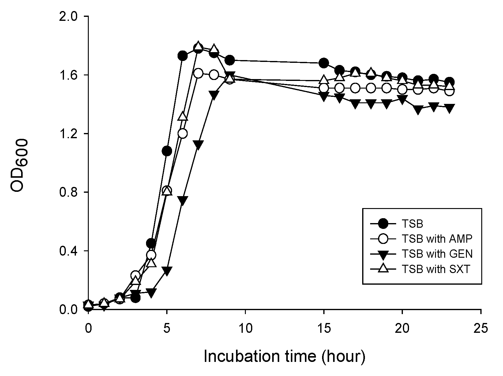
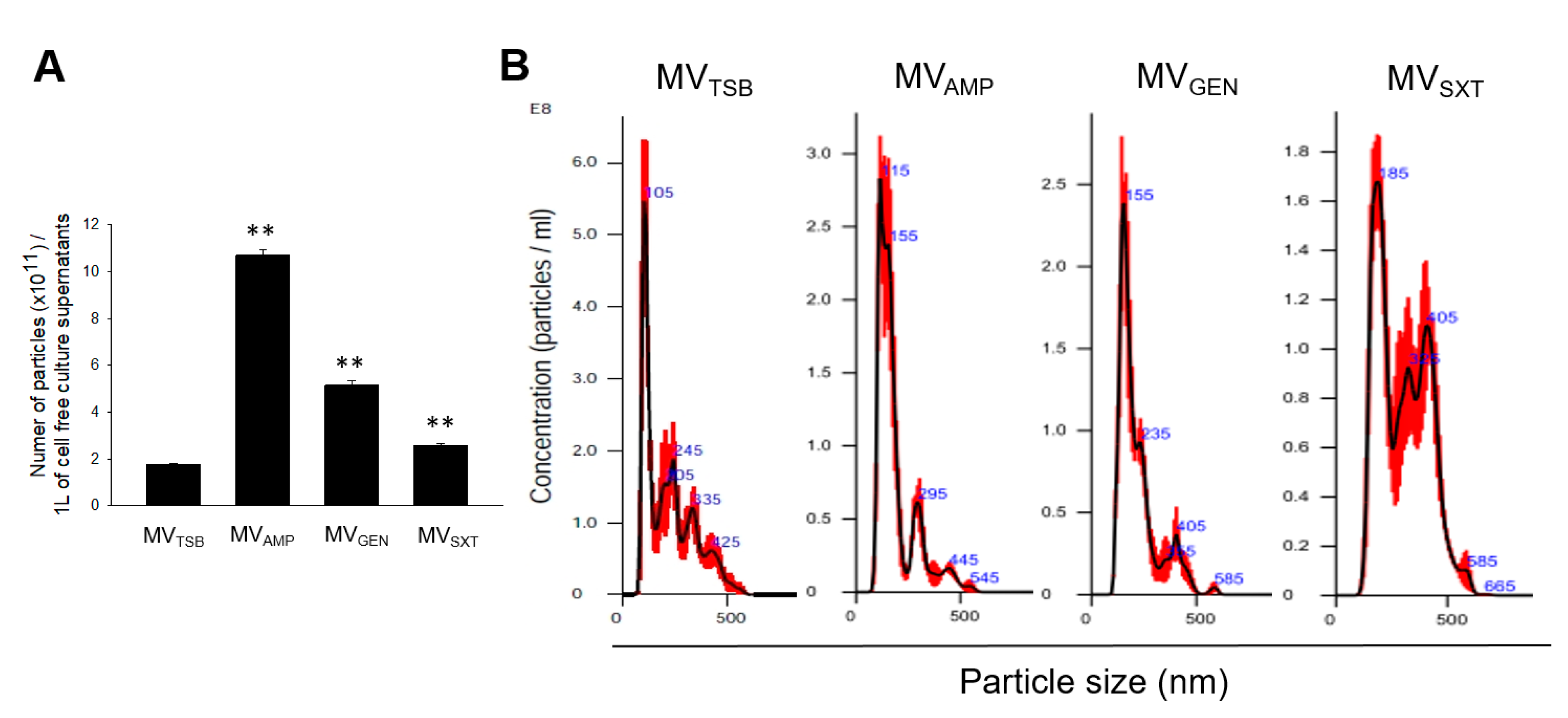
3.1. MV Production in L. monocytogenes Cultured with Sub-Inhibitory Concentrations of Antibiotics

To determine effects of sub-inhibitory concentrations of antibiotics on MV production, L. monocytogenes was cultured in TSB without antibiotics or TSB supplemented with 1/2 MIC of AMP, GEN, or SXT with shaking to reach late exponential phase at OD600 of 1.4 (Figure 1).
MVs were isolated from bacterial culture in TSB without antibiotics (MVTSB) or in TSB with AMP (MVAMP), with GEN (MVGEN), or with SXT (MVSXT). MV samples of MVTSB, MVAMP, MVGEN, and MVSXT obtained from 1 L of cell free culture supernatants contained 1.78 × 10¹¹, 10.7 × 10¹¹, 5.15 × 10¹¹, and 2.59 × 10¹¹ particles, respectively, based on NTA (Figure 2A). Mean sizes of MVTSB, MVAMP, MVGEN, and MVSXT were 106.3 ± 3.7 nm, 132.6 ± 12.4 nm, 156.9 ± 6.9 nm, and 194.4 ± 14.8 nm, respectively (Figure 2B). These results suggest that sub-inhibitory concentrations of antibiotics such as AMP, GEN, and SXT used to treat listeriosis can affect the biogenesis of MVs in L. monocytogenes.

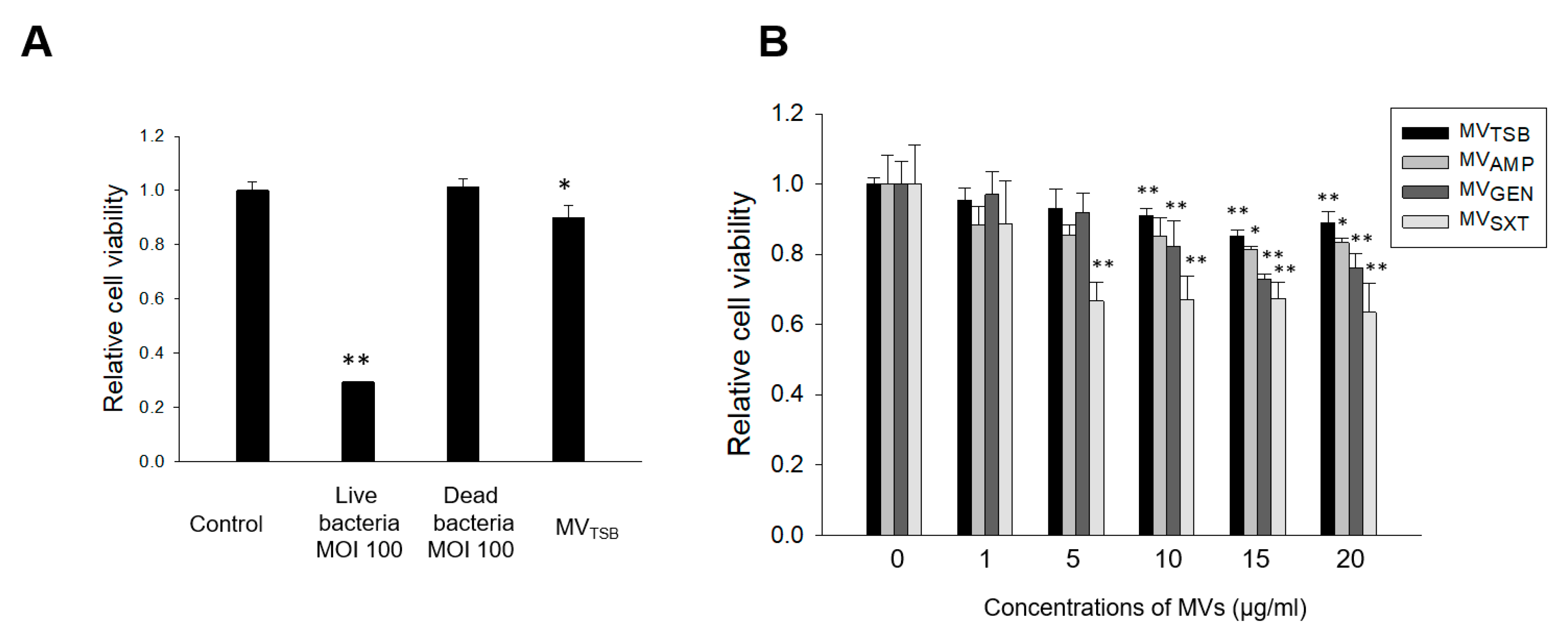
3.2. Host Cell Cytotoxicity Induced by MVs from L. monocytogenes Cultured with Sub-Inhibitory Concentrations of Antibiotics

To determine whether live L. monocytogenes, dead L. monocytogenes, and bacteria-derived MVs could induce cytotoxicity to human colon epithelial cells, Caco-2 cells were treated with live bacteria at multiplicity of infection (MOI) 100, dead bacteria at MOI 100, or 10 μg/mL of MVTSB for 24 h. Cell viability was then analyzed using the MTT assay. Results revealed that live bacteria and 10 μg/mL of MVTSB, but not dead bacteria, induced cytotoxicity to Caco-2 cells (Figure 3A). Next, to determine whether L. monocytogenes MVs could induce cytotoxicity in a dose-dependent manner, Caco-2 cells were treated with various concentrations (1~20 μg/mL) of different MVs for 24 h. No cytotoxicity was observed for cells treated with 1/2 MICs of AMP, GEN, and SXT for 24 h (Figure S1). Cytotoxicity was induced in Caco-2 cells treated with ≥5 µg/mL of MVSXT, ≥10 µg/mL of MVGEN and MVTSB, and ≥15 µg/mL of MVAMP when the cytotoxicity was compared to untreated (no antibiotics) control cells (Figure 3B). MVs derived from L. monocytogenes cultured under three antibiotic stress conditions were more cytotoxic at ≥ 10 µg/mL than MVTSB at the same concentration. Among different MVs, MVSXT exhibited the highest cytotoxicity. These results suggest that L. monocytogenes exposed to sub-inhibitory concentrations of SXT can produce more cytotoxic MVs than bacteria under exposure to sub-inhibitory concentrations of AMP or GEN or no antibiotics.

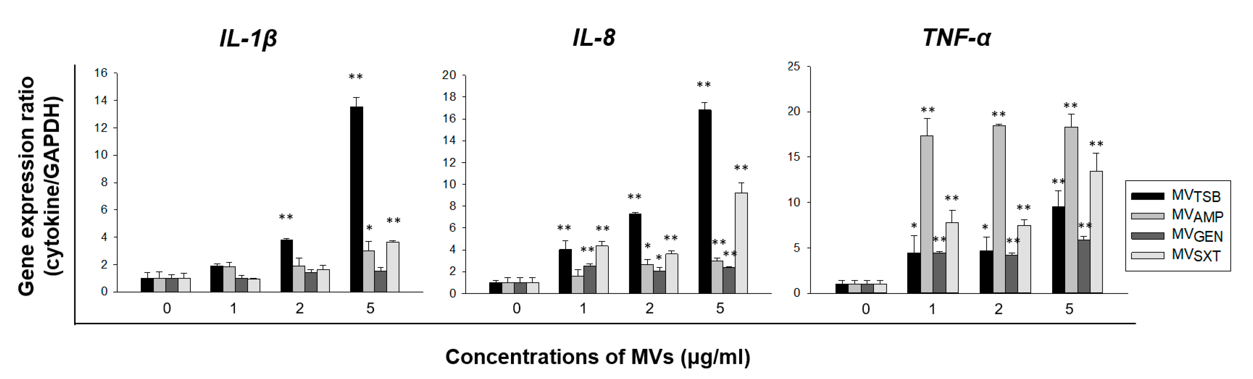
3.3. Difference in Ability to Induce an Innate Immune Response by MVs from L. monocytogenes Cultured with Sub-Inhibitory Concentrations of Antibiotics

To determine whether MVs from L. monocytogenes cultured under antibiotic stress conditions could trigger an innate immune response in vitro, Caco-2 cells were treated with 1, 2, or 5 μg/mL of MVTSB, MVAMP, MVGEN, or MVSXT for 4 h. Expression levels of pro-inflammatory cytokine genes (such as IL-1β and TNF-α) and chemokine gene such as IL-8 were analyzed using qPCR. As shown in Figure 4, MVTSB significantly stimulated the expression of IL-1β and IL-8 genes compared with other MVs, whereas the expression level of TNF-α gene was significantly higher in cells treated with MVAMP than in cells treated with MVTSB, MVGEN, or MVSXT. MVSXT induced more expression of TNF-α gene than MVTSB. These results suggest that sub-inhibitory concentrations of antibiotics can alter MV-induced pro-inflammatory responses in Caco-2 cells. Furthermore, sub-inhibitory concentrations of AMP affected the ability of L. monocytogenes MVs to induce pro-inflammatory response by altering the expression level of TNF-α in Caco-2 cells.

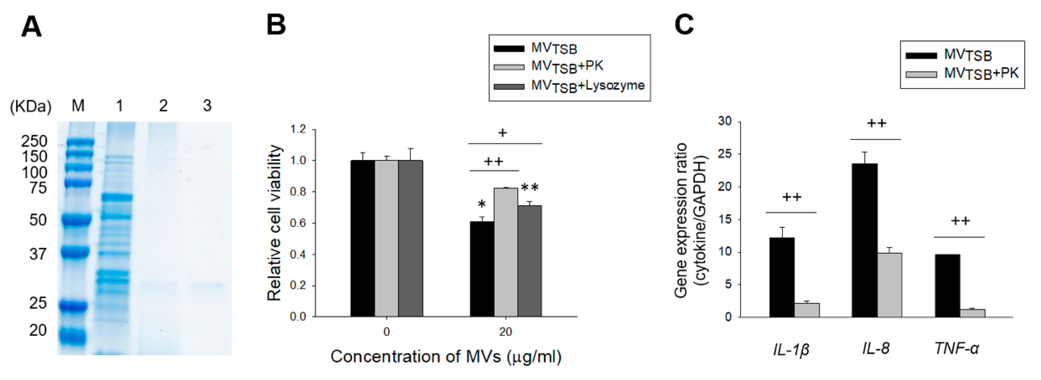
3.4. MV Components Responsible for Cytotoxicity and Innate Immune Response in Host Cells

To determine which L. monocytogenes MV components (proteins or peptidoglycans) were responsible for the cytotoxicity and innate immune response in host cells, MVTSB were treated with proteinase K or lysozyme. As shown in Figure 5A, proteins in MVTSB were degraded by proteinase K treatment. After Caco-2 cells were incubated with intact, proteinase K-treated, or lysozyme-treated MVTSB for 24 h, cytotoxicity to Caco-2 cells was significantly decreased for MVTSB incubated with proteinase K or lysozyme compared to intact MVTSB (Figure 5B). These results suggest that both proteins and peptidoglycans in MVs are responsible for the cytotoxicity of MVs to host cells. Next, Caco-2 cells were incubated with intact or proteinase K-treated MVTSB for 4 h. Expression levels of IL-1β, IL-8, and TNF-α genes were then assessed using qPCR. Expression levels of all tested cytokine genes were significantly decreased in cells incubated with proteinase K-treated MVTSB as compared to those in cells incubated with intact MVTSB (Figure 5C). These results suggest that proteins in MVs are partly responsible for the pro-inflammatory responses in Caco-2 cells.

4. Discussion

The present study demonstrated that L. monocytogenes exposed to sub-inhibitory concentrations of antibiotics used to treat listeriosis produced more MVs than bacteria cultured under no antibiotics in vitro. Furthermore, MVs obtained from bacterial culture under different antibiotics induced different host cell responses regarding cytotoxicity and pro-inflammatory responses. These results suggest that sub-inhibitory concentrations of antibiotics may alter MV-mediated host pathology involved in L. monocytogenes infection.
In previous studies, we have shown that L. monocytogenes can produce MVs during in vitro culture and that this microorganism can actively utilize MVs as a secretion system to interact with host cells or as a cargo of virulence factors [16,17,21]. The size, shape, and amount of L. monocytogenes MVs varied depending on transcription factor σB, bacterial growth stage, and stress type [16,17]. Sigma B is directly responsible for the production of L. monocytogenes MVs [16]. Wild-type strain produced nine times more MVs than ΔsigB mutant strain cultured under carbon starvation stress conditions, whereas salt stress condition did not affect MV production [16,17]. In the present study, sub-inhibitory concentration of antibiotics such as AMP, GEN, or SXT stimulated the production of L. monocytogenes MVs. Especially, bacteria cultured in the presence of AMP produced six times more MV particles than bacteria cultured in the presence of GEN or SXT or no antibiotics (Figure 2A). AMP can inhibit bacterial cell wall synthesis [4]. Biagini et al. [18] have shown that sub-inhibitory concentration of penicillin can stimulate MV production in S. pyogenes by weakening cell walls. Cell wall inhibitors such as β-lactams exhibit more profound effects on MV production in L. monocytogenes than protein synthesis inhibitor GEN or folate pathway inhibitor SXT. In addition, MV size in L. monocytogenes was different between antibiotic and no antibiotic conditions. MVTSB sizes were measured to have an average of 106 nm in diameter, whereas sizes of MVs produced by L. monocytogenes under antibiotic stress conditions ranged from 132 to 194 nm in diameter (Figure 2B). This suggests that MV sizes produced under antibiotic stress conditions are much bigger than MVs produced under no antibiotics. Interestingly, MVSXT exhibited the largest size among different MVs. In Gram-negative bacterium Burkholderia cepacia, MVs obtained from bacterial culture under sub-inhibitory concentrations of SXT also showed the biggest size compared to MVs from bacterial culture under sub-inhibitory concentrations of meropenem or ceftazidime [22]. These results suggest that sub-inhibitory concentrations of folate pathway inhibitors may produce bigger size of EVs in both Gram-positive and Gram-negative bacteria than other classes of antibiotics. Our results suggest that L. monocytogenes can regulate the biogenesis of MVs in different ways when bacteria are exposed to different antibiotics.
We have previously shown that MVs isolated from L. monocytogenes cultured with or without salt stress to early exponential phase (OD600 of 0.5) are not cytotoxic to Caco-2 cells, although they can increase cell viability when cells are treated with MVs ≤ 30 μg/mL [21]. However, in the present study, MVs from L. monocytogenes cultured with or without antibiotics to late exponential phase (OD600 of 1.4) did induce cytotoxicity to Caco-2 host cells in a dose-dependent manner (Figure 3B). Furthermore, the ability of MVs to induce cytotoxicity was different between MVs obtained from different antibiotic stress conditions. MVs produced under antibiotic stress conditions (MVAMP, MVGEN, and MVSXT) were more cytotoxic than MVs produced under no antibiotic conditions. MVSXT showed the highest cytotoxicity among four L. monocytogenes MVs. We have previously shown that salt stress can regulate global protein profile of L. monocytogenes MVs [17]. These results suggest that bacterial culture conditions or bacterial growth stages may regulate molecular components associated with cytotoxicity of L. monocytogenes MVs to host cells. As shown in Figure 5B, cytotoxicities of both proteinase K- and lysozyme treated-L. monocytogenes MVs to host cells were significantly reduced compared to those of intact MVs. Twenty μg/mL of MVTBS significantly induced cell cytotoxicity. However, relative cell viability was lower than that shown in Figure 3B (0.61 ± 0.03 vs. 0.89 ± 0.03). The size of the microplate (96 vs. 24-well microplate) used to measure cytotoxicity might has an influence on the difference of viabilities. These results indicate that proteins and peptidoglycans are directly responsible for the cytotoxicity of L. monocytogenes MVs. Although specific cytotoxic factors in L. monocytogenes MVs were not identified in this study, L. monocytogenes exposed to sub-inhibitory concentrations of antibiotics produced more cytotoxic MVs than bacteria under no antibiotic conditions.
Interaction of L. monocytogenes with intestinal epithelial cells can stimulate the production of pro-inflammatory cytokines such as TNF-α and IL-6 and chemokines such as IL-8 and monocyte chemoattractant protein (MCP)-1 to initiate mucosal inflammatory response [23]. In our previous study [21], we have shown that L. monocytogenes MVs (10 μg/mL) can stimulate the expression of IL-1β, IL-6, TNF-α, IL-8, MCP-1, and MIP-1α genes in Caco-2 cells. Interestingly, expression levels of IL-1β, IL-6, and TNF-α genes in cells treated with MVs produced under no salt stress condition were significantly higher than those in cells treated with MVs produced under salt stress condition. However, expression levels of chemokine genes including IL-8, MCP-1, and MIP-1α were higher in cells treated with MVs produced under salt stress than in cells treated with MVs produced under no salt stress. In the present study, we determined whether L. monocytogenes MVs produced under antibiotic stress conditions stimulated the expression of IL-1β, TNF-α, and IL-8 genes in Caco-2 cells. In agreement with the previous study, MVTSB stimulated the expression IL-1β, TNF-α, and IL-8 genes, although expression levels of these genes in Caco-2 cells treated with MVs produced with antibiotics were significantly different from those in Caco-2 cells treated with MVs produced without antibiotics. Expression levels of IL-1β and IL-8 genes were the highest level in cells treated with MVTSB, whereas the expression level of TNF-α gene was the highest in cells treated with MVAMP (Figure 4). These results suggest that antibiotic stress may alter the pathogenesis of L. monocytogenes by regulating pro-inflammatory responses in epithelial cells responding to secreted MVs. MVs produced by Gram-positive bacteria contain many pathogen-associated molecular patterns (PAMPs) such as peptidoglycans, proteins, and nucleic acid [13,24,25]. These PAMP molecules can be recognized by pattern recognition receptors expressed in host cells and activate the innate immune response through the induction of a variety of pro-inflammatory cytokines [26]. As shown in Figure 5C, MVTSB treated with proteinase K significantly inhibited the expression of IL-1β, TNF-α, and IL-8 genes as compared to intact MVs. Treatment of MVs derived from Enterococcus faecium with proteinase K also significantly reduced the expression of pro-inflammatory cytokine and chemokine genes in Caco-2 cells as compared to treatment with intact MVs [27]. These findings suggest that proteins in the surface or lumens of L. monocytogenes MVs are largely responsible for pro-inflammatory responses in host cells. However, other components in MVs involved in pro-inflammatory responses need to be identified because expression levels of IL-1β, IL-8, and TNF-α genes are not completely inhibited by proteinase K-treated MVs. Our results support that gastrointestinal stresses such as salt or antibiotics may contribute to the diversity of PAMPs or cargo molecules in L. monocytogenes MVs involved in pro-inflammatory responses in host cells.
To treat listeriosis, antimicrobial is essential. However, resistance of L. monocytogenes to commonly used antibiotics has emerged [5,28,29]. The first antibiotic-resistance strain of L. monocytogenes was reported in 1988 [30]. Since then, many resistant strains have been isolated from food, environment, and clinical cases. A recent study has reported that most of isolates of multidrug-resistant L. monocytogenes strains isolated from beef cattle and are resistant to AMP, penicillin, and erythromycin [31]. The present study analyzed the pathology of host cells induced by MVs secreted from L. monocytogenes cultured under different antibiotic conditions. Sub-inhibitory concentrations of AMP, GEN, and SXT increased the production of MVs in L. monocytogenes. Especially, MVSXT exhibited a strong cytotoxic activity towards intestinal epithelial cells, whereas MVAMP induced strong pro-inflammatory responses via the expression of TNF-α gene. Our findings may help us understand the biogenesis of MVs and their contribution to pathogenesis of L. monocytogenes under antibiotic stress conditions that may encounter during an in vivo infection. The challenge of future studies is to identify cargo molecules associated with host cell cytotoxicity and pro-inflammatory responses in L. monocytogenes MVs.

Supplementary Materials

The following are available online at https://0-www-mdpi-com.brum.beds.ac.uk/2073-4425/12/3/415/s1, Figure S1: Viabilities of Caco-2 cells treated with 1/2 MICs of antibiotics.

Author Contributions

Conceptualization, J.-C.L. and J.-H.S.; methodology, J.-H.W., S.K., T.L. and J.-C.L.; formal analysis, J.-H.W., S.K., T.L., J.-C.L. and J.-H.S.; resources, J.-H.W., S.K., T.L., J.-C.L. and J.-H.S.; writing—original draft preparation, J.-C.L. and J.-H.S.; writing—review and editing, J.-C.L. and J.-H.S.; supervision, J.-C.L. and J.-H.S.; funding acquisition, J.-H.S. All authors have read and agreed to the published version of the manuscript.

Funding

This research was supported by a grant (2018R1D1A3B07045611) of the Basic Science Research Program through the National Research Foundation (NRF) funded by the Ministry of Education, Republic of Korea.

Conflicts of Interest

The authors declare no conflict of interest.

References

  1. Schlech, W.F., 3rd. Foodborne listeriosis. Clin. Infect. Dis. Off. Publ. Infect. Dis. Soc. Am. 2000, 31, 770–775. [Google Scholar] [CrossRef]
  2. Vazquez-Boland, J.A.; Kuhn, M.; Berche, P.; Chakraborty, T.; Dominguez-Bernal, G.; Goebel, W.; Gonzalez-Zorn, B.; Wehland, J.; Kreft, J. Listeria pathogenesis and molecular virulence determinants. Clin. Microbiol. Rev. 2001, 14, 584–640. [Google Scholar] [CrossRef] [Green Version]
  3. Temple, M.E.; Nahata, M.C. Treatment of listeriosis. Ann. Pharmacother. 2000, 34, 656–661. [Google Scholar] [CrossRef]
  4. Hof, H.; Nichterlein, T.; Kretschmar, M. Management of listeriosis. Clin. Microbiol. Rev. 1997, 10, 345–357. [Google Scholar] [CrossRef] [PubMed]
  5. Charpentier, E.; Courvalin, P. Antibiotic resistance in Listeria spp. Antimicrob. Agents Chemother. 1999, 43, 2103–2108. [Google Scholar] [CrossRef] [Green Version]
  6. Janakiraman, V. Listeriosis in pregnancy: Diagnosis, treatment, and prevention. Rev. Obstet. Gynecol. 2008, 1, 179–185. [Google Scholar] [PubMed]
  7. Spitzer, P.G.; Hammer, S.M.; Karchmer, A.W. Treatment of Listeria monocytogenes infection with trimethoprim-sulfamethoxazole: Case report and review of the literature. Rev. Infect. Dis. 1986, 8, 427–430. [Google Scholar] [CrossRef] [PubMed]
  8. Scheer, M.S.; Hirschman, S.Z. Oral and ambulatory therapy of Listeria bacteremia and meningitis with trimethoprim-sulfamethoxazole. Mt. Sinai J. Med. N. Y. 1982, 49, 411–414. [Google Scholar]
  9. Bishop, D.G.; Work, E. An extracellular glycolipid produced by Escherichia coli grown under lysine-limiting conditions. Biochem. J. 1965, 96, 567–576. [Google Scholar] [CrossRef] [PubMed] [Green Version]
  10. Knox, K.W.; Vesk, M.; Work, E. Relation between excreted lipopolysaccharide complexes and surface structures of a lysine-limited culture of Escherichia coli. J. Bacteriol. 1966, 92, 1206–1217. [Google Scholar] [CrossRef] [Green Version]
  11. Kuehn, M.J.; Kesty, N.C. Bacterial outer membrane vesicles and the host-pathogen interaction. Genes Dev. 2005, 19, 2645–2655. [Google Scholar] [CrossRef] [Green Version]
  12. Kulp, A.; Kuehn, M.J. Biological functions and biogenesis of secreted bacterial outer membrane vesicles. Annu. Rev. Microbiol. 2010, 64, 163–184. [Google Scholar] [CrossRef] [PubMed] [Green Version]
  13. Lee, E.Y.; Choi, D.Y.; Kim, D.K.; Kim, J.W.; Park, J.O.; Kim, S.; Kim, S.H.; Desiderio, D.M.; Kim, Y.K.; Kim, K.P.; et al. Gram-positive bacteria produce membrane vesicles: Proteomics-based characterization of Staphylococcus aureus-derived membrane vesicles. Proteomics 2009, 9, 5425–5436. [Google Scholar] [CrossRef]
  14. Rivera, J.; Cordero, R.J.; Nakouzi, A.S.; Frases, S.; Nicola, A.; Casadevall, A. Bacillus anthracis produces membrane-derived vesicles containing biologically active toxins. Proc. Natl. Acad. Sci. USA 2010, 107, 19002–19007. [Google Scholar] [CrossRef] [PubMed] [Green Version]
  15. Nicholas, A.; Jeon, H.; Selasi, G.N.; Na, S.H.; Kwon, H.I.; Kim, Y.J.; Choi, C.W.; Kim, S.I.; Lee, J.C. Clostridium difficile-derived membrane vesicles induce the expression of pro-inflammatory cytokine genes and cytotoxicity in colonic epithelial cells in vitro. Microb. Pathog. 2017, 107, 6–11. [Google Scholar] [CrossRef]
  16. Lee, J.H.; Choi, C.W.; Lee, T.; Kim, S.I.; Lee, J.C.; Shin, J.H. Transcription factor σB plays an important role in the production of extracellular membrane-derived vesicles in Listeria monocytogenes. PLoS ONE 2013, 8, e73196. [Google Scholar] [CrossRef]
  17. Lee, T.; Jun, S.H.; Choi, C.W.; Kim, S.I.; Lee, J.C.; Shin, J.H. Salt stress affects global protein expression profiles of extracellular membrane-derived vesicles of Listeria monocytogenes. Microb. Pathog. 2018, 115, 272–279. [Google Scholar] [CrossRef]
  18. Biagini, M.; Garibaldi, M.; Aprea, S.; Pezzicoli, A.; Doro, F.; Becherelli, M.; Taddei, A.R.; Tani, C.; Tavarini, S.; Mora, M.; et al. The Human Pathogen Streptococcus pyogenes Releases Lipoproteins as Lipoprotein-rich Membrane Vesicles. Mol. Cell. Proteom. Mcp 2015, 14, 2138–2149. [Google Scholar] [CrossRef] [PubMed] [Green Version]
  19. Andreoni, F.; Toyofuku, M.; Menzi, C.; Kalawong, R.; Mairpady Shambat, S.; Francois, P.; Zinkernagel, A.S.; Eberl, L. Antibiotics Stimulate Formation of Vesicles in Staphylococcus aureus in both Phage-Dependent and -Independent Fashions and via Different Routes. Antimicrob. Agents Chemother. 2019, 63, e01418–e01439. [Google Scholar] [CrossRef] [PubMed] [Green Version]
  20. Clinical and Laboratory Standards Institute. Performance Standards for Antimicrobial Susceptibility Testing; M100 ED29:2019; CLSI: Wayne, PA, USA, 2019. [Google Scholar]
  21. Jun, S.H.; Lee, T.; Lee, J.C.; Shin, J.H. Different epithelial cell response to membrane vesicles produced by Listeria monocytogenes cultured with or without salt stress. Microb. Pathog. 2019, 133, 103554. [Google Scholar] [CrossRef]
  22. Kim, S.Y.; Kim, M.H.; Son, J.H.; Kim, S.I.; Yun, S.H.; Kim, K.; Kim, S.; Shin, M.; Lee, J.C. Outer membrane vesicles produced by Burkholderia cepacia cultured with subinhibitory concentrations of ceftazidime enhance pro-inflammatory responses. Virulence 2020, 11, 995–1005. [Google Scholar] [CrossRef]
  23. Jung, H.C.; Eckmann, L.; Yang, S.K.; Panja, A.; Fierer, J.; Morzycka-Wroblewska, E.; Kagnoff, M.F. A distinct array of proinflammatory cytokines is expressed in human colon epithelial cells in response to bacterial invasion. J. Clin. Investig. 1995, 95, 55–65. [Google Scholar] [CrossRef] [PubMed] [Green Version]
  24. Jun, S.H.; Lee, J.H.; Kim, S.I.; Choi, C.W.; Park, T.I.; Jung, H.R.; Cho, J.W.; Kim, S.H.; Lee, J.C. Staphylococcus aureus-derived membrane vesicles exacerbate skin inflammation in atopic dermatitis. Clin. Exp. Allergy J. Br. Soc. Allergy Clin. Immunol. 2017, 47, 85–96. [Google Scholar] [CrossRef] [PubMed]
  25. Liu, Y.; Defourny, K.A.Y.; Smid, E.J.; Abee, T. Gram-Positive Bacterial Extracellular Vesicles and Their Impact on Health and Disease. Front. Microbiol. 2018, 9, 1502. [Google Scholar] [CrossRef] [PubMed] [Green Version]
  26. Kumar, H.; Kawai, T.; Akira, S. Pathogen recognition by the innate immune system. Int. Rev. Immunol. 2011, 30, 16–34. [Google Scholar] [CrossRef]
  27. Kim, M.H.; Kim, S.Y.; Son, J.H.; Kim, S.I.; Lee, H.; Kim, S.; Shin, M.; Lee, J.C. Production of Membrane Vesicles by Enterococcus faecium Cultured With or Without Subinhibitory Concentrations of Antibiotics and Their Pathological Effects on Epithelial Cells. Front. Cell. Infect. Microbiol. 2019, 9, 295. [Google Scholar] [CrossRef] [Green Version]
  28. Lungu, B.; O’Bryan, C.A.; Muthaiyan, A.; Milillo, S.R.; Johnson, M.G.; Crandall, P.G.; Ricke, S.C. Listeria monocytogenes: Antibiotic resistance in food production. Foodborne Pathog. Dis. 2011, 8, 569–578. [Google Scholar] [CrossRef] [PubMed]
  29. Luque-Sastre, L.; Arroyo, C.; Fox, E.M.; McMahon, B.J.; Bai, L.; Li, F.; Fanning, S. Antimicrobial Resistance in Listeria Species. Microbiol. Spectr. 2018, 6. [Google Scholar]
  30. Poyart-Salmeron, C.; Carlier, C.; Trieu-Cuot, P.; Courtieu, A.L.; Courvalin, P. Transferable plasmid-mediated antibiotic resistance in Listeria monocytogenes. Lancet 1990, 335, 1422–1426. [Google Scholar] [CrossRef]
  31. Obaidat, M.M. Prevalence and antimicrobial resistance of Listeria monocytogenes, Salmonella enterica and Escherichia coli O157:H7 in imported beef cattle in Jordan. Comp. Immunol. Microbiol. Infect. Dis. 2020, 70, 101447. [Google Scholar] [CrossRef]
Figure 1. Growth of L. monocytogenes under antibiotic stress conditions. Bacteria were cultured in tryptic soy broth (TSB) without antibiotics, TSB with 0.25 μg/mL ampicillin (AMP), TSB with 0.25 μg/mL gentamicin (GEN), or TSB with 0.25/1.25 μg/mL trimethoprim/sulfamethoxazole (SXT) at 37 °C with shaking. Bacterial growth was measured at OD600 at indicated time points. Data are representative of three independent experiments with similar results.
Genes 12 00415 g001
Figure 2. Production of membrane vesicles (MVs) in L. monocytogenes cultured under antibiotic stress conditions. Bacteria were grown to late exponential phase (OD600 of 1.4). MVs were then isolated from bacterial culture supernatants. Nano particle tracking analysis (NTA) was performed to determine the number and size of MVs. (A) Particle numbers of MVTSB, MVAMP, MVGEN, and MVSXT in 1 L of cell free culture supernatants. Data are presented as mean ± SD of three independent experiments. **, p < 0.01 compared to MVTSB. (B) Size distribution of MVTSB, MVAMP, MVGEN, and MVSXT based on NTA. Data are representative of three independent experiments with similar results.
Genes 12 00415 g002
Figure 3. Cytotoxicities of bacteria and MVs from L. monocytogenes cultured with or without antibiotics to Caco-2 cells. (A) L. monocytogenes was cultured in TSB to reach 1.0 at OD600. Caco-2 cells were then treated with live bacteria at multiplicity of infection (MOI) 100, dead bacteria at MOI 100, and MVTSB (10 μg/mL) for 24 h. Dead bacteria were prepared by fixation with 10% formalin for 20 h. (B) Caco-2 cells were treated with various concentration of MVAMP, MVGEN, or MVSXT for 24 h. Cell viability was determined using an MTT assay. Data are presented as mean ± SD of three independent experiments. *, p < 0.05; **, p < 0.01 compared to untreated control cells.
Genes 12 00415 g003
Figure 4. Expression levels of pro-inflammatory cytokine and chemokine genes in Caco-2 cells treated with L. monocytogenes MVs produced with or without antibiotics. Caco-2 cells were treated with various concentrations of L. monocytogenes MVs for 4 h. Gene expression was then assessed via qPCR. Data are presented as mean ± SD of three independent experiments. *, p < 0.05; **, p < 0.01 compared to untreated control cells.
Genes 12 00415 g004
Figure 5. Host cell responses to proteinase K-treated L. monocytogenes MVs. (A) SDS-PAGE analysis of MV proteins. L. monocytogenes MVs were treated with 0.1 mg/mL proteinase K for 3 h at 50 °C for the degradation of MV proteins. M, molecular weight marker; 1, intact MVTSB; 2, proteinase K-treated MVTSB; 3, proteinase K. (B) Caco-2 cells were incubated with intact (MVTSB), proteinase K-treated MVs (MVTSB + PK), or lysozyme-treated MVs (MVTSB + Lysozyme) for 24 h. Cell viability was then determined using an MTT assay. Data are presented as mean ± SD of three independent experiments. *, p < 0.05; **, p < 0.01 compared to untreated control cells. +, p < 0.05; ++, p < 0.01 compared to 20 µg/mL of MVTSB. (C) Caco-2 Cells were incubated with intact or proteinase K-treated MVTSB for 4 h. Gene expression was then assessed using qPCR. Data are presented as mean ± SD of three independent experiments. ++, p < 0.01 compared to intact MVTSB.
Genes 12 00415 g005
Publisher’s Note: MDPI stays neutral with regard to jurisdictional claims in published maps and institutional affiliations.

