Article

A Missense Mutation in the KLF7 Gene Is a Potential Candidate Variant for Congenital Deafness in Australian Stumpy Tail Cattle Dogs

1
Institute of Veterinary Medicine, University of Goettingen, 37077 Göttingen, Germany
2
School of Veterinary Science, The University of Queensland, Gatton, QLD 4343, Australia
*
Author to whom correspondence should be addressed.
These authors have equally contributed.
Submission received: 19 November 2020 / Revised: 21 March 2021 / Accepted: 23 March 2021 / Published: 24 March 2021
(This article belongs to the Special Issue Canine Genetics 2)

Abstract

:
Congenital deafness is prevalent among modern dog breeds, including Australian Stumpy Tail Cattle Dogs (ASCD). However, in ASCD, no causative gene has been identified so far. Therefore, we performed a genome-wide association study (GWAS) and whole genome sequencing (WGS) of affected and normal individuals. For GWAS, 3 bilateral deaf ASCDs, 43 herding dogs, and one unaffected ASCD were used, resulting in 13 significantly associated loci on 6 chromosomes, i.e., CFA3, 8, 17, 23, 28, and 37. CFA37 harbored a region with the most significant association (−log10(9.54 × 10⁻²¹) = 20.02) as well as 7 of the 13 associated loci. For whole genome sequencing, the same three affected ASCDs and one unaffected ASCD were used. The WGS data were compared with 722 canine controls and filtered for protein coding and non-synonymous variants, resulting in four missense variants present only in the affected dogs. Using effect prediction tools, two variants remained with predicted deleterious effects within the Heart development protein with EGF like domains 1 (HEG1) gene (NC_006615.3: g.28028412G>C; XP_022269716.1: p.His531Asp) and Kruppel-like factor 7 (KLF7) gene (NC_006619.3: g.15562684G>A; XP_022270984.1: p.Leu173Phe). Due to its function as a regulator in heart and vessel formation and cardiovascular development, HEG1 was excluded as a candidate gene. On the other hand, KLF7 plays a crucial role in the nervous system, is expressed in the otic placode, and is reported to be involved in inner ear development. 55 additional ASCD samples (28 deaf and 27 normal hearing dogs) were genotyped for the KLF7 variant, and the variant remained significantly associated with deafness in ASCD (p = 0.014). Furthermore, 24 dogs with heterozygous or homozygous mutations were detected, including 18 deaf dogs. The penetrance was calculated to be 0.75, which is in agreement with previous reports. In conclusion, KLF7 is a promising candidate gene causative for ASCD deafness.

1. Introduction

Deafness can cause several inconveniences for dogs (Canis familiaris, CFA), as more attention is required to avoid undetected danger. Deaf dogs are not suitable as working dogs because their training is more challenging than for normal hearing dogs. In addition, they are more likely to be startled and show more tendency to bite [1]. More than 100 modern dog breeds have been reported to be affected by congenital deafness [2]. Hence, deafness seems to be a common disorder among dogs, particularly in breeds such as the Dalmatian, Bull Terrier, English Setter, English Cocker Spaniel, and Australian Cattle Dog [3]. Hearing loss or deafness can be categorized mainly by five criteria in dogs: (1) Cause (genetic or nongenetic, inherited or acquired); (2) association with other diseases or phenotypes (syndromic or non-syndromic); (3) number of affected ears (unilateral or bilateral); (4) degree of loss (partial or total); and (5) site of pathology (peripheral or central) [4]. Peripheral deafness can also be classified as inherited or acquired, congenital or late onset, and sensorineural or conductive. In dogs, three classifications of deafness are commonly seen, including inherited congenital sensorineural, acquired later-onset sensorineural, and acquired later-onset conductive deafness [5].
In dogs, congenital sensorineural deafness is common, resulting in total deafness in young puppies that is either unilateral or bilateral. Sensorineural deafness results from dysfunction of cochlea or spiral ganglion. While it can be a degenerative process that relates to aging, noise trauma, exposure to therapeutic drugs that have ototoxic side effects, and chronic conditions [6], it is frequently inherited and so linked to one or more genetic mutations. Some morphological studies in dogs showed congenital sensorineural deafness manifested hypoplasia or aplasia of the sensory cells in the organ of Corti, stria vascularis, macula saccule, solidification, and calcification of tectorial membrane [7,8]. Congenital sensorineural deafness is usually, but not always, related to pigmentation genes in some breeds [3].
Diagnosis of canine deafness usually consists of behavioral or electrodiagnostic testing. The behavioral testing is often unreliable, especially for the unilateral deafness or partial hearing impairment cases. The response of dogs may be affected by psychology (e.g., anxiety or loss of interest) and other senses (e.g., visual cues, vibration, or even air movement) [9]. The brainstem auditory evoked response (BAER) is the averaged record of the electrical activity of the auditory pathway in response to externally applied acoustic stimuli [10]. Compared with behavioral testing, the BAER test is an objective diagnostic method, with the advantages of being easy to record, noninvasive, safe, short test time, and giving reliable results [11].
The Australian Stumpy Tail Cattle Dog (ASCD) is a unique breed with a natural bob-tail, which should be distinguished from the Australian Cattle Dog breed. ASCD is alert, watchful and obedient, and talented in working and controlling cattle. It has been recognized as a standardized breed since 1963 by the Australian National Kennel Council. For a long time, general opinion held that the origins of the Australian Stumpy Tail Cattle Dog arose from European herding dogs and the Australian Dingo. However, recently it has been suggested that the ancestors of the Australian Stumpy Tail Cattle Dog and the Australian Cattle Dog, sharing a common origin, arrived in Australia with early free settlers, as their unidentified companions, between 1788 and c. 1800 (Clark, Noreen R. A Dog for the Job. (in prep. 2020)). Each pup should undergo a BAER test because this breed has a high deafness prevalence (https://www.akc.org/dog-breeds/australian-stump-tail-cattle-dog/ (accessed on 24 March 2021)). A research study of 315 ASCDs showed the incidence of congenital sensorineural deafness was 17.8% [12]. There was no evidence that congenital sensorineural deafness in ASCD has a left/right asymmetry or a sex-specific pattern, but there was a significant correlation between red (over blue) coat color and deafness [12].
No unique causative variants have been identified so far for any dog breeds, possibly in part due to the fact that deafness appears to be a comparatively heterogenous disease as described above. In addition, there are several hypotheses about the inheritance pattern of congenital sensorineural deafness (reviewed by [1]). In Border Collies, for instance, Ubiquitin Specific Peptidase 31 (USP31) and RB Binding Protein 6 (RBBP6) have been associated with adult-onset deafness [13], whereas in the Doberman Pinscher, an insertion in Protein Tyrosine Phosphatase Receptor Type Q (PTPRQ) and a missense variant in Myosin VIIA (MYO7A) have been shown to be causative for a form of deafness that includes vestibular disease [14,15]. Although chromosome 2 (CFA2), 6, 14, 17, 27, and 29 have been associated with hearing loss in Dalmatians, no causative variants have been identified so far [16].
In ASCD, congenital sensorineural deafness has been linked to a chromosomal region on CFA10 [12]. However, within a potential candidate gene Sry-related Hmg-box gene 10 (SOX10) located in this region, no causative alterations were detected. A recent genome-wide association study (GWAS) reported 14 chromosomes that were significantly associated with deafness in three canine breeds, and CFA3 was significantly associated with bilateral deafness in Australian Cattle Dogs [17]. In this study, three suggestive candidate genes near significantly associated regions were detected in these three dog breeds, including ATPase Na+/K+ Transporting Subunit Alpha 4 (ATP1A4), Transformation/Transcription Domain Associated Protein (TRRAP), and Potassium Inwardly Rectifying Channel Subfamily J Member 10 (KCNJ10) [17]. However, none have been convincingly identified as causative mutations.
To extend the identification of potential candidate genes causing deafness in ASCD we performed a genome-wide association study and whole genome sequencing (WGS) in deaf ASCD. We identified a unique missense variant in Kruppel-like factor 7 (KLF7) gene significantly associated with deafness in ASCDs. This variant was absent in 722 dogs of bioproject PRJN448733 (see below). As KLF7 plays an important role in the nervous system, is expressed in the inner ear, and seems to be involved in inner ear development [18,19], it was a convincing candidate for ASCD deafness.

2. Materials and Methods

2.1. Ethical Statement

The collection of dog blood samples was done by S. Sommerlad at the time of BAER testing. The collection of samples was approved by the ‘‘Niedersächsisches Landesamt für Verbraucherschutz und Lebensmittelsicherheit” (33.19-42502-05-15A506) according to §8a Abs. 1 Nr. 2 of the TierSchG. All ASCDs were tested and sampled under approval of The University of Queensland’s Animal Ethics Committee.

2.2. Phenotyping and Samples

Fifty-nine Australian Stumpy Tail Cattle Dogs (Table S1) from a previous study [12] were used in this study. BAER testing was performed on 59 dogs [20], 28 were normal hearing dogs and 31 were diagnosed as deaf, of which 10 were bilateral deaf, 12 were left-sided deaf, and 9 were right-sided deaf (Table S1). Three bilaterally deaf ASCDs (#217, #253 and #330), and one control dog with normal hearing (#326) were used for next generation sequencing. Dog #326 was a littermate of #330. These four dogs were female and red in color; all but #330 had a speckled coat. DNA was extracted using a salting-out method as described [12]. All samples were pseudonymized using internal IDs. Furthermore, data from two repository were used in this study. One repository contain Variant Call Format (VCF) data of 722 canine individuals (https://0-www-ncbi-nlm-nih-gov.brum.beds.ac.uk/bioproject/PRJNA448733 (accessed on 24 March 2021)) [21]. It consists of 144 established breeds, 11 samples with mixed breed, 26 samples with unknown breed status, 104 village and feral dogs from different regions, and 54 wild canids from six species. An additional dataset consisted of 590 samples including 582 dogs from 126 breeds and 8 wolves (https://www.ebi.ac.uk/ena/data/view/PRJEB32865 (accessed on 24 March 2021)) [22].

2.3. Next Generation Sequencing and Variant Calling

A total of 1.0 μg DNA per ASCD sample was used as input material for the DNA library preparations. Sequencing libraries were generated using NEBNext® DNA Library Prep Kit following manufacturer’s recommendations and indices were added to each sample. The genomic DNA was randomly fragmented to a size of 350bp by shearing, then DNA fragments were end polished, A-tailed, and ligated with the NEBNext adapter for Illumina sequencing, and further PCR enriched by P5 and indexed P7 oligos. The PCR products were purified (AMPure XP system) and resulting libraries were analyzed for size distribution by Agilent 2100 Bioanalyzer and quantified using real-time PCR. For #217, #253, #326, #330, a total of 599,770,692, 723,624,660, 743,641,356, 620,101,998 raw reads were obtained, respectively. Corresponding coverages were around 40× (paired-end reads, 2 × 150 bp).
Raw sequence data were aligned to dog genome CanFam3.1 using BWA 0.7.17 [23]. SAMtools 1.9 were used for format change and sorting of sequences [24]. Duplicates were marked by PICARD (http://broadinstitute.github.io/picard/ (accessed on 24 March 2021)). Variant calling was performed by GATK 4.1.3 with best practice pipeline [25].

2.4. Genome Wide Association Analysis

We used the VCF data obtained in the previous step for GWAS analysis. Three deaf dogs (#217, #253, #330) were used as cases. As ASCD is utilized for control and herding of cattle according to its breed standard (http://www.fci.be/Nomenclature/Standards/351g01-en.pdf (accessed on 24 March 2021)), VCFs of 43 herding dogs from 15 breeds (Australian Cattle Dog, Bearded Collie, Belgian Malinois, Belgian Sheepdog, Belgian Tervuren, Berger Blanc Suisse, Berger Picard, Border Collie, Bouvier des Flandres, Entlebucher Sennenhund, Finnish Lapphund, German Shepherd Dog, Pembroke Welsh Corgi, Shetland Sheepdog, Spanish Water Dog) were extracted from the publicly available 722 canine VCF repository (https://0-www-ncbi-nlm-nih-gov.brum.beds.ac.uk/bioproject/PRJNA448733 (accessed on 24 March 2021)) [21]. Sample selection criteria were the same as described [21]. A total of 43 herding dogs and the normal hearing dog #326 were chosen as controls (Table S2). The VCF files of the 43 herding dogs and 4 ASCDs were merged by BCFtools 1.9 [24]. Filtering was done using VCFtools 0.1.13 with options --max-alleles 2, --min-alleles 2, --min-meanDP 20, --minQ 20, --minGQ 20, --remove-indels, --max-missing 0.95, --maf 0.05, --hwe 0.001 [26]. After filtering, 857,343 variants remained and were further pruned by Linkage Disequilibrium with –indep 1000 3 1 function in PLINK 1.90 [27]. The final data set consisted of 20,656 single nucleotide polymorphisms (SNPs). Principal component analysis (PCA) was performed using EIGENSOFT package [28]. GEMMA 0.98 was used for association analysis by case–control setting (3 deaf cases vs. one normal hearing ASCD and 43 herding dogs as controls) [29]. A univariate linear mixed model with sex, 5 principal components, and relatedness of 47 dog individuals for corrections was applied for the association test. Bonferroni threshold −log10P (0.01/20,656) = 6.32 was utilized. Qqman package was used to generate Manhattan and quantile–quantile (QQ) plots [30]. The genomic inflation factor lambda was calculated with formula lambda = median (qchisq(1-p, 1))/qchisq(0.5, 1) where p is a vector of p values.

2.5. Next Generation Sequencing Data Analysis for Identification of Associated Variants

Data after variant calling were analyzed with SNP & Variation Suite 8.8.3 (Golden Helix Inc., Bozeman, MT, USA). SNPs and indels were set to missing with read depth ≤ 10, genotype quality ≤ 15, alt read ratios for Ref_Ref ≥ 0.15, Ref_Alt outside 0.3 to 0.7, Alt_Alt ≤ 0.85. Variants were analyzed using autosomal recessive and dominant models, respectively. In the autosomal recessive filtering model, 3 deaf ASCDs were set as Alt_Alt, control ASCD as Ref_Ref or Alt_Ref. In the autosomal dominant filtering model, the 3 deaf ASCDs were set as Alt_Alt or Alt_Ref and controls as Ref_Ref. To further narrow the range of candidate variants, we compared the common variants of deaf ASCDs with 722 canine genomes to identify private variants. The shared variants in the three deaf dogs were filtered by BCFtools 1.9 with ‘isec’ option. Private variants were annotated using SnpEFF software [31] to determine high (loss of function) and moderate (missense) impact variants (Ensembl transcripts release 101). These functional variants were further checked by Integrative Genome Viewer (IGV) software to obtain real high quality variants [32]. Variant effects were predicted by SIFT [33], PolyPhen-2 [34], and PROVEAN [35].

2.6. Genotyping of KLF7 Variant in Australian Stumpy Tail Cattle Dogs

Targeted genotyping of the KLF7 missense variant was performed in 59 ASCDs by PCR amplification using primers cfa_KLF7_Ex3_F (5′-AGACTCTCTCAGCCGTGGAT-3′) and cfa_KLF7_Ex3_R (5′-GGCCAACTTGTACCACTACCT-3′), resulting in a 295 bp fragment. Genotyping of PCR products were implemented by RFLP analysis after cleavage with the restriction enzyme HinP1I (NEB). The wild type allele was cleaved into two fragments, 236 bp and 59 bp, while the homozygous mutant remained uncut. Frequency distribution for alleles and genotypes was calculated using Fisher’s Exact Test in these 59 ASCDs. Allelic and genotypic odds ratios were calculated according to [36].

2.7. Investigation of Human Deafness Genes in 3 Deaf Australian Stumpy Tail Cattle Dogs

Human hearing loss or deafness genes were queried using online software GLAD4U with “hearing loss” and/or “deafness” as keywords [37]. After combining the three query results, 346 genes were chosen for further analysis (Table S3). The variants of these gene regions (including 1000 bp up- and downstream regions) were extracted by BCFtools from VCF files of the three deaf ASCDs and annotated by SnpEFF software. Variants with high (loss of function) and moderate (missense) impacts were selected for further analysis (Ensembl transcripts release 101). The genotype information of the chosen variants was further checked in 722 canines.

3. Results

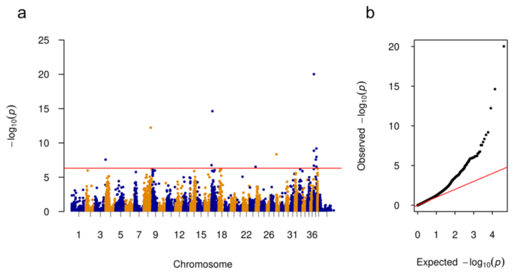
3.1. Genome Wide Association Analysis

The analysis was done using three bilateral deaf female dogs from three different litters. The hearing status of the individuals determined using BAER is shown in Table 1 and Table S1.
The three affected ASCDs were compared with 44 control dogs. 13 SNPs on 6 chromosomes (CFA3, 8, 17, 23, 28, 37) above the Bonferroni significance level were identified. The QQ-plot indicated that some associations might be due to population substructure. Associated SNPs are shown in Figure 1 and summarized in Table 2. The majority of the significantly associated SNPs (7/13) were located on CFA37 including SNP chr37:44793 (position according to CanFam3.1) with the highest −log10p-value = 20.02. A search for large structural variants (SVs) flanking the significantly associated regions on CFA3, 8, 17, 23, 28, and 37 using IGV was unsuccessful.

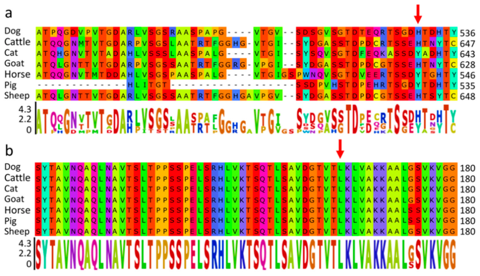
3.2. Whole Genome Sequencing Reveals Four Potential Variants

To further locate the candidate variants, next generation sequencing was performed in 3 deaf ASCDs (#217, #253, #330) and 1 normal hearing ASCD (#326). After quality control, a total of 4,208,002 SNPs and 2,298,760 indels were detected. According to previous deafness studies, sequence data were initially analyzed using a recessive model of inheritance. Using this model, 129,383 SNPs and 51,942 indels were detected. Using only variants that had been annotated and verified as mRNA transcripts (Ensembl release 101), 338 SNPs and 523 indels remained (Table S4). After filtering these variants against the 722 dog database, none of the homozygous Alt_Alt genotypes were exclusively present in the deaf ASCDs (Table S4). As there were no reports about such a high prevalence of deafness in the 722 control dogs and it can be assumed that the majority of the controls were hearing, these variants were presumably not causative.
As no associated variants were found using the recessive inheritance model, a dominant inheritance model was applied. In this analysis, private variants only present in the three deaf ASCDs (Alt_Alt and Alt_Ref) compared to the 722 controls (Ref_Ref) were filtered, resulting in 270,980 SNPs and 351,927 indels. After quality control and functional annotating, 167 protein-changing variants (58 SNPs and 109 indels) remained (Table S5). These variants were further filtered against #326 (normal hearing littermate of #330) assuming that this dog should be homozygous wild type under the supposed model. After this step, four missense variants remained as potential causative candidates (Table 3). Within the 722 control dogs, no homozygous Alt_Alt or heterozygous carriers were detected for these 4 missense variants. In an additional dataset consisting of 590 dog samples, only two heterozygous individuals (Brussels Griffon dogs) were determined for the Microtubule associated protein 6 (MAP6) gene variant. To deduce which of the variants could be causative for deafness, protein function prediction tools were used. As shown in Table 4 only the variants in Heart development protein with EGF like domains 1 (HEG1) and KLF7 were predicted to be deleterious by at least two of the prediction tools.
To further confirm the causative possibilities of the two remaining variants, their amino acid conservation was analyzed in the same 7 species. The missense variant in HEG1 gene (NC_006615.3: g.28028412G>C) resulted in an amino acid exchange of p.His531Asp (XP_022269716.1). In KLF7 gene (NC_006619.3: g.15562684G>A), the variant led to an exchange of p.Leu173Phe (XP_022270984.1). Especially in KFL7, the amino acid position seems to be highly conserved across several different species, as shown in Figure 2.

3.3. Genotyping of KLF7 Variant in ASCDs

To verify the association of the KLF7 variant with ASCD congenital deafness, 27 normal hearing and 28 deaf ASCDs (21 unilaterally and 7 bilaterally deaf dogs) were used to investigate the KLF7 variant genotype distribution. As summarized in Table 5, 59 ASCDs including the 4 whole genome sequenced dogs were used to check the association of the KLF7 missense variant with ASCD deafness. Four dogs were homozygous carriers (A_A) and 14 heterozygous (A_G) among the 31 deaf ASCDs. Within the 28 normal hearing ASCDs, 5 heterozygous and one homozygous carrier were detected. The penetrance of ASCD deafness was calculated to be 0.75. As determined by Fisher’s exact test, homozygosity for the KLF7 variant was significantly associated with congenital deafness (p = 0.014). The odds ratioAA = 6.8 (95% CI [0.68, 67.25]), i.e., homozygous carriers are 6.8 times more likely to be deaf than wild type.

4. Discussion

Deafness is a common disorder among dogs, and the observed prevalence is highest in Dalmatians (29.9%) [3] and 17.8% in ASCD [12]. Even selective breeding based on deafness phenotyping decreased the prevalence in Dalmatians only to 17.8% [40]. Several other dog breeds also show rather high prevalence rates (>10%), e.g., Australian Cattle Dog and Bull Terrier [3]. To accelerate the decline of overall prevalence of congenital sensorineural deafness, it would be important to identify the genetic cause of the disorder to enable informed breeding.
We used four ASCD DNA samples from a previous study of deafness in Australian Stumpy Tail Cattle Dogs for GWAS and WGS analysis. The previous study used a genome screen with 325 microsatellite (290 were used for linkage mapping) to determine a significantly linked deafness region on CFA10 [12]. However, SOX10, the only potential candidate gene in this region, had to be excluded, as it did not harbor any causative variants. Another promising candidate in the CFA10 region, i.e., Trio- and f-actin-binding protein (TRIOBP), had also to be excluded. In the above mentioned study, deafness was reported to be autosomal recessive inherited with incomplete penetrance [12]. As shown before, GWAS with multiple breeds can improve the accuracy of causative variant mapping [41,42]. Our analysis provided evidence for at least six highly associated chromosomal regions. However, due to the small number of affected dogs, some associated regions might have resulted from the close relationship of the dogs. This can be seen in the QQ-plot which showed convincing evidence for an association with some indication of a population substructure. In our study, more than half of the significant associated SNPs (7 out of 13) were located on CFA37, including the most significantly related SNP (chr37:44793, p = 9.54 × 10²¹). In a recent study of Dalmatian deafness, signals were also detected in this region [17]. However, there was no associated peak on CFA37 reported in the previous microsatellite-based study in ASCD. A possible explanation could be that there were only five microsatellite markers on CFA37, one of which had a low degree of polymorphism (3 alleles, PIC 0.5) [12]. This might have been insufficient to detect associations on this chromosome. An alternative explanation is that ASCD deafness may be heterogeneous. There may be more than one variant causing congenital deafness in this breed, and using limited family associations may reveal private mutations. Further genotyping analysis in a wider range of affected (28) and unaffected (27) ASCDs revealed that the KLF7 missense variant was still significantly associated with congenital deafness (Table 5). Furthermore, the penetrance of deafness in ASCD calculated based on the KLF7 variant was 0.75, which was in agreement with the previously calculated penetrance of 0.72 [12]. Altered allele (A) frequency is 24.58% (Table 5). If we take penetrance into consideration, the deafness frequency is (24.58% × 0.75) = 18.4%, which is also close to the previous investigation of 17.8% overall ASCD breed deafness frequency [12]. Several homozygous wild type individuals were detected among the deaf ASCDs suggesting additional genetic risk factors. This was not surprising, as canine congenital deafness seems to be a complex disorder and different regions were detected in other GWASs for deafness so far [17].
According to our GWAS, functional relationships with deafness of genes near the significantly associated loci on most chromosomes were unapparent (Table 2). Only the region on CFA37 was further supported by WGS. In the initial GWAS 651 variants on chromosome 37 (between CFA37:7217 to CFA37:30803691) were identified (Figure 1). Variant CFA37:15503029T>C with a p-value of 8.61 × 10⁶ was only 12,534 bp distant from KLF7. To evaluate LD over-pruning and potential effects on resolution, we repeated the GWAS using less stringent pruning parameters (--indep 1000 5 4). This increased the number of associated variants to 60,746. In agreement with the previous analysis, a variant with −log10p-value = 14.68 at position CFA37:15463045 remained in the vicinity of KLF7 (Table S6) and a significantly associated region spanning from CFA37:15463045 to CFA37:16433709 was detected harboring KLF7 (CFA37:15515563-15607345). As expected, a further reduction of pruning stringency resulted in more chromosomal regions above the significant threshold (Figure S1). However, especially on CFA10, no significantly associated variants were identified.
In addition, we applied whole genome sequencing of the deaf dogs and used a large number of available canine whole genome sequence data as controls to improve the accuracy and efficiency of causative variant identification. Several GWAS of canine complex hereditary deafness failed to identify causative variants with the exception of two associated genes (MYO7A, PTPRQ) causative for a specific form of canine congenital bilateral deafness with vestibular disease [14,15].
For next generation sequence analysis in the present study, functional variants within coding regions were primarily considered due to their direct impact on protein function [43]. We filtered all variants using an autosomal recessive model, however, no functional variants fulfilled this mode of inheritance. Again, the chromosomal region 1 Mb up- and downstream of SOX10 (CFA10:25680441-27690530) was checked using IGV, but no deafness associated variants including larger structural variants were identified. After WGS analysis and variant effect prediction, only two missense variants within HEG1 and KLF7 remained. HEG1 is involved in cardiovascular development [44] and therefore seemed unlikely to be involved in the development of deafness. However, the candidate variant (NC_006619.3: g.15562684G>A) in KLF7 (CFA37:15515563-15607345) was close to the significantly associated SNP CFA37:16399127 (p = 2.66 × 10⁸) (Table 2). KLF7 is a zinc finger transcription factor and has been reported to play a role in the nervous system and is vital for neuronal morphogenesis that could function in axon outgrowth [18]. KLF7 was suggested to have potential functions in neurogenesis of mice, like neuronal differentiation and maturation [45]. KLF7 was also found to promote axon regeneration [46]. Furthermore, KLF7 is required for the development of sensory neurons [47], and it has been reported to play roles in neurotransmission and synaptic vesicle trafficking [48]. These two processes have important influences on the auditory system, and therefore disruption of KLF7 could lead to hearing impairment and dysfunction [49]. Indeed, KLF7 was confirmed to be expressed in the otic placode which will develop into ears, indicating KLF7 could have an effect on ear development [19]. KLF7 was also detected to be a fibroblast growth factor (FGF) responsive factor in ear progenitor induction processes, which implies it may be involved in early ear induction [50]. KLF7 has been considered as one high quality candidate gene for human branchio-oto-renal syndrome, which is an autosomal dominant disease with hearing loss as one clinical sign [51]. KLF7 was the nearby gene (50,519 bp distance) of one significant signal in adult hearing difficulty GWAS [52]. One recent GWAS of hearing-related traits with up to 330,759 individuals (UK Biobank) revealed 31 significant genomic risk loci for adult hearing difficulty, KLF7 was also detected to be significantly associated [53]. Furthermore, the protein sequence segments surrounding KLF7 variant are much more conserved than that of HEG1 among the same 7 species (Figure 2). Recently, KLF7 has been reported to directly regulate GATA Binding Protein 3 (GATA3) expression [54]. GATA3 is expressed in the otic placode and is involved in inner ear development [55]. Though the interaction between KLF7 and GATA3 was reported in chicken adipogenesis, KLF7 is quite conserved among several species (Figure 2). Knockdown of Paired Box Protein Pax-2 (PAX2) (inner ear development gene) led to a significant up-regulation of both KLF7 and GATA3 expression [19], which implies KLF7 and GATA3 are probably involved in the same pathway. Furthermore, GATA3 is the causative gene for human hypoparathyroidism, deafness, and renal dysplasia (HDR) syndrome [56]. Therefore, KLF7 could interact with GATA3 during the development of inner ear, and defects in KLF7 could affect GATA3 normal expression patterns in otic placode. This may be a potential cause of hearing loss in ASCD cases. The incomplete penetrance presented by the KLF7 variant in deafness may be related to its role as a transcription factor that is involved in a specific part of the hearing pathway. Our findings could provide clues for the functional analysis of the KLF7 in inner ear development. Functional analysis of KLF7 regarding ear development may provide further evidence for its role in deafness.
Another intriguing possible pathway is suggested by the finding of a KLF binding site upstream of the M promoter of Microphthalmia-associated transcription factor (M-MITF) that induces gene expression changes in humans [57]. Although the aforementioned study was related to melanoma development, M-MITF has been identified as the locus responsible for white coat patterning in dogs [58]. Hereditary deafness has been reported to be associated with white pigmentation in several species, e.g., by affecting M-MITF isoform expression in pigs [59] and cows [60] as well as humans [61]. Canine deafness was also linked with white pigmentation due to the merle and piebald locus [62]. Congenital sensorineural deafness of English Bull Terrier is predominant in individuals with white coat color [63]. Similarly, congenital hereditary sensorineural deafness in the Australian Cattle Dog was negatively associated with bilateral facial masks, also individuals with pigmented body patches showed a lower risk of deafness [64]. An inverse association of pigmented head patches and congenital sensorineural deafness was also observed in Dalmatians, while on the other hand, a positive correlation was detected with blue irises [65,66,67,68,69,70,71]. In ASCD, congenital sensorineural deafness was moderately significant associated with red/blue coat color, but not with speckling and facial masks [12]. However, no functional alterations in genes related to coat color or pigmentation were detected after filtering for case–control setting in the present study. Thus far, no causative variants within genes involved in pigmentation have been identified in canine deafness. Some pigmentation genes have actually been excluded as candidates in different dog breeds, e.g., c-Kit (KIT) and melanocyte protein 17 (SILV) [72,73]. An alternative explanation is that deafness caused by dysfunctions of other biological processes may be more common, such as ear development and morphogenesis. This is highly relevant in the Gene Ontology (GO) category analysis of potential canine hereditary deafness genes [2]. In our study, KLF7 was reported to participate in inner ear development processes [50]. There is good evidence here that the KLF7 variant contributes to deafness, but the genotyping data supports the view that this is a multigene/multifactorial disease, and so this is one contributing mutation.

5. Conclusions

In summary, a missense variant within KLF7 gene has been identified to be significantly associated with congenital deafness in Australian Stumpy Tail Cattle Dogs. As KLF7 gene was reported to be expressed in the inner ear and associated with human hearing difficulties, our findings could provide clues for further elucidating novel genetic causes for human hearing loss.

Supplementary Materials

The following are available online at https://0-www-mdpi-com.brum.beds.ac.uk/article/10.3390/genes12040467/s1, Figure S1. Manhattan and QQ plots of the Genome Wide Association Analysis (GWAS) for ASCD deafness. Table S1: KLF7 variant genotypes of 59 ASCDs. Table S2: Dog breeds of herding group. Table S3: Human deafness related genes. Table S4: Variants of recessive inheritance model and annotated with Ensembl release 101. Table S5: Private variants and annotated with Ensembl release 101. Table S6: Significantly associated SNPs above Bonferroni significance threshold (6.78).

Author Contributions

F.X.: Investigation, visualization, writing—original draft. S.S. (Shuwen Shan): Investigation, visualization, writing—original draft. S.S. (Susan Sommerlad): Resources, writing—review and editing. J.M.S.: Resources, writing—review and editing. B.B.: Conceptualization, resources, project administration, funding acquisition, supervision, writing—original draft, review and editing. All authors have read and agreed to the published version of the manuscript.

Funding

This research received no external funding.

Institutional Review Board Statement

The collection of dog blood samples was done by S. Sommerlad at the time of BAER testing. The collection of samples was approved by the ‘‘Niedersächsisches Landesamt für Verbraucherschutz und Lebensmittelsicherheit” (33.19-42502-05-15A506) according to §8a Abs. 1 Nr. 2 of the TierSchG. All ASCDs were tested and sampled under approval of The University of Queensland’s Animal Ethics Committee.

Informed Consent Statement

Informed consent was obtained from all subjects involved in the study.

Data Availability Statement

The data presented in this study are openly available in Center for Open Science (OSF) reference number https://osf.io/z36ap (accessed on 24 March 2021).

Acknowledgments

S. Shan and F. Xu were supported by a fellowship of the China Scholarship Council (CSC). The authors would like to thank Dogs Queensland and the participating Australian Stumpy Tail Cattle Dog breeders of Queensland and New South Wales Australia, for their cooperation.

Conflicts of Interest

The authors declare no conflict of interest.

References

  1. Rak, S.G.; Distl, O. Congenital sensorineural deafness in dogs: A molecular genetic approach toward unravelling the responsible genes. Vet. J. 2005, 169, 188–196. [Google Scholar] [CrossRef] [PubMed]
  2. Kelly-Smith, M.; Strain, G.M. STRING Data Mining of GWAS Data in Canine Hereditary Pigment-Associated Deafness. Vet. Anim. Sci. 2020, 9, 100118. [Google Scholar] [CrossRef] [PubMed]
  3. Strain, G.M. Deafness prevalence and pigmentation and gender associations in dog breeds at risk. Vet. J. 2004, 167, 23–32. [Google Scholar] [CrossRef]
  4. Strain, G.M. The genetics of deafness in domestic animals. Front. Vet. Sci. 2015, 2, 29. [Google Scholar] [CrossRef]
  5. Strain, G.M. Aetiology, prevalence and diagnosis of deafness in dogs and cats. Br. Vet. J. 1996, 152, 17–36. [Google Scholar] [CrossRef]
  6. Cunningham, L.L.; Tucci, D.L. Hearing loss in adults. N. Engl. J. Med. 2017, 377, 2465–2473. [Google Scholar] [CrossRef]
  7. Hiraide, F.; Paparella, M.M. Histopathology of the temporal bones of deaf dogs. Auris Nasus Larynx 1988, 15, 97–104. [Google Scholar] [CrossRef]
  8. Coppens, A.; Resibois, A.; Poncelet, L. Bilateral deafness in a maltese terrier and a great pyrenean puppy: Inner ear morphology. J. Comp. Pathol. 2000, 122, 223–228. [Google Scholar] [CrossRef] [PubMed]
  9. Strain, G.M. Hereditary deafness in dogs and cats: Causes, prevalence, and current research. Strain 2003, 225, 578–9758. [Google Scholar]
  10. Sims, M.H. Electrodiagnostic evaluation of auditory function. Vet. Clin. N. Am. Small Anim. Pract. 1988, 18, 913–944. [Google Scholar] [CrossRef]
  11. Wilson, W.J.; Mills, P.C. Brainstem auditory-evoked response in dogs. Am. J. Vet. Res. 2005, 66, 2177–2187. [Google Scholar] [CrossRef]
  12. Sommerlad, S.; McRae, A.F.; McDonald, B.; Johnstone, I.; Cuttell, L.; Seddon, J.M.; O’Leary, C.A. Congenital sensorineural deafness in Australian stumpy-tail cattle dogs is an autosomal recessive trait that maps to CFA10. PLoS ONE 2010, 5, e13364. [Google Scholar] [CrossRef] [PubMed]
  13. Yokoyama, J.S.; Lam, E.T.; Ruhe, A.L.; Erdman, C.A.; Robertson, K.R.; Webb, A.A.; Williams, D.C.; Chang, M.L.; Hytönen, M.K.; Lohi, H. Variation in genes related to cochlear biology is strongly associated with adult-onset deafness in border collies. PLoS Genet. 2012, 8, e13364. [Google Scholar] [CrossRef]
  14. Guevar, J.; Olby, N.J.; Meurs, K.M.; Yost, O.; Friedenberg, S.G. Deafness and vestibular dysfunction in a Doberman Pinscher puppy associated with a mutation in the PTPRQ gene. J. Vet. Intern. Med. 2018, 32, 665–669. [Google Scholar] [CrossRef]
  15. Webb, A.A.; Ruhe, A.L.; Neff, M.W. A missense mutation in MYO7A is associated with bilateral deafness and vestibular dysfunction in the Doberman pinscher breed. Can. J. Vet. Res. 2019, 83, 142–148. [Google Scholar]
  16. Kluth, S.; Distl, O. Congenital sensorineural deafness in Dalmatian dogs associated with quantitative trait loci. PLoS ONE 2013, 8, e1002898. [Google Scholar] [CrossRef]
  17. Hayward, J.J.; Kelly-Smith, M.; Boyko, A.R.; Burmeister, L.; De Risio, L.; Mellersh, C.; Freeman, J.; Strain, G.M. A genome-wide association study of deafness in three canine breeds. PLoS ONE 2020, 15, e0232900. [Google Scholar] [CrossRef]
  18. Laub, F.; Lei, L.; Sumiyoshi, H.; Kajimura, D.; Dragomir, C.; Smaldone, S.; Puche, A.C.; Petros, T.J.; Mason, C.; Parada, L.F. Transcription factor KLF7 is important for neuronal morphogenesis in selected regions of the nervous system. Mol. Cell. Biol. 2005, 25, 5699–5711. [Google Scholar] [CrossRef]
  19. Chen, J.; Tambalo, M.; Barembaum, M.; Ranganathan, R.; Simões-Costa, M.; Bronner, M.E.; Streit, A. A systems-level approach reveals new gene regulatory modules in the developing ear. Development 2017, 144, 1531–1543. [Google Scholar] [CrossRef]
  20. Strain, G. Brainstem auditory evoked response (BAER). Deaf. Dogs Cats 2011, 83–107. [Google Scholar] [CrossRef]
  21. Plassais, J.; Kim, J.; Davis, B.W.; Karyadi, D.M.; Hogan, A.N.; Harris, A.C.; Decker, B.; Parker, H.G.; Ostrander, E.A. Whole genome sequencing of canids reveals genomic regions under selection and variants influencing morphology. Nat. Commun. 2019, 10, 1–14. [Google Scholar] [CrossRef]
  22. Jagannathan, V.; Drögemüller, C.; Leeb, T.; Consortium, D.B.V.D.; Aguirre, G.; André, C.; Bannasch, D.; Becker, D.; Davis, B.; Ekenstedt, K. A comprehensive biomedical variant catalogue based on whole genome sequences of 582 dogs and eight wolves. Anim. Genet. 2019, 50, 695–704. [Google Scholar] [CrossRef]
  23. Li, H.; Durbin, R. Fast and accurate long-read alignment with Burrows–Wheeler transform. Bioinformatics 2010, 26, 589–595. [Google Scholar] [CrossRef]
  24. Li, H.; Handsaker, B.; Wysoker, A.; Fennell, T.; Ruan, J.; Homer, N.; Marth, G.; Abecasis, G.; Durbin, R. The sequence alignment/map format and SAMtools. Bioinformatics 2009, 25, 2078–2079. [Google Scholar] [CrossRef]
  25. McKenna, A.; Hanna, M.; Banks, E.; Sivachenko, A.; Cibulskis, K.; Kernytsky, A.; Garimella, K.; Altshuler, D.; Gabriel, S.; Daly, M. The Genome Analysis Toolkit: A MapReduce framework for analyzing next-generation DNA sequencing data. Genome Res. 2010, 20, 1297–1303. [Google Scholar] [CrossRef]
  26. Danecek, P.; Auton, A.; Abecasis, G.; Albers, C.A.; Banks, E.; DePristo, M.A.; Handsaker, R.E.; Lunter, G.; Marth, G.T.; Sherry, S.T. The variant call format and VCFtools. Bioinformatics 2011, 27, 2156–2158. [Google Scholar] [CrossRef]
  27. Purcell, S.; Neale, B.; Todd-Brown, K.; Thomas, L.; Ferreira, M.A.; Bender, D.; Maller, J.; Sklar, P.; De Bakker, P.I.; Daly, M.J. PLINK: A tool set for whole-genome association and population-based linkage analyses. Am. J. Hum. Genet. 2007, 81, 559–575. [Google Scholar] [CrossRef]
  28. Price, A.L.; Patterson, N.J.; Plenge, R.M.; Weinblatt, M.E.; Shadick, N.A.; Reich, D. Principal components analysis corrects for stratification in genome-wide association studies. Nat. Genet. 2006, 38, 904–909. [Google Scholar] [CrossRef]
  29. Zhou, X.; Stephens, M. Genome-wide efficient mixed-model analysis for association studies. Nat. Genet. 2012, 44, 821–824. [Google Scholar] [CrossRef]
  30. Turner, S.D. qqman: An R package for visualizing GWAS results using QQ and manhattan plots. Biorxiv 2014, 005165. [Google Scholar] [CrossRef]
  31. Cingolani, P.; Platts, A.; Wang, L.L.; Coon, M.; Nguyen, T.; Wang, L.; Land, S.J.; Lu, X.; Ruden, D.M. A program for annotating and predicting the effects of single nucleotide polymorphisms, SnpEff: SNPs in the genome of Drosophila melanogaster strain w1118; iso-2; iso-3. Fly 2012, 6, 80–92. [Google Scholar] [CrossRef] [PubMed]
  32. Thorvaldsdóttir, H.; Robinson, J.T.; Mesirov, J.P. Integrative Genomics Viewer (IGV): High-performance genomics data visualization and exploration. Brief. Bioinform. 2013, 14, 178–192. [Google Scholar] [CrossRef]
  33. Kumar, P.; Henikoff, S.; Ng, P.C. Predicting the effects of coding non-synonymous variants on protein function using the SIFT algorithm. Nat. Protoc. 2009, 4, 1073. [Google Scholar] [CrossRef]
  34. Adzhubei, I.; Jordan, D.M.; Sunyaev, S.R. Predicting functional effect of human missense mutations using PolyPhen-2. Curr. Protoc. Hum. Genet. 2013, 76, 7–20. [Google Scholar] [CrossRef]
  35. Choi, Y.; Chan, A.P. PROVEAN web server: A tool to predict the functional effect of amino acid substitutions and indels. Bioinformatics 2015, 31, 2745–2747. [Google Scholar] [CrossRef]
  36. Clarke, G.M.; Anderson, C.A.; Pettersson, F.H.; Cardon, L.R.; Morris, A.P.; Zondervan, K.T. Basic statistical analysis in genetic case-control studies. Nat. Protoc. 2011, 6, 121–133. [Google Scholar] [CrossRef] [PubMed]
  37. Jourquin, J.; Duncan, D.; Shi, Z.; Zhang, B. GLAD4U: Deriving and prioritizing gene lists from PubMed literature. BMC Genom. 2012, 13, S20. [Google Scholar] [CrossRef]
  38. Taylor, W. Residual colours: A proposal for aminochromography. Protein Eng. 1997, 10, 743–746. [Google Scholar] [CrossRef]
  39. Schneider, T.D.; Stephens, R.M. Sequence logos: A new way to display consensus sequences. Nucleic Acids Res. 1990, 18, 6097–6100. [Google Scholar] [CrossRef] [PubMed]
  40. Lewis, T.; Freeman, J.; De Risio, L. Decline in prevalence of congenital sensorineural deafness in Dalmatian dogs in the United Kingdom. J. Vet. Intern. Med. 2020, 34, 1524–1531. [Google Scholar] [CrossRef]
  41. Daetwyler, H.D.; Capitan, A.; Pausch, H.; Stothard, P.; Van Binsbergen, R.; Brøndum, R.F.; Liao, X.; Djari, A.; Rodriguez, S.C.; Grohs, C. Whole-genome sequencing of 234 bulls facilitates mapping of monogenic and complex traits in cattle. Nat. Genet. 2014, 46, 858. [Google Scholar] [CrossRef]
  42. Raven, L.-A.; Cocks, B.G.; Hayes, B.J. Multibreed genome wide association can improve precision of mapping causative variants underlying milk production in dairy cattle. BMC Genom. 2014, 15, 62. [Google Scholar] [CrossRef]
  43. Yan, J.; Wang, X. Detection of Disease-associated Mutations and Biomarkers Using Next-generation Sequencing. Detect. Methods Precis. Med. 2020, 18, 119. [Google Scholar]
  44. Donat, S.; Lourenço, M.; Paolini, A.; Otten, C.; Renz, M.; Abdelilah-Seyfried, S. Heg1 and Ccm1/2 proteins control endocardial mechanosensitivity during zebrafish valvulogenesis. Elife 2018, 7, e28939. [Google Scholar] [CrossRef] [PubMed]
  45. Laub, F.; Aldabe, R.; Friedrich Jr, V.; Ohnishi, S.; Yoshida, T.; Ramirez, F. Developmental expression of mouse Krüppel-like transcription factor KLF7 suggests a potential role in neurogenesis. Dev. Biol. 2001, 233, 305–318. [Google Scholar] [CrossRef]
  46. Blackmore, M.G.; Wang, Z.; Lerch, J.K.; Motti, D.; Zhang, Y.P.; Shields, C.B.; Lee, J.K.; Goldberg, J.L.; Lemmon, V.P.; Bixby, J.L. Krüppel-like Factor 7 engineered for transcriptional activation promotes axon regeneration in the adult corticospinal tract. Proc. Natl. Acad. Sci. USA 2012, 109, 7517–7522. [Google Scholar] [CrossRef]
  47. Lei, L.; Laub, F.; Lush, M.; Romero, M.; Zhou, J.; Luikart, B.; Klesse, L.; Ramirez, F.; Parada, L.F. The zinc finger transcription factor Klf7 is required for TrkA gene expression and development of nociceptive sensory neurons. Genes Dev. 2005, 19, 1354–1364. [Google Scholar] [CrossRef]
  48. Kajimura, D.; Dragomir, C.; Ramirez, F.; Laub, F. Identification of genes regulated by transcription factor KLF7 in differentiating olfactory sensory neurons. Gene 2007, 388, 34–42. [Google Scholar] [CrossRef] [PubMed]
  49. Akçimen, F.; Vural, A.; Durmuş, H.; Çakar, A.; Houlden, H.; Parman, Y.G.; Başak, A.N. A novel homozygous FBXO38 variant causes an early-onset distal hereditary motor neuronopathy type IID. J. Hum. Genet. 2019, 64, 1141–1144. [Google Scholar] [CrossRef] [PubMed]
  50. Tambalo, M.; Anwar, M.; Ahmed, M.; Streit, A. Enhancer activation by FGF signalling during otic induction. Dev. Biol. 2020, 457, 69–82. [Google Scholar] [CrossRef]
  51. Brophy, P.D.; Alasti, F.; Darbro, B.W.; Clarke, J.; Nishimura, C.; Cobb, B.; Smith, R.J.; Manak, J.R. Genome-wide copy number variation analysis of a Branchio-oto-renal syndrome cohort identifies a recombination hotspot and implicates new candidate genes. Hum. Genet. 2013, 132, 1339–1350. [Google Scholar] [CrossRef]
  52. Wells, H.R.; Freidin, M.B.; Abidin, F.N.Z.; Payton, A.; Dawes, P.; Munro, K.J.; Morton, C.C.; Moore, D.R.; Dawson, S.J.; Williams, F.M. GWAS Identifies 44 Independent Associated Genomic Loci for Self-Reported Adult Hearing Difficulty in UK Biobank. Am. J. Hum. Genet. 2019, 105, 788–802. [Google Scholar] [CrossRef]
  53. Kalra, G.; Milon, B.; Casella, A.M.; Herb, B.R.; Humphries, E.; Song, Y.; Rose, K.P.; Hertzano, R.; Ament, S.A. Biological insights from multi-omic analysis of 31 genomic risk loci for adult hearing difficulty. PLoS Genet. 2020, 16, e1009025. [Google Scholar] [CrossRef]
  54. Sun, Y.; Jin, Z.; Zhang, X.; Cui, T.; Zhang, W.; Shao, S.; Li, H.; Wang, N. GATA binding protein 3 is a direct target of Kruppel-like transcription factor 7 and inhibits chicken adipogenesis. Front. Physiol. 2020, 11, 610. [Google Scholar] [CrossRef]
  55. Lawoko-Kerali, G.; Rivolta, M.N.; Holley, M. Expression of the transcription factors GATA3 and Pax2 during development of the mammalian inner ear. J. Comp. Neurol. 2002, 442, 378–391. [Google Scholar] [CrossRef] [PubMed]
  56. Van Esch, H.; Groenen, P.; Nesbit, M.A.; Schuffenhauer, S.; Lichtner, P.; Vanderlinden, G.; Harding, B.; Beetz, R.; Bilous, R.W.; Holdaway, I. GATA3 haplo-insufficiency causes human HDR syndrome. Nature 2000, 406, 419–422. [Google Scholar] [CrossRef] [PubMed]
  57. Pierrat, M.-J.; Marsaud, V.; Mauviel, A.; Javelaud, D. Expression of microphthalmia-associated transcription factor (MITF), which is critical for melanoma progression, is inhibited by both transcription factor GLI2 and transforming growth factor-β. J. Biol. Chem. 2012, 287, 17996–18004. [Google Scholar] [CrossRef]
  58. Karlsson, E.K.; Baranowska, I.; Wade, C.M.; Hillbertz, N.H.S.; Zody, M.C.; Anderson, N.; Biagi, T.M.; Patterson, N.; Pielberg, G.R.; Kulbokas, E.J. Efficient mapping of mendelian traits in dogs through genome-wide association. Nat. Genet. 2007, 39, 1321–1328. [Google Scholar] [CrossRef] [PubMed]
  59. Chen, L.; Guo, W.; Ren, L.; Yang, M.; Zhao, Y.; Guo, Z.; Yi, H.; Li, M.; Hu, Y.; Long, X. A de novo silencer causes elimination of MITF-M expression and profound hearing loss in pigs. BMC Biol. 2016, 14, 1–15. [Google Scholar] [CrossRef] [PubMed]
  60. Philipp, U.; Lupp, B.; Mömke, S.; Stein, V.; Tipold, A.; Eule, J.C.; Rehage, J.; Distl, O. A MITF mutation associated with a dominant white phenotype and bilateral deafness in German Fleckvieh cattle. PLoS ONE 2011, 6, e28857. [Google Scholar] [CrossRef]
  61. Tassabehji, M.; Newton, V.E.; Read, A.P. Waardenburg syndrome type 2 caused by mutations in the human microphthalmia (MITF) gene. Nat. Genet. 1994, 8, 251–255. [Google Scholar] [CrossRef]
  62. Strain, G.M. Canine deafness. Vet. Clin. Small Anim. Pract. 2012, 42, 1209–1224. [Google Scholar] [CrossRef]
  63. De Risio, L.; Freeman, J.; Lewis, T. Prevalence, heritability and genetic correlations of congenital sensorineural deafness and coat pigmentation phenotype in the English bull terrier. BMC Vet. Res. 2016, 12, 146. [Google Scholar] [CrossRef] [PubMed]
  64. Sommerlad, S.F.; Morton, J.M.; Haile-Mariam, M.; Johnstone, I.; Seddon, J.M.; O’Leary, C.A. Prevalence of congenital hereditary sensorineural deafness in Australian Cattle Dogs and associations with coat characteristics and sex. BMC Vet. Res. 2012, 8, 202. [Google Scholar] [CrossRef]
  65. Strain, G.M.; Kearney, M.T.; Gignac, I.J.; Levesque, D.C.; Nelson, H.J.; Tedford, B.L.; Remsen, L.G. Brainstem auditory-evoked potential assessment of congenital deafness in Dalmatians: Associations with phenotypic markers. J. Vet. Intern. Med. 1992, 6, 175–182. [Google Scholar] [CrossRef]
  66. Greibrokk, T. Hereditary deafness in the Dalmation: Relationship to eye and coat color. Journal 1995, 30, 170–176. [Google Scholar]
  67. Famula, T.; Oberbauer, A.; Sousa, C. A threshold model analysis of deafness in Dalmatians. Mamm. Genome 1996, 7, 650–653. [Google Scholar] [CrossRef] [PubMed]
  68. Wood, J.; Lakhani, K. Prevalence and prevention of deafness in the Dalmatian—Assessing the effect of parental hearing status and gender using ordinary logistic and generalized random litter effect models. Vet. J. 1997, 154, 121–133. [Google Scholar] [CrossRef]
  69. Muhle, A.C.; Jaggy, A.; Stricker, C.; Steffen, F.; Dolf, G.; Busato, A.; Kornberg, M.; Mariscoli, M.; Srenk, P.; Gaillard, C. Further contributions to the genetic aspect of congenital sensorineural deafness in Dalmatians. Vet. J. 2002, 163, 311–318. [Google Scholar] [CrossRef] [PubMed]
  70. Juraschko, K.; Meyer-Lindenberg, A.; Nolte, I.; Distl, O. Analysis of systematic effects on congenital sensorineural deafness in German Dalmatian dogs. Vet. J. 2003, 166, 164–169. [Google Scholar] [CrossRef]
  71. Cargill, E.; Famula, T.; Strain, G.; Murphy, K. Heritability and segregation analysis of deafness in US Dalmatians. Genetics 2004, 166, 1385–1393. [Google Scholar] [CrossRef] [PubMed]
  72. Metallinos, D.; Rine, J. Exclusion of EDNRB and KIT as the basis for white spotting in Border Collies. Genome Biol. 2000, 1, 1–4. [Google Scholar] [CrossRef] [PubMed]
  73. Stritzel, S.; Wohlke, A.; Distl, O. Elimination of SILV as a candidate for congenital sensorineural deafness in Dalmatian dogs. Anim. Genet. 2007, 38, 662. [Google Scholar] [CrossRef] [PubMed]
Figure 1. Manhattan and quantile–quantile (QQ) plots illustrating deafness associated chromosomal regions. (a) The Manhattan plot shows on the y-axis the negative log-base-10 of the p value for each of the polymorphisms in the genome (along the x-axis), when tested for differences in frequency between 3 bilateral deaf dogs (cases) and 44 controls (1 × ASCD, 43 herding dogs of 15 dog breeds). The red line indicates the Bonferroni significance threshold (−log10(0.01/20,656) = 6.32). (b) The QQ plot depicts the distribution of p-values of the genome-wide association study (GWAS) analysis and genomic inflation factor lambda is 1.20.
Genes 12 00467 g001
Figure 2. Cross-species comparison of variant amino acid positions in HEG1 and KLF7. Partial protein sequences of HEG1 (a) and KLF7 (b) flanking the variant amino acid positions were aligned using ClustalW (https://www.ebi.ac.uk/Tools/msa/clustalo/ (accessed on 24 March 2021)). The variant positions are highlighted with a red arrow. Residual color scheme was referred from [38], sequence logos are shown according to [39].
Genes 12 00467 g002
Table 1. BAER (brainstem auditory evoked response) results of 4 Australian Stumpy Tail Cattle Dogs (ASCDs).
IDGenderCoat ColourBAER Test Results
217FemaleRed speckledBilaterally Deaf
253FemaleRed speckledBilaterally Deaf
330FemaleRedBilaterally Deaf
326FemaleRed speckledNormal Hearing
Table 2. Significantly associated SNPs above Bonferroni significance threshold (6.32).
CFAPositionp-ValueNearby GenesDistance (bp)
390,987,9322.67 × 10−8LCORL186,575
862,032,8635.93 × 10−13DGLUCY19,884
171,977,3431.73 × 10−7EIPR10
179,456,1332.34 × 10−15TRIB2204,307
2350,096,3143.04 × 10−7KCNAB10
2821,5164.50 × 10−9PTPN2042,882
3713,3932.04 × 10−7WDR75144,007
3744,7939.54 × 10−21WDR75112,607
3780,4381.36 × 10−9WDR7576,962
3716,399,1272.66 × 10−8CRYGD25,757
3722,102,3926.48 × 10−10ABCA1234,340
3722,579,9832.93 × 10−7FN157,573
3722,711,6971.10 × 10−8FN1189,287
Table 3. Genotype information of four potential causative variants for ASCD deafness.
ChrHGVS Genome Position (a)Variant TypeGene (b)#217#253#330#326
13NC_006595.3:g.60805542 C>Tmissense variantGCC_TC_TC_TC_C
21NC_006603.3:g.23019999 C>Tmissense variantMAP6C_TC_TC_TC_C
33NC_006615.3:g.28028412 G>Cmissense variantHEG1G_CG_CG_CG_G
37NC_006619.3:g.15562684 G>Amissense variantKLF7A_AA_AA_GG_G
(a) Positions according to CanFam3.1; (b) GC: GC vitamin D binding protein, MAP6: Microtubule associated protein 6, HEG1: Heart development protein with EGF like domains 1, KLF7: Kruppel-like factor 7.
Table 4. Variant effect predicted by SIFT, PolyPhen-2, and PROVEAN.
GeneAmino Acid ExchangeSIFTPolyphen-2PROVEAN
GCp.Gly389RrgToleratedBenignNeutral
MAP6p.Arg486CysAffect protein functionBenignNeutral
HEG1p.His531AspAffect protein functionUnknownDeleterious
KLF7p.Leu173PheAffect protein functionPossibly damagingNeutral
SIFT: https://sift.bii.a-star.edu.sg (accessed on 24 March 2021), Polyphen-2: http://genetics.bwh.harvard.edu/pph2/index.shtml (accessed on 24 March 2021), PROVEAN: http://provean.jcvi.org/index.php (accessed on 24 March 2021).
Table 5. Genotype distribution of KLF7 variant in 31 deaf and 28 normal hearing ASCDs dogs.
PhenotypeG_GA_GA_ATotal NumberP(c)
Unilaterally deaf10101210.054
Bilaterally deaf343100.010
Deafness (uni (a) + bi (b))13144310.014
Normal hearing225128
(a) uni: Unilaterally deaf; (b) bi: Bilaterally deaf; (c) p-value using Fisher’s exact test.
Publisher’s Note: MDPI stays neutral with regard to jurisdictional claims in published maps and institutional affiliations.

