Article

Novel Autosomal Recessive Splice-Altering Variant in PRKD1 Is Associated with Congenital Heart Disease

1
Developmental Medicine Department, King Abdullah International Medical Research Center, King Saud Bin Abdulaziz University for Health Sciences, King Abdulaziz Medical City, Ministry of National Guard- Health Affairs (MNG-HA), Riyadh 11481, Saudi Arabia
2
KACST-BWH Centre of Excellence for Biomedicine, Joint Centers of Excellence Program, King Abdulaziz City for Science and Technology (KACST), Riyadh 11442, Saudi Arabia
3
Saudi Human Genome Project (SHGP), King Abdulaziz City for Science and Technology (KACST), Satellite Lab at King Abdulaziz Medical City (KAMC), Ministry of National Guard Health Affairs (MNG-HA), Riyadh 11481, Saudi Arabia
4
Department of Cardiac Sciences, Ministry of the National Guard—Health Affairs, King Abdullah International Medical Research Center, King Saud bin Abdulaziz University for Health Sciences, Riyadh 11481, Saudi Arabia
5
Centogene GmbH, 18055 Rostock, Germany
*
Author to whom correspondence should be addressed.
Submission received: 22 March 2021 / Revised: 14 April 2021 / Accepted: 18 April 2021 / Published: 21 April 2021
(This article belongs to the Collection Genotype-Phenotype Study in Disease)

Abstract

:
Congenital heart defects (CHDs) are the most common types of birth defects, and global incidence of CHDs is on the rise. Despite the prevalence of CHDs, the genetic determinants of the defects are still in the process of being identified. Herein, we report a consanguineous Saudi family with three CHD affected daughters. We used whole exome sequencing (WES) to investigate the genetic cause of CHDs in the affected daughters. We found that all affected individuals were homozygous for a novel splice-altering variant (NM_001330069.1: c.265-1G>T) of PRKD1, which encodes a calcium/calmodulin-dependent protein kinase in the heart. The homozygous variant was found in the affected patients with Pulmonary Stenosis (PS), Truncus Arteriosis (TA), and Atrial Septal Defect (ASD). Based on the family’s pedigree, the variant acts in an autosomal recessive manner, which makes it the second autosomal recessive variant of PRKD1 to be identified with a link to CHDs, while all other previously described variants act dominantly. Interestingly, the father of the affected daughters was also homozygous for the variant, though he was asymptomatic of CHDs himself. Since both of his sisters had CHDs as well, this raises the possibility that the novel PRKD1 variant may undergo autosomal recessive inheritance mode with gender limitation. This finding confirms that CHD can be associated with both dominant and recessive mutations of the PRKD1 gene, and it provides a new insight to genotype–phenotype association between PRKD1 and CHDs. To our knowledge, this is the first report of this specific PRKD1 mutation associated with CHDs.

1. Introduction

Congenital heart defects (CHDs) are the most common type of birth defects [1], caused by abnormal development of the heart or heart vessels during fetal development. Clinically, CHD manifests in diverse syndromic and non-syndromic phenotypes. There are at least eighteen different recognized CHDs in which the walls, valves, or the blood vessels surrounding the heart are affected, including Ventricular Septal Defect (VSD), Tetralogy of Fallot, Atrial Septal Defect (ASD), Aortic Valve Stenosis (AVS), and Pulmonary Valve Stenosis (PVS) [2]. Globally, CHD prevalence is estimated to range between 19 and 75 cases per 1000 live births and is increasing [3]. CHDs have a multifactorial etiology including both heritable and environmental factors. Genetic causes can be identified in about 20–30% of CHD cases, recognized to be an underestimate [4], while only about 2% of CHDs are attributed to known environmental risk factors such as diabetes during pregnancy [5]. The severity of CHD symptoms and their impact varies according to the nature of the heart defect, with some mild forms, CHD appear with no symptoms [6], while other severe forms require critical surgical interventions, prescribed medications, regular checkups, and monitoring throughout the patients’ lives. These, in turn, have a drastic effect on the patient’s quality of life; moreover, is associated with a substantial global burden [7,8].
The increased incidence of CHDs, in particular pulmonic stenosis, VSD, ASD, and AVSD [9] is prominently high among inbred populations, suggesting that recessive genetic components may play a role. Genomic sequencing approaches have been indispensable to expose the inherited genetic causes of CHDs among multiple related individuals and have significantly contributed to the basic understanding of cardiac morphogenesis and the development of molecular inspired therapeutic and preventative procedures. These approaches have led to the discovery of novel CHD-associated genes, including NRP1, PRDM1, ADAMTS19 and, of particular importance to our study, PRKD1 [10,11,12,13]. So far, only one study revealed the autosomal recessive mode of inheritance in CHDs related to PRKD1 [11].
In the present study, we report the identification of an autosomal recessive PRKD1 variant associated with CHDs in three female children of a consanguineous Saudi family based on whole-exome sequencing (WES). The affected individuals exhibited various cardiac anomalies including pulmonic stenosis, tricuspid regurgitation, and truncus arteriosus. WES revealed a novel PRKD1 splice variant in the affected individuals. To the best of our knowledge, this is the first report of autosomal recessive PRKD1-related cardiac phenotypes associated with this particular splice variant.

2. Materials and Methods

2.1. Patient Description and Ethical Considerations

The proband patient in this study is a 10-year-old female belonging to a Saudi consanguineous family (III-3, Figure 1A). The family consists of two healthy parents and five children (4 affected females and 1 healthy male). The proband has been diagnosed with pulmonary stenosis and tricuspid regurgitation. Her younger sister (III-2), 9-year-old, suffers from pulmonic stenosis (PS), truncus arteriosus (TA), and ventricular septal defect (VSD). Both siblings were diagnosed and treated at the Department of Pediatric Cardiology, King Abdulaziz Medical City, Ministry of National Guard Health Affairs, Riyadh, Saudi Arabia (Figure 1B,C). The other two affected females (III-3, III-5), 7- and 3-year-old, respectively, showed mild cardiac anomalies that healed on their own. Table 1 summaries the clinical features and medical history of the patients involved in this study. Similarly, the father’s two sisters (II-2 and II-3) suffered from congenital heart anomalies, specifically a septal defect and the condition healed spontaneously without medical intervention in both cases. Ethical approval for this study was obtained from the Institutional Review Board Committee at King Abdullah International Medical Research Centre, Riyadh, and a written consent form was signed by the patients’ parents.

2.2. Genomic DNA Extraction

DNA was extracted from blood samples utilizing a QIAamp DNA Micro kit (Hilden, Germany). DNA quantification and quality checking were performed using a NanoDrop™ spectrophotometer following standard procedures.

2.3. Whole-Exome Sequencing (WES)

WES was performed through Centogene Company (Centogene GmbH, Rostock, Germany). DNA samples of six family members, consisting of three affected individuals (III- 1, III-2, and III-3), both parents (II-1, II-2), and unaffected sibling (III-4). DNA was sequenced using an Illumina platform (Illumina, Inc., San Diego, CA, USA). RNA capture baits against approximately 60 Mb of the human exome (covering > 99% of regions in the Consensus Coding Sequence, Gencode databases, and RefSeq) were used to enrich for genomic regions of interest. Genomic DNA was fragmented using the SureSelect Human All Exon V6 kit (Agilent Technologies, Santa Clara, CA, USA). The generated library was then sequenced to obtain an average of 100× depth of coverage, and approximately 97% of the targeted bases were covered more than 10×.

2.4. Variant Annotation and Filtering

Data interpretation and analysis were carried out by Centogene. The analysis was performed using an end-to-end in-house bioinformatics pipeline with applications including base calling and alignment of reads to the GRCh37/hg19 (GRCh37; http://genome.ucsc.edu/ (accessed on 22 February 2019)) genome assembly. The analysis cascade has been previously described [11]. In summary, primary filtering out of low-quality reads and possible artifacts, and the subsequent annotation of variants was applied. All detected variants were screened against all disease-causing variants reported in the Human Mutations Database (www.hgmd.cf.ac.uk/ac/index.php (accessed on 22 February 2019)), the Centogene mutation database (CentoMD) and ClinVar (https://0-www-ncbi-nlm-nih-gov.brum.beds.ac.uk/clinvar/ (accessed on 22 February 2019)). Evaluation of variants focused on coding exons and the flanking +/−20 intronic bases. All pertinent inheritance patterns and the family history, as well as clinical information, were used to evaluate and eventually identify the relevant variants. Only variations within genes potentially related to the proband’s medical condition were reported.

2.5. Sanger Sequencing

The identified PRKD1 variant (c.265-1G>T) was subjected to Sanger sequencing for all the family members under investigation using standard methods (Centogene GmbH, Rostock, Germany).

2.6. mRNA Analysis

To enable analysis of the PRKD1 transcript, separate blood samples were collected using the PAXgene® Blood RNA Tube system (BD Biosciences, Franklin Lakes, NJ, USA). The samples were used to prepare mRNA according to the manufacturer’s instructions. Subsequent PCRs were carried out using standard reagents and equipment. We used Primer3Plus software for primer design (http://www.bioinformatics.nl/cgi-bin/primer3plus/primer3plus.cgi (accessed on 22 November 2020)). We applied a forward primer that binds in exon 1 and a reverse primer that binds in exon 3 (Table 2). The size of the expected product was 351 bp. Templates for the PCRs were from the index, from the heterozygous mother and from a wild-type control.

3. Results

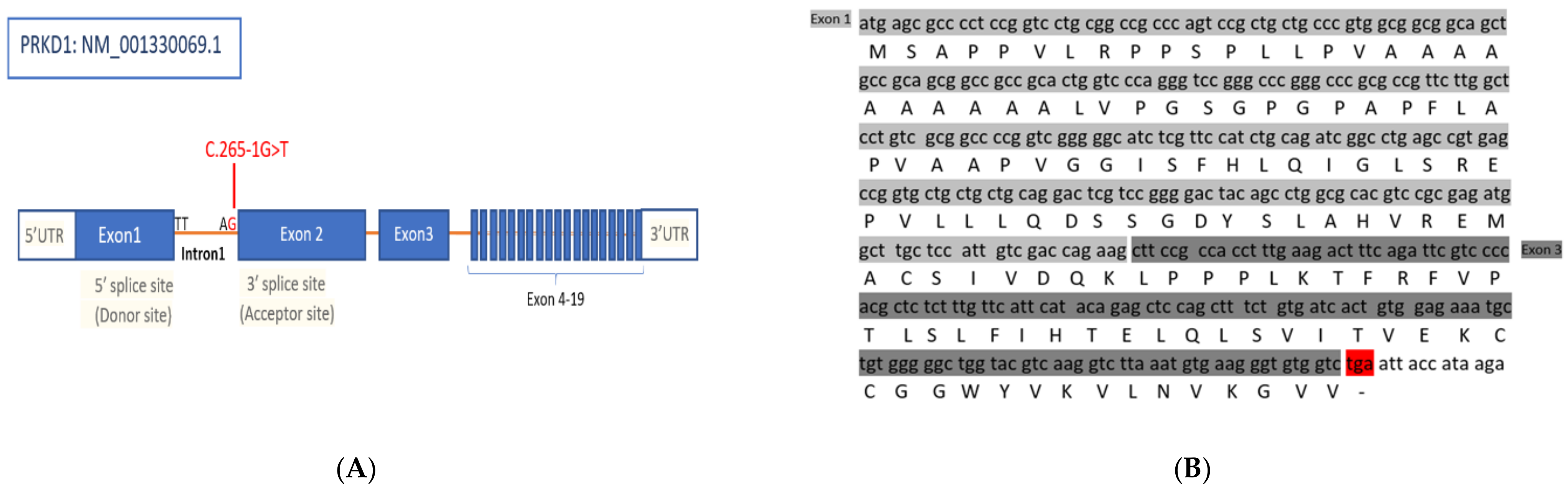
3.1. Identification of a Splice Site Mutation in PRKD1

In this study WES was used to investigate the genetic cause behind this clinical presentation. Our analysis revealed a PRKD1 variant of uncertain significance associated with the disease, which has a single nucleotide substitution (c.265-1G>T) located inintron1 (Figure 2). All three affected females as well as the unaffected father were homozygous for this variant, while the mother and unaffected son were heterozygous (Figure 1). The PRKD1 variant c.265-1G>T is predicted to disrupt a highly conserved acceptor splice site of exon 2 which would interfere with spliceosome recognition and assembly; and resulting in incorrect splicing. This variant in PRKD1 has neither been documented in gnomAD nor in HGMD and Clinvar, is, thus, novel. Erroneous splicing events including those caused by mutations in pre-mRNA templates were the main cause of disease in several previously documented cases [14]. We assume that this exon skipping in PRKD1 transcription may be the cause of the CHDs in this family.

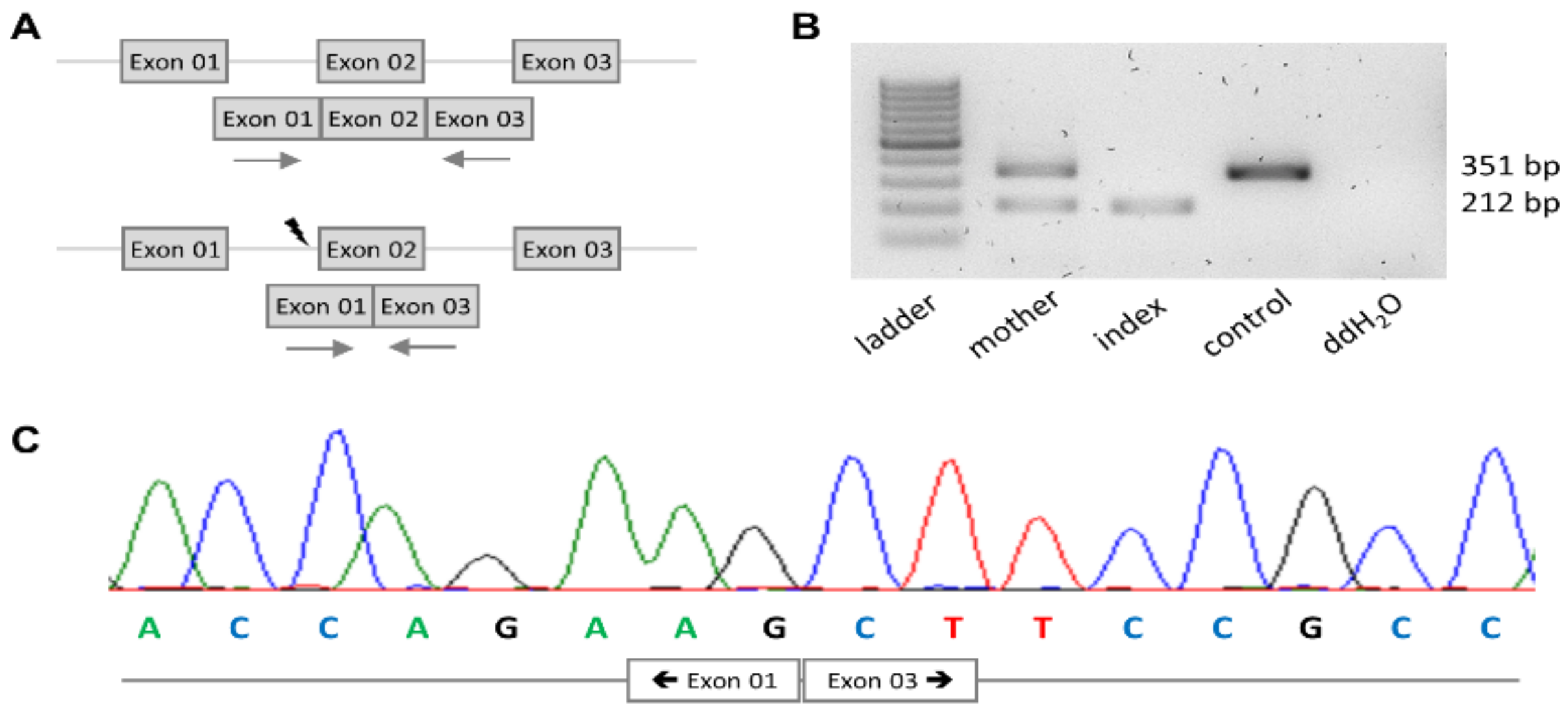
3.2. PRKD1 Splicing Analysis

The identified variant affected the canonical splice acceptor of exon 2 of PRKD1. To determine whether it has an effect on splicing of blood RNA, we applied an exon 2-spanning RT-PCR (Figure 3A). A sample from a wild-type control yielded a single band of the expected size of 351 bp being more intense (Figure 3B). In contrast, a single band of approximately 212 bp was observed when using a sample from the index. In the heterozygous mother, both bands were seen, with the larger one being more intense (Figure 3b). Sequencing of the band from the index revealed exon 1 sequence to be directly followed by exon 3 sequence (Figure 3C). This observation was consistent with skipping of exon 2. The resulting deletion of 139 bp at mRNA level confers a frameshift at protein level and, in turn, generates a premature stop codon in exon 3. A translated protein would thus be severely truncated. Moreover, the mutant PRKD1 mRNA is predicted to undergo nonsense-mediated mRNA decay [15]. Evidence for the latter to occur is in fact provided by the lower intensity of the smaller mutant band in RT-PCR (Figure 3B). Collectively, these observations render c.265-1G>T a classical loss-of-function variant.

4. Discussion

Discovery of the genetic basis of CHDs has been proven to be challenging. Studies have shown that most syndromic CHD can be caused by chromosomal anomalies [16], copy number variations [17], single gene or unknown factors. However, fine mapping of copy number variations (CNVs) in patients with isolated CHD has been used to identify candidate genes that contribute to the origin of pediatric heart disease. The majority of CHD with monogenic inheritance is associated with the known—syndromic presentation. Furthermore, most of them occur sporadically, and families show a clear monogenic mode of inheritance of non-syndromic CHD and cardiac anomalies have been found to result from changes in single or multiple genes with either dominant or recessive mode of inheritance [18]. Phenotypically, both single and multiple genes defects can be translated into syndromic or non-syndromic CHDs. The majority of CHD patients exhibit an isolated non-syndromic CHD phenotype despite this fact, the genetic mechanisms underlying isolated CHD cases remain incompletely understood [4]. Evidence for the genetic basis of isolated CHD comes from familial clustering of cases [19]. In the last two decades, research has primarily focused on gene discovery in non-syndromic forms of CHD, and the list of gene mutations that result in isolated CHDs is rapidly expanding, yet the evidence is variable for each gene [20]. The first genes linked to non-syndromic CHD were the transcription factors including NKX2.5 and GATA4 [18]. Since the involvement of those genes was discovered, a large number of other genes have also been implicated in isolated CHDs, including the transcription factors (TBX1, TBX2, TBX3, TBX5, and MEF2) were observed [21,22] as well as several genes that function in crucial cell signaling pathways like ZIC3, NOTCH1 [23] BMP-2, BMP-4, and TGF- β, and SOX17 [24,25,26].
Here, we present a comprehensive report of clinical and molecular characterization of a consanguineous family of Saudi origin, demonstrating autosomal recessive inheritance of the non-syndromic form of CHD. The affected members showed multiple cardiac phenotypes including pulmonary stenosis, truncus arteriosus and septal defects. By analyzing the WES data, we identified a rare homozygous variant in the PRKD1 gene in the affected members of the family. The PRKD1 gene is mapped to Chr14q1. This gene encodes a protein kinase called PKD1. In addition, PKD2 and PKD3 are two related proteins. They are encoded by PRKD2 and PRKD3 genes located on 19q13.32 and 2p22.2, respectively. The three proteins have different subcellular localizations and tissue expression patterns and thus carry out independent and diverse functions [27,28]. PKD1 is the major PKD protein in the heart [28]. Upon activation, PKD1 phosphorylates class II histone deacetylases (HDACs), resulting in their disassociation from the myocyte enhancer factor-2 (MEF2) transcription factor. This PKD1 induced disassociation relieves MEF2 repression and thus induces the activation of its targeted genes [24]. MEF2 is expressed in cardiogenic precursor cells and in differentiated cardiomyocytes and is considered as an essential regulator of cardiac myogenesis and the right ventricular development [27]. Unfortunately, the heart phenotype of prkd1−/− is uncharacterized in mice because the PRKD1 complete knockout caused embryonic lethality [28]. However, Mef2c−/− mice models were found to have abnormalities in heart tube looping, cushion formation and complex vascular malformations [29,30,31,32], suggesting that the protein truncating PRKD1 variant may be responsible for the CHD phenotype through inability to relieve MEF2 repression.
In addition, there is increasing evidence that PKD-mediated signaling pathway plays an important role in the cardiovascular system, particularly in the regulation of myocardial contraction, hypertrophy, and pathological cardiac remodeling. Moreover, several gain- and loss- of-function mutations in PRKD1 have been reported to interfere with normal cardiac morphogenesis [11,31]. It has been shown that PRKD1 is a potential regulatory factor involved in cardiac neural crest cells (NCCc) migration and patterning at the developing arches of the heart [33]. Therefore, loss-of-function mutations in the PRKD1 gene is predicted to contribute to the pathophysiology of congenital cardiac defects such as TA and Aberrant origin of the right subclavian artery. This is consistent with the TA phenotype identified in our patients with the truncating PRKD1 mutation. We believe that PRKD1 is the most compelling candidate for TA pathogenesis in our patients [34].
Some PRKD1 variants have been linked to various heart defects, such as truncus arteriosus, conotruncal heart defects and many other cardiac anomalies as listed in Table 3. Sifrim et al. reported that missense mutations in the PRKD1 gene were significantly enriched in patients with congenital heart defects and ectodermal dysplasia (CHDED) [33]. They identified de novo missense mutations in PRKD1 in three syndromic CHD cases. These mutations included an amino acid substitution (p.Le299Trp) within the CysII rich regulatory domain of PKD1 and a (p.Gly592Arg) mutation within the PKD1 catalytic domain. Both mutations suggested a dominant gain of function in PRKD1.Thus, an autosomal dominant PRKD1-associated OMIM phenotype termed congenital heart defects, and ectodermal dysplasia was recognized (OMIM: 617364). Jin et al. also reported few dominant loss-of-function heterozygous mutations in PRKD1, in one patient with heterotaxy syndrome and in two patients with conotruncal heart defect (CTD) [10].
The pedigree of the affected family suggests that this novel PRKD1 variant is autosomal recessive. Both parents were unaffected, while three out of four of their children had CHDs. All affected daughters were homozygous for the variant, while their unaffected brother was heterozygous (Figure 1). Although several autosomal dominant PRKD1 mutations associated with CHDs have been reported [8,16], only one autosomal recessive CHD-associated PRKD1 variant has previously been identified in Saudi patients [11], this finding suggests that recessive variants in the dominant gene PRKD1 should not be ruled out before careful consideration. Despite the prevalence of autosomal dominant PRKD1-associated phenotypes, recessive forms were not previously observed. Monies et al. (2017) identified recessive variants for 11 different genes for which only dominant diseases had been reported [35].
Interestingly, homozygosity for the PRKD1 variant (NM_001330069.1: c.265-1G>T) reported here was also detected in the unaffected father. It is possible he might have had a mild CHD phenotype which was cleared at an early age. However, it is also possible that the PRKD1 variant is gender-limited, as only female family members, including the three daughters and the father’s two sisters, were diagnosed with CHDs. Notably, Shaheen at al. also have reported a recessive PRKD1 variant in two female patients [11]. This would not be the first time that sex limitation was considered as a possibility for the genetic determinants of heart disease. Tsagaris et al. posited that cardiac disease in a large family was probably caused by a sex-limited autosomal dominant gene [36]. Additionally, Monteleone and Fagan discovered CHDs in all males in three generations of a family and hypothesized that it was either due to X-linked recessive inheritance or autosomal dominant inheritance with sex limitation [36]. It is unclear whether the novel PRKD1 variant is truly sex-limited, but it is remarkable that all affected individuals were females and though the father was homozygous for the variant, he showed no CHD phenotype. More research is needed to determine what role, if any, sex plays in the described novel autosomal recessive PRKD1 variant-associated CHD phenotypes.
The PRKD1 variant reported in this study, identifies the genetic cause of non-syndromic CHD in a family of multiple affected children. Thus, we report a novel autosomal recessive PRKD1 variant linked to CHDs. The detection of a homozygous variant in this gene indicates a possible genetic diagnosis of PRKD1-associated cardiac phenotypes with an autosomal recessive mode of inheritance.

5. Conclusions

Taken altogether, the present study of a Saudi consanguineous family with isolated CHD supports a previous study arguing that biallelic truncating mutation in the PRKD1 gene may cause non-syndromic CHD. Our finding expands the mutation spectrum of the PRKD1 gene and demonstrates the substantial impact of consanguineous populations in revealing monogenic forms of complex disorders.

Author Contributions

Conceptualization, M.A. (Manal Alaamery) and S.M.; methodology, M.A. (Manal Alaamery) and S.M.; validation, M.A. (Manal Alaamery), S.M., and C.B.; formal analysis, M.A. (Manal Alaamery) and S.M.; investigation, M.A. (Manal Alaamery), M.A. (Maha Albeladi), and S.M.; resources, M.A. (Manal Alaamery) and F.A.; data curation, M.A. (Manal Alaamery), S.M., F.A., C.B., F.C., and M.S.K.; writing—original draft preparation, M.A. (Manal Alaamery), S.M., and M.A. (Maha Albeladi); writing—review and editing, S.M., M.A. (Manal Alaamery), N.A., M.A. (Maha Albeladi), C.B., and K.D.K.; visualization, M.A. (Manal Alaamery) and S.M.; supervision, M.A. (Manal Alaamery) and S.M.; project administration, S.M. and M.A. (Manal Alaamery); funding acquisition, M.A. (Manal Alaamery). All authors have read and agreed to the published version of the manuscript.

Funding

This research was funded by King Abdullah International Medical research Center, grant number RC 15/140.

Institutional Review Board Statement

The study was conducted according to the guidelines of the Declaration of Helsinki and approved by the Institutional Review Board at King Abdullah International Medical Research Centre, Riyadh; (RC 15/140 approved on the 15 May 2016).

Informed Consent Statement

Informed consent was obtained from all subjects involved in the study.

Data Availability Statement

The data presented in this study are available in this article.

Acknowledgments

We would like to thank all study participants and their families for contributing to this study. In addition, we thank all the developmental medicine department members for their contributions.

Conflicts of Interest

K.D.K. and C.B. are employees of Centogene GmbH (Rostock, Germany).

Abbreviations

LOFloss-of-function
NAnot applicable
NRnot reported
VSDVentricle septal defect
ASDAtrial septal defect
AVSDAtrioventricular septal defect
DORVDouble outlet right ventricle
LSVCLeft superior vena cava
SDSSudden death syndrome

References

  1. Mendis, S.; Puska, P.; Norrving, B.; Organization, W.H.; Federation, W.H.; Organization, W.S. Global Atlas on Cardiovascular Disease Prevention and Control; World Health Organization: Geneva, Switzerland, 2011; ISBN 978-92-4-456437-0. [Google Scholar]
  2. Hoffman, J.I.E.; Kaplan, S. The Incidence of Congenital Heart Disease. J. Am. Coll. Cardiol. 2002, 39, 1890–1900. [Google Scholar] [CrossRef] [Green Version]
  3. Liu, Y.; Chen, S.; Zühlke, L.; Black, G.C.; Choy, M.-K.; Li, N.; Keavney, B.D. Global Birth Prevalence of Congenital Heart Defects 1970–2017: Updated Systematic Review and Meta-Analysis of 260 Studies. Int. J. Epidemiol. 2019, 48, 455–463. [Google Scholar] [CrossRef]
  4. Nees, S.N.; Chung, W.K. The Genetics of Isolated Congenital Heart Disease. Am. J. Med. Genet. Part C Semin. Med. Genet. 2019. [Google Scholar] [CrossRef] [PubMed]
  5. Ferencz, C.; Rubin, J.D.; McCarter, R.J.; Brenner, J.I.; Neill, C.A.; Perry, L.W.; Hepner, S.I.; Downing, J.W. Congenital Heart Disease: Prevalence at Livebirth. The Baltimore-Washington Infant Study. Am. J. Epidemiol. 1985, 121, 31–36. [Google Scholar] [CrossRef] [PubMed]
  6. Bruneau, B.G. The Developmental Genetics of Congenital Heart Disease. Nature 2008, 451, 943–948. [Google Scholar] [CrossRef]
  7. 69 Economic Burden of Congenital Heart Disease on Families|Pediatric Research. Available online: https://0-www-nature-com.brum.beds.ac.uk/articles/pr2005321 (accessed on 10 March 2021).
  8. Roth, G.A.; Abate, D.; Abate, K.H.; Abay, S.M.; Abbafati, C.; Abbasi, N.; Abbastabar, H.; Abd-Allah, F.; Abdela, J.; Abdelalim, A.; et al. Global, Regional, and National Age-Sex-Specific Mortality for 282 Causes of Death in 195 Countries and Territories, 1980–2017: A Systematic Analysis for the Global Burden of Disease Study 2017. Lancet 2018, 392, 1736–1788. [Google Scholar] [CrossRef] [Green Version]
  9. Becker, S.M.; Al Halees, Z.; Molina, C.; Paterson, R.M. Consanguinity and Congenital Heart Disease in Saudi Arabia. Am. J. Med. Genet. 2001, 99, 8–13. [Google Scholar] [CrossRef]
  10. Jin, S.C.; Homsy, J.; Zaidi, S.; Lu, Q.; Morton, S.; DePalma, S.R.; Zeng, X.; Qi, H.; Chang, W.; Sierant, M.C.; et al. Contribution of Rare Inherited and de Novo Variants in 2871 Congenital Heart Disease Probands. Nat. Genet. 2017, 49, 1593–1601. [Google Scholar] [CrossRef] [Green Version]
  11. Shaheen, R.; Al Hashem, A.; Alghamdi, M.H.; Seidahmad, M.Z.; Wakil, S.M.; Dagriri, K.; Keavney, B.; Goodship, J.; Alyousif, S.; Al-Habshan, F.M.; et al. Positional Mapping of PRKD1, NRP1 and PRDM1 as Novel Candidate Disease Genes in Truncus Arteriosus. J. Med. Genet. 2015, 52, 322–329. [Google Scholar] [CrossRef] [PubMed]
  12. Shieh, J.T.C.; Bittles, A.H.; Hudgins, L. Consanguinity and the Risk of Congenital Heart Disease. Am. J. Med. Genet. A 2012, 158A, 1236–1241. [Google Scholar] [CrossRef] [PubMed] [Green Version]
  13. Massadeh, S.; Alhashem, A.; van de Laar, I.M.B.H.; Alhabshan, F.; Ordonez, N.; Alawbathani, S.; Khan, S.; Kabbani, M.S.; Chaikhouni, F.; Sheereen, A.; et al. ADAMTS19-Associated Heart Valve Defects: Novel Genetic Variants Consolidating a Recognizable Cardiac Phenotype. Clin. Genet. 2020, 98, 56–63. [Google Scholar] [CrossRef]
  14. Anna, A.; Monika, G. Splicing Mutations in Human Genetic Disorders: Examples, Detection, and Confirmation. J. Appl. Genet. 2018, 59, 253–268. [Google Scholar] [CrossRef] [PubMed] [Green Version]
  15. Supek, F.; Lehner, B.; Lindeboom, R.G.H. To NMD or Not To NMD: Nonsense-Mediated MRNA Decay in Cancer and Other Genetic Diseases. Trends Genet. 2020. [Google Scholar] [CrossRef]
  16. Hartman, R.J.; Rasmussen, S.A.; Botto, L.D.; Riehle-Colarusso, T.; Martin, C.L.; Cragan, J.D.; Shin, M.; Correa, A. The Contribution of Chromosomal Abnormalities to Congenital Heart Defects: A Population-Based Study. Pediatr. Cardiol. 2011, 32, 1147–1157. [Google Scholar] [CrossRef]
  17. Soemedi, R.; Wilson, I.J.; Bentham, J.; Darlay, R.; Töpf, A.; Zelenika, D.; Cosgrove, C.; Setchfield, K.; Thornborough, C.; Granados-Riveron, J.; et al. Contribution of Global Rare Copy-Number Variants to the Risk of Sporadic Congenital Heart Disease. Am. J. Hum. Genet. 2012, 91, 489–501. [Google Scholar] [CrossRef] [Green Version]
  18. Wessels, M.W.; Willems, P.J. Genetic Factors in Non-Syndromic Congenital Heart Malformations. Clin. Genet. 2010, 78, 103–123. [Google Scholar] [CrossRef] [PubMed]
  19. Fahed, A.C.; Nemer, G.M. Genetic Causes of Syndromic and Non-Syndromic Congenital Heart Disease. Mutat. Hum. Genet. Dis. 2012. [Google Scholar] [CrossRef] [Green Version]
  20. Butler, T.L.; Esposito, G.; Blue, G.M.; Cole, A.D.; Costa, M.W.; Waddell, L.B.; Walizada, G.; Sholler, G.F.; Kirk, E.P.; Feneley, M.; et al. GATA4 Mutations in 357 Unrelated Patients with Congenital Heart Malformation. Genet. Test Mol. Biomark. 2010, 14, 797–802. [Google Scholar] [CrossRef]
  21. Zaidi, S.; Brueckner, M. Genetics and Genomics of Congenital Heart Disease. Circ. Res. 2017, 120, 923–940. [Google Scholar] [CrossRef]
  22. Xie, H.; Zhang, E.; Hong, N.; Fu, Q.; Li, F.; Chen, S.; Yu, Y.; Sun, K. Identification of TBX2 and TBX3 Variants in Patients with Conotruncal Heart Defects by Target Sequencing. Hum. Genom. 2018, 12, 44. [Google Scholar] [CrossRef] [PubMed]
  23. Preuss, C.; Capredon, M.; Wünnemann, F.; Chetaille, P.; Prince, A.; Godard, B.; Leclerc, S.; Sobreira, N.; Ling, H.; Awadalla, P.; et al. Family Based Whole Exome Sequencing Reveals the Multifaceted Role of Notch Signaling in Congenital Heart Disease. PLoS Genet. 2016, 12, e1006335. [Google Scholar] [CrossRef]
  24. Armstrong Ehrin, J. Bischoff Joyce Heart Valve Development. Circ. Res. 2004, 95, 459–470. [Google Scholar] [CrossRef] [Green Version]
  25. Nakajima, Y.; Yamagishi, T.; Hokari, S.; Nakamura, H. Mechanisms Involved in Valvuloseptal Endocardial Cushion Formation in Early Cardiogenesis: Roles of Transforming Growth Factor (TGF)-Beta and Bone Morphogenetic Protein (BMP). Anat. Rec. 2000, 258, 119–127. [Google Scholar] [CrossRef]
  26. Zhu, N.; Welch, C.L.; Wang, J.; Allen, P.M.; Gonzaga-Jauregui, C.; Ma, L.; King, A.K.; Krishnan, U.; Rosenzweig, E.B.; Ivy, D.D.; et al. Rare Variants in SOX17 Are Associated with Pulmonary Arterial Hypertension with Congenital Heart Disease. Genom. Med. 2018, 10, 56. [Google Scholar] [CrossRef]
  27. Avkiran, M.; Rowland, A.J.; Cuello, F.; Haworth, R.S. Protein Kinase D in the Cardiovascular System: Emerging Roles in Health and Disease. Circ. Res. 2008, 102, 157–163. [Google Scholar] [CrossRef] [PubMed] [Green Version]
  28. Qiu, W.; Steinberg, S.F. Phos-Tag SDS-PAGE Resolves Agonist- and Isoform-Specific Activation Patterns for PKD2 and PKD3 in Cardiomyocytes and Cardiac Fibroblasts. J. Mol. Cell Cardiol. 2016, 99, 14–22. [Google Scholar] [CrossRef] [Green Version]
  29. Edmondson, D.G.; Lyons, G.E.; Martin, J.F.; Olson, E.N. Mef2 Gene Expression Marks the Cardiac and Skeletal Muscle Lineages during Mouse Embryogenesis. Development 1994, 120, 1251–1263. [Google Scholar]
  30. Fielitz, J.; Kim, M.-S.; Shelton, J.M.; Qi, X.; Hill, J.A.; Richardson, J.A.; Bassel-Duby, R.; Olson, E.N. Requirement of Protein Kinase D1 for Pathological Cardiac Remodeling. Proc. Natl. Acad. Sci. USA 2008, 105, 3059–3063. [Google Scholar] [CrossRef] [PubMed] [Green Version]
  31. Lin, Q.; Schwarz, J.; Bucana, C.; Olson, E.N. Control of Mouse Cardiac Morphogenesis and Myogenesis by Transcription Factor MEF2C. Science 1997, 276, 1404–1407. [Google Scholar] [CrossRef] [Green Version]
  32. Bi, W.; Drake, C.J.; Schwarz, J.J. The Transcription Factor MEF2C-Null Mouse Exhibits Complex Vascular Malformations and Reduced Cardiac Expression of Angiopoietin 1 and VEGF. Dev. Biol. 1999, 211, 255–267. [Google Scholar] [CrossRef] [PubMed] [Green Version]
  33. Sifrim, A.; Hitz, M.-P.; Wilsdon, A.; Breckpot, J.; Turki, S.H.A.; Thienpont, B.; McRae, J.; Fitzgerald, T.W.; Singh, T.; Swaminathan, G.J.; et al. Distinct Genetic Architectures for Syndromic and Nonsyndromic Congenital Heart Defects Identified by Exome Sequencing. Nat. Genet. 2016, 48, 1060–1065. [Google Scholar] [CrossRef] [PubMed]
  34. Schussler, O.; Gharibeh, L.; Mootoosamy, P.; Murith, N.; Tien, V.; Rougemont, A.-L.; Sologashvili, T.; Suuronen, E.; Lecarpentier, Y.; Ruel, M. Cardiac Neural Crest Cells: Their Rhombomeric Specification, Migration, and Association with Heart and Great Vessel Anomalies. Cell Mol. Neurobiol. 2021, 41, 403–429. [Google Scholar] [CrossRef] [PubMed]
  35. Monies, D.; Maddirevula, S.; Kurdi, W.; Alanazy, M.H.; Alkhalidi, H.; Al-Owain, M.; Sulaiman, R.A.; Faqeih, E.; Goljan, E.; Ibrahim, N.; et al. Autozygosity Reveals Recessive Mutations and Novel Mechanisms in Dominant Genes: Implications in Variant Interpretation. Genet. Med. 2017, 19, 1144–1150. [Google Scholar] [CrossRef] [PubMed] [Green Version]
  36. Tsagaris, T.J.; Bustamante, R.A.; Friesendorff, R.A. Familial Heart Disease. Dis. Chest 1967, 52, 153–158. [Google Scholar] [CrossRef] [PubMed]
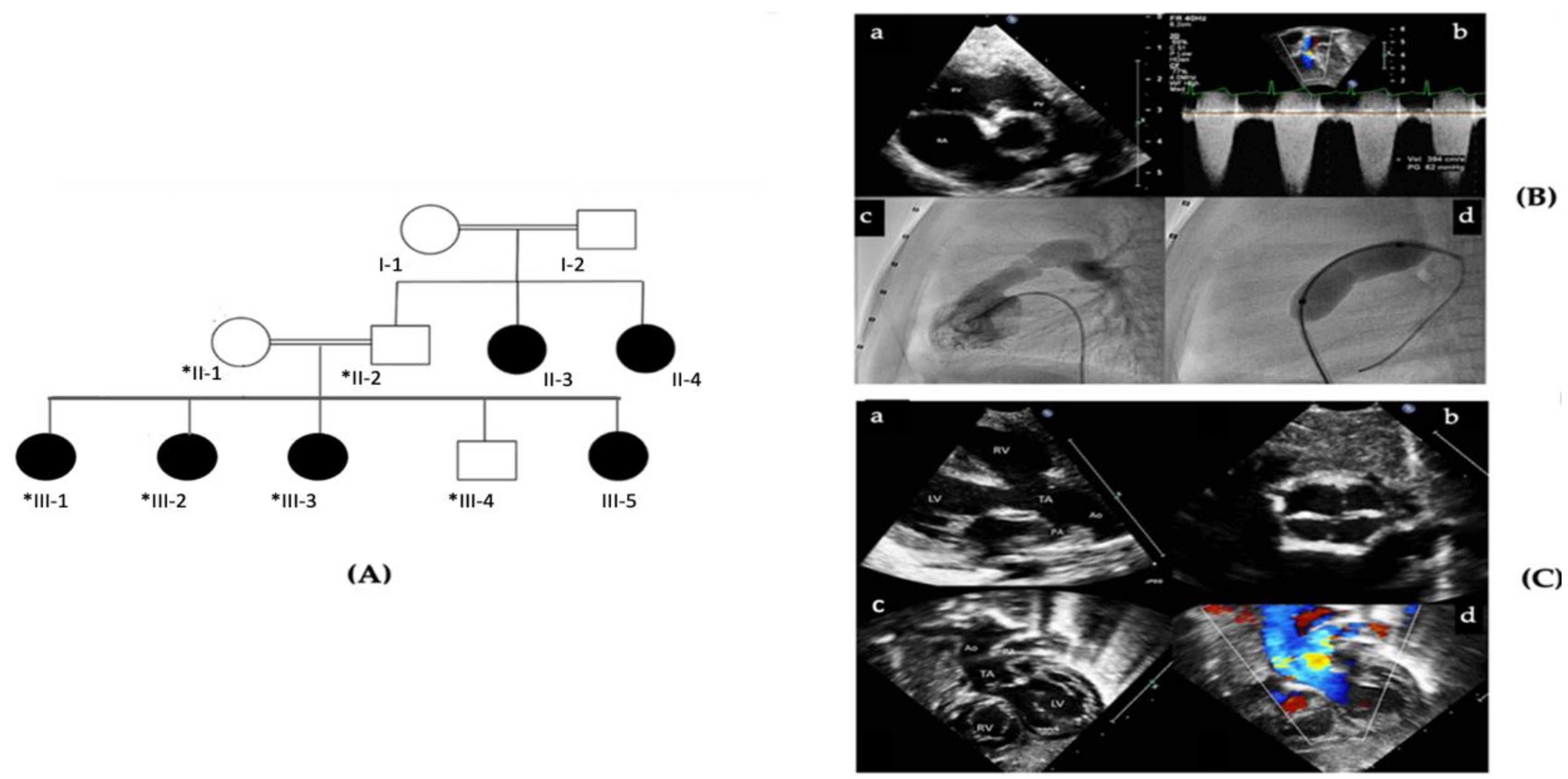
Figure 1. (A) Pedigree of a consanguineous family with members affected with congenital heart defects. Pedigree depicts an autosomal recessive mode of inheritance. Double lines are indicative of consanguineous union. Females and males are represented with circles and square symbols, respectively. Clear symbols signify healthy individuals, and filled symbols signify individuals affected with cardiac diseases. +/+ denotes individual with (c.265-1G>T) homozygous mutation in the PRKD1 gene and +/− denotes carriers for a heterozygous (c.265-1G>T) PRKD1 variant. The individual numbers labelled with asterisks indicates the samples which are available for the studies. (B) Echo images from the proband patient (III-1) with pulmonary stenosis. a: Parasternal short axis view showing thickened and stenotic pulmonary valve. b: Doppler measurement of the flow velocity across the pulmonary valve with maximum peak gradient of 62mmHg. c: Right ventricular angiography showing thickened and stenotic pulmonary valve. d: Balloon dilatation of the pulmonary valve with a waist at the valve area. (C) Echo images from patient III-2 with Truncus arteriosus. a: Parasternal long axis view showing a large ventricular septal defect with truncus arteriosus, aorta (Ao) and pulmonary artery (PA). b: Short axis image showing the quadricuspid truncal valve. c, d: Subcostal 2D and colour images showing the truncus arteriosus and its division to aorta (Ao) and pulmonary artery (PA). LV: left ventricle. RV: right ventricle.
Genes 12 00612 g001
Figure 2. Schematic representation of PRKD1. (A) structure of the PRKD1 gene and the splice mutation position (c.265-1G>T) in the variant reported in this study. (B) Nucleotide sequence and predicted amino acid sequence alignment of PRKD1 variant. The exon 2 skipping event detected in PRKD1 cDNA (NM_0011330069.1: C.265-1 G>T), is predicted to truncate PRKD1 transcript by the formation of a premature stop codon highlighted in red.
Genes 12 00612 g002
Figure 3. Analysis of the PRKD1 transcript. (A) Strategy to amplify the relevant cDNA region of PRKD1, wildtype and mutant scheme on top and bottom, respectively. The flash indicated the position of the variant; arrows indicate PCR primers. (B) Results of the reverse transcription PCR. (C) Sanger sequence chromatogram of the PRKD1 transcript in index revealing the absence of exon 2.
Genes 12 00612 g003
Table 1. Clinical features of family members with the (c.265-1G>T) PRKD1 variant.
Patient II-3, II-4Patient III-1Patient III-2Patient III-3
AgeNA1096
GenderFemalesFemaleFemaleFemale
WeightNA4029.6NA
HeightNA148138.5NA
Cardiovascular SymptomsNAAsymptomaticRespiratory distressAsymptomatic
Clinical DiagnosisSeptal Defects (not specified)Pulmonary stenosisTA type II and VSDASD
Mental DevelopmentNormalNormalNormalNormal
Neurological AbnormalitiesNormalNormalNormalNormal
Motor developmentNormalNormalNormalNormal
Abbreviations; NA: not available, TA: Truncus arteriosus VSD: Ventricle septal defect, ASD: Atrial septal defect.
Table 2. Primer sequences for detecting the consequences of the PRKD1 variant (c.265-1G>T) at mRNA level.
PrimerSequence
ForwardGCATCTCGTTCCATCTGCAG (Exon01)
ReverseCTCCACAGTGATCACAGAAAGC (Exon03)
Table 3. Summary of the PRKD1 variants reported in the CHD Literature.
PRKD1 VariantsMutationInheritanceCHD ClassificationGender of Human SubjectsClinical DiagnosisRef.
NM_001330069.1: c.265-1G>TDisruption of acceptor splice side of Exon 2 (Exon skipping event lead to premature stop codon)—(LOF)RecessiveNon-syndromic CHDFemalePS, Tricuspid regurgitationThis Study
FemaleTA, VSD
FemaleASD
NAMaleUnaffected
NAMale/FemaleHealthy control
NM_002742.2: c.1852 C>THomozygous Truncating Mutations in PRKD1(LOF)RecessiveNon-syndromic CHDTwo FemalesTruncus arteriosusShaheen et al., 2015 [11]
NM_002742.2: c.1774 G>ADe novo missense mutations p. Gly592Arg (Gain of function mutation)DominantSyndromic CHDTwins
Males
Pulmonary valvar abnormality. Coupled with hypoglycemia, jaundice and hypothermia, Delayed speech and language development, Microcephaly, Bilateral conductive hearing impairment, Ectodermal dysplasia, Lipson syndrome.Sifrim et al., 2016 [33]
AVSD, Hypotonia, Scoliosis,
NM_002742.2: c.896 T>GDe novo missense mutations p. leu299Trp (Gain of function mutation)DominantSyndromic CHDMaleAttention deficit hyperactivity disorder, Microcephaly, Arnold-Chiari type I, Microcephaly, Nystagmus
Chr14: 30,108,080 G>APremature stop codon (p.R243X)DominantNRHetrotaxy syndrome,
Ebstein anomaly, L-loop corrected transposition
Jin et al., 2017 [10]
Chr14: 30,066,751 G>APremature stop codon (p.Q794X)DominantNRASD, secundum, patent ductus arteriosus, pulmonary stenosis, valvar
Chr14: 30,046,444 T>AStop-less mutation (p.X913C)DominantNR Aberrant right subclavian artery, abnormal branching left aortic arch, Aortic stenosis, Bicommissural aortic valve, DORV, LSVC, SDS, tubular hypoplasia of aorta, VSD,
Chr14: 30,100,011 G>APremature Stop codon (p.Q537X)DominantNR Healthy Control
ENSG00000184304 G>TDe-novo Missense MutationDominant ASD, secundum, pulmonary stenosis, Tricuspid stenosis | VSD
Publisher’s Note: MDPI stays neutral with regard to jurisdictional claims in published maps and institutional affiliations.

