Case Report

Congenital Cataracts and Gut Dysmotility in a DYNC1H1 Dyneinopathy Patient

by
Rose Gelineau-Morel
1,†,
Marshall Lukacs
2,†,
K. Nicole Weaver
2,
Robert B. Hufnagel
2,
Donald L. Gilbert
1 and
Rolf W. Stottmann
2,3,*
1
Division of Neurology, Cincinnati Children’s Hospital Medical Center, Cincinnati, OH 45229, USA
2
Division of Human Genetics, Cincinnati Children’s Hospital Medical Center, Cincinnati, OH 45229, USA
3
Division of Developmental Biology, Cincinnati Children’s Hospital Medical Center, Cincinnati, OH 45229, USA
*
Author to whom correspondence should be addressed.
These authors contributed equally to this work.
Submission received: 4 May 2016 / Revised: 20 September 2016 / Accepted: 7 October 2016 / Published: 14 October 2016
(This article belongs to the Section Human Genomics and Genetic Diseases)

Abstract

:
Whole exome sequencing continues to end the diagnostic odyssey for a number of patients and expands our knowledge of phenotypes associated with gene mutations. We describe an 11-year-old female patient with a constellation of symptoms including congenital cataracts, gut dysmotility, sensory neuropathy, and bifrontal polymicrogyria. Whole exome sequencing was performed and identified a de novo heterozygous missense mutation in the ATPase motor domain of cytoplasmic dynein heavy chain 1 (DYNC1H1), which is known to be involved in neuronal migration and retrograde axonal transport. The mutation was found to be highly damaging by multiple prediction programs. The residue is highly conserved, and reported mutations in this gene result in a variety of phenotypes similar to that of our patient. We report only the second case of congenital cataracts and the first of gut dysmotility in a patient with DYNC1H1, thus expanding the spectrum of disease seen in DYNC1H1 dyneinopathies.

1. Introduction

DYNC1H1 (OMIM #600112) encodes the heavy chain 1 of cytoplasmic dynein and controls microtubule binding as well as the recruitment of dynein components involved in retrograde axonal transport in neurons [1,2]. Mutations in DYNC1H1 are associated with a wide spectrum of clinical manifestations including spinal muscular atrophy (SMA) [1,3,4,5], hereditary motor and sensory neuropathies [6], hereditary spastic paraplegia [7], malformations of cortical development (MCD), epilepsy, and intellectual disability [2,8,9]. Axonal defects are likely due to DYNC1H1’s role as the sole motor unit responsible for retrograde transport in axons [10]. Neuronal migration defects are likely due to DYNC1H1’s direct association with LIS1 (Lissencephaly 1/platelet-activating factor acetylhydrolase isoform 1B, alpha subunit, PAFAH1B1) which is known to be critically required for normal neuronal migration during cortical development [9,11]. Reports of pediatric patients with DYNC1H1 mutations have previously been published. The authors of those reports describe patients with a variety of symptoms including developmental delay, epilepsy, muscle weakness and wasting, microcephaly, spastic quadriplegia, foot deformities, and motor and sensory neuropathies [10]. Magnetic Resonance Imaging of those patients identified cortical malformations including pachygyria and polymicrogyria. Here we describe a patient with a DYNC1H1 variant and symptoms not previously associated with this gene. Our observations expand the spectrum of manifestations of DYNC1H1 mutations in humans.

2. Materials and Methods

We describe a patient under the care of physicians at Cincinnati Children’s Hospital Medical Center (CCHMC). All patient and parents were enrolled in a study upon informed written consent and assent as approved by the CCHMC Institutional Review Board (2014-3789, approved on 24 September 2015). Exome sequencing was performed at the CCHMC DNA Sequencing and Genotyping Core. In brief, genomic DNA was enriched with the NimbleGen EZ Exome V2 kit (Roche NimbleGen, Madison, WI, USA) and the exome library was sequenced using Illumina’s Hi Seq 2000 (Illumina, San Diego, CA, USA). Alignment and variant detection was performed using the Broad Institute’s web-based Genome Analysis Toolkit (GATK; [12]). Patient examinations were performed following best-practice guidelines and standard equipment.

3. Results

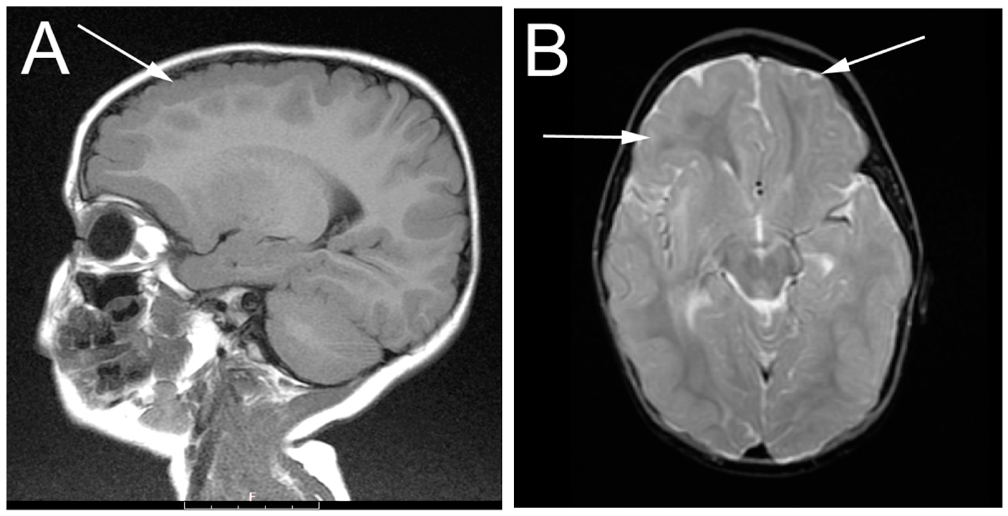
3.1. Patient Description
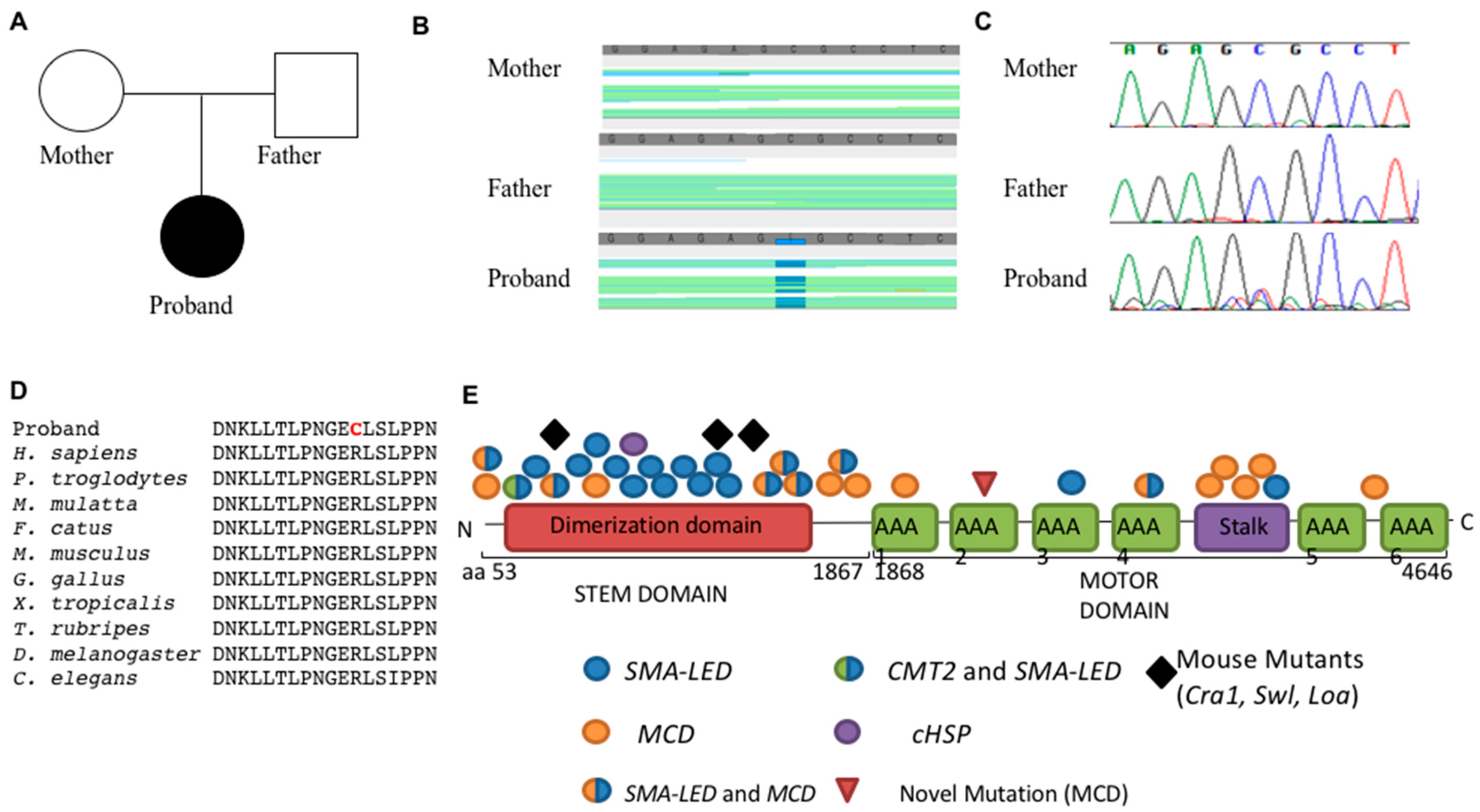
The proband is an 11-year-old female from a generally uncomplicated pregnancy and born full term via forceps assisted vaginal delivery secondary to cephalopelvic disproportion. Perinatally, she was noted to have hypotonia with poor weight gain and difficulty feeding. She initially required nasogastric feeds at seven months and, ultimately, gastric tube placement at one year of age. Extensive medical evaluation was significant for antroduodenal motility study performed at 17 months, demonstrated mild neuropathic changes in the stomach and small bowel, as well as post-prandial hypo-motility, consistent with pseudo-obstruction. She continued to have dysphagia, with choking and gagging, and chronic constipation. Upper GI, endoscopy, and swallow study did not indicate paralysis or weakness of pharyngeal muscles, dysmotility, or aspiration. Her constipation was managed with motility agents and laxatives, and she was gradually able to take more of her nutrition by mouth, although she continues to require some nutrition via her feeding tube. She was also found to have congenital cataracts, requiring bilateral implantation of intraocular lenses, as well as surgery to correct strabismus. A TORCH screen (Toxoplasma gondii, Other viruses, Rubella, Cytomegalovirus, and Herpes simplex) to assess infectious causes of congenital cataracts in utero was negative.
Early behavioral intervention started at eight months. The girl became able to sit independently by two years of age, and walk at three years of age. She began to speak single words at age two, and simple sentences around age six. The patient presented for neurological evaluation at two years of age with concerns for seizures. As her routine EEG was normal, no antiseizure medication was prescribed. Muscle biopsy demonstrated neuropathic changes with large fiber groups consistent with denervation/re-innervation. Nerve conduction/EMG studies were not performed. Electroencephalogram (EEG) analysis at that time revealed diffuse bilateral epileptiform discharges with a bifrontal predominance. Magnetic resonance imaging (MRI) of the brain demonstrated symmetric bilateral polymicrogyria of the frontal lobe involving the superior and middle frontal gyri. There was hypo-myelination of subcortical white matter associated with the dysplastic areas. No other areas of polymicrogyria were seen and otherwise the brain, ventricles, and extra-axial spaces were normal in appearance (Figure 1).
She was lost to follow-up, but re-presented at age 10 with episodes of loss of consciousness, loss of tone, vomiting, and tachycardia lasting 5–10 s with post-ictal drowsiness. These episodes once again raised concerns that the patient was experiencing seizures. No dysmorphic features have been noted in the patient. There were no abnormal cardiac findings and no organomegaly. Neurologically, the patient exhibited cognitive and language impairment, with limited speech and cooperation on examination. Cognitive function was too low for formal IQ testing. She also exhibited impaired social skills, anxiety, and stereotypies. A motor exam revealed hypotonia, normal reflexes, and normal strength. Although the parents reported decreased sensitivity to extreme temperatures, sensory examination was not considered reliable due to her cognition.

3.2. Genetics

Given the constellation of symptoms combined with findings of polymicrogyria on MRI, we hypothesized a genetic etiology for the phenotypes seen in this patient. We performed next-generation exome sequencing on the patient as well as both parents. Non-synonymous coding variants were identified and compared to those found in control databases, including, NHLBI’s ESP6500 exome data [13], the 1000 Genomes Project [14], the EXAC project (Exome aggregation consortium, 2015) and an internal CCHMC sequencing control data control cohort. We analyzed the variant list for autosomal dominant de novo, homozygous recessive, and compound heterozygous mutations using three independent analysis platforms. All three analysis methods (Golden Helix Sample Variation Suite, Ingenuity Variant Analysis, and manual curation) identified the same likely candidate. The number of variants at each step for one of our analyses is shown (Table 1). Similar results were obtained from each.
We identified a de novo autosomal mutation in cytoplasmic dynein heavy chain 1 (DYNC1H1; c.6994C > T; p.R2332C; Figure 2C) in the proband. Sanger sequencing confirmed the mutation was present in the proband, but neither parent (Figure 2D). DYNC1H1 is cytoplasmic dynein heavy chain and contains three major domains, the stalk that binds microtubules, the motor ATPase domain that provides the energy for motor function, and the stem domain that binds molecular cargo for transport. This de novo missense mutation in the second AAA ATPase subunit of the DYNC1H1 motor domain was predicted to be deleterious by multiple bioinformatics prediction algorithms, including PolyPhen (1.0, “probably damaging” [15]) and MutationTaster (“disease-causing” [16]). In addition, the arginine residue at this position in DYNC1H1 is highly conserved through nematodes (Figure 2D). The patient’s symptoms are consistent with a variety of the phenotypes observed in other patients and mice with mutations in DYNC1H1 with the notable addition of congenital cataracts and gut dysmotility [10].

4. Discussion

The spectrum of disease caused by mutations in DYNC1H1 has become evident with the advent of wide-spread genomic sequencing. We present here a patient with a mutation in the motor domain of DYNC1H1 with a varied clinical presentation including failure to thrive, oral dysphagia, gut dysmotility, congenital cataracts, and developmental delay. MRI studies demonstrated bifrontal polymicrogyria. While the broad findings of developmental delay and polymicrogyria are consistent with previously reported patients with mutations in DYNC1H1, this report suggests other systems can be affected by dynein mutations. A very recent report also identified this mutation in a patient with many similar phenotypes, including congenital cataracts, microcephaly and failure to thrive [17]. Additionally, a patient with hereditary spastic paraplegia was found to develop bilateral cataracts in childhood [7]. As DYNC1H1 is expressed in the elongating fiber cells of the lens and possibly traffics vesicles along microtubules for reorganization of the fiber cell, it is possible that mutations in the motor domain disrupt the development of the lens and result in cataracts [18]. In addition, this is the first report of a DYNC1H1 patient to display gut dysmotility with evidence of a mild enteric neuropathy. While the role of DYNC1H1 in the enteric nervous system or neural crest cell migration into the gut is unknown, previously reported patients with mutations in DYNC1H1 have exhibited peripheral motor and sensory neuropathies, which may be related to the proband’s phenotype [1]. Tissue-specific loss-of-function mutations in DYNC1H1 will aide in determining the true requirement of DYNC1H1 in the lens and enteric nervous system.
The mechanism underlying the wide range of clinical presentations in patients with DYNC1H1 mutations has yet to be determined. Several mouse models with loss of Dync1h1 function exist. The first homozygous knockout Dync1h1 mouse died in early gestation (prior to embryonic day 8.5) due to abnormalities in the Golgi apparatus, endosome, lysosome, and massive defects in mitosis in the inner cell mass [19]. Later, ENU mutagenesis screens produced two independent point mutations in the stem domain of Dync1h1. Legs-at-odd-angles (Loa) and Cramping1 (Cra1) heterozygous mice display a dominant motor weakness and sensory neuropathy phenotype characterized by a massive decrease in motor neurons (20%–50%) by gestational age E18.5 leading to paralysis and death within 24 h [20,21]. A subsequent analysis showed that Loa mutant mice also had neuronal migration defects in the cortex [22]. A radiation-induced knockout, Sprawling (Swl), contains a 9 bp deletion in the stem domain of Dync1h1 resulting in a dominant phenotype with early onset sensory neuropathy and loss of muscle spindles in hind-limb muscles [23]. However, to date a mouse mutant with mutations in the Dync1h1 motor domain has not been identified. Our patient’s phenotype is consistent with other patients with mutations in the motor domain, producing a largely MCD/epilepsy phenotype. As previously reported [1,24], patients with mutations in the stem domain are significantly more likely to develop motor and sensory neuropathies, while patients with mutations in the motor domain are more likely to develop malformations of cortical development and epilepsy, though these genotypes show some degree of overlapping phenotypes (Figure 2E). Scoto et al. recently highlighted the overlap in phenotypes of tail and motor domain mutations in a large cohort of SMA-LED patients with comorbid MCD [24]. As only the tail domain has been mutated in mouse models of a dyneinopathy, targeted mutations involving only the motor domain in mice will shed light on the role of each domain in the disease.

Acknowledgments

This study was supported by a grant from the National Institutes of Health (R.W.S. R01NS085023). The funding source had no role in the collection, analysis or interpretation of the date, preparation of the manuscript and/or the decision to submit for publication.

Author Contributions

R.G.M., D.L.G. and R.W.S. conceived and designed the experiments; R.G.M., M.L., K.N.W., R.B.H. and R.W.S. analyzed the data; R.G.M., M.L., D.L.G. and R.W.S. wrote the paper.

Conflicts of Interest

The authors declare no conflict of interest.

Abbreviations

The following abbreviations are used in this manuscript:
CCHMCCincinnati Children’s Hospital Medical Center
cHSPHereditary Spastic Paraplegia
CMT2Charcot-Marie-Tooth Type 2
DYNC1H1cytoplasmic dynein heavy chain 1
EEGElectroencephalogram
EXACExome aggregation consortium
GATKGenome Analysis Toolkit
MCDmalformations of cortical development
MRIMagnetic Resonance Imaging
PAFAH1B1platelet-activating factor acetylhydrolase isoform 1B, alpha subunit
SMAspinal muscular atrophy
SMA-LEDSpinal Muscular Atrophy-Lower Extremity Dominant
TORCHToxoplasma gondii, Other viruses, Rubella, Cytomegalovirus, and Herpes simplex

References

  1. Peeters, K.; Bervoets, S.; Chamova, T.; Litvinenko, I.; de Vriendt, E.; Bichev, S.; Kancheva, D.; Mitev, V.; Kennerson, M.; Timmerman, V.; et al. Novel mutations in the DYNC1H1 tail domain refine the genetic and clinical spectrum of dyneinopathies. Hum. Mutat. 2015, 36, 287–291. [Google Scholar] [CrossRef] [PubMed]
  2. Poirier, K.; Lebrun, N.; Broix, L.; Tian, G.; Saillour, Y.; Boscheron, C.; Parrini, E.; Valence, S.; Pierre, B.S.; Oger, M.; et al. Mutations in TUBG1, DYNC1H1, KIF5C and KIF2A cause malformations of cortical development and microcephaly. Nat. Genet. 2013, 45, 639–647. [Google Scholar] [CrossRef] [PubMed] [Green Version]
  3. Harms, M.B.; Ori-McKenney, K.M.; Scoto, M.; Tuck, E.P.; Bell, S.; Ma, D.; Masi, S.; Allred, P.; Al-Lozi, M.; Reilly, M.M.; et al. Mutations in the tail domain of DYNC1H1 cause dominant spinal muscular atrophy. Neurology 2012, 78, 1714–1720. [Google Scholar] [CrossRef] [PubMed]
  4. Fiorillo, C.; Moro, F.; Yi, J.; Weil, S.; Brisca, G.; Astrea, G.; Severino, M.; Romano, A.; Battini, R.; Rossi, A.; et al. Novel dynein DYNC1H1 neck and motor domain mutations link distal spinal muscular atrophy and abnormal cortical development. Hum. Mutat. 2014, 35, 298–302. [Google Scholar] [CrossRef] [PubMed]
  5. Tsurusaki, Y.; Saitoh, S.; Tomizawa, K.; Sudo, A.; Asahina, N.; Shiraishi, H.; Ito, J.; Tanaka, H.; Doi, H.; Saitsu, H.; et al. A DYNC1H1 mutation causes a dominant spinal muscular atrophy with lower extremity predominance. Neurogenetics 2012, 13, 327–332. [Google Scholar] [CrossRef] [PubMed]
  6. Weedon, M.N.; Hastings, R.; Caswell, R.; Xie, W.; Paszkiewicz, K.; Antoniadi, T.; Williams, M.; King, C.; Greenhalgh, L.; Newbury-Ecob, R.; et al. Exome sequencing identifies a DYNC1H1 mutation in a large pedigree with dominant axonal Charcot-Marie-Tooth disease. Am. J. Hum. Genet. 2011, 89, 308–312. [Google Scholar] [CrossRef] [PubMed]
  7. Strickland, A.V.; Schabhuttl, M.; Offenbacher, H.; Synofzik, M.; Hauser, N.S.; Brunner-Krainz, M.; Gruber-Sedlmayr, U.; Moore, S.A.; Windhager, R.; Bender, B.; et al. Mutation screen reveals novel variants and expands the phenotypes associated with DYNC1H1. J. Neurol. 2015, 262, 2124–2134. [Google Scholar] [CrossRef] [PubMed]
  8. Vissers, L.E.; de Ligt, J.; Gilissen, C.; Janssen, I.; Steehouwer, M.; de Vries, P.; van Lier, B.; Arts, P.; Wieskamp, N.; del Rosario, M.; et al. A de novo paradigm for mental retardation. Nat. Genet. 2010, 42, 1109–1112. [Google Scholar] [CrossRef] [PubMed]
  9. Willemsen, M.H.; Vissers, L.E.; Willemsen, M.A.; van Bon, B.W.; Kroes, T.; de Ligt, J.; de Vries, B.B.; Schoots, J.; Lugtenberg, D.; Hamel, B.C.; et al. Mutations in DYNC1H1 cause severe intellectual disability with neuronal migration defects. J. Med. Genet. 2012, 49, 179–183. [Google Scholar] [CrossRef] [PubMed]
  10. Schiavo, G.; Greensmith, L.; Hafezparast, M.; Fisher, E.M. Cytoplasmic dynein heavy chain: The servant of many masters. Trends Neurosci. 2013, 36, 641–651. [Google Scholar] [CrossRef] [PubMed]
  11. Tsai, J.W.; Bremner, K.H.; Vallee, R.B. Dual subcellular roles for LIS1 and dynein in radial neuronal migration in live brain tissue. Nat. Neurosci. 2007, 10, 970–979. [Google Scholar] [CrossRef] [PubMed]
  12. McKenna, A.; Hanna, M.; Banks, E.; Sivachenko, A.; Cibulskis, K.; Kernytsky, A.; Garimella, K.; Altshuler, D.; Gabriel, S.; Daly, M.; et al. The Genome Analysis Toolkit: A MapReduce framework for analyzing next-generation DNA sequencing data. Genome Res. 2010, 20, 1297–1303. [Google Scholar] [CrossRef] [PubMed]
  13. Fu, W.; O’Connor, T.D.; Jun, G.; Kang, H.M.; Abecasis, G.; Leal, S.M.; Gabriel, S.; Rieder, M.J.; Altshuler, D.; Shendure, J.; et al. Analysis of 6515 exomes reveals the recent origin of most human protein-coding variants. Nature 2013, 493, 216–220. [Google Scholar] [CrossRef] [PubMed]
  14. 1000 Genomes Project Consortium; Abecasis, G.R.; Altshuler, D.; Auton, A.; Brooks, L.D.; Durbin, R.M.; Gibbs, R.A.; Hurles, M.E.; McVean, G.A. A map of human genome variation from population-scale sequencing. Nature 2010, 467, 1061–1073. [Google Scholar] [PubMed]
  15. Adzhubei, I.A.; Schmidt, S.; Peshkin, L.; Ramensky, V.E.; Gerasimova, A.; Bork, P.; Kondrashov, A.S.; Sunyaev, S.R. A method and server for predicting damaging missense mutations. Nat. Methods 2010, 7, 248–249. [Google Scholar] [CrossRef] [PubMed]
  16. Schwarz, J.M.; Cooper, D.N.; Schuelke, M.; Seelow, D. MutationTaster2: Mutation prediction for the deep-sequencing age. Nat. Methods 2014, 11, 361–362. [Google Scholar] [CrossRef] [PubMed]
  17. Zhu, X.; Petrovski, S.; Xie, P.; Ruzzo, E.K.; Lu, Y.F.; McSweeney, K.M.; Ben-Zeev, B.; Nissenkorn, A.; Anikster, Y.; Oz-Levi, D.; et al. Whole-exome sequencing in undiagnosed genetic diseases: Interpreting 119 trios. Genet. Med. 2015, 17, 774–781. [Google Scholar] [CrossRef] [PubMed]
  18. Lo, W.K.; Wen, X.J.; Zhou, C.J. Microtubule configuration and membranous vesicle transport in elongating fiber cells of the rat lens. Exp. Eye Res. 2003, 77, 615–626. [Google Scholar] [CrossRef]
  19. Harada, A.; Takei, Y.; Kanai, Y.; Tanaka, Y.; Nonaka, S.; Hirokawa, N. Golgi vesiculation and lysosome dispersion in cells lacking cytoplasmic dynein. J. Cell Biol. 1998, 141, 51–59. [Google Scholar] [CrossRef] [PubMed] [Green Version]
  20. El-Kadi, A.M.; Bros-Facer, V.; Deng, W.; Philpott, A.; Stoddart, E.; Banks, G.; Jackson, G.S.; Fisher, E.M.; Duchen, M.R.; Greensmith, L.; et al. The legs at odd angles (Loa) mutation in cytoplasmic dynein ameliorates mitochondrial function in SOD1G93A mouse model for motor neuron disease. J. Biol. Chem. 2010, 285, 18627–18639. [Google Scholar] [PubMed]
  21. De Angelis, M.H.; Flaswinkel, H.; Fuchs, H.; Rathkolb, B.; Soewarto, D.; Marschall, S.; Heffner, S.; Pargent, W.; Wuensch, K.; Jung, M.; et al. Genome-wide, large-scale production of mutant mice by ENU mutagenesis. Nat. Genet. 2000, 25, 444–447. [Google Scholar] [CrossRef] [PubMed]
  22. Ori-McKenney, K.M.; Vallee, R.B. Neuronal migration defects in the Loa dynein mutant mouse. Neural Dev. 2011. [Google Scholar] [CrossRef] [PubMed]
  23. Chen, X.J.; Levedakou, E.N.; Millen, K.J.; Wollmann, R.L.; Soliven, B.; Popko, B. Proprioceptive sensory neuropathy in mice with a mutation in the cytoplasmic Dynein heavy chain 1 gene. J. Neurosci. 2007, 27, 14515–14524. [Google Scholar] [CrossRef] [PubMed]
  24. Scoto, M.; Rossor, A.M.; Harms, M.B.; Cirak, S.; Calissano, M.; Robb, S.; Manzur, A.Y.; Martínez Arroyo, A.; Rodriguez Sanz, A.; Mansour, S.; et al. Novel mutations expand the clinical spectrum of DYNC1H1-associated spinal muscular atrophy. Neurology 2015, 84, 668–679. [Google Scholar] [CrossRef] [PubMed]
Figure 1. Magnetic resonance imaging (MRI) imaging of the patient at two years of age showing frontal lobe bilateral polymicrogyria (highlighted by arrows). (A) sagittal plane; (B) transverse plane.
Genes 07 00085 g001
Figure 2. (A) Pedigree of family; (B) Whole exome sequencing identified a heterozygous C > T mutation in DYNC1H1 confirmed by Sanger sequencing (C); (D) High conservation of the affected residue is shown through multiple species; (E) Summary of DYNC1H1 mutations. (AAA: ATPase Associated with diverse cellular activities domain; SMA-LED: Spinal Muscular Atrophy-Lower Extremity Dominant; MCD: Malformation of Cortical Development; CMT2: Charcot-Marie-Tooth Type 2; cHSP: Hereditary Spastic Paraplegia; Cra1: Cramping1, Swl: Sprawling, Loa: Legs-at-odd-angles).
Genes 07 00085 g002
Table 1. Exome Sequencing Analysis.
Familial Variant Analysis# of Variants
Total Variants114,284
Quality Control > 20 and Read Depth > 1096,646
De Novo Variant Analysis
Variants with MAF < 0.01 (Exac, 1000 genomes project, NHLBI ESP6500 exome data)13,452
Coding, non-synonymous variants1192
De novo mutations70
Variants with alt allele > 0.3 freq. in proband9
Variants supported by manual inspection of bam files1 (DYNC1H1)
Homozygous Recessive Analysis
Variants with MAF < 0.03 (Exac, 1000 genomes project, NHLBI ESP6500 exome data)16,546
Coding, non-synonymous variants1799
Homozygous recessive mutations11
Variants supported by manual inspection of bam files9
Remove variants seen in homozygotes in Exac1
Gene causes human disease not seen in proband1
Compound Heterozygous Analysis
Variants with MAF < 0.03 (Exac, 1000 genomes project, NHLBI ESP6500 exome data)16,546
Coding, non-synonymous variants1799
Genes Represented with compound heterozygous mutations16
Remove genes for which variants are seen as homozygotes in Exac3
Gene known to causes human disease not seen in proband2
Known gene expression not consistent with disease in proband1

