Review

B Cell Epitope-Based Vaccination Therapy

1
Department of Molecular Life Science, Tokai University School of Medicine, 143, Shimokasuya, Isehara, Kanagawa, 259-1193, Japan
2
Department of Breast and Endocrine Surgery, Tokai University School of Medicine, 143, Shimokasuya, Isehara, Kanagawa, 259-1193, Japan
*
Author to whom correspondence should be addressed.
Submission received: 13 March 2015 / Revised: 31 July 2015 / Accepted: 7 August 2015 / Published: 14 August 2015

Abstract

:
Currently, many peptide vaccines are undergoing clinical studies. Most of these vaccines were developed to activate cytotoxic T cells; however, the response is not robust. Unlike vaccines, anti-cancer antibodies based on passive immunity have been approved as a standard treatment. Since passive immunity is more effective in tumor treatment, the evidence suggests that limited B cell epitope-based peptide vaccines may have similar activity. Nevertheless, such peptide vaccines have not been intensively developed primarily because humoral immunity is thought to be preferable to cancer progression. B cells secrete cytokines, which suppress immune functions. This review discusses the possibility of therapeutic antibody induction by a peptide vaccine and the role of active and passive B cell immunity in cancer patients. We also discuss the use of humanized mice as a pre-clinical model. The necessity of a better understanding of the activity of B cells in cancer is also discussed.

1. Introduction

While a long history of cancer research has clarified many of the mechanisms underlying cancer immunoediting, a T cell-based peptide vaccine that results in cytotoxic T cell activation is not considered an effective component of anti-cancer therapy [1,2]. The mechanisms by which the immune response is reduced by cancer are being studied intensively. In contrast, passive immunity conferred by therapeutic antibody treatment, which involves the transfusion of specific antibodies, is effective against several types of cancers. One representative is Herceptin (Trastuzumab), which is now given to breast cancer patients overexpressing Her2. Compared to passive immunization, quality of life might be improved by active immunization, which would lead to the production of anti-cancer T cells and antibodies; however, clinical tests have failed to report sufficient responses in a wide variety of patient populations. Recently, T cell exhaustion and immune checkpoint molecule expression have been studied as candidates that cause peptide vaccines to be ineffective. In particular, immune checkpoints in the early naive and late effector phases may suppress cancer cell rejection. If these checkpoints are removed, then rejection may occur without further induction of antigen-specific T effector cells or antibodies. If so, then a tumor antigen-specific immune reaction may already be poised. Otherwise, B cell epitope-based vaccines may help to induce anti-cancer effects in patients overexpressing Her2. In this review, we will discuss this issue.

2. Peptide Vaccines Induce Specific Cytotoxic T Cells

Regarding patient selection, major histocompatibility complex MHC restriction is critical for small peptides. Several algorithms to predict the affinity of peptides to human and mouse MHCs are currently available. We used the following three algorithms: SYFPEITHI, developed by Rammensee H. G. et al. (http://www.syfpeithi.de/) [3], BioInformatics and Molecular Analysis Section (BIMAS), an HLA class I and class II epitope-prediction algorithm based on a database developed by Parker K. C. et al. (http://www-bimas.cit.nih.gov/molbio/hla_bind/) [4] for HLA class I prediction and on Immune Epitope Database (IEDB) and, an analysis resource developed for HLA class II prediction by Peters B. et al. (http://www.immuneepitope.org/) [5]. Peptides predicted to have the highest affinity to an adequate MHC might not always activate specific T cells because high-affinity T cells can be removed by negative selection in the thymus, as Her2 is an auto-antigen. Thus, affinity prediction must be further developed before it can be used to select peptides for cancer vaccines.
Although many cancer vaccines have shown therapeutic benefits in several recent clinical trials, the vaccines did not cause striking tumor regression [6,7,8,9,10]. Since many review articles have discussed peptide vaccines [11,12,13], we will briefly summarize these vaccines here. Peptide vaccines were developed primarily to induce killer T cell activation using class I-restricted peptides presented on antigen-presenting cells such as dendritic cells (DCs). DCs were developed from patient bone marrow hematopoietic cells, and the peptides were pulsed on the DCs. Alternatively, a cancer antigen-derived peptide was emulsified with adjuvants, such as Freund’s incomplete adjuvant (IFA), and injected subcutaneously. As shown in Figure 1A, the peptide is taken up and presented by dendritic cells in vivo. The adjuvant helps to activate the DCs. Thereafter, the peptide antigen-presenting DCs activate peptide-specific helper T (Th) cells via the class II major histocompatibility complex (MHC). Simultaneously, the DCs cross-present the peptide antigens on the class I MHC and activate cytotoxic T (Tc) cells.
During the early studies of tumor antigens, immunologists did not have any information regarding T cell exhaustion. Therefore, many peptide vaccines were developed to activate only killer T cells in vivo. After their insufficient function was revealed, next-generation peptide vaccines were developed based on new concepts. In Japan, several clinical tests are now ongoing or have recently been completed, although none has passed phase III clinical trials. The next-generation peptide vaccines include multivalent longer peptides with class I and class II MHC epitopes, multi-peptide vaccines consisting of class I and class II epitopes, a peptide cocktail, and hybrid peptides. In clinical tests, their efficacy was somewhat limited, although the reason was not strictly noted. Clinical trials from 2008–2012 that examined next-generation peptide vaccines were reviewed by Yamada et al. and newer information is provided by Li et al. [14,15].
Figure 1. Anti-cancer pathways. (A) Apoptotic tumor cells are digested by dendritic cells (DCs). The tumor antigens are presented by class II MHCs and activate tumor-specific helper T (Th) cells. Tumor-associated antigens are also cross-presented by class I MHCs. Class I MHC-presented antigens activate cytotoxic T (Tc) cells, and Th cells support this activation. As a result, Tc cells can attack tumor cells. (B) Passively induced monoclonal anti-tumor antibodies recognize tumors which express high level of tumor-associated antigens on their surface, leading to opsonization. The opsonized tumor cells are injured by NK cells and neutrophils through antibody-depended cell cytotoxicity (ADCC). (C) A peptide vaccine with highly limited epitopes induces Th activation. Following peptide antigen presentation by class II MHCs on DCs, Th cells are activated and signal B cells to secrete peptide-specific antibodies. The antibodies may induce ADCC similarly to passive humoral immunity.
Antibodies 04 00225 g001
For breast cancer peptide vaccines, Mittendorf et al. reported that the clinical trials of peptide vaccine immunoadjuvant therapy (E75 and GP2; the epitope peptide is shown in Figure 2) were effective for recurrence prevention [16]. These peptides are basically class I HLA-restricted peptides that activate killer T cells in the patients. However, the vaccine trials showed that the activation of only killer T cells could not maintain efficiency without a booster, while the activation of helper T cells helped to maintain efficiency for longer periods. The dosing is also extremely important for obtaining the highest efficiency. The mechanisms of these phenomena can be explained by the above-mentioned hypothesis. However, curiously, lower Her2 expression such as Her2 1+ and Her2 2+ was preferable for peptide vaccination. The patients with Her2 expression at the 3+ level required Herceptin treatment together with peptide vaccination [17]. This finding suggests that effective antibody secretion by the peptide instead of Herceptin administration may be effective for tumor suppression. Activation of cellular immunity without enhancing immune-suppressive secretion of cytokines such as interleukin-10 (IL-10) would be a better option.
Figure 2. The structures of Her2 and the Her2 peptide vaccine epitopes. The structures of Her2 and the breast cancer vaccine epitopes are shown, as reviewed by Roscoski Jr [18].
Antibodies 04 00225 g002

3. Why Can Peptide Vaccines Not Fully Induce Cellular Immunity to Reject Established Cancerous Conditions?

Although peptides may activate T cells in vitro, an immune regulation system called ‘cancer immune escape’ may act soon after activation, preventing complete rejection [19,20]. Recently, peptide vaccine was reported to induce CD8 T cell exhaustion at the site of injection of Freund’s incomplete adjuvant (IFA) [21,22]. As Hailemichael reported, peptide/IFA vaccination primed tumor-specific CD8 T cells, which did not accumulate in tumors but accumulated at the persisting, antigen-rich vaccination site. Once at the vaccination site, the primed T cells became dysfunctional and underwent antigen-driven, interferon-γ (IFN-γ)- and Fas ligand (FasL)-mediated apoptosis, resulting in hyporesponsiveness to subsequent vaccination. The primary reason for this response might be the use of FIA, but no definite answer has been obtained. One of the most frequently-used adjuvants is GM-CSF, a white blood cell growth factor [23,24]. Immunization with these peptides may require a different adjuvant to prevent negative effects.
If specific T cells are suppressed as described above, then cancer invasion may be promoted during cancer immunoediting. Cancer immunoediting allows tumors to eliminate, equilibrate and escape despite the expression of tumor-associated and tumor-specific antigens [20]. Cancer escape involves the induction of regulatory T (Treg) cells or accumulation of IL-10 or TGF-β secreted by tumor tissues, apoptosis induction through FasL signaling to Fas-expressing T cells, and class I MHC downregulation of tumor cells for evasion of T cell surveillance [25,26]. Recent research has revealed immune checkpoint molecules such as cytotoxic T lymphocyte-associated protein 4 (CTLA-4) during the early phase and programmed death 1 (PD-1) during the memory phase [27,28]. CTLA-4 is a homologue of CD28 and is expressed exclusively on T cells [29,30]. CTLA-4 downregulates the activation of T cells in the early phase, but the details of its function are not clear. PD-1 is an immunoglobulin superfamily protein that downregulates effector phase T cells [31,32]. During the effector phase, continuously activated T cells express PD-1 and induce anergy or apoptosis when PD-1 binds to programmed death 1 ligand (PD-L1) expressed by tumor cells in the cancer microenvironment [33,34].
Many other mechanisms that reduce the Tc cell-based immune reaction may exist and suppress anti-tumor effects. These data suggest a new mode of cancer treatment, as described below.

4. Immune Checkpoint Antibodies

Tc cell activity is regulated by the cancer microenvironment. Therefore, inhibition of immune checkpoints controlling the cancer microenvironment and killer T cell dysfunction has been attempted. Antibodies against PD-1, CTLA-4 and PD-L1 have been used as molecular targeting agents, as they inhibit the exhaustion of CD8 T cells infiltrating cancer tissues [28,35,36,37,38,39]. Interestingly, PD-1 expression by lymphocytes in tumor parenchyma and PD-L1 expression in the tumor invasive margin correlate positively with the effectiveness of these antibodies, along with CD8 T cell infiltration [40]. This evidence suggests that when CD8 T cells infiltrate a tumor or the tumor stroma, although the T cells are induced to be anergic by the PD-1/PD-L1 pathway, the antibody can disturb the interaction of PD-1/PD-L1 and effectively induce anti-cancer effects. These results suggest that maintenance of cancer-specific T cell competence is important for enhancing anti-tumor immunity. Many excellent reviews have been published regarding this issue [32,37,41,42].
Today, several immune checkpoint-blocking antibodies such as Ipilimumab (FDA approved anti-CTLA-4 antibody, [43]), nivolumab (BMS-936558) (Phase III trial; anti-PD-1 antibody [44,45]) and avelumab (MSB0010718C) (Phase III trial; anti-PD-L1 antibody [46]) were approved or under the clinical trial, as they can induce high anti-cancer immunity and low side effects. Other candidates involving not only CD28/CTLA-Ig family members but also TNF superfamily members are under the clinical trials.
These immune checkpoint antibodies may be highly effective when used in combination with pre-existing peptide vaccines, while no reports have been published regarding the successful induction of anti-cancer effects with a simple cancer antigen-related peptide.

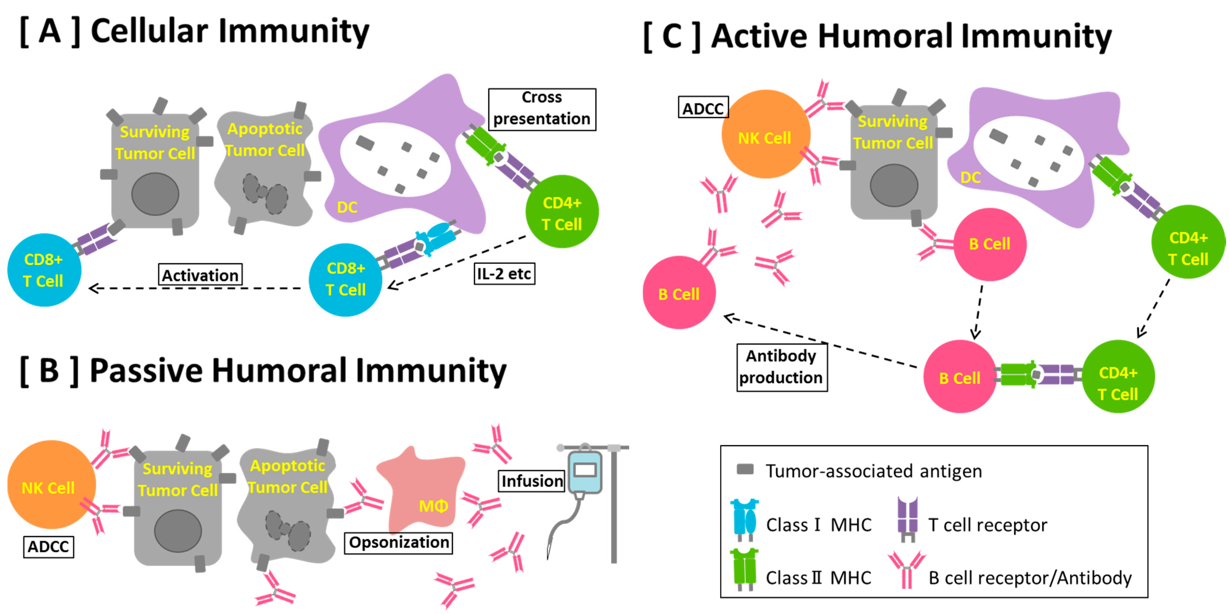
5. Anti-Cancer Antibody

In parallel with peptide vaccines, molecular targeting reagents to mobilize antibody-based passive immunity have been developed, starting with Herceptin and rituximab, which are now standard cancer treatments that significantly induce anti-tumor effects. The specific antigen of Herceptin, Her2, is overexpressed not only on breast cancer cells but also in ovarian and lung cancer [47,48,49,50,51]. Herceptin is a humanized monoclonal antibody that was approved in 1998 and that improved patient survival significantly. Although the exact mechanism is unclear, Herceptin can inhibit the signaling pathways involving HIF1α and Erk by masking the Her2 molecule and can react with the innate immune system via Fc receptors, possibly inducing antibody-dependent cellular cytotoxicity (ADCC) [52], antibody-dependent cellular phagocytosis (ADCP) [53] and complement-dependent cytotoxicity (CDC) [54]. Herceptin also suppresses Her2 homodimer and heterodimer formation. Moreover, this antibody may activate natural killer (NK) cells and other components of the immune system, which may be suppressed gradually [55] (Figure 1b). NK cells are involved in NK cell-mediated ADCC [56]. NK cells also secrete a high level of IFN-γ, which may help cancer cells to re-express class I MHC molecules. Consequently, the immune response may shift to Th1-dominated immunity. Clarifying which pathway is involved in Herceptin therapy has been difficult. Breast cancer progression of patients expressing Her2 at high levels is suppressed when they are treated with Herceptin.
Following the success of Herceptin, other therapeutic antibodies were developed. Rituximab, humanized anti-CD20; cetuximab, a chimeric anti-Her1 antibody for colorectal cancer [57]; alemtuzumab, a humanized anti-CD52 antibody for chronic lymphocytic leukaemia; and bevacizumab, a humanized anti-VEGF antibody for colorectal and lung cancer, have all been approved [58]. Pertuzumab, another anti-Her2 monoclonal antibody, was approved by the FDA in 2012 [59,60]. Other antibodies are currently awaiting approval.
Herceptin, which recognizes only one cancer antigen, is effective in Her2-positive breast cancer patients likely because the progression of this cancer uniquely depends on overexpression of a single antigen. Although Her2 is also overexpressed in other cancers such as ovarian cancer, Herceptin is not always an effective reagent. This evidence suggests that humoral immunity is not always sufficient to control tumor progression by a single tumor antigen.

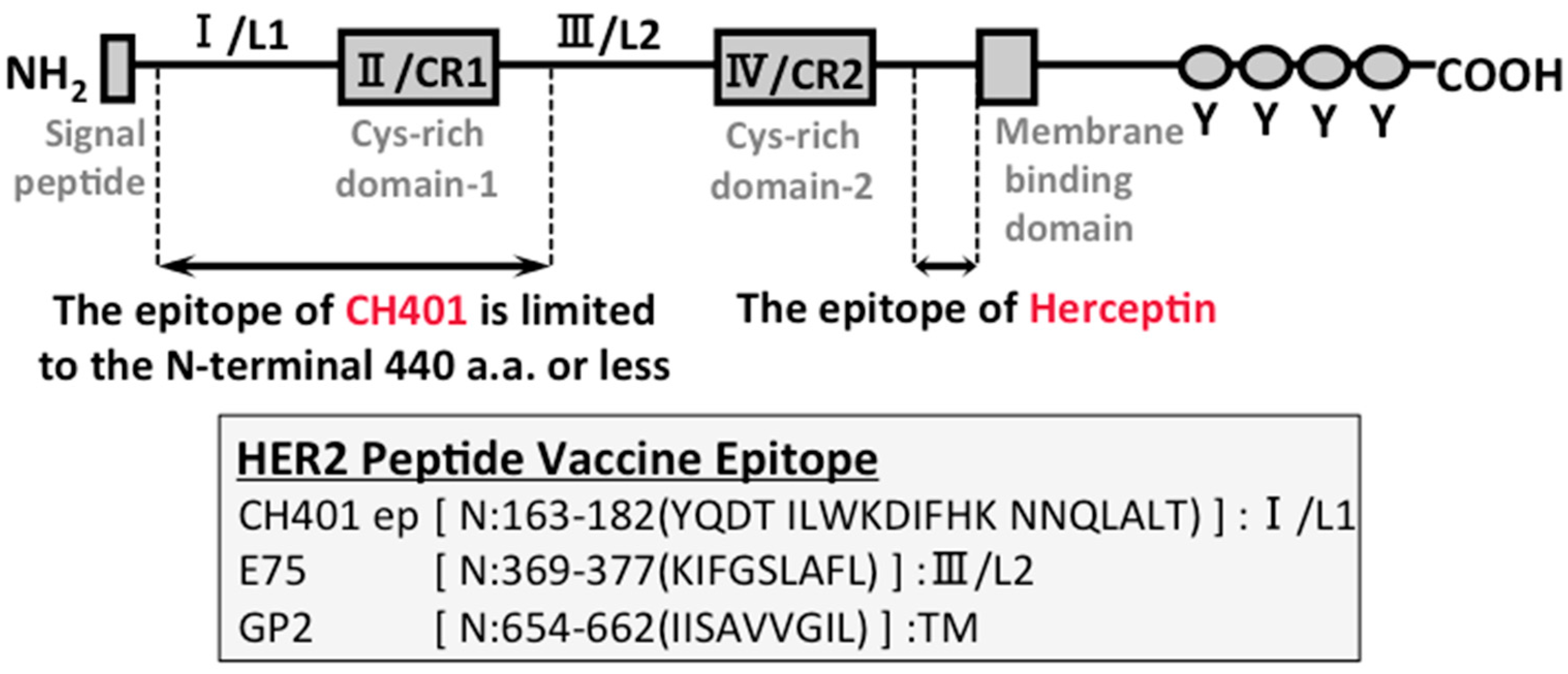
6. Why is Herceptin Successful?

To activate B cell-based anti-cancer immunity, the tumor antigen must be expressed on the surface of cancer cells. In particular, for antibody-based passive immune treatment to be effective, the antigen should be recognized by specific antibodies. Therefore, the tumor antigens must be expressed in large quantities. As shown in Figure 3, if the tumor antigen is not expressed on the surface, then no specific antibodies will be induced in vivo (Figure 3A). Low expression of tumor antigens may induce antibody binding but may not induce an antibody-mediated response (Figure 3B). Infusion of antibodies against a tumor antigen induces apoptosis in cells expressing high levels of that antigen (Figure 3C). Patients with high Her2 expression are responsive to Herceptin because the number of Her2 antibody molecules bound to the antigen per cancer cell becomes extremely high, inducing signaling to innate cytotoxic cells and thus cytotoxicity, such as ADCC, ADCP, and CDC. In addition to antibody-mediated cytotoxicity, strong signaling is reported to induce NK and CD8 T cell functions [61,62,63].
Thus, such effects may be induced by other antibody-based immunotherapies. To induce cytolytic activity in antibody-based passive immunotherapy, tumor antigens must be expressed. Similarly, a B cell epitope-based peptide vaccine using a protein other than Her2 may also require high expression of tumor antigens (Figure 3D).
A specific antibody that attacks cancer cells induces fundamental anti-cancer effects, which should make a peptide-based vaccine effective. Since Her2 is closely correlated with carcinogenesis [64], suppression of the Her2 signal may be particularly important. Tumor antigens that are not involved in carcinogenesis may have lower efficacy.
Figure 3. Antigen expression determines the effect of antibody-based passive immunity. (A) The tumor antigens are not expressed on the surface of cancer cells. Consequently, antibodies cannot bind to cancer cells, and anti-cancer immunity is not induced. (B) The tumor antigens are expressed on the surface of cancer cells; however, the antibody response is not sufficient to induce strong anti-cancer immunity. (C) After infusion of monoclonal antibodies, enough antibodies bind to cancer cells via cancer antigens to induce anti-cancer immunity. (D) Immunization with cancer-specific peptides might induce anti-cancer immunity similar to those produced by monoclonal antibody drugs.
Antibodies 04 00225 g003

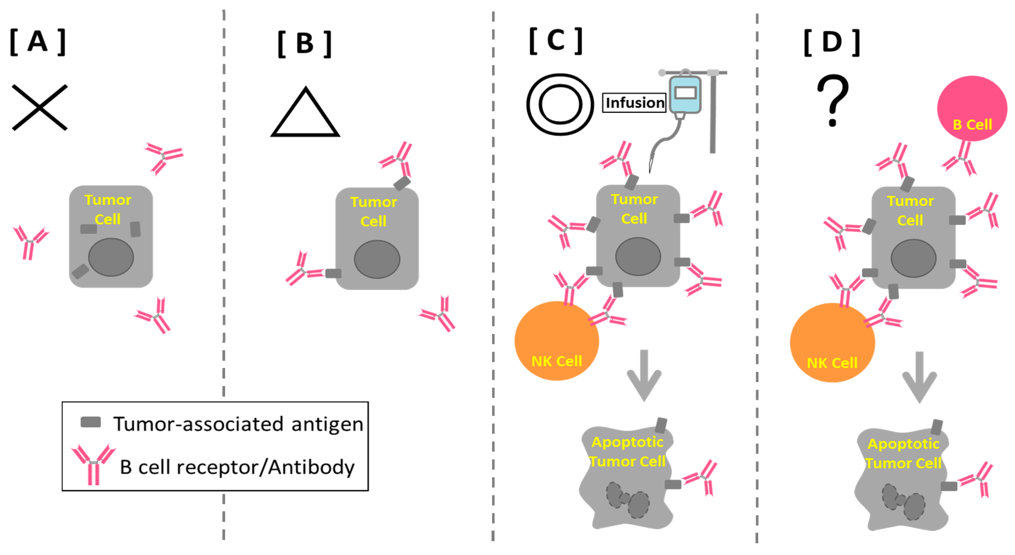
7. B Cell-based Peptide Vaccine

Peptide vaccines with class II HLA epitopes have entered clinical studies because Th cells are necessary for inducing cytotoxicity of cytotoxic T cells [65,66]; however, most of these studies have not involved antibody-based vaccines because of the importance of cytotoxic T cell activation in anti-tumor effects. The humoral immunity induced by peptide vaccines was thought to be supportive in classical studies of tumor immunity [66,67]; however, carcinomas, which show some response to passive antibody-based immunotherapy, as in breast cancer, may respond to an antibody-based peptide vaccine if the peptide is limited to the effective epitope (Figure 1c). An active peptide-vaccine therapy will increase patient quality of life by saving time and money, as frequent antibody infusions would not be needed. However, designing of antibody-based vaccine is difficult even if the antigens are infectious microbes. The clinical trial of respiratory syncytial virus (RSV) vaccine used an epitope selected strictly to contain the epitopes of both B cells and T cells. In the clinical trials, the effect of vaccination on RSV did not reach statistical significance [68,69].
Miller et al. developed B cell-based EGFR (Her1) and Her2 vaccines [70,71,72,73,74,75,76]. Her2 is a well-known antigen that is highly expressed on breast cancer cells in ~20% of patients. As Her1 is also highly expressed on the surface of cancer cells, their recognition by antibodies has promise. These authors employed computer-aided analysis [77] with crystal structure analysis to identify potential B cell epitopes in these target proteins, developed ligand-binding domain-specific peptides and demonstrated that the vaccine inhibits tumor growth in vitro and in vivo.
We are also developing a B cell epitope-based Her2 peptide vaccine, CH401MAP. While Miller’s group prepared the vaccines by predicting B cell epitopes of cancer antigens, we determined the epitope of an anti-Her2 monoclonal antibody, CH401, which was developed by Dr. Imai’s group by epitope mapping [78,79]. The identified epitope differed from that of Herceptin and other known Her2 antibodies (Figure 2). CH401 was reported to induce apoptosis in Her2-positive cancer cells [80]. We determined the epitope and reported that the 20 amino acid multiple antigen peptide (CH401MAP), together with Freund’s adjuvant, can induce specific antibodies in mice and humanized mice with human immune cells reconstituted in immunodeficient mice [79,81].
Moreover, various algorithms were used to predict that the 20 amino acid peptide could be presented by the HLAs of most Japanese breast cancer patients. A Her2 peptide with such a high ratio of affinity with the patients’ HLAs has never been reported. PBMCs of breast cancer patients responded to this peptide by secreting various cytokines [82] and retained the ability to secrete CH401MAP-specific antibodies, although the PBMCs could not enhance specific antibody secretion after peptide stimulation [83]. From these data, we speculate that this peptide may function as a peptide vaccine primarily to induce a specific antibody. Because this peptide was also predicted to possess class I HLA affinity, it may be effective for cytotoxic T cell induction.

8. B Cells and Cancer Immunity

In contrast, an increase in B cells has been reported without a direct correlation with cancer prognosis [84]. However, the effect of the increase in the B cell ratio on cancer progression is unclear. Since we reported that the PBMCs of breast cancer patients are rather unresponsive to the Her2 peptide, the newly developed B cell function may be somewhat reduced by the cancer environment. From the presented evidence, the patients may need to be selected for maintained B cell function to achieve a B cell epitope-based vaccine.
An increase in the B cell ratio may indicate thymus malfunction, which induces the suppression of T cell differentiation. This phenomenon might induce peripheral T lymphopenia, which increases the relative number of B cells in PBMCs; otherwise, T cells may be actively trapped or killed in the periphery, resulting in an increase in the relative number of B cells. In this case, lymphocytes may accumulate in the tumor parenchyma or invasive margin and then undergo apoptosis. Humoral immunity cannot be fully activated without Th cells; however, anti-tumor humoral immunity may not be reduced even when cytotoxic T cells are limited in their response. ADCC can be induced by neutrophils and by NK cells if specific antibodies are produced.

9. Humanized Mouse Model for Peptide Vaccination

Before B cell epitope-based peptide vaccines enter clinical trials, pre-clinical models may be preferable for evaluating efficacy because the activation level of T cells cannot be easily predicted by the available algorithms. The therapeutic super-agonist CD28 antibody TGN1412 was developed using mouse and monkey models and caused antibody-induced cytokine storms in healthy donors in clinical trials [85]. This accident suggested that risk could not be predicted by the available animal models and that side effects are an important possibility.
Humanized mice can be established by transplanting human tissues into severely-immunodeficient mice, such as NOD-SCID IL2Rγ (null) mice (NOG mice; Central Institute for Experimental Animals) [86] and NSG (Jackson) mice [87]. These mice are used primarily as pre-clinical models. Humanized mice with human immunity can be established by transplanting human hematopoietic stem cells (HSCs) or human peripheral blood mononuclear cells (PBMCs) into severely-immunodeficient mice. Immunization of humanized mice can clarify whether immune-competent cells can react with specific peptide antigens [88,89,90,91]. Although these humanized mice cannot completely mimic human immunity, some response can be detected [81,92]. We found that CH401MAP could induce specific antibodies in humanized mice transplanted with human cord blood stem cells. The BLT mouse, which is established by transplanting human fetal bone, liver, and HSCs, may also show some improvement [93]. The Central Institute for Experimental Animals and The Jackson Lab have improved upon humanized mouse systems by expressing additional human genes such as human cytokines and human leukocyte antigens [89]. If the transplantation and maintenance of patient PBMCs become possible, then the function of B cells may be estimated using these humanized mouse systems. The development of these systems is promising for the selection of patients for peptide vaccination.

10. Future Views

Peptide vaccines that induce B cell epitope-specific polyclonal antibodies may be useful against cancers that express high levels of surface tumor antigens and that respond to passive antibody treatment. These vaccines may be most effective when used in combination with antibodies against immune checkpoint molecules such as ipilimumab, nivolumab, and avelumab. To evaluate such molecular targeting treatments, pre-clinical model animals such as humanized mice should be adapted.

Acknowledgements

I would like to acknowledge our collaborators, Noriaki Hirayama and Hidetoshi Inoko, for their help in discussing this issue. We appreciate the contribution by members of the Kametani laboratory for the described results.

Author Contributions

Asuka Miyamoto and Banri Tsuda prepared portions of the manuscript, Yoshie Kametani and Yutaka Tokuda designed the manuscript, and Yoshie Kametani wrote the paper.

Conflicts of Interest

The authors declare that no conflicts of interest exist.

References

  1. Stagg, J.; Andre, F.; Loi, S. Immunomodulation via chemotherapy and targeted therapy: A new paradigm in breast cancer therapy? Breast Care 2012, 7, 2670272. [Google Scholar] [CrossRef] [PubMed]
  2. Andre, F.; Dieci, M.; Dubsky, P.; Sotiriou, C.; Curigliano, G.; Denkert, C.; Loi, S. Molecular pathways: Involvement of immune pathways in the therapeutic response and outcome in breast cancer. Clin. Cancer Res. 2013, 19, 28–33. [Google Scholar] [CrossRef] [PubMed]
  3. Rammensee, H.; Bachmann, J.; Emmerich, N.P.; Bachor, O.A.; Stevanovic, S. Syfpeithi: Database for mhc ligands and peptide motifs. Immunogenetics 1999, 50, 213–219. [Google Scholar] [CrossRef] [PubMed]
  4. Parker, K.C.; Bednarek, M.A.; Coligan, J.E. Scheme for ranking potential hla-a2 binding peptides based on independent binding of individual peptide side-chains. J. Immunol. 1994, 152, 163–175. [Google Scholar] [PubMed]
  5. Peters, B.; Sidney, J.; Bourne, P.; Bui, H.H.; Buus, S.; Doh, G.; Fleri, W.; Kronenberg, M.; Kubo, R.; Lund, O.; et al. The immune epitope database and analysis resource: From vision to blueprint. PLoS Biol. 2005, 3, e91. [Google Scholar] [CrossRef] [PubMed] [Green Version]
  6. Schwartzentruber, D.J.; Lawson, D.H.; Richards, J.M.; Conry, R.M.; Miller, D.M.; Treisman, J.; Gailani, F.; Riley, L.; Conlon, K.; Pockaj, B.; et al. Gp100 peptide vaccine and interleukin-2 in patients with advanced melanoma. N. Engl. J. Med. 2011, 364, 2119–2127. [Google Scholar] [CrossRef] [PubMed]
  7. Kantoff, P.W.; Higano, C.S.; Shore, N.D.; Berger, E.R.; Small, E.J.; Penson, D.F.; Redfern, C.H.; Ferrari, A.C.; Dreicer, R.; Sims, R.B.; et al. Sipuleucel-t immunotherapy for castration-resistant prostate cancer. N. Engl. J. Med. 2010, 363, 411–422. [Google Scholar] [CrossRef] [PubMed]
  8. Kenter, G.G.; Welters, M.J.; Valentijn, A.R.; Lowik, M.J.; Berends-van der Meer, D.M.; Vloon, A.P.; Essahsah, F.; Fathers, L.M.; Offringa, R.; Drijfhout, J.W.; et al. Vaccination against hpv-16 oncoproteins for vulvar intraepithelial neoplasia. N. Engl. J. Med. 2009, 361, 1838–1847. [Google Scholar] [CrossRef] [PubMed]
  9. Schuster, S.J.; Neelapu, S.S.; Gause, B.L.; Janik, J.E.; Muggia, F.M.; Gockerman, J.P.; Winter, J.N.; Flowers, C.R.; Nikcevich, D.A.; Sotomayor, E.M.; et al. Vaccination with patient-specific tumor-derived antigen in first remission improves disease-free survival in follicular lymphoma. J. Clin. Oncol. 2011, 29, 2787–2794. [Google Scholar] [CrossRef] [PubMed]
  10. Lizee, G.; Overwijk, W.W.; Radvanyi, L.; Gao, J.; Sharma, P.; Hwu, P. Harnessing the power of the immune system to target cancer. Ann. Rev. Med. 2013, 64, 71–90. [Google Scholar] [CrossRef] [PubMed]
  11. Harao, M.; Mittendorf, E.A.; Radvanyi, L.G. Peptide-based vaccination and induction of cd8+ t-cell responses against tumor antigens in breast cancer. BioDrugs 2015, 29, 15–30. [Google Scholar] [CrossRef] [PubMed]
  12. Baxter, D. Active and passive immunization for cancer. Hum. Vaccines Immunother. 2014, 10, 2123–2129. [Google Scholar] [CrossRef] [PubMed]
  13. Schneble, E.J.; Berry, J.S.; Trappey, F.A.; Clifton, G.T.; Ponniah, S.; Mittendorf, E.; Peoples, G.E. The her2 peptide nelipepimut-s (e75) vaccine (neuvax) in breast cancer patients at risk for recurrence: Correlation of immunologic data with clinical response. Immunotherapy 2014, 6, 519–531. [Google Scholar] [CrossRef] [PubMed]
  14. Yamada, A.; Sasada, T.; Noguchi, M.; Itoh, K. Next-generation peptide vaccines for advanced cancer. Cancer Sci. 2013, 104, 15–21. [Google Scholar] [CrossRef] [PubMed]
  15. Li, W.; Joshi, M.; Singhania, S.; Ramsey, K.; Murthy, S. Peptide vaccine: Progress and challenges. Vaccines 2014, 2, 515–536. [Google Scholar] [CrossRef]
  16. Mittendorf, E.; Clifton, G.T.; Holmes, J.; Clive, K.; Patil, R.; Benavides, L.; Gates, J.; Sears, A.; Stojadinovic, A.; Ponniah, S.; et al. Clinical trial results of the her-2/neu (e75) vaccine to prevent breast cancer recurrence in high-risk patients: From us millitary cancer institute clinical trials group study i-01 and i-02. Cancer 2012, 118, 2594–2602. [Google Scholar] [CrossRef] [PubMed]
  17. Clive, K.; Tyler, J.; Travis Clifton, G.; Holmes, J.; Ponniah, S.; Peoples, G.; Mittendorf, E. The gp2 peptide: A her2/neu-based breast cancer vaccine. J. Surg. Oncol. 2012, 105, 452–458. [Google Scholar] [CrossRef] [PubMed]
  18. Roskoski, R., Jr. The erbb/her family of protein-tyrosine kinases and cancer. Pharm. Res. 2014, 79, 34–74. [Google Scholar] [CrossRef] [PubMed]
  19. Hanahan, D.; Weinberg, R.A. Hallmarks of cancer: The next generation. Cell 2011, 144, 646–674. [Google Scholar] [CrossRef] [PubMed]
  20. Schreiber, R.D.; Old, L.J.; Smyth, M.J. Cancer immunoediting: Integrating immunity's roles in cancer suppression and promotion. Science 2011, 331, 1565–1570. [Google Scholar] [CrossRef] [PubMed]
  21. Hailemichael, Y.; Dai, Z.; Jaffarzad, N.; Ye, Y.; Medina, M.A.; Huang, X.F.; Dorta-Estremera, S.M.; Greeley, N.R.; Nitti, G.; Peng, W.; et al. Persistent antigen at vaccination sites induces tumor-specific cd8(+) t cell sequestration, dysfunction and deletion. Nat. Med. 2013, 19, 465–472. [Google Scholar] [CrossRef] [PubMed]
  22. Cerezo, D.; Pena, M.J.; Mijares, M.; Martinez, G.; Blanca, I.; De Sanctis, J.B. Peptide vaccines for cancer therapy. Recent Patents Inflam. Allergy Drug Discov. 2015, 9, 38–45. [Google Scholar] [CrossRef]
  23. Chang, D.Z.; Lomazow, W.; Joy Somberg, C.; Stan, R.; Perales, M.A. Granulocyte-macrophage colony stimulating factor: An adjuvant for cancer vaccines. Hematology 2004, 9, 207–215. [Google Scholar] [CrossRef] [PubMed]
  24. Allison, A.C. Immunological adjuvants and their modes of action. Arch. Immunol. Ther. Exp. 1997, 45, 141–147. [Google Scholar]
  25. Roychoudhuri, R.; Eil, R.L.; Restifo, N.P. The interplay of effector and regulatory T cells in cancer. Curr. Opin. Immunol. 2015, 33c, 101–111. [Google Scholar] [CrossRef] [PubMed]
  26. Whiteside, T.L. The role of death receptor ligands in shaping tumor microenvironment. Immunol. Invest. 2007, 36, 25–46. [Google Scholar] [CrossRef] [PubMed]
  27. Nirschl, C.J.; Drake, C.G. Molecular pathways: Coexpression of immune checkpoint molecules: Signaling pathways and implications for cancer immunotherapy. Clin. Cancer Res. 2013, 19, 4917–4924. [Google Scholar] [CrossRef] [PubMed]
  28. Pardoll, D.M. The blockade of immune checkpoints in cancer immunotherapy. Nat. Rev. Cancer 2012, 12, 252–264. [Google Scholar] [CrossRef] [PubMed]
  29. Linsley, P.S.; Ledbetter, J.A. The role of the cd28 receptor during t cell responses to antigen. Ann. Rev. Immunol. 1993, 11, 191–212. [Google Scholar] [CrossRef] [PubMed]
  30. Brunet, J.F.; Denizot, F.; Golstein, P. A differential molecular biology search for genes preferentially expressed in functional t lymphocytes: The ctla genes. Immunol. Rev. 1988, 103, 21–36. [Google Scholar] [CrossRef] [PubMed]
  31. Okazaki, T.; Iwai, Y.; Honjo, T. New regulatory co-receptors: Inducible co-stimulator and pd-1. Curr. Opin. Immunol. 2002, 14, 779–782. [Google Scholar] [CrossRef]
  32. Okazaki, T.; Chikuma, S.; Iwai, Y.; Fagarasan, S.; Honjo, T. A rheostat for immune responses: The unique properties of pd-1 and their advantages for clinical application. Nat. Immunol. 2013, 14, 1212–1218. [Google Scholar] [CrossRef] [PubMed] [Green Version]
  33. Carreno, B.M.; Collins, M. The b7 family of ligands and its receptors: New pathways for costimulation and inhibition of immune responses. Ann. Rev. Immunol. 2002, 20, 29–53. [Google Scholar] [CrossRef] [PubMed]
  34. Blank, C.; Gajewski, T.F.; Mackensen, A. Interaction of pd-l1 on tumor cells with pd-1 on tumor-specific t cells as a mechanism of immune evasion: Implications for tumor immunotherapy. Cancer Immunol. Immun. 2005, 54, 307–314. [Google Scholar] [CrossRef] [PubMed]
  35. Hodi, F.S.; Butler, M.; Oble, D.A.; Seiden, M.V.; Haluska, F.G.; Kruse, A.; Macrae, S.; Nelson, M.; Canning, C.; Lowy, I.; et al. Immunologic and clinical effects of antibody blockade of cytotoxic t lymphocyte-associated antigen 4 in previously vaccinated cancer patients. Proc. Natl. Acad. Sci. USA 2008, 105, 3005–3010. [Google Scholar] [CrossRef] [PubMed]
  36. Ribas, A. Clinical development of the anti-ctla-4 antibody tremelimumab. Semin. Oncol. 2010, 37, 450–454. [Google Scholar] [CrossRef] [PubMed]
  37. Naidoo, J.; Page, D.B.; Wolchok, J.D. Immune modulation for cancer therapy. Br. J. Cancer 2014, 111, 2214–2219. [Google Scholar] [CrossRef] [PubMed]
  38. Ostrand-Rosenberg, S.; Horn, L.; Haile, S. The programmed death-1 immune-suppressive pathway: Barrier to antitumor immunity. J. Immunol. 2014, 193, 3835–3841. [Google Scholar] [CrossRef] [PubMed]
  39. Postow, M.; Callahan, M.; Wolchok, J. Immune checkpoint blockade in cancer therapy. J. Clin. Oncol. 2015, 33, 1974–1982. [Google Scholar] [CrossRef] [PubMed]
  40. Tumeh, P.; Harview, C.; Yearley, J.; Shintaku, I.; Taylor, E.; Robert, L.; Chmielowski, B.; Spasic, M.; Henry, G.; Ciobanu, V.; et al. Pd-1 blockade induces responses by inhibiting adaptive immune resistance. Nature 2014, 515, 568–571. [Google Scholar] [CrossRef] [PubMed]
  41. Gunturi, A.; McDermott, D.F. Nivolumab for the treatment of cancer. Expert Opin. Invest. Drug. 2015, 24, 253–260. [Google Scholar] [CrossRef] [PubMed]
  42. Momtaz, P.; Postow, M.A. Immunologic checkpoints in cancer therapy: Focus on the programmed death-1 (pd-1) receptor pathway. Pharmacogenomics Pers. Med. 2014, 7, 357–365. [Google Scholar]
  43. Wolchok, J.; Hodi, F.; Weber, J.; Allison, J.; Urba, W.; Robert, C.; O'Day, S.; Hoos, A.; Humphrey, R.; Berman, D.; et al. Development of ipilimumab: A novel immunotherapeutic approach for the treatment of advanced melanoma. Ann. NY Acad. Sci. 2013, 1291, 1–13. [Google Scholar] [CrossRef] [PubMed]
  44. Wolchok, J.; Kluger, H.; Callahan, M.; Postow, M.; Rizvi, N.; Lesokhin, A.; Segal, N.; Ariyan, C.; Gordon, R.; Reed, K.; et al. Nivolumab plus ipilimumab in advanced melanoma. N. Engl. J. Med. 2013, 369, 122–133. [Google Scholar] [CrossRef] [PubMed]
  45. Ansell, S.; Lesokhin, A.; Borrello, I.; Halwani, A.; Scott, E.; Gutierrez, M.; Schuster, S.; Millenson, M.; Cattry, D.; Freeman, G.; et al. Pd-1 blockade with nivolumab in relapsed or refractory hodgkin's lymphoma. N. Engl. J. Med. 2015, 372, 311–319. [Google Scholar] [CrossRef] [PubMed]
  46. Boyerinas, B.; Jochems, C.; Fantini, M.; Heery, C.; Gulley, J.; Tsang, K.; Schlom, J. Antibody-dependent cellular cytotoxicity activity of a novel anti-pd-l1 antibody avelumab (msb0010718c) on human tumor cells. Cancer Immunol. Res. 2015. [Google Scholar] [CrossRef] [PubMed]
  47. Kern, J.A.; Schwartz, D.A.; Nordberg, J.E.; Weiner, D.B.; Greene, M.I.; Torney, L.; Robinson, R.A. P185neu expression in human lung adenocarcinomas predicts shortened survival. Cancer Res. 1990, 50, 5184–5187. [Google Scholar] [PubMed]
  48. Berchuck, A.; Rodriguez, G.; Kinney, R.B.; Soper, J.T.; Dodge, R.K.; Clarke-Pearson, D.L.; Bast, R.C., Jr. Overexpression of her-2/neu in endometrial cancer is associated with advanced stage disease. Am. J. Obstet. Gynecol. 1991, 164, 15–21. [Google Scholar] [CrossRef]
  49. Cirisano, F.D.; Karlan, B.Y. The role of the her-2/neu oncogene in gynecologic cancers. J. Soc. Gynecol. Invest. 1996, 3, 99–105. [Google Scholar] [CrossRef]
  50. Morrison, C.; Zanagnolo, V.; Ramirez, N.; Cohn, D.E.; Kelbick, N.; Copeland, L.; Maxwell, G.L.; Fowler, J.M. Her-2 is an independent prognostic factor in endometrial cancer: Association with outcome in a large cohort of surgically staged patients. J. Clin. Oncol. 2006, 24, 2376–2385. [Google Scholar] [CrossRef] [PubMed]
  51. Yano, T.; Doi, T.; Ohtsu, A.; Boku, N.; Hashizume, K.; Nakanishi, M.; Ochiai, A. Comparison of her2 gene amplification assessed by fluorescence in situ hybridization and her2 protein expression assessed by immunohistochemistry in gastric cancer. Oncol. Rep. 2006, 15, 65–71. [Google Scholar] [CrossRef] [PubMed]
  52. Weiner, L.M.; Surana, R.; Wang, S. Monoclonal antibodies: Versatile platforms for cancer immunotherapy. Nat. Rev. Immunol. 2010, 10, 317–327. [Google Scholar] [CrossRef] [PubMed]
  53. Winiarska, M.; Glodkowska-Mrowka, E.; Bil, J.; Golab, J. Molecular mechanisms of the antitumor effects of anti-cd20 antibodies. Front. Biosci. 2011, 16, 277–306. [Google Scholar] [CrossRef]
  54. Zipfel, P.F.; Skerka, C. Complement regulators and inhibitory proteins. Nat. Rev. Immunol. 2009, 9, 729–740. [Google Scholar] [CrossRef] [PubMed]
  55. De, P.; Hasmann, M.; Leyland-Jones, B. Molecular determinants of trastuzumab efficacy: What is their clinical relevance? Cancer Treat. Rev. 2013, 39, 925–934. [Google Scholar] [CrossRef] [PubMed]
  56. Marechal, R.; De Schutter, J.; Nagy, N.; Demetter, P.; Lemmers, A.; Deviere, J.; Salmon, I.; Tejpar, S.; Van Laethem, J.L. Putative contribution of cd56 positive cells in cetuximab treatment efficacy in first-line metastatic colorectal cancer patients. BMC Cancer 2010, 10, 340. [Google Scholar] [CrossRef] [PubMed]
  57. Mach, J.P. Introduction to monoclonal antibodies. Cancer Immun. 2012, 12, 11. [Google Scholar] [PubMed]
  58. Oldham, R.K.; Dillman, R.O. Monoclonal antibodies in cancer therapy: 25 years of progress. J. Clin. Oncol. 2008, 26, 1774–1777. [Google Scholar] [CrossRef] [PubMed]
  59. Slamon, D.; Eiermann, W.; Robert, N.; Pienkowski, T.; Martin, M.; Press, M.; Mackey, J.; Glaspy, J.; Chan, A.; Pawlicki, M.; et al. Adjuvant trastuzumab in her2-positive breast cancer. N. Engl. J. Med. 2011, 365, 1273–1283. [Google Scholar] [CrossRef] [PubMed]
  60. Baselga, J.; Cortes, J.; Kim, S.B.; Im, S.A.; Hegg, R.; Im, Y.H.; Roman, L.; Pedrini, J.L.; Pienkowski, T.; Knott, A.; et al. Pertuzumab plus trastuzumab plus docetaxel for metastatic breast cancer. N. Engl. J. Med. 2012, 366, 109–119. [Google Scholar] [CrossRef] [PubMed]
  61. zum Buschenfelde, C.M.; Hermann, C.; Schmidt, B.; Peschel, C.; Bernhard, H. Antihuman epidermal growth factor receptor 2 (her2) monoclonal antibody trastuzumab enhances cytolytic activity of class i-restricted her2-specific T lymphocytes against her2-overexpressing tumor cells. Cancer Res. 2002, 62, 2244–2247. [Google Scholar]
  62. Banerjee, D.; Matthews, P.; Matayeva, E.; Kaufman, J.L.; Steinman, R.M.; Dhodapkar, K.M. Enhanced T-cell responses to glioma cells coated with the anti-egf receptor antibody and targeted to activating fcgammars on human dendritic cells. J. Immunother. 2008, 31, 113–120. [Google Scholar] [CrossRef] [PubMed]
  63. Arnould, L.; Gelly, M.; Penault-Llorca, F.; Benoit, L.; Bonnetain, F.; Migeon, C.; Cabaret, V.; Fermeaux, V.; Bertheau, P.; Garnier, J.; et al. Trastuzumab-based treatment of her2-positive breast cancer: An antibody-dependent cellular cytotoxicity mechanism? Br. J. Cancer 2006, 94, 259–267. [Google Scholar] [CrossRef] [PubMed]
  64. Neve, R.M.; Lane, H.A.; Hynes, N.E. The role of overexpressed her2 in transformation. Ann. Oncol. 2001, 12 (Suppl. 1), S9–S13. [Google Scholar] [CrossRef] [PubMed]
  65. Paschen, A.; Eichmuller, S.; Schadendorf, D. Identification of tumor antigens and T-cell epitopes, and its clinical application. Cancer Immunol. Immun. 2004, 53, 196–203. [Google Scholar] [CrossRef] [PubMed]
  66. Voskens, C.J.; Strome, S.E.; Sewell, D.A. Synthetic peptide-based cancer vaccines: Lessons learned and hurdles to overcome. Curr. Mol. Med. 2009, 9, 683–693. [Google Scholar] [CrossRef] [PubMed]
  67. Pilla, L.; Rivoltini, L.; Patuzzo, R.; Marrari, A.; Valdagni, R.; Parmiani, G. Multipeptide vaccination in cancer patients. Exp. Opin. Biol. Ther. 2009, 9, 1043–1055. [Google Scholar] [CrossRef] [PubMed]
  68. Simoes, E.; Tan, D.; Ohlsson, A.; Sales, V.; Wang, E. Respiratory syncytial virus vaccine: A systematic overview with emphasis on respiratory syncytial virus subunit vaccines. Vaccine 2002, 20, 954–960. [Google Scholar] [CrossRef]
  69. Beck, A.; Klinguer-Hamour, C.; Bussat, M.-C.; Champion, T.; Haeuw, J.-F.; Goetsch, L.; Wurch, T.; Sugawara, M.; Milon, A.; van Dorsselaer, A.; et al. Peptides as tools and drugs for immunotherapies. J. Peptide Sci. 2007, 13, 588–602. [Google Scholar] [CrossRef] [PubMed]
  70. Foy, K.C.; Wygle, R.M.; Miller, M.J.; Overholser, J.P.; Bekaii-Saab, T.; Kaumaya, P.T. Peptide vaccines and peptidomimetics of egfr (her-1) ligand binding domain inhibit cancer cell growth in vitro and in vivo. J. Imunol. 2013, 191, 217–227. [Google Scholar] [CrossRef] [PubMed]
  71. Miller, M.J.; Foy, K.C.; Kaumaya, P.T. Cancer immunotherapy: Present status, future perspective, and a new paradigm of peptide immunotherapeutics. Discov. Med. 2013, 15, 166–176. [Google Scholar] [PubMed]
  72. Dakappagari, N.K.; Douglas, D.B.; Triozzi, P.L.; Stevens, V.C.; Kaumaya, P.T. Prevention of mammary tumors with a chimeric her-2 b-cell epitope peptide vaccine. Cancer Res. 2000, 60, 3782–3789. [Google Scholar] [PubMed]
  73. Dakappagari, N.K.; Pyles, J.; Parihar, R.; Carson, W.E.; Young, D.C.; Kaumaya, P.T. A chimeric multi-human epidermal growth factor receptor-2 b cell epitope peptide vaccine mediates superior antitumor responses. J. Immunol. 2003, 170, 4242–4253. [Google Scholar] [CrossRef] [PubMed]
  74. Dakappagari, N.K.; Lute, K.D.; Rawale, S.; Steele, J.T.; Allen, S.D.; Phillips, G.; Reilly, R.T.; Kaumaya, P.T. Conformational her-2/neu b-cell epitope peptide vaccine designed to incorporate two native disulfide bonds enhances tumor cell binding and antitumor activities. J. Biol. Chem. 2005, 280, 54–63. [Google Scholar] [CrossRef] [PubMed]
  75. Dakappagari, N.K.; Sundaram, R.; Rawale, S.; Liner, A.; Galloway, D.R.; Kaumaya, P.T. Intracellular delivery of a novel multiepitope peptide vaccine by an amphipathic peptide carrier enhances cytotoxic t-cell responses in hla-a*201 mice. J. Peptide Res. 2005, 65, 189–199. [Google Scholar] [CrossRef] [PubMed]
  76. Kaumaya, P.T.; Foy, K.C.; Garrett, J.; Rawale, S.V.; Vicari, D.; Thurmond, J.M.; Lamb, T.; Mani, A.; Kane, Y.; Balint, C.R.; et al. Phase i active immunotherapy with combination of two chimeric, human epidermal growth factor receptor 2, b-cell epitopes fused to a promiscuous t-cell epitope in patients with metastatic and/or recurrent solid tumors. J. Clin. Oncol. 2009, 27, 5270–5277. [Google Scholar] [CrossRef] [PubMed]
  77. Sundaram, R.; Lynch, M.; Rawale, S.; Sun, Y.; Kazanji, M.; Kaumaya, P. De novo design of peptide immunogens that mimic the coiled coil region of human t-cell leukemia virus type-1 glycoprotein 21 transmembrane subunit for induction of native protein reactive neutralizing antibodies. J. Biol. Chem. 2004, 279, 24141–24151. [Google Scholar] [CrossRef] [PubMed]
  78. Hinoda, Y.; Sasaki, S.; Ishida, T.; Imai, K. Monoclonal antibodies as effective therapeutic agents for solid tumors. Cancer Sci. 2004, 95, 621–625. [Google Scholar] [CrossRef] [PubMed]
  79. Miyako, H.; Kametani, Y.; Katano, I.; Ito, R.; Tsuda, B.; Furukawa, A.; Saito, Y.; Ishikawa, D.; Ogino, K.; Sasaki, S.; et al. Antitumor effect of new her2 peptide vaccination based on b cell epitope. Anticancer Res. 2011, 31, 3361–3368. [Google Scholar] [PubMed]
  80. Ishida, T.; Tsujisaki, M.; Hinoda, Y.; Imai, K.; Yachi, A. Establishment and characterization of mouse-human chimeric monoclonal antibody to erbb-2 product. Jpn. J. Cancer Res. 1994, 85, 172–178. [Google Scholar] [CrossRef] [PubMed]
  81. Kametani, Y.; Shiina, M.; Katano, I.; Ito, R.; Ando, K.; Toyama, K.; Tsukamoto, H.; Matsumura, T.; Saito, Y.; Ishikawa, D.; et al. Development of human-human hybridoma from anti-her-2 peptide-producing b cells in immunized nog mouse. Exp. Hematol. 2006, 34, 1240–1248. [Google Scholar] [CrossRef] [PubMed]
  82. Tsuda, B.; Kametani, Y.; Goto, Y.; Saito, Y.; Suzuki, Y.; Habu, S.; Inoko, H.; Tokuda, Y. A human b cell receptor epitope-based erbb-2 peptide (n:163-182) with pan-reactivity to the t cells of japanese breast cancer patients. J. Vaccines Vaccination 2012, 3, 159. [Google Scholar] [CrossRef]
  83. Tsuda, B.; Kametani, Y.; Miyamoto, A.; Miyako, H.; Kumaki, N.; Ogiy, R.; Oshitanai, R.; Terao, M.; Morioka, T.; Niikura, N.; et al. The effect of peptide treatment on the hla-binding and antibody production in peripheral blood mononuclear cells obtained from japanese breast cancer patients. J. Vaccines Vaccination 2015, in press. [Google Scholar]
  84. Babusíková, O.; Novotná, L.; Korec, S.; Schneková, K.; Turková, D.; Havránková, M.; Plentová, K. Change in the proportion of t and b lymphocytes in human malignant neoplasia in relation to the clinical stage. Neoplasma 1975, 22, 413–421. [Google Scholar] [PubMed]
  85. Hunig, T. The storm has cleared: Lessons from the cd28 superagonist tgn1412 trial. Nat. Rev. Immunol. 2012, 12, 317–318. [Google Scholar] [CrossRef] [PubMed]
  86. Ito, M.; Kobayashi, K.; Nakahata, T. Nod/shi-scid il2rgamma(null) (nog) mice more appropriate for humanized mouse models. Curr. Top. Microbiol. Immunol. 2008, 324, 53–76. [Google Scholar] [PubMed]
  87. Brehm, M.A.; Wiles, M.V.; Greiner, D.L.; Shultz, L.D. Generation of improved humanized mouse models for human infectious diseases. J. Immunol. Methods 2014, 410, 3–17. [Google Scholar] [CrossRef] [PubMed]
  88. Ito, R.; Shiina, M.; Saito, Y.; Tokuda, Y.; Kametani, Y.; Habu, S. Antigen-specific antibody production of human b cells in nog mice reconstituted with the human immune system. Curr. Top. Microbiol. Immunol. 2008, 324, 95–107. [Google Scholar] [PubMed]
  89. Ito, R.; Takahashi, T.; Katano, I.; Ito, M. Current advances in humanized mouse models. Cell. Mol. Immunol. 2012, 9, 208–214. [Google Scholar] [CrossRef] [PubMed]
  90. Brehm, M.A.; Shultz, L.D.; Luban, J.; Greiner, D.L. Overcoming current limitations in humanized mouse research. J. Infect. Dis. 2013, 208 (Suppl. 2), S125–S130. [Google Scholar] [CrossRef] [PubMed]
  91. Aryee, K.E.; Shultz, L.D.; Brehm, M.A. Immunodeficient mouse model for human hematopoietic stem cell engraftment and immune system development. Method. Mol. Biol. 2014, 1185, 267–278. [Google Scholar]
  92. Matsumura, T.; Kametani, Y.; Ando, K.; Hirano, Y.; Katano, I.; Ito, R.; Shiina, M.; Tsukamoto, H.; Saito, Y.; Tokuda, Y.; et al. Functional cd5+ b cells develop predominantly in the spleen of nod/scid/gammac(null) (nog) mice transplanted either with human umbilical cord blood, bone marrow, or mobilized peripheral blood cd34+ cells. Exp. Hematol. 2003, 31, 789–797. [Google Scholar] [CrossRef]
  93. Wege, A.K.; Melkus, M.W.; Denton, P.W.; Estes, J.D.; Garcia, J.V. Functional and phenotypic characterization of the humanized blt mouse model. Curr. Top. Microbiol. Immunol. 2008, 324, 149–165. [Google Scholar] [PubMed]

