From Anti-SARS-CoV-2 Immune Responses to COVID-19 via Molecular Mimicry

Department of Biosciences, Biotechnologies, and Biopharmaceutics, University of Bari, 70125 Bari, Italy
Antibodies 2020, 9(3), 33; https://0-doi-org.brum.beds.ac.uk/10.3390/antib9030033
Submission received: 13 June 2020
/
Revised: 4 July 2020
/
Accepted: 9 July 2020
/
Published: 16 July 2020
(This article belongs to the Special Issue Antibodies, B Cell Responses and Immune Responses to SARS-CoV-2 Infections)
Abstract
:Aim: To define the autoimmune potential of Severe Acute Respiratory Syndrome Coronavirus 2 (SARS-CoV-2) infection. Methods: Experimentally validated epitopes cataloged at the Immune Epitope DataBase (IEDB) and present in SARS-CoV-2 were analyzed for peptide sharing with the human proteome. Results: Immunoreactive epitopes present in SARS-CoV-2 were mostly composed of peptide sequences present in human proteins that—when altered, mutated, deficient or, however, improperly functioning—may associate with a wide range of disorders, from respiratory distress to multiple organ failure. Conclusions: This study represents a starting point or hint for future scientific–clinical investigations and suggests a range of possible protein targets of autoimmunity in SARS-CoV-2 infection. From an experimental perspective, the results warrant the testing of patients’ sera for autoantibodies against these protein targets. Clinically, the results warrant a stringent surveillance on the future pathologic sequelae of the current SARS-CoV-2 pandemic.
1. Introduction
It is well known that coronavirus (re)infections have a pathologic immune potential. Indeed:
- In 2008, it was reported [6] that prior immunization with SARS-CoV nucleocapsid protein N causes severe pneumonia in mice infected with SARS-CoV;
- In 2012, immunization with SARS coronavirus vaccines was found to lead to lung immunopathology on challenge with the SARS virus [7];
- In 2016, Agrawal et al. [8] demonstrated that immunization with inactivated Middle East Respiratory Syndrome (MERS) coronavirus vaccine leads to lung immunopathology on challenge with live virus. Moreover, the Authors documented that immunopathology with SARS-CoV vaccines occurred for whole-virus vaccines, subunit vaccines, different inactivation methods, different preparation substrates, and with recombinant surface spike protein. Finally, the Authors also underscored that, even if studies with vector vaccines point to the nucleoprotein N as responsible for the immunopathological effects and indicate that the S protein might be free of risk, nonetheless, also recombinant spike protein induced the immunopathology [6,7,8,9].
However, the molecular and cellular basis for how SARS-CoVs might impact on the host immune system resulting in dysfunctional immune responses, severe morbidity, and mortality remain obscure [1,10].
Following the current SARS-CoV-2 pandemic, cross-reactivity was proposed as a mechanism that might explain the immunopathology associated with the coronavirus infection [11]. The rationale is that the sharing of peptide motifs between SARS-CoV-2 and human proteins might evoke immune responses capable of hitting not only the virus but also the human proteins with consequent autoimmune pathologic sequelae in the human host. As a matter of fact, the study [11] called attention to a vast and specific peptide sharing between SARS-CoV-2 spike glycoprotein and alveolar surfactant-related proteins, in this way addressing the issue of why SARS-CoV-2 so heavily attacks the respiratory system.
In the wake of such results, in order to validate (or, as well, invalidate) the cross-reactivity hypothesis, investigation was expanded here by analyzing the peptide sharing between the human host and immunoreactive epitopes that are also present in SARS-CoV-2. Actually, the mere sharing of peptide sequences between pathogens and human proteins might be of little significance whether it remained sterile of cross-reactive autoimmune reactions. On the contrary, a peptide sharing between immunoreactive pathogen-derived epitopes and human proteins would assume the value of a proof of concept of cross-reactivity as a mechanism contributing to the pathogen-induced diseases.
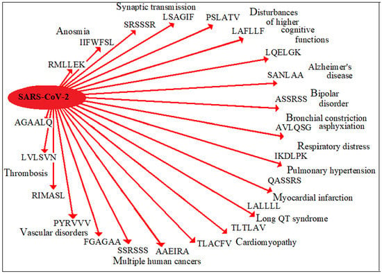
In this scientific framework, using the hexapeptide as an antigenic and immunogenic unit [12,13,14,15], immunoreactive epitopes that are present in SARS-CoV-2 were analyzed for matches with the human proteome. The results confirm a vast peptide commonality that involves human proteins implied in pulmonary insufficiency, neurological disorders, cardiac and vascular alterations, pregnancy dysfunctions, multiple cancers and anosmia, among others.
2. Materials and Methods
2.1. SARS-CoV-2 Epitopes
Analyses were conducted on an immunome composed of 233 linear epitopes that were experimentally validated, present in the proteins of SARS-CoV, and cataloged in the IEDB [16]. The experimentally validated 233 epitopes have been described in detail by Ahmed et al. [17], map identically in SARS-CoV-2 proteins, and are listed in Supplementary Materials Table S1.
The hexapeptide was used as a measurement unit to define minimal epitopic sequences. Indeed, 5–6 amino acids (aa) can form a sufficient minimal determinant for epitope–paratope interaction [12,13]. Likewise, T-cell epitopes contain a core of 5–6 aa that are involved in antigenicity and immunogenicity [13,14,15] and are flanked by NH2- and COOH– terminus aa that act as anchor motifs.
2.2. Analyses of the Peptide Sharing between SARS-CoV-2 Epitopes and the Human Proteome
The SARS-CoV-2 epitope sequences were dissected into hexapeptides which overlapped each other by five aa residues. The resulting 733 hexapeptides were analyzed for occurrence(s) within human proteins using the Pir Peptide Match program [18].
Human proteins involved in the hexapeptide sharing were analyzed for function/diseases using UniProtKB, PubMed, and OMIM resources.
2.3. Calculation of the Expected Value for Hexapeptide Sharing
The expected value for hexapeptide sharing between two proteins can be calculated by considering the number of all possible hexapeptides, N. Since in a hexapeptide each residue can be any of the 20 aa, the number of all possible hexapeptides N is given by N = 206 = 64 × 106. Then, the number of the expected occurrences is directly proportional to the number of hexapeptides in the two proteins and inversely proportional to N. Assuming that the number of hexapeptides in the two proteins is ≪N and neglecting the relative abundance of aa, we obtain a formula derived by approximation, where the expected number of hexapeptides is 1/N or 20−6.
3. Results
3.1. Numerical Description of the Peptide Sharing between SARS-CoV-2 Epitopes and the Human Proteome
Table 1 reports that 230 out of the 733 hexapeptides composing the analyzed 233 immuno- reactive epitopes occurred among 460 human proteins. Many of these shared sequences recurred more times. For instance, the zinc finger protein ZNF265—a splice factor that is important in renin mRNA processing and stability [19]—shares the hexapeptide SRSSSR with the viral epitope SQASSRSSSR (IEDB ID: 60380). The hexapeptide SRSSSR recurs three times in the zinc finger protein, exactly at aa positions: 211-216, 241-246, and 256-261. Then, including multiple occurrences, on the whole the hexapeptides shared with the human proteins amount to 505.
3.2. Distribution of the Shared Hexapeptides through the SARS-CoV-2 Epitopes
For reasons of synthesis, Table 2 reports the distribution of the shared hexapeptides relatively to a sample (n = 58) of SARS-CoV-2 epitopes. In the viral epitope sequences, the hexapeptides shared with human proteins are marked in capital letter format. The distribution of the shared hexapeptides throughout the entire set of 233 SARS-CoV-2 epitopes is described in Supplementary Materials Table S2.
Table 2 documents that numerous immunoreactive SARS-CoV-2 epitopes are composed mostly or, in many instances, uniquely of peptide sequences shared with human proteins. As an example, among the many, the epitope IEDB ID 4936, which is present in the viral nucleocapsid protein and corresponds to the aa sequence ATEGALNTPK, results from the consecutive succession/overlapping of 6 mers present in human proteins too.
Taken together, Table 1, Table 2, and Table S2 highlight the unexpectedness of the peptide overlap between the human proteome and the immunoreactive viral epitopes derived from IEDB, described by Ahmed et al. [17] and analyzed here. From a mathematical point of view, if one considers that the probability of a hexapeptide to occur in 2 proteins is ~20−6 (or 1 out of 64,000,000 or 0.000000015625), then the number of hexapeptides (namely, 505, including multiple occurrences) shared between the SARS-CoV-2 immunome and human proteins is surprisingly high and can be explained only on the basis of evolutionary processes [20].
In the context of autoimmunity, Table 1, Table 2, and Table S2 concretize the effective possibility of cross-reactions between SARS-CoV-2 and the human proteome, given the fact that the immunological information unit in terms of both immunogenicity and antigenicity is contained in a space formed by 5–6 aa residues [12,13,14,15].
3.3. Distribution of the Hexapeptide Sharing through the Human Proteins and the Potential Diseasome
As quantified in Table 1, hexapeptides from immunoreactive viral epitopes occur across 460 human proteins. The 460 human proteins are listed and synthetically described in Supplementary Materials Table S3. The 460 human proteins are involved in metabolic, developmental, and regulatory cellular functions and—when mutated, modified, deficient or, however, improperly functioning—may lead to altered functions and more or less severe pathologies in the human organism. Obvious reasons of space prevent a protein-by-protein analysis of all of them and, here, only a few proteins (given by the UniProt name in italic and shared peptides in parentheses) and the related pathologies that could arise in case of cross reactivity are dealt with as follows.
- Pulmonary disorders
Molecular mimicry between SARS-CoV-2 spike glycoprotein and alveolar surfactants-related proteins have already been described [11]. Additional proteins that–if hit–can alter lung functions are:
- Mothers against decapentaplegic homolog 9 protein (QASSRS), alterations of which may lead to pulmonary hypertension with proliferating endothelial cells in pulmonary arterioles, right ventricular failure, and death [21];
- Adenylate cyclase type 9 (KQLSSN) that is expressed in multiple cells of the lung with expression highest in airway smooth muscle [24];
- Acetylcholinesterase (AVLQSG) where imbalances in the neurotransmitter acetylcholine relate to neurological conditions, such as Alzheimer’s disease, Parkinson’s disease, and myastenia gravis; irreversible inhibition of acetylcholinesterase may lead to muscular paralysis, convulsions, bronchial constriction, and death by asphyxiation [25].
Cancer of the lung and other organs:
Cardiac disorders:
- Low-density lipoprotein receptor-related protein 8 (LALLLL), alterations of which can lead to myocardial infarction [37];
- Dol-P-Glc:Glc(2)Man(9)GlcNAc(2)-PP-Dol alpha-1,2-glucosyltransferase (TLTLAV) is implicated in susceptibility to the long QT syndrome [38];
- Presenilin-2 (TLACFV) relates to dilated cardiomyopathy and heart failure [39];
- Nuclear receptor coactivator 6 (PSLATV) can cause dilated cardiomyopathy [42];
- Latent-transforming growth factor beta-binding protein 3 (LALLLL) can associate with skin thickening, cardiac valvular thickening, tracheal stenosis, and respiratory insufficiency [43].
Vascular disorders:
- Ephrin-B3 (LALLLL) is involved in blood pressure control and vascular smooth muscle cell contractility [44];
- Endoglin (LVLSVN) is required for normal structure and integrity of adult vasculature [45];
- Filamin-A (PYRVVV), alterations of filamin-A can cause disorders related to the vascular system and to a large phenotypic spectrum of disorders such as deafness, urogenital defects, malformations, intestinal obstruction, constipation, recurrent vomiting, and diarrhea [48];
- Glomulin (RIMASL) is crucial in vascular morphogenesis—especially in cutaneous veins [49].
Autoinflammatory syndrome:
Coagulopathies:
- In addition, cross-reactive peptide sharing involving additional proteins can further affect the level of thrombomodulin. Indeed, cross-reactions with retinoic acid receptor RXR-alpha (LGFSTG) and prostaglandin G/H synthase 2 (TVLLKE) might completely eliminate thrombomodulin from blood circulation because (1) retinoic acid receptor RXR-alpha promotes the thrombomodulin gene transcription [55] and (2) prostaglandin G/H synthase 2 stimulates the expression of functionally active thrombomodulin in human smooth muscle cells [56].
- Adding up to such pro-thrombotic scenario, it is also worth of mention the potential cross-reactivity with tyrosine-protein kinase JAK2 (LLDDFV) that is involved in myelofibrosis, myeloid leukocytosis, and thrombocytosis with excessive platelet production resulting in increased numbers of circulating platelets, hemorrhages, and thrombotic episodes [57,58,59].
Neurological disorders:
- Circadian locomotor output cycles protein kaput (ASSRSS) relates to bipolar disorder [62];
- Adenosine receptor A1 (VLPPLL), where sleep is significantly attenuated by the loss of adenosine A1 receptor expression [63];
- Calcium/calmodulin-dependent protein kinase kinase 2 (PSLATV) is linked to disturbances of higher cognitive functions, such as working memory and executive function. as well as schizophrenia [67];
- Endoplasmic reticulum mannosyl-oligosaccharide 1,2-alpha-mannosidase (LAFLLF) can cause mental retardation [68];
- Glutaminase kidney isoform, mitochondrial (LQELGK) can associate with epileptic encephalopathy, infantile cataract, skin abnormalities leukocytoclasia at the surface of the dermis, focal vacuolar alterations, hyperkeratosis, parakeratosis, glutamate excess, and impaired intellectual development, global developmental delay, progressive ataxia, and elevated glutamine [69,70,71];
- Mitochondrial glutamate carrier 1 (RLQSLQ) relates to neonatal myoclonic epilepsy [72].
Moreover, and remarkably, the voltage-gated calcium channel gamma subunits 2, 3, 4, 6, and 8 contain epitopic hexapeptides (Table 3). Voltage-gated calcium channels control cellular calcium entry in response to membrane potential changes [73,74], and gamma subunits 2, 3, 4, and 8 regulate α-Amino-3-hydroxy- 5-Methyl-4-isoxazolePropionic Acid Receptor (or AMPA receptor or AMPAR) and are collectively known as Transmembrane AMPAR Regulatory Proteins (TARPs) [75]. AMPARs are involved in the fast synaptic transmission in the central nervous system and are altered in many psychological and neurological disorders such as schizophrenia, depression and Parkinson’s disease [76].
Anosmia:
As a final note, this Results Section closes with one disorder that is one of the first symptoms to appear in COVID-19, namely, anosmia. The role of anosmia as a first symptom is clearly justified by the fact that seven olfactory receptors (i.e., proteins related to the smell) [77] share hexapeptides with the viral epitopes (Table 4).
4. Discussion
This study shows that hexapeptides from immunoreactive epitopes present in SARS-CoV-2 are widespread among a high number of human proteins. Such a peptide sharing implies the possibility of cross-reactions and, consequently, as discussed in the Results (Section 3), of a vast phenotypic constellation of diseases, from pneumonia and neurological disorders to cardio-vascular alterations and coagulopathies. Hence, this study appears to offer scientific hints to explain the clinical fact that SARS-CoV-2 infection is capable of triggering so many and so different pathologies in so many and so different organs of the human host [78].
On the whole, the data indicate that—besides possible virus-induced multi-organ direct cytopathic effects and other possible pathogenic mechanisms—self-reactive antibodies may be at the root of the pathologic scenario that accompanies SARS-CoV-2 infection. In fact, previous research focusing on SARS-CoV, which similarly to SARS-CoV-2 causes a respiratory failure syndrome, showed that anti-spike protein IgGs can cause lung damage by directly affecting inflammatory mechanisms, promoting the release of pro-inflammatory cytokines, such as IL-8, as well as macrophagic tissue infiltration [79]. A similar effect of SARS-CoV-2 on inflammatory response has already been proposed [80]. Then it appears reasonable to hypothesize that autoantibody-mediated macrophagic and complement activation and autoantibody-dependent cell-mediated cytotoxicity mechanisms might be some of the mechanisms behind the well-documented multi-organ damage in COVID-19, thus representing one of many possible links between adaptive and innate immunity in the pathogenesis of the disease.
Moreover, as reviewed by Vabret et al. [81], it has been shown that high antibody response can associate with more severe clinical cases. This had also been seen in the previous SARS-CoV-1 epidemic, where neutralizing antibody titers were found to be significantly higher in deceased patients compared to patients who had recovered [82] and might lead to suspect antibody-dependent enhancement phenomena.
Of note, the present data also indicate that most possibly the damage caused by SARS-CoV-2 might not end with the end of the pandemic. Indeed, the peptide sharing between SARS-CoV-2 epitopes and specific tumor suppressor proteins, and, in general, many tumor-related proteins [26,27,28,29,30,31,32,33,34,35,36], theoretically predicts, in the absence of current clinical data, that a morbidity/mortality increase in various cancers might follow the current SARS-CoV-2 pandemic.
As a conclusive note, it is mandatory also to observe that this study largely undervalues the potential risk of cross-reactivity between SARS-CoV-2 immunome and human proteins. Indeed, analyses were conducted on linear epitopes and used the hexapeptide as an immune unit probe. Then, if one considered conformational epitopes and used the pentapeptide as a minimal immune determinant [13,83], the number of viral versus human commonalities would increase exponentially. Given this premise, the present results call researchers and clinicians for a common research effort to study the autoimmune pathogenicity connected to the anti-SARS-CoV-2 immune response and suggests, once more, that using entire antigens in anti-SARS-CoV-2 vaccine formulations might lead to autoimmune manifestations and adverse events [84]. Actually, the present data further support the fundamental concept that only “non-self” peptides can lead to safe and efficacious immunotherapies [85,86,87].
Supplementary Materials
The following are available online at https://0-www-mdpi-com.brum.beds.ac.uk/2073-4468/9/3/33/s1, Table S1: SARS-CoV-2 immunome consisting of 233 immunopositive linear epitopes, assembled from IEDB [16], described by Ahmed et al. [17], and listed according to IEDB ID number. Table S2: Hexapeptide sharing between 233 epitopes present in SARS-CoV-2 and human proteins. Table S3: List and short description of 460 human proteins that share hexapeptides with the 233 SARS-CoV-2 epitopes.
Funding
This research received no funding.
Conflicts of Interest
The author declares no conflict of interest.
References
- Cameron, M.J.; Bermejo-Martin, J.F.; Danesh, A.; Muller, M.P.; Kelvin, D.J. Human immunopathogenesis of severe acute respiratory syndrome (SARS). Virus Res. 2008, 133, 13–19. [Google Scholar] [CrossRef] [PubMed]
- Nicholls, J.M.; Poon, L.L.; Lee, K.C.; Ng, W.F.; Lai, S.T.; Leung, C.Y.; Chu, C.M.; Hui, P.K.; Mak, K.L.; Lim, W.; et al. Lung pathology of fatal severe acute respiratory syndrome. Lancet 2003, 361, 1773–1778. [Google Scholar] [CrossRef] [Green Version]
- Huang, K.J.; Su, I.J.; Theron, M.; Wu, Y.C.; Lai, S.K.; Liu, C.C.; Lei, H.Y. An interferon-gamma-related cytokine storm in SARS patients. J. Med. Virol. 2005, 75, 185–194. [Google Scholar] [CrossRef] [PubMed]
- Lau, Y.L.; Peiris, J.S. Pathogenesis of severe acute respiratory syndrome. Curr. Opin. Immunol. 2005, 17, 404–410. [Google Scholar] [CrossRef]
- Wong, C.K.; Lam, C.W.; Wu, A.K.; Ip, W.K.; Lee, N.L.; Chan, I.H.; Lit, L.C.; Hui, D.S.; Chan, M.H.; Chung, S.S.; et al. Plasma inflammatory cytokines and chemokines in severe acute respiratory syndrome. Clin. Exp. Immunol. 2004, 136, 95–103. [Google Scholar] [CrossRef] [Green Version]
- Yasui, F.; Kai, C.; Kitabatake, M.; Inoue, S.; Yoneda, M.; Yokochi, S.; Kase, R.; Sekiguchi, S.; Morita, K.; Hishima, T.; et al. Prior immunization with severe acute respiratory syndrome (SARS)-associated coronavirus (SARS-CoV) nucleocapsid protein causes severe pneumonia in mice infected with SARS-CoV. J. Immunol. 2008, 181, 6337–6348. [Google Scholar] [CrossRef] [Green Version]
- Tseng, C.T.; Sbrana, E.; Iwata-Yoshikawa, N.; Newman, P.C.; Garron, T.; Atmar, R.L.; Peters, C.J.; Couch, R.B. Immunization with SARS coronavirus vaccines leads to pulmonary immunopathology on challenge with the SARS virus. PLoS ONE 2012, 7, e35421. [Google Scholar] [CrossRef]
- Agrawal, A.S.; Tao, X.; Algaissi, A.; Garron, T.; Narayanan, K.; Peng, B.H.; Couch, R.B.; Tseng, C.T. Immunization with inactivated Middle East Respiratory Syndrome coronavirus vaccine leads to lung immunopathology on challenge with live virus. Hum. Vaccin. Immunother. 2016, 12, 2351–2356. [Google Scholar] [CrossRef]
- Deming, D.; Sheahan, T.; Heise, M.; Yount, B.; Davis, N.; Sims, A.; Suthar, M.; Harkema, J.; Whitmore, A.; Pickles, R.; et al. Vaccine efficacy in senescent mice challenged with recombinant SARS-CoV bearing epidemic and zoonotic spike variants. PLoS Med. 2006, 3, 2359–2375. [Google Scholar] [CrossRef] [Green Version]
- Tay, M.Z.; Poh, C.M.; Rénia, L.; MacAry, P.A.; Ng, L.F.P. The trinity of COVID-19: Immunity, inflammation and intervention. Nat. Rev. Immunol. 2020. [Google Scholar] [CrossRef]
- Kanduc, D.; Shoenfeld, Y. On the molecular determinants of the SARS-CoV-2 attack. Clin. Immunol. 2020, 215, 108426. [Google Scholar] [CrossRef]
- Pieczenik, G. Are the universes of antibodies and antigens symmetrical? Reprod. Biomed. Online 2003, 6, 154–156. [Google Scholar] [CrossRef]
- Kanduc, D. Pentapeptides as minimal functional units in cell biology and immunology. Curr. Protein Pept. Sci. 2013, 14, 111–120. [Google Scholar] [CrossRef] [PubMed]
- Reddehase, M.J.; Rothbard, J.B.; Koszinowski, U.H. A pentapeptide as minimal antigenic determinant for MHC class I-restricted T lymphocytes. Nature 1989, 337, 651–653. [Google Scholar] [CrossRef] [PubMed] [Green Version]
- Raychaudhuri, S.; Sandor, C.; Stahl, E.A.; Freudenberg, J.; Lee, H.S.; Jia, X.; Alfredsson, L.; Padyukov, L.; Klareskog, L.; Worthington, J.; et al. Five amino acids in three HLA proteins explain most of the association between MHC and seropositive rheumatoid arthritis. Nat. Genet. 2012, 44, 291–296. [Google Scholar] [CrossRef]
- Vita, R.; Mahajan, S.; Overton, J.A.; Dhanda, S.K.; Martini, S.; Cantrell, J.R.; Wheeler, D.K.; Sette, A.; Peters, B. The Immune Epitope Database (IEDB): 2018 update. Nucleic Acids Res. 2019, 47, D339–D343. Available online: www.iedb.org (accessed on 7 July 2020). [CrossRef] [Green Version]
- Ahmed, S.F.; Quadeer, A.A.; McKay, M.R. Preliminary Identification of Potential Vaccine Targets for the COVID-19 Coronavirus (SARS-CoV-2) Based on SARS-CoV Immunological Studies. Viruses 2020, 12, 254. [Google Scholar] [CrossRef] [Green Version]
- Chen, C.; Li, Z.; Huang, H.; Suzek, B.E.; Wu, C.H.; UniProt Consortium. A fast Peptide Match service for UniProt knowledgebase. Bioinformatics 2013, 29, 2808–2809. Available online: https://proteininformationresource.org/pirwww/dbinfo/ (accessed on 7 July 2020). [CrossRef] [Green Version]
- Morris, B.; Adams, D.J.; van der Weyden, L. Renin gene expression: The switch and the fingers. Clin. Exp. Pharmacol. Physiol. 2001, 28, 1044–1047. [Google Scholar] [CrossRef]
- Kanduc, D. The comparative biochemistry of viruses and humans: An evolutionary path towards autoimmunity. Biol. Chem. 2019, 400, 629–638. [Google Scholar] [CrossRef]
- Nasim, M.T.; Ogo, T.; Ahmed, M.; Randall, R.; Chowdhury, H.M.; Snape, K.M.; Bradshaw, T.Y.; Southgate, L.; Lee, G.J.; Jackson, I.; et al. Molecular genetic characterization of SMAD signaling molecules in pulmonary arterial hypertension. Hum. Mutat. 2011, 32, 1385–1389. [Google Scholar] [CrossRef] [Green Version]
- Huang, X.; Dai, Z.; Cai, L.; Sun, K.; Cho, J.; Albertine, K.H.; Malik, A.B.; Schraufnagel, D.E.; Zhao, Y.Y. Endothelial p110γPI3K mediates endothelial regeneration and vascular repair after inflammatory vascular injury. Circulation 2016, 133, 1093–1103. [Google Scholar] [CrossRef] [PubMed] [Green Version]
- Kazi, A.S.; Tao, J.Q.; Feinstein, S.I.; Zhang, L.; Fisher, A.B.; Bates, S.R. Role of the PI3-kinase signaling pathway in trafficking of the surfactant protein A receptor P63 (CKAP4) on type II pneumocytes. Am. J. Physiol. Lung Cell. Mol. Physiol. 2010, 299, L794–L807. [Google Scholar] [CrossRef] [Green Version]
- Teixeira, H.M.; Alcantara-Neves, N.M.; Barreto, M.; Figueiredo, C.A.; Costa, R.S. Adenylyl cyclase type 9 gene polymorphisms are associated with asthma and allergy in Brazilian children. Mol. Immunol. 2017, 82, 137–1145. [Google Scholar] [CrossRef] [PubMed]
- Blackburn, T.P. Actions of drugs on the autonomic nervous system. In Pharmacology for Chemists: Drug Discovery in Context; Hill, R., Kenakin, T., Blackburn, T.P., Eds.; The Royal Society of Chemistry: London, UK, 2018; pp. 73–129. ISBN 978-1-78262-142-3. [Google Scholar]
- Hill, D.A.; Ivanovich, J.; Priest, J.R.; Gurnett, C.A.; Dehner, L.P.; Desruisseau, D.; Jarzembowski, J.A.; Wikenheiser-Brokamp, K.A.; Suarez, B.K.; Whelan, A.J.; et al. DICER1 mutations in familial pleuropulmonary blastoma. Science 2009, 325, 965. [Google Scholar] [CrossRef] [Green Version]
- Zhang, S.; Wang, Y.; Dai, S.D.; Wang, E.H. Down-regulation of NKD1 increases the invasive potential of non-small-cell lung cancer and correlates with a poor prognosis. BMC Cancer 2011, 11, 186. [Google Scholar] [CrossRef] [PubMed] [Green Version]
- Lv, Z.D.; Zhang, L.; Liu, X.P.; Jin, L.Y.; Dong, Q.; Li, F.N.; Wang, H.B.; Kong, B. NKD1 down-regulation is associated with poor prognosis in breast invasive ductal carcinoma. Int. J. Clin. Exp. Pathol. 2015, 8, 4015–4021. [Google Scholar]
- Harada, H.; Nagai, H.; Tsuneizumi, M.; Mikami, I.; Sugano, S.; Emi, M. Identification of DMC1, a novel gene in the TOC region on 17q25.1 that shows loss of expression in multiple human cancers. J. Hum. Genet. 2001, 46, 90–95. [Google Scholar] [CrossRef]
- Bott, M.; Brevet, M.; Taylor, B.S.; Shimizu, S.; Ito, T.; Wang, L.; Creaney, J.; Lake, R.A.; Zakowski, M.F.; Reva, B.; et al. The nuclear deubiquitinase BAP1 is commonly inactivated by somatic mutations and 3p21.1 losses in malignant pleural mesothelioma. Nat. Genet. 2011, 43, 668–672. [Google Scholar] [CrossRef]
- Testa, J.R.; Cheung, M.; Pei, J.; Below, J.E.; Tan, Y.; Sementino, E.; Cox, N.J.; Dogan, A.U.; Pass, H.I.; Trusa, S.; et al. Germline BAP1 mutations predispose to malignant mesothelioma. Nat. Genet. 2011, 43, 1022–1025. [Google Scholar] [CrossRef] [Green Version]
- Wiesner, T.; Obenauf, A.C.; Murali, R.; Fried, I.; Griewank, K.G.; Ulz, P.; Windpassinger, C.; Wackernagel, W.; Loy, S.; Wolf, I.; et al. Germline mutations in BAP1 predispose to melanocytic tumors. Nat. Genet. 2011, 43, 1018–1021. [Google Scholar] [CrossRef] [PubMed] [Green Version]
- Shi, Y.; Ouyang, P.; Sugrue, S.P. Characterization of the gene encoding pinin/DRS/memA and evidence for its potential tumor suppressor function. Oncogene 2000, 19, 289–297. [Google Scholar] [CrossRef] [PubMed] [Green Version]
- Lalonde, J.P.; Lim, R.; Ingley, E.; Tilbrook, P.A.; Thompson, M.J.; McCulloch, R.; Beaumont, J.G.; Wicking, C.; Eyre, H.J.; Sutherland, G.R.; et al. HLS5, a novel RBCC (ring finger, B box, coiled-coil) family member isolated from a hemopoietic lineage switch, is a candidate tumor suppressor. J. Biol. Chem. 2004, 279, 8181–8189. [Google Scholar] [CrossRef] [PubMed] [Green Version]
- Kong, R.; Yi, F.; Wen, P.; Liu, J.; Chen, X.; Ren, J.; Li, X.; Shang, Y.; Nie, Y.; Wu, K.; et al. Myo9b is a key player in SLIT/ROBO-mediated lung tumor suppression. J. Clin. Invest. 2015, 125, 4407–4420. [Google Scholar] [CrossRef] [Green Version]
- Tschan, M.P.; Gullberg, U.; Shan, D.; Torbett, B.E.; Fey, M.F.; Tobler, A. The hDMP1 tumor suppressor is a new WT1 target in myeloid leukemias. Leukemia 2008, 22, 1087–1090. [Google Scholar] [CrossRef] [Green Version]
- Shen, G.Q.; Li, L.; Girelli, D.; Seidelmann, S.B.; Rao, S.; Fan, C.; Park, J.E.; Xi, Q.; Li, J.; Hu, Y.; et al. An LRP8 variant is associated with familial and premature coronary artery disease and myocardial infarction. Am. J. Hum. Genet. 2007, 81, 780–791. [Google Scholar] [CrossRef] [Green Version]
- Hayashi, K.; Fujino, N.; Ino, H.; Uchiyama, K.; Sakata, K.; Konno, T.; Masuta, E.; Funada, A.; Sakamoto, Y.; Tsubokawa, T.; et al. A KCR1 variant implicated in susceptibility to the long QT syndrome. J. Mol. Cell. Cardiol. 2011, 50, 50–57. [Google Scholar] [CrossRef]
- Li, D.; Parks, S.B.; Kushner, J.D.; Nauman, D.; Burgess, D.; Ludwigsen, S.; Partain, J.; Nixon, R.R.; Allen, C.N.; Irwin, R.P.; et al. Mutations of presenilin genes in dilated cardiomyopathy and heart failure. Am. J. Hum. Genet. 2006, 79, 1030–1039. [Google Scholar] [CrossRef] [Green Version]
- Szabadosova, V.; Boronova, I.; Ferenc, P.; Tothova, I.; Bernasovska, J.; Zigova, M.; Kmec, J.; Bernasovsky, I. Analysis of selected genes associated with cardiomyopathy by next-generation sequencing. J. Clin. Lab. Anal. 2018, 32. [Google Scholar] [CrossRef]
- Zhou, C.; Li, C.; Zhou, B.; Sun, H.; Koullourou, V.; Holt, I.; Puckelwartz, M.J.; Warren, D.T.; Hayward, R.; Lin, Z.; et al. Novel nesprin-1 mutations associated with dilated cardiomyopathy cause nuclear envelope disruption and defects in myogenesis. Hum. Mol. Genet. 2017, 26, 2258–2276. [Google Scholar] [CrossRef]
- Roh, J.I.; Cheong, C.; Sung, Y.H.; Lee, J.; Oh, J.; Lee, B.S.; Lee, J.E.; Gho, Y.S.; Kim, D.K.; Park, C.B.; et al. Perturbation of NCOA6 leads to dilated cardiomyopathy. Cell Rep. 2014, 8, 991–998. [Google Scholar] [CrossRef] [PubMed] [Green Version]
- McInerney-Leo, A.M.; Le Goff, C.; Leo, P.J.; Kenna, T.J.; Keith, P.; Harris, J.E.; Steer, R.; Bole-Feysot, C.; Nitschke, P.; Kielty, C.; et al. Mutations in LTBP3 cause acromicric dysplasia and geleophysic dysplasia. J. Med. Genet. 2016, 53, 457–464. [Google Scholar] [CrossRef]
- Wang, Y.; Wu, Z.; Luo, H.; Peng, J.; Raelson, J.; Ehret, G.B.; Munroe, P.B.; Stoyanova, E.; Qin, Z.; Cloutier, G.; et al. The role of GRIP1 and ephrin B3 in blood pressure control and vascular smooth muscle cell contractility. Sci. Rep. 2016, 6, 38976. [Google Scholar] [CrossRef] [PubMed]
- McAllister, K.A.; Grogg, K.M.; Johnson, D.W.; Gallione, C.J.; Baldwin, M.A.; Jackson, C.E.; Helmbold, E.A.; Markel, D.S.; McKinnon, W.C.; Murrell, J.; et al. Endoglin, a TGF-beta binding protein of endothelial cells, is the gene for hereditary haemorrhagic telangiectasia type 1. Nat. Genet. 1994, 8, 345–351. [Google Scholar] [CrossRef] [PubMed] [Green Version]
- Zhang, M.C.; He, L.; Giro, M.; Yong, S.L.; Tiller, G.E.; Davidson, J.M. Cutis laxa arising from frameshift mutations in exon 30 of the elastin gene (ELN). J. Biol. Chem. 1999, 274, 981–986. [Google Scholar] [CrossRef] [PubMed] [Green Version]
- Urbán, Z.; Michels, V.V.; Thibodeau, S.N.; Davis, E.C.; Bonnefont, J.P.; Munnich, A.; Eyskens, B.; Gewillig, M.; Devriendt, K.; Boyd, C.D. Isolated supravalvular aortic stenosis: Functional haploinsufficiency of the elastin gene as a result of nonsense-mediated decay. Hum. Genet. 2000, 106, 577–588. [Google Scholar] [CrossRef] [PubMed]
- Reinstein, E.; Frentz, S.; Morgan, T.; García-Miñaúr, S.; Leventer, R.J.; McGillivray, G.; Pariani, M.; van der Steen, A.; Pope, M.; Holder-Espinasse, M.; et al. Vascular and connective tissue anomalies associated with X-linked periventricular heterotopia due to mutations in Filamin A. Eur. J. Hum. Genet. 2013, 21, 494–502. [Google Scholar] [CrossRef]
- Brouillard, P.; Boon, L.M.; Mulliken, J.B.; Enjolras, O.; Ghassibé, M.; Warman, M.L.; Tan, O.T.; Olsen, B.R.; Vikkula, M. Mutations in a novel factor, glomulin, are responsible for glomuvenous malformations (“glomangiomas”). Am. J. Hum. Genet. 2002, 70, 866–874. [Google Scholar] [CrossRef] [Green Version]
- McDermott, M.F.; Aksentijevich, I.; Galon, J.; McDermott, E.M.; Ogunkolade, B.W.; Centola, M.; Mansfield, E.; Gadina, M.; Karenko, L.; Pettersson, T.; et al. Germline mutations in the extracellular domains of the 55 kDa TNF receptor, TNFR1, define a family of dominantly inherited autoinflammatory syndromes. Cell 1999, 97, 133–144. [Google Scholar] [CrossRef]
- Aganna, E.; Hammond, L.; Hawkins, P.N.; Aldea, A.; McKee, S.A.; van Amstel, H.K.; Mischung, C.; Kusuhara, K.; Saulsbury, F.T.; Lachmann, H.J.; et al. Heterogeneity among patients with tumor necrosis factor receptor-associated periodic syndrome phenotypes. Arthritis Rheum. 2003, 48, 2632–2644. [Google Scholar] [CrossRef]
- Esmon, C.T.; Owen, W.G. The discovery of thrombomodulin. J. Thromb. Haemost. 2004, 2, 209–213. [Google Scholar] [CrossRef]
- Ito, T.; Thachil, J.; Asakura, H.; Levy, J.H.; Iba, T. Thrombomodulin in disseminated intravascular coagulation and other critical conditions-a multi-faceted anticoagulant protein with therapeutic potential. Crit. Care 2019, 23, 280. [Google Scholar] [CrossRef] [PubMed] [Green Version]
- Isermann, B.; Sood, R.; Pawlinski, R.; Zogg, M.; Kalloway, S.; Degen, J.L.; Mackman, N.; Weiler, H. The thrombomodulin-protein C system is essential for the maintenance of pregnancy. Nat. Med. 2003, 9, 331–337. [Google Scholar] [CrossRef]
- Horie, S.; Ishii, H.; Matsumoto, F.; Kusano, M.; Kizaki, K.; Matsuda, J.; Kazama, M. Acceleration of thrombomodulin gene transcription by retinoic acid: Retinoic acid receptors and Sp1 regulate the promoter activity through interactions with two different sequences in the 5’-flanking region of human gene. J. Biol. Chem. 2001, 276, 2440–2450. [Google Scholar] [CrossRef] [PubMed] [Green Version]
- Rabausch, K.; Bretschneider, E.; Sarbia, M.; Meyer-Kirchrath, J.; Censarek, P.; Pape, R.; Fischer, J.W.; Schrör, K.; Weber, A.A. Regulation of thrombomodulin expression in human vascular smooth muscle cells by COX-2-derived prostaglandins. Circ. Res 2005, 96, e1–e6. [Google Scholar] [CrossRef] [PubMed]
- Mead, A.J.; Rugless, M.J.; Jacobsen, S.E.; Schuh, A. Germline JAK2 mutation in a family with hereditary thrombocytosis. N. Engl. J. Med. 2012, 366, 967–969. [Google Scholar] [CrossRef] [PubMed]
- Baxter, E.J.; Scott, L.M.; Campbell, P.J.; East, C.; Fourouclas, N.; Swanton, S.; Vassiliou, G.S.; Bench, A.J.; Boyd, E.M.; Curtin, N.; et al. Acquired mutation of the tyrosine kinase JAK2 in human myeloproliferative disorders. Lancet 2005, 365, 1054–1061. [Google Scholar] [CrossRef]
- Kralovics, R.; Passamonti, F.; Buser, A.S.; Teo, S.S.; Tiedt, R.; Passweg, J.R.; Tichelli, A.; Cazzola, M.; Skoda, R.C. A gain-of-function mutation of JAK2 in myeloproliferative disorders. N. Engl. J. Med. 2005, 352, 1779–1790. [Google Scholar] [CrossRef] [Green Version]
- Nicholas, B.; Rudrasingham, V.; Nash, S.; Kirov, G.; Owen, M.J.; Wimpory, D.C. Association of Per1 and Npas2 with autistic disorder: Support for the clock genes/social timing hypothesis. Mol. Psychiatry. 2007, 12, 581–592. [Google Scholar] [CrossRef] [PubMed] [Green Version]
- Yang, Z.; Matsumoto, A.; Nakayama, K.; Jimbo, E.F.; Kojima, K.; Nagata, K.; Iwamoto, S.; Yamagata, T. Circadian-relevant genes are highly polymorphic in autism spectrum disorder patients. Brain Dev. 2016, 38, 91–99. [Google Scholar] [CrossRef]
- McClung, C.A. Role for the Clock gene in bipolar disorder. Cold Spring Harb. Symp. Quant. Biol. 2007, 72, 637–644. [Google Scholar] [CrossRef] [PubMed] [Green Version]
- Bjorness, T.E.; Kelly, C.L.; Gao, T.; Poffenberger, V.; Greene, R.W. Control and function of the homeostatic sleep response by adenosine A1 receptors. J. Neurosci. 2009, 29, 1267–1276. [Google Scholar] [CrossRef] [PubMed]
- Ribases, M.; Gratacos, M.; Badia, A.; Jimenez, L.; Solano, R.; Vallejo, J.; Fernandez-Aranda, F.; Estivill, X. Contribution of NTRK2 to the genetic susceptibility to anorexia nervosa, harm avoidance and minimum body mass index. Mol. Psychiatry 2005, 10, 851–860. [Google Scholar] [CrossRef] [Green Version]
- Chen, Z.; Simmons, M.S.; Perry, R.T.; Wiener, H.W.; Harrell, L.E.; Go, R.C. Genetic association of neurotrophic tyrosine kinase receptor type 2 (NTRK2) with Alzheimer’s disease. Am. J. Med. Genet. B. Neuropsychiatr. Genet. 2008, 147, 363–369. [Google Scholar] [CrossRef] [PubMed]
- Torres, C.M.; Siebert, M.; Bock, H.; Mota, S.M.; Krammer, B.R.; Duarte, J.Á.; Bragatti, J.A.; Castan, J.U.; de Castro, L.A.; Saraiva-Pereira, M.L.; et al. NTRK2 (TrkB gene)variants and temporal lobe epilepsy: A genetic association study. Epilepsy Res. 2017, 137, 1–8. [Google Scholar] [CrossRef]
- Yu, P.; Chen, X.; Zhao, W.; Zhang, Z.; Zhang, Q.; Han, B.; Zhai, J.; Chen, M.; Du, B.; Deng, X.; et al. Effect of rs1063843 in the CAMKK2 gene on the dorsolateral prefrontal cortex. Hum. Brain Mapp. 2016, 37, 2398–2406. [Google Scholar] [CrossRef]
- Rafiq, M.A.; Kuss, A.W.; Puettmann, L.; Noor, A.; Ramiah, A.; Ali, G.; Hu, H.; Kerio, N.A.; Xiang, Y.; Garshasbi, M.; et al. Mutations in the alpha 1,2-mannosidase gene, MAN1B1, cause autosomal-recessive intellectual disability. Am. J. Hum. Genet. 2011, 89, 176–182. [Google Scholar] [CrossRef] [Green Version]
- Rumping, L.; Büttner, B.; Maier, O.; Rehmann, H.; Lequin, M.; Schlump, J.U.; Schmitt, B.; Schiebergen-Bronkhorst, B.; Prinsen, H.C.M.T.; Losa, M.; et al. Identification of a loss-of-function mutation in the context of glutaminase deficiency and neonatal epileptic encephalopathy. JAMA Neurol. 2019, 76, 342–350. [Google Scholar] [CrossRef] [Green Version]
- Rumping, L.; Tessadori, F.; Pouwels, P.J.W.; Vringer, E.; Wijnen, J.P.; Bhogal, A.A.; Savelberg, S.M.C.; Duran, K.J.; Bakkers, M.J.G.; Ramos, R.J.J.; et al. GLS hyperactivity causes glutamate excess, infantile cataract and profound developmental delay. Hum. Mol. Genet. 2019, 28, 96–104. [Google Scholar] [CrossRef]
- Van Kuilenburg, A.B.P.; Tarailo-Graovac, M.; Richmond, P.A.; Drögemöller, B.I.; Pouladi, M.A.; Leen, R.; Brand-Arzamendi, K.; Dobritzsch, D.; Dolzhenko, E.; Eberle, M.A.; et al. Glutaminase deficiency caused by short tandem repeat expansion in GLS. N. Engl. J. Med. 2019, 380, 1433–1441. [Google Scholar] [CrossRef]
- Molinari, F.; Raas-Rothschild, A.; Rio, M.; Fiermonte, G.; Encha-Razavi, F.; Palmieri, L.; Palmieri, F.; Ben-Neriah, Z.; Kadhom, N.; Vekemans, M.; et al. Impaired mitochondrial glutamate transport in autosomal recessive neonatal myoclonic epilepsy. Am. J. Hum. Genet. 2005, 76, 334–339. [Google Scholar] [CrossRef] [PubMed] [Green Version]
- Catterall, W.A.; Lenaeus, M.J.; Gamal El-Din, T.M. Structure and Pharmacology of Voltage-Gated Sodium and Calcium Channels. Annu. Rev. Pharmacol. Toxicol. 2020, 60, 133–154. [Google Scholar] [CrossRef] [PubMed] [Green Version]
- Zamponi, G.W.; Striessnig, J.; Koschak, A.; Dolphin, A.C. The physiology, pathology, and pharmacology of voltage-gated calcium channels and their future therapeutic potential. Pharmacol. Rev. 2015, 67, 821–870. [Google Scholar] [CrossRef] [PubMed] [Green Version]
- Chen, R.S.; Deng, T.C.; Garcia, T.; Sellers, Z.M.; Best, P.M. Calcium channel gamma subunits: A functionally diverse protein family. Cell. Biochem. Biophys. 2007, 47, 178–186. [Google Scholar] [CrossRef]
- Payne, H.L. The role of transmembrane AMPA receptor regulatory proteins (TARPs) in neurotransmission and receptor trafficking. Mol. Membr. Biol. 2008, 25, 353–362. [Google Scholar] [CrossRef]
- Malnic, B.; Godfrey, P.A.; Buck, L.B. The human olfactory receptor gene family. Proc. Natl. Acad. Sci. USA 2004, 101, 2584–2589. [Google Scholar] [CrossRef] [Green Version]
- Di Gennaro, F.; Pizzol, D.; Marotta, C.; Antunes, M.; Racalbuto, V.; Veronese, N.; Smith, L. Coronavirus Diseases (COVID-19) current status and future perspectives: A narrative review. Int. J. Environ. Res. Public. Health 2020, 17, 2690. [Google Scholar] [CrossRef] [Green Version]
- Liu, L.; Wei, Q.; Lin, Q.; Fang, J.; Wang, H.; Kwok, H.; Tang, H.; Nishiura, K.; Peng, J.; Tan, Z.; et al. Anti–spike IgG causes severe acute lung injury by skewing macrophage responses during acute SARS-CoV infection. JCI Insight 2019, 4, e123158. [Google Scholar] [CrossRef]
- Fu, Y.; Cheng, Y.; Wu, Y. Understanding SARS-CoV-2-mediated inflammatory re-sponses: From mechanisms to potential therapeutic tools. Virol. Sin. 2020, 1–6. [Google Scholar] [CrossRef] [Green Version]
- Vabret, N.; Britton, G.J.; Gruber, C.; Hegde, S.; Kim, J.; Kuksin, M.; Levantovsky, R.; Malle, L.; Moreira, A.; Park, M.D.; et al. Immunology of COVID-19: Current State of the Science. Immunity 2020, 52, 910–941. [Google Scholar] [CrossRef]
- Zhang, L.; Zhang, F.; Yu, W.; He, T.; Yu, J.; Yi, C.E.; Ba, L.; Li, W.; Farzan, M.; Chen, Z.; et al. Antibody responses against SARS coronavirus are correlated with disease outcome of infected individuals. J. Med. Virol. 2006, 78, 1–8. [Google Scholar] [CrossRef] [Green Version]
- Kanduc, D. Homology, similarity, and identity in peptide epitope immunodefinition. J. Pept. Sci. 2012, 18, 487–494. [Google Scholar] [CrossRef] [PubMed]
- Kanduc, D. Peptide cross-reactivity: The original sin of vaccines. Front. Biosci. 2012, 4, 1393–1401. [Google Scholar] [CrossRef] [PubMed] [Green Version]
- Kanduc, D. “Self-nonself” peptides in the design of vaccines. Curr. Pharm. Des. 2009, 15, 3283–3289. [Google Scholar] [CrossRef] [PubMed]
- Kanduc, D. Immunogenicity, immunopathogenicity, and immunotolerance in one graph. Anticancer Agents Med. Chem. 2015, 15, 1264–1268. [Google Scholar] [CrossRef]
- Kanduc, D.; Shoenfeld, Y. From HBV to HPV: Designing vaccines for extensive and intensive vaccination campaigns worldwide. Autoimmun. Rev. 2016, 15, 1054–1061. [Google Scholar] [CrossRef]

Table 1.
Hexapeptide sharing between 233 epitopes present in SARS-CoV-2 and human proteins.
| Hexapeptides composing the 233 epitopes | 733 |
| Hexapeptides shared with the human proteome | 230 |
| Hexapeptides shared with the human proteome (including multiple occurrences) | 505 |
| Human proteins involved in the sharing | 460 |
Table 2.
SARS-CoV-2 epitopes with sequences shared with human proteins marked in capital letters.
| A 1 | B 2 | C 3 | A 1 | B 2 | C 3 |
|---|---|---|---|---|---|
| 956 | N | AEGSRGGSQA | 37,515 | N | lLLLDRLNql |
| 999 | S | aeiRASANLA | 37,544 | S | lLLQYGSfc |
| 1220 | S | aevqidrli | 37,583 | orf1b | Llmpiltlt |
| 1221 | S | aevqidrlit | 37,724 | S | LLQYGSfct |
| 1349 | ORF7b | AFLLFLVLI | 37,766 | orf1a | LLSAGIFGA |
| 1350 | ORF7b | AFLLFLVLIMLIIFw | 38,043 | orf1b | Lmierfvsl |
| 1946 | orf1a | AIILASFSA | 38,353 | S | lntLVKQLSSNFGAi |
| 2027 | orf1b | aimtrclav | 38,831 | S | LQDVVNQNAQALNTL |
| 2431 | N | ALALLLLDr | 38,855 | S | Lqipfamqm |
| 2682 | orf1b | ALLADKfpv | 38,874 | orf1b | LQLGFSTGv |
| 2801 | S | alntlvkql | 38,881 | N | LQLPQGttl |
| 2802 | N | ALNTPKdhi | 38,990 | S | lqslqtyvtQQLIRA |
| 2855 | orf1a | aLRANSAvk | 39,003 | S | lqtyvtQQLIRAAEI |
| 2998 | orf1a | alweiqqvv | 39,576 | N | Lsprwyfyy |
| 3589 | S | aphgvvflhv | 40,459 | orf1b | LVLSVNpyv |
| 3810 | N | APSASAFFgm | 40,677 | M | Lwllwpvtl |
| 3939 | S | aQALNTLvk | 40,685 | M | lWPVTLAcf |
| 3956 | N | AQFAPSASA | 41,962 | ORF7b | mLIIFWFSL |
| 3982 | S | aqkfnGLTVLPPLLT | 42,093 | orf1b | Mlwckdghv |
| 4307 | N | asaffgmsr | 42,128 | orf1b | Mmisagfsl |
| 4321 | S | ASANLAATk | 42,260 | orf1a | Mpaswvmri |
| 4936 | N | ATEGALNTPK | 42,648 | N | msriGMEVTPSGTWl |
| 5149 | M | aTSRTLSYY | 42,873 | S | mTSCCSCLk |
| 5150 | M | aTSRTLSYYK | 42,972 | orf1b | mvMCGGSLyv |
| 5209 | orf1b | ATVVIGtsk | 43,024 | M | mwSFNPETni |
| 5447 | orf1a | AVLQSGFRK | 44,501 | N | nkhidayktFPPTEP |
| 5908 | S | ayrfngiGVTQNVly | 44,814 | S | nlnESLIDL |
| 6184 | ORF7a | celyhyqecv | 44,913 | M | nlVIGFLFL |
| 6668 | S | cmTSCCSCLk | 60,380 | N | sQASSRSSSR |
Table 3.
Hexapeptide sharing between SARS-CoV-2 epitopes and TARPs.
| Shared Hexapeptide(s) | TARP |
|---|---|
| LSAGIF, SRSSSR | gamma-2 subunit |
| LSAGIF | gamma-3 subunit |
| SRSSSR | gamma-4 subunit |
| LGAGCF | gamma-6 subunit |
| SRSSSR | gamma-8 subunit |
Table 4.
Hexapeptide sharing between SARS-CoV-2 epitopes and olfactory receptors.
| Shared Hexapeptide | Olfactory Receptor |
|---|---|
| AIILAS | 2B11 |
| AIILAS | 2W1 |
| SVLLSM | 51G1 |
| SVLLSM | 51G2 |
| RMLLEK | 51J1 |
| IFWFSL | 52N5 |
| IIFWFSL | 7D4 |
© 2020 by the author. Licensee MDPI, Basel, Switzerland. This article is an open access article distributed under the terms and conditions of the Creative Commons Attribution (CC BY) license (http://creativecommons.org/licenses/by/4.0/).
Share and Cite
MDPI and ACS Style
Kanduc, D. From Anti-SARS-CoV-2 Immune Responses to COVID-19 via Molecular Mimicry. Antibodies 2020, 9, 33. https://0-doi-org.brum.beds.ac.uk/10.3390/antib9030033
AMA Style
Kanduc D. From Anti-SARS-CoV-2 Immune Responses to COVID-19 via Molecular Mimicry. Antibodies. 2020; 9(3):33. https://0-doi-org.brum.beds.ac.uk/10.3390/antib9030033
Chicago/Turabian StyleKanduc, Darja. 2020. "From Anti-SARS-CoV-2 Immune Responses to COVID-19 via Molecular Mimicry" Antibodies 9, no. 3: 33. https://0-doi-org.brum.beds.ac.uk/10.3390/antib9030033
Note that from the first issue of 2016, this journal uses article numbers instead of page numbers. See further details here.