Article

Two Faces of Water in the Formation and Stabilization of Multicomponent Crystals of Zwitterionic Drug-Like Compounds

by
Artem O. Surov
1,
Nikita A. Vasilev
1,
Andrei V. Churakov
2,
Olga D. Parashchuk
3,
Sergei V. Artobolevskii
4,
Oleg A. Alatortsev
4,
Denis E. Makhrov
4 and
Mikhail V. Vener
2,4,*
1
G.A. Krestov Institute of Solution Chemistry of RAS, 153045 Ivanovo, Russia
2
N.S. Kurnakov Institute of General and Inorganic Chemistry of RAS, 119991 Moscow, Russia
3
Faculty of Physics, Lomonosov Moscow State University, 119991 Moscow, Russia
4
Department of Quantum Chemistry, D. Mendeleev University of Chemical Technology, 125047 Moscow, Russia
*
Author to whom correspondence should be addressed.
Submission received: 14 February 2021 / Revised: 26 February 2021 / Accepted: 3 March 2021 / Published: 6 March 2021
(This article belongs to the Special Issue Quantum Chemistry)

Abstract

:
Two new hydrated multicomponent crystals of zwitterionic 2-aminonicotinic acid with maleic and fumaric acids have been obtained and thoroughly characterized by a variety of experimental (X-ray analysis and terahertz Raman spectroscopy) and theoretical periodic density functional theory calculations, followed by Bader analysis of the crystalline electron density) techniques. It has been found that the Raman-active band in the region of 300 cm−1 is due to the vibrations of the intramolecular O-H...O bond in the maleate anion. The energy/enthalpy of the intermolecular hydrogen bonds was estimated by several empirical approaches. An analysis of the interaction networks reflects the structure-directing role of the water molecule in the examined multicomponent crystals. A general scheme has been proposed to explain the proton transfer between the components during the formation of multicomponent crystals in water. Water molecules were found to play the key role in this process, forming a “water wire” between the COOH group of the dicarboxylic acid and the COO group of the zwitterion and the rendering crystal lattice of the considered multicomponent crystals.

1. Introduction

Most pharmaceutical compounds and materials for technological applications are designed and produced as organic molecular crystals [1,2]. The fundamental physicochemical properties and efficiency of these materials mainly depend on the nature of intermolecular interactions that are responsible for overall packing arrangements of the molecules or ions in periodic structures. Thus, the ability to control and modify the crystalline environment of a material without affecting its intrinsic chemical properties is of great importance to the development of new solid pharmaceutical forms and molecular devices [3,4]. Changing the packing arrangement of molecules in the solid state by introducing an additional component via the formation of a multicomponent crystal is a powerful strategy for improving and fine-tuning the most critical properties of a material, including its aqueous solubility and dissolution rate, physical stability, bioavailability, permeability, mechanical strength, melting point, etc. [5,6,7,8,9,10]. The main strategy of deliberate design of multicomponent crystals relies on the concept of supramolecular synthons [11,12], which are defined as spatial arrangements of intermolecular interactions [13] that occur in a repeatable and predictable fashion, regardless of the availability of other functional groups [14]. The identification and understanding of appropriate intermolecular interactions that govern and control the molecular assembly through supramolecular synthons are the basis of crystal engineering [15,16]. The major drawback of the synthon approach to the crystal structure description, however, is that it does not account for the strength and/or importance of various interactions in controlling the resulting crystal structure. Since the packing patterns in single- and multi-component crystals are the result of the fine balance between all the noncovalent interactions in the material, a systematic quantitative assessment of the strength and nature of intermolecular forces responsible for the formation of supramolecular synthons is crucial as it provides a deeper insight into the fundamental principles that drive the formation of multicomponent molecular crystals and determine their properties.
A lot of attention has been recently paid to multicomponent crystals containing zwitterions and/or ions of drug-like compounds [17,18,19,20,21,22]. In such crystals, an 8-membered cycle with two short (strong) N+–H…O– bonds can be realized (Figure 2 in Reference [19]). This synthon, denoted as R22(8), has practically never been studied before [23,24,25], despite the fact that its energy is quite high (>50 kJ/mol [20]). The mechanism of formation of this synthon seems obvious, namely, it is formed through a proton transfer between an acid and a base along the N...H...O bond [19]. If one of the components is a zwitterion, the mechanism of proton transfer is more complicated and can involve solvent molecules (water or alcohol), which are often included in the resulting multicomponent crystals [18,20]. The transfer of an excess proton along the water wires has been studied in detail in many theoretical works [26,27,28], due to its realization in bio-systems [29,30,31]. As far as we know, there have been no theoretical works describing the proton transfer from dicarboxylic acid to a zwitterion of a drug-like compound in a polar protic solvent.
This work has three aims:
(i).
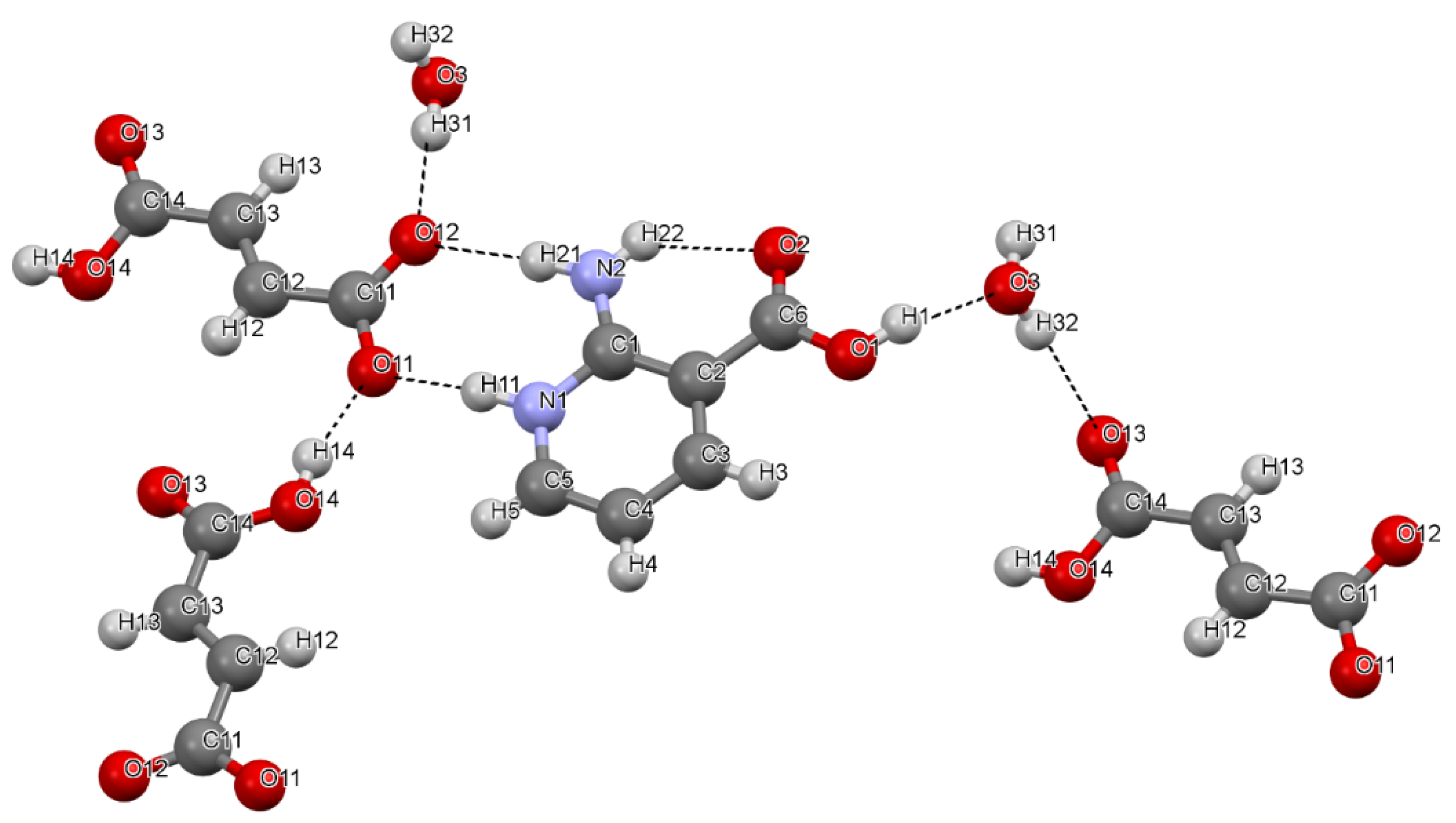
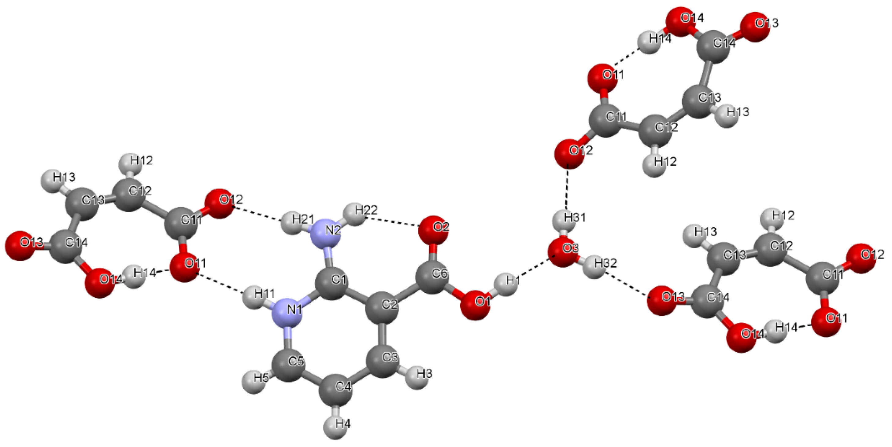
To characterize the structure and hydrogen bond (H-bond) network in two multicomponent crystals—[2AmNic+Mle+H2O] (1:1:1) and [2AmNic+Fum+H2O] (1:1:1)—by X-ray analysis, terahertz Raman spectroscopy, and periodic density functional theory (DFT) calculations. 2AmNic denotes 2-amino-nicotinic acid, while Mle and Fum stand for maleic and fumaric acids, respectively.
(ii).
To reveal the structure-directing role of the water molecule in the considered crystals.
(iii).
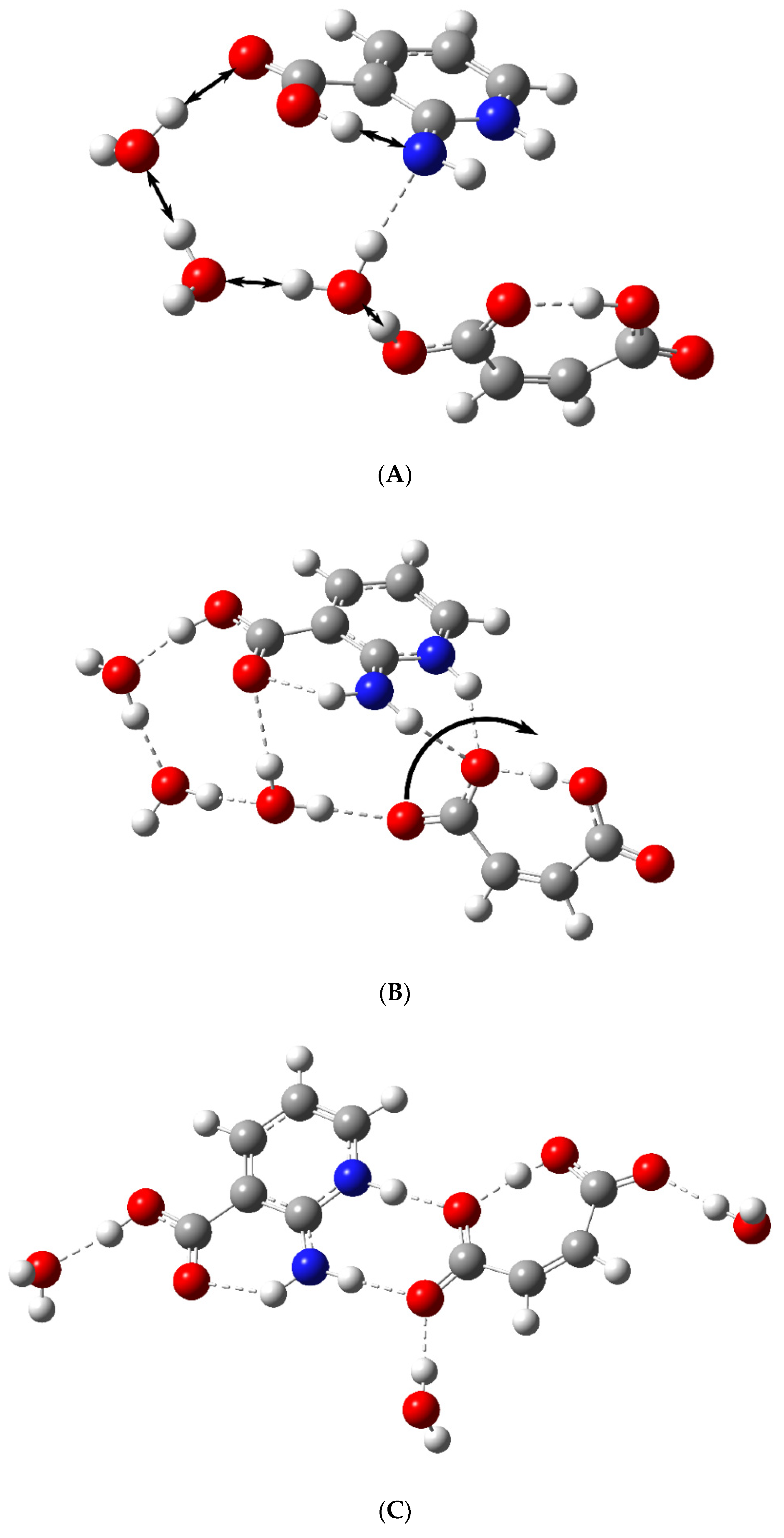
To theoretically substantiate the scheme of proton transfer from the dicarboxylic acid to the zwitterion by means of water wires.

2. Materials and Methods

2.1. Compounds and Solvents

The 2-aminonicotinic acid (C6H6N2O2, 98%) was purchased from Sigma-Aldrich, and the maleic (C4H4O4, 98%) and fumaric acids (C4H4O4, 98%) were bought from Merck. The solvents were purchased from various suppliers and were used as received without further purification.

2.2. Preparation Procedures

The grinding experiments were performed using a Fritsch planetary micro-mill, model Pulverisette 7, in 12 mL agate grinding jars with ten 5 mm agate balls at a rate of 500 rpm for 50 min. In a typical experiment, 100–120 mg of an equimolar 2-aminonicotinic acid/salt former mixture were placed into a grinding jar, and 40–50 μL of water or a water/methanol mixture (1:1 v:v) were added with a micropipette. In another method, 200 mg of a 1:1 mixture of 2-aminonicotinic acid and a salt former were suspended in 3 mL of water and were left to be stirred on a magnetic stirrer at room temperature overnight. The precipitate was filtered from the solution and dried at room temperature. The identification of the solid forms obtained by different methods and estimation the solvent content were carried out by the X-ray powder diffraction (Supplementary Figures S1 and S2) and thermal analysis (Supplementary Figures S3 and S4).
The diffraction quality single crystals of fumarate and maleate salts of 2-aminonicotinic acid were obtained by dissolving 100 mg of a stoichiometric 1:1 mixture of the components in 12 mL of H2O at 60 °C. After complete dissolution, the solution was gently cooled to the room temperature, covered by Parafilm with a few small holes pierced in it, and left for the solvent to evaporate. Small colorless crystals appeared in the solution after 5–7 days.

2.3. Thermal Analysis

2.3.1. Differential Scanning Calorimetry (DSC)

The thermal analysis was carried out using a differential scanning calorimeter with a refrigerated cooling system (Perkin Elmer DSC 4000, Waltham, MA, USA). The sample was heated in a sealed aluminum sample holder at a rate of 10 °C·min−1 in a nitrogen atmosphere. The unit was calibrated with indium and zinc standards. The accuracy of the weighing procedure was ±0.01 mg.

2.3.2. Thermogravimetric Analysis (TGA)

The TGA was performed on a TG 209 F1 Iris thermomicrobalance (Netzsch, Selb, Germany). Approximately 10 mg of the sample was added to a platinum crucible. The samples were heated at a constant heating rate of 10 °C·min−1 and purged throughout the experiment with a dry argon stream at 30 mL·min−1.

2.4. Single Crystal and Powder X-ray Diffraction (XRD) Experiments

The single-crystal XRD data were collected on a SMART APEX II diffractometer (Bruker AXS, Karlsruhe, Germany) using graphite-monochromated MoKα radiation (λ = 0.71073 Å). Absorption corrections based on measurements of equivalent reflections were applied [32]. The structures were solved by direct methods and refined by full-matrix least-squares on F2 with anisotropic thermal parameters for all the non-hydrogen atoms [33]. All the hydrogen atoms were found from a difference Fourier map and refined isotropically. The crystallographic data for [2AmNic+Mle+H2O] (1:1:1) and [2AmNic+Fum+H2O] (1:1:1) were deposited by the Cambridge Crystallographic Data Centre as supplementary publications numbered 2,053,450 and 2,053,451. This information can be obtained free of charge from the Cambridge Crystallographic Data Centre at www.ccdc.cam.ac.uk/data_request/cif.
The X-ray powder diffraction (XRPD) data of the bulk materials were recorded under ambient conditions in Bragg-Brentano geometry with a Bruker D2 Phaser diffractometer equipped with a second-generation LynxEye detector with CuKα radiation (λ = 1.5406 Å).

2.5. Raman Spectroscopy

For the Raman measurements, all the powders were compressed into tablets. The Raman measurements in the spectral range of 10–440 cm−1 were performed using a Raman microscope with the excitation wavelength 633 nm, provided by a He–Ne laser with the maximum power of 17 mW (inVia and RL633, Renishaw plc, Spectroscopy Product Division, Old Town Wotton-Under-Edge, Gloucestershire, UK). The 50× objective lens (Leica DM 2500 M, NA = 0.75, Leica Mikrosysteme Vertrieb GmbHMikroskopie und HistologieErnst-Leitz-Strasse 17-37, Wetzlar, Germany) was used. The measurements were made with a built-in double monochromator with dispersion subtraction in the confocal regime (NExT monochromator, Renishaw plc, Spectroscopy Product Division, Old Town Wotton-Under-Edge, Gloucestershire, UK). The acquisition time and number of accumulations for the Raman spectra were adjusted to maximize the signal-to-noise ratio with the minimal sample degradation. All the spectra for the powder samples were measured at several points and then averaged to reduce the anisotropy effect on the Raman spectra. The background from the Raman spectra was subtracted by the cubic spline interpolation method. All the spectra were divided by the number of accumulations and acquisition time. The dips in the spectra at wavenumbers of 23 cm−1 and 304 cm−1 are the artefacts of the measurements associated with the presence of dust particles on the NExT monochromator mirrors.

2.6. Periodic (Solid-State) DFT Computations

In the CRYSTAL17 calculations [34], the B3LYP (Becke 3-parameter, Lee-Yang-Parr) [35,36] and PBE (Perdew-Burke-Ernzerhof) [37] functionals were employed with 6-31G** all-electron Gaussian-type localized orbital basis sets. The London dispersion interactions were taken into account by introducing the D3 correction with Becke-Jones damping (PBE-D3) developed by Grimme et al. [38,39]. The structural relaxations were limited to the positional parameters of the atoms. In all cases, the experimental crystal structure with normalized X-H bond lengths was used as the starting point for geometry optimization. Further details of the calculations are given in Section S1 of Supplementary Materials.
The metric parameters of the H-bonded fragments in the considered crystals are better reproduced by B3LYP than PBE-D3 (Table 1 and Table 2). The enthalpies/energies of intermolecular H-bonds calculated using the B3LYP and PBE-D3 approximations are compared in Supplementary Table S1. In accord with the literature [25], PBE-D3 overestimates the H-bonded energy. Thus, the B3LYP/6-31G** approximation was used to calculate the Raman spectra and estimate the H-bond energies in this work.
Table 1. Distances (Å) between the heavy atoms involved in the formation of intermolecular H-bonds and the angle (degree) of the O…H-X fragment, X = O or N, in [2AmNic+Fum+H2O] (1:1:1). The experiment vs. the theoretical value. The periodic DFT computations were performed at different levels of approximation with fixed unit parameters.
Fragment 1ExperimentPBE-D3/6-31G**B3LYP/6-31G**
O12…H21-N22.877 (175.7)2.848 (176.4)2.867 (176.0)
O11…H11-N12.662 (175.3)2.668 (173.5)2.681 (173.7)
O12…H31-O32.702 (174.4)2.684 (177.9)2.697 (178.0)
O13…H32-O32.859 (167.8)2.801 (172.2)2.828 (171.2)
O11…H14-O142.559 (164.0)2.540 (160.6)2.547 (161.9)
O3…H1-O12.582 (165.9)2.545 (165.4)2.571 (165.9
1 See Figure 1, the atomic numbering is borrowed from the cif file.
Figure 1. A fragment of the [2AmNic+Fum+H2O] (1:1:1) crystal structure, illustration of hydrogen bond patterns.
Symmetry 13 00425 g001
Table 2. Distances (Å) between the heavy atoms involved in the formation of inter- and intra-molecular H-bonds and the angle (degree) of the O…H-X fragment, X = O or N, in [2AmNic+Mle+H2O] (1:1:1). The experiment vs. the theoretical values.
Fragment 1ExperimentPBE-D3/6-31G**B3LYP/6-31G**
O12…H21-N22.816 (176.4)2.804 (170.4)2.811 (171.7)
O11…H11-N12.816 (170.1)2.816 (173.7)2.818 (172.8)
O12…H31-O32.717 (177.2)2.701 (176.0)2.706 (176.4)
O13…H32-O32.771 (177.5)2.701 (173.4)2.706 (177.6)
O3…H1-O12.567 (173.0)2.536 (173.6)2.550 (174.4)
O11…H14-O14 (intra)2.460 (174.9)2.462 (176.1)2.460 (174.9)
1 See Figure 2, the atomic numbering is borrowed from the cif file.
Figure 2. A fragment of the [2AmNic+Mle+H2O] (1:1:1) crystal structure, illustration of hydrogen bond patterns.
Symmetry 13 00425 g002

3. Results

3.1. Crystal Structure and H-Bond Network

The relevant crystallographic data for the multicomponent crystals are presented in Supplementary Materials Table S2. The [2AmNic+Fum+H2O] (1:1:1) crystal has a layered (ribbon) structure. In addition to the R22(8) synthon, the dicarboxylic acid anion is stabilized in the layer by two intermolecular O–…H-O bonds, which form both the oxygen atoms of the COO group, when interacting with the H2O molecule, and the COOH group of the fumaric acid (Figure 1 and Table 1). According to Reference [40], the latter H-bond can be considered short (Table 1). A water molecule forms three H-bonds: two as a proton donor and one as an acceptor (Figure 1). Two H-bonds formed by the water molecule lie in the layer, while the third interacts with the fumaric acid molecule in an adjacent layer.
The [2AmNic+Mle+H2O] (1:1:1) crystal does not have a layered (ribbon) structure. This may be due to the presence of an intramolecular H-bond in the maleate anion. As a result, this crystal contains one H-bond less per 1:1:1 trimer than the [2AmNic+Fum+H2O] crystal (Table 1 and Table 2). In both crystals, the water molecule forms three H-bonds (Figure 2), and one of them is short (Table 2). A characteristic feature of the H-bond network in the considered crystals is bifurcate H-bonds formed by the COO group of the dicarboxylic acids. In contrast to Reference [20], all the H-bonds formed by the COO groups are “classical” and rather strong (see Section 3.2). It should be noted that compounds with C=O and P=O groups quite often form bifurcate H-bonds in molecular crystals [41,42], while the formation of such bonds by the COO group is a rather rare phenomenon. Both crystals have a large number of intermolecular H-bonds, with the COO group proton participating in the formation of short (strong) intermolecular H-bonds.
A maleate anion has a very short and practically linear intramolecular O…H–O bond (c.f. Tables 1 and 2 in Reference [43]). To identify possible spectral features of this H-bond, we recorded a terahertz Raman spectrum of the two crystals as well as crystalline fumaric acid (Supplementary Figures S5–S7) and compared it with that of crystalline maleic acid (Figure 4 in Reference [20]). When comparing the spectra of the two crystalline acids, we came to the conclusion that the band at 320 cm−1 was due to the vibrations of the intramolecular O…H–O bond. The visualization of this vibration (Supplementary Figure S8) supported this conclusion. The Raman spectrum of the [2AmNic+Mle+H2O] crystal also exhibits a band in the region of 300 cm−1 (Supplementary Figure S2). It follows from Figure 3 that this is caused by vibrations of two intramolecular H-bonds: O…H–O in the maleate anion and O…H–N in the 2-aminonicotinic acid.
Unlike crystalline [2AmNic+Fum+H2O] (1:1:1), two N+–H…O– bonds in the 8-membered cycle of the [2AmNic+Mle+H2O] (1:1:1) crystal are almost equivalent (Table 2). This phenomenon can be explained by the presence of an intramolecular H-bond in the maleate ion. To substantiate this assumption, we compared the frequency and shape of the stretching vibrations of the N+–H groups in heterodimers of fumaric and maleic anions with a 2-amino-nicotinic acid cation (Supplementary Figure S9). In accordance with the literature data [44], there is strong coupling between the intra- and inter-molecular H-bonds formed by the oxygen of the CO2 group.

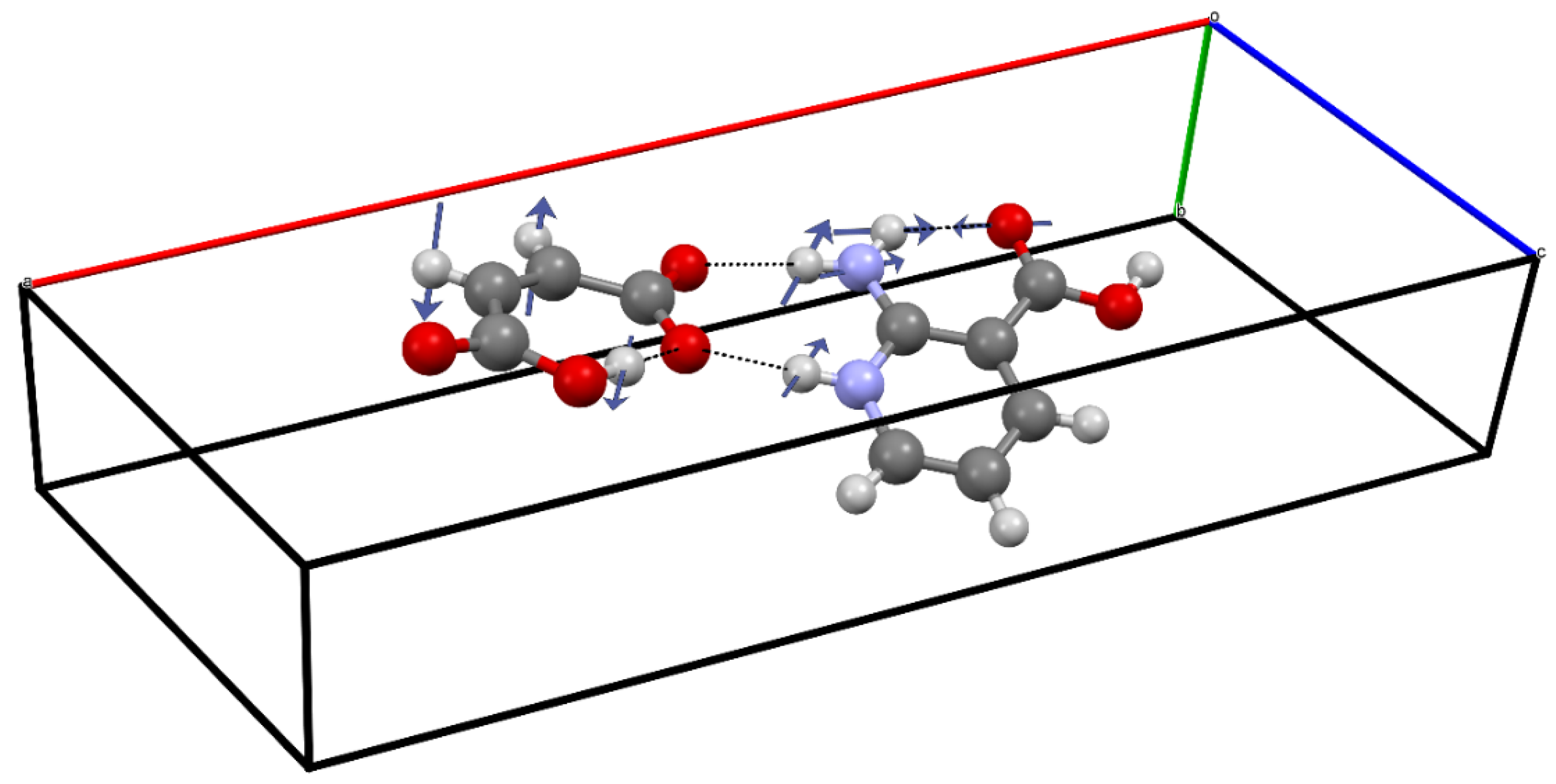
3.2. The Structure-Directing Role of the Water Molecule

The molecules or ions that make up multicomponent crystals are held together by various noncovalent interactions, including H-bonds, halogen bonds, and π⋯π stacking [40,45,46,47,48,49,50]. The fine balance between these intermolecular forces is mainly responsible for the physicochemical properties of crystalline materials and plays an important role in determining their packing arrangements and morphology [51]. Although all types of intermolecular interactions contribute to the ultimate stability of the crystal structure, intermolecular H-bonds often play a more prominent role than others due to their strength and directionality [52,53,54,55], tailoring the supramolecular architectures of multicomponent crystals and enabling a crystal engineering strategy to be applied [12,56,57]. There are two major groups of multicomponent molecular crystals: cocrystals (that are made from different neutral chemical entities) [6,58] and organic salts (that consist of charged species of components) [8,59]. The lattice energies vary from ~160 to ~300 kJ/mol, both for co-crystals [60,61,62,63,64] and for organic salts [19,65,66,67,68]. It should be noted that the estimation of the lattice energy of organic salts is not straightforward [19].
To elucidate the role of water in the formation of the structure of the considered crystals, we calculated the contribution of the H-bonds formed by a water molecule to the total energy of the intermolecular H-bonds per a 1:1:1 structural unit. Several schemes for estimating the energy (enthalpy) of intermolecular H-bonds in crystals have been proposed in the literature. In most cases, empirical approaches that are used relate the energy of an intermolecular interaction with a certain electron density parameter at the bond critical point [69,70,71]. In this case, the calculated values of the electron density, the values of the parameters derived from the precise X-ray diffraction data, and hybrid approaches are used [72]. This gives rise to well-founded criticism [73,74]. To obtain reliable values of the H-bond energies/enthalpies, we used several approaches, two of which estimated the intermolecular H-bond enthalpy from the spectroscopic [75] and metric [76] characteristics of these bonds in the crystals. It should be noted that to estimate the energy of intramolecular H-bonds in the solid state requires the use of other empirical approaches [77,78].
The results are shown in Table 3. In accordance with the literature data [79,80], all the approaches yield values of the energies/enthalpies of weak and moderate H-bonds [40] that are in good agreement with each other. Significant differences in the calculated values are observed only in short (strong) H-bonds (R(O…O) < 2.6 Å), which is caused by the contribution of the covalent component to the energy of these bonds [81,82]. All the schemes for estimating energies/enthalpies allow us to conclude that the total energy of hydrogen bonds formed by water molecules is greater than the energy of the R22(8) synthon. According to all of the approaches, the total enthalpies/energies of the H-bonds are about 40% for [2AmNic+Fum+H2O] and 50% for [2AmNic+Mle+H2O], respectively. This allows us to conclude that the water molecule determines the structure of the considered multicomponent crystals.

4. Discussion

Due to the structural features, many medicinal and bioactive compounds are in the zwitterionic form both in the crystal and in the solution at pH values characteristic of physiological fluids [83,84]. The presence of acidic and basic functional groups in the molecule structure with close pKa values (the difference is less than 3 units) leads to the formation of amphoteric or zwitterionic compounds. Many zwitterionic medicinal compounds have a high melting point, which is explained by strong intermolecular interactions (primarily H-bonds and dipole–dipole contacts) between the charged fragments of the crystal molecules. Due to the high energy of the crystal lattice and the permanent intramolecular multipole moment, a large number of zwitterionic compounds are poorly soluble in both polar and non-polar solvents [85,86]. In addition, due to the poor membrane permeability, zwitterions have a low absorption rate compared to neutral and even ionized forms, which results in limited bioavailability [84,87,88]. One of the most common methods to solve this problem is salt formation with various organic or inorganic counterions [89]. The formation of a salt with a zwitterionic compound, in most cases, makes the product melting point lower compared to that of the initial zwitterionic form as there are fewer dipole–dipole interactions so that the solubility in polar and non-polar solvents and bioavailability improve [86,90,91]. Despite the large number of publications devoted to the preparation and study of salts of zwitterionic compounds, the process of proton transfer from an acid to a zwitterionic molecule during their formation remains poorly understood.
We chose dicarboxylic acid as it could be used to describe the possible pathway of proton transfer from its COOH group to the COO group of AmNicAc in water. The molecule of maleic acid seems to be the most suitable as its second acidic proton is involved in the formation of the intramolecular H-bond. Fumaric acid is assumed to have a similar proton transfer pathway, but the presence of a second COOH group makes the theoretical model much more complicated. The starting structure was a trimer of maleic acid, 2-amino nicotinic acid, and water (1:1:1), to which we added a minimum number of water molecules that was necessary for proton transfer. It turned out that two additional water molecules were enough to implement the process. These molecules interact with the atoms of the 1:1:1 structure or with each other through H-bonds, the energy of which is much higher than that of the H-bonds in bulk water (the reason for the “strengthening” of the intermolecular H-bonds is the acidic proton of the COOH group and the COO group). The calculations were carried out in the discrete-continuum approximation [92,93,94,95] using the Gaussian16 program [96]. The bulk water was described by the polarizable continuum model [96]. The calculations were carried out in the B3LYP/6-311++G** approximation.
The initial structure is shown in Figure 4A. In accordance with the literature data [26], the acidic proton goes to the neighboring water molecule and then, by the “relay mechanism”, moves to the COO group of the amino acid. As a result of the synchronous transfer of the “acidic” proton along the H-bonds chain (along the water wire) and the intramolecular transition of the proton in the N...H...O fragment, the structure in Figure 4B is formed. Then, the maleate ion rotates by ~90 degrees and the “first” solvation shell is rearranged, i.e., the structure in Figure 4C is formed, which is very close to the structure realized in the crystal, see Figure 2.
The process scheme is shown below. The relative stability of the structures is given in parentheses (the sum of the electronic and zero-point energies) in kJ/mol:
4A (60.6) -> 4B (19.1) -> 4C (0.0)
This process can be modeled by ab initio molecular dynamics simulations using relatively small cells [97]. However, such modeling is beyond the scope of this work.

5. Conclusions

The structure and H-bond network in two multicomponent crystals—[2AmNic+Fum+H2O] (1:1:1) and [2AmNic+Mle+H2O] (1:1:1)—are characterized by X-ray analysis, terahertz Raman spectroscopy, and periodic DFT calculations. The intramolecular H-bonds cause the appearance of a Raman-active band around 300 cm−1 in the [2AmNic+Mle+H2O] (1:1:1) crystal. The total enthalpy of the intermolecular H-bonds in these crystals, estimated per a 1:1:1 structural unit, is about 160 kJ/mol; moreover, the water molecule accounts for about 90 kJ/mol. This allows us to conclude that the water molecule determines the structure of the considered multicomponent crystals. A scheme of the transfer of a dicarboxylic acid proton to a zwitterionic amino acid molecule in the process of the [2AmNic+Fum+H2O] (1:1:1) and [2AmNic+Mle+H2O] (1:1:1) formation in the polar protic solvent is proposed. Water molecules were found to play the key role in this process, forming a “water wire” between the COOH group of the dicarboxylic acid and the COO group of the zwitterion.

Supplementary Materials

The following are available online at https://0-www-mdpi-com.brum.beds.ac.uk/2073-8994/13/3/425/s1, Section S1: Computational details; Table S1: Theoretical values of the enthalpy, ∆HHB, and energy, EHB, of intermolecular H-bonds in the [2AmNic+Fum+H2O] (1:1:1) crystal evaluated using several empirical approaches; Figures S1 and S2: calculated and experimental PXRD patterns; Figures S3 and S4: DSC and TG traces; Figure S5: Raman spectrum of the [2AmNic+Fum+H2O] (1:1:1) crystal. Experiment (black line) vs. B3LYP/6-31G** computations (red bars); Figure S6: Raman spectrum of the [2AmNic+Mle+H2O] (1:1:1) crystal. Experiment (black line) vs. B3LYP/6-31G** computations (red bars); Figure S7: Raman spectrum of crystalline fumaric acid. Experiment (black line) vs. B3LYP/6-31G** computations (red bars); Figure S8: Schematic representation of the Raman active vibration at 337 cm−1 of crystalline maleic acid, evaluated using periodic DFT computations at the B3LYP/6-31G** level; Figure S9: Schematic representation of the IR active vibration at 2524 cm−1 of the fumaric acid-2-amino-nicotinic acid zwitterion heterodimer (the upper panel) and 2636 cm−1 of the maleic acid-2-amino-nicotinic acid zwitterion (the lower panel), evaluated using DFT computations at the B3LYP/6-31G** level; Table S2: Crystallographic data for [2AmNic+Fum+H2O] (1:1:1) and [2AmNic+Mle+H2O] (1:1:1) multicomponent crystals.

Author Contributions

Conceptualization, A.O.S. and M.V.V.; experimental methodology, A.O.S. and N.A.V.; theoretical methodology, M.V.V.; investigation, A.O.S., N.A.V., A.V.C., O.D.P., S.V.A., O.A.A., D.E.M. and M.V.V.; single-crystal XRD experiment, A.V.C.; Raman spectroscopy, O.D.P.; writing and visualization, A.O.S. and M.V.V.; supervision, M.V.V. and A.O.S.; project administration, A.O.S.; funding acquisition, A.O.S. All authors have read and agreed to the published version of the manuscript.

Funding

The work was supported by the Russian Science Foundation (project No. 19-73-10005).

Institutional Review Board Statement

Not applicable.

Informed Consent Statement

Not applicable.

Data Availability Statement

The Raman spectra and I/O files are available from the respective author upon reasonable request.

Acknowledgments

The experimental Raman spectra were obtained at Lomonosov Moscow State University using the equipment purchased under the Lomonosov Moscow State University Program of Development. M.V.V. thanks Alexander P. Voronin (G.A. Krestov Institute of Solution Chemistry of RAS) for performing the Bader analysis of the crystal electron density.

Conflicts of Interest

The authors declare no conflict of interest.

References

  1. Schwoerer, M.; Wolf, H.C. Organic Molecular Solids; Wiley: Weinheim, Germany, 2008. [Google Scholar]
  2. Qiu, Y.; Chen, Y.; Zhang, G.G.Z.; Yu, L.; Mantri, R.V. Developing Solid Oral Dosage Forms: Pharmaceutical Theory and Practice; Elsevier Science: London, UK, 2016. [Google Scholar]
  3. Sathisaran, I.; Dalvi, S.V. Engineering Cocrystals of Poorly Water-Soluble Drugs to Enhance Dissolution in Aqueous Medium. Pharmaceutics 2018, 10, 108. [Google Scholar] [CrossRef] [PubMed] [Green Version]
  4. Sun, L.; Wang, Y.; Yang, F.; Zhang, X.; Hu, W. Cocrystal Engineering: A Collaborative Strategy toward Functional Materials. Adv. Mater. 2019, 31, 1902328. [Google Scholar] [CrossRef]
  5. Kavanagh, O.N.; Croker, D.M.; Walker, G.M.; Zaworotko, M.J. Pharmaceutical cocrystals: From serendipity to design to application. Drug Discov. Today 2019, 24, 796–804. [Google Scholar] [CrossRef] [Green Version]
  6. Duggirala, N.K.; Perry, M.L.; Almarsson, Ö.; Zaworotko, M.J. Pharmaceutical cocrystals: Along the path to improved medicines. Chem. Commun. 2016, 52, 640–655. [Google Scholar] [CrossRef] [PubMed]
  7. Bolla, G.; Nangia, A. Pharmaceutical cocrystals: Walking the talk. Chem. Commun. 2016, 52, 8342–8360. [Google Scholar] [CrossRef] [PubMed]
  8. Berry, D.J.; Steed, J.W. Pharmaceutical cocrystals, salts and multicomponent systems; intermolecular interactions and property based design. Adv. Drug Del. Rev. 2017, 117, 3–24. [Google Scholar] [CrossRef] [PubMed] [Green Version]
  9. Aakeröy, C.B.; Hussain, I.; Desper, J. 2-Acetaminopyridine:  A Highly Effective Cocrystallizing Agent. Cryst. Growth Des. 2006, 6, 474–480. [Google Scholar] [CrossRef]
  10. Aakeröy, C.B.; Forbes, S.; Desper, J. Altering physical properties of pharmaceutical co-crystals in a systematic manner. CrystEngComm 2014, 16, 5870–5877. [Google Scholar] [CrossRef] [Green Version]
  11. Walsh, R.D.B.; Bradner, M.W.; Fleischman, S.; Morales, L.A.; Moulton, B.; Rodríguez-Hornedo, N.; Zaworotko, M.J. Crystal engineering of the composition of pharmaceutical phases. Chem. Commun. 2003, 2, 186–187. [Google Scholar] [CrossRef] [PubMed]
  12. Corpinot, M.K.; Bučar, D.-K. A Practical Guide to the Design of Molecular Crystals. Cryst. Growth Des. 2019, 19, 1426–1453. [Google Scholar] [CrossRef] [Green Version]
  13. Desiraju, G.R. Supramolecular Synthons in Crystal Engineering—A New Organic Synthesis. Angew. Chem. Int. Ed. Engl. 1995, 34, 2311–2327. [Google Scholar] [CrossRef]
  14. Lemmerer, A.; Bernstein, J.; Kahlenberg, V. One-pot covalent and supramolecular synthesis of pharmaceutical co-crystals using the API isoniazid: A potential supramolecular reagent. CrystEngComm 2010, 12, 2856–2864. [Google Scholar] [CrossRef]
  15. Desiraju, G.R. Crystal Engineering: From Molecule to Crystal. J. Am. Chem. Soc. 2013, 135, 9952–9967. [Google Scholar] [CrossRef] [PubMed]
  16. Desiraju, G.R. Crystal Engineering: A Holistic View. Angew. Chem. Int. Ed. 2007, 46, 8342–8356. [Google Scholar] [CrossRef] [PubMed]
  17. Surov, A.O.; Voronin, A.P.; Vener, M.V.; Churakov, A.V.; Perlovich, G.L. Specific features of supramolecular organisation and hydrogen bonding in proline cocrystals: A case study of fenamates and diclofenac. CrystEngComm 2018, 20, 6970–6981. [Google Scholar] [CrossRef]
  18. Bolla, G.; Nangia, A. Novel pharmaceutical salts of albendazole. CrystEngComm 2018, 20, 6394–6405. [Google Scholar] [CrossRef] [Green Version]
  19. Voronin, A.P.; Surov, A.O.; Churakov, A.V.; Parashchuk, O.D.; Rykounov, A.A.; Vener, M.V. Combined X-ray Crystallographic, IR/Raman Spectroscopic, and Periodic DFT Investigations of New Multicomponent Crystalline Forms of Anthelmintic Drugs: A Case Study of Carbendazim Maleate. Molecules 2020, 25, 2386. [Google Scholar] [CrossRef] [PubMed]
  20. Mirzaei, M.; Sadeghi, F.; Molčanov, K.; Zarȩba, J.K.; Gomila, R.M.; Frontera, A. Recurrent Supramolecular Motifs in a Series of Acid–Base Adducts Based on Pyridine-2,5-Dicarboxylic Acid N-Oxide and Organic Bases: Inter- and Intramolecular Hydrogen Bonding. Cryst. Growth Des. 2020, 20, 1738–1751. [Google Scholar] [CrossRef]
  21. Yadav, B.; Balasubramanian, S.; Chavan, R.B.; Thipparaboina, R.; Naidu, V.G.M.; Shastri, N.R. Hepatoprotective Cocrystals and Salts of Riluzole: Prediction, Synthesis, Solid State Characterization, and Evaluation. Cryst. Growth Des. 2018, 18, 1047–1061. [Google Scholar] [CrossRef]
  22. Thomas, S.P.; Kumar, V.; Alhameedi, K.; Guru Row, T.N. Non-Classical Synthons: Supramolecular Recognition by S…O Chalcogen Bonding in Molecular Complexes of Riluzole. Chem. Eur. J. 2019, 25, 3591–3597. [Google Scholar] [CrossRef] [PubMed]
  23. Babu, N.J.; Reddy, L.S.; Nangia, A. Amide−N-Oxide Heterosynthon and Amide Dimer Homosynthon in Cocrystals of Carboxamide Drugs and Pyridine N-Oxides. Mol. Pharm. 2007, 4, 417–434. [Google Scholar] [CrossRef] [PubMed]
  24. Dunitz, J.D.; Gavezzotti, A. Supramolecular Synthons: Validation and Ranking of Intermolecular Interaction Energies. Cryst. Growth Des. 2012, 12, 5873–5877. [Google Scholar] [CrossRef]
  25. Vener, M.V.; Levina, E.O.; Koloskov, O.A.; Rykounov, A.A.; Voronin, A.P.; Tsirelson, V.G. Evaluation of the lattice energy of the two-component molecular crystals using solid-state density functional theory. Cryst. Growth Des. 2014, 14, 4997–5003. [Google Scholar] [CrossRef]
  26. Xu, J.; Zhang, Y.; Voth, G.A. Infrared Spectrum of the Hydrated Proton in Water. J. Phys. Chem. Lett. 2011, 2, 81–86. [Google Scholar] [CrossRef] [PubMed]
  27. Kaila, V.R.I.; Hummer, G. Energetics and dynamics of proton transfer reactions along short water wires. PCCP 2011, 13, 13207–13215. [Google Scholar] [CrossRef] [PubMed] [Green Version]
  28. Zatula, A.S.; Ryding, M.J.; Uggerud, E. Concerted proton migration along short hydrogen bonded water bridges in bipyridine–water clusters. PCCP 2012, 14, 13907–13909. [Google Scholar] [CrossRef] [PubMed]
  29. Freier, E.; Wolf, S.; Gerwert, K. Proton transfer via a transient linear water-molecule chain in a membrane protein. Proc. Natl. Acad. Sci. USA 2011, 108, 11435–11439. [Google Scholar] [CrossRef] [Green Version]
  30. Di Donato, M.; van Wilderen, L.J.G.W.; Van Stokkum, I.H.M.; Stuart, T.C.; Kennis, J.T.M.; Hellingwerf, K.J.; van Grondelle, R.; Groot, M.L. Proton transfer events in GFP. PCCP 2011, 13, 16295–16305. [Google Scholar] [CrossRef] [PubMed]
  31. Grigorenko, B.; Polyakov, I.; Nemukhin, A. Mechanisms of ATP to cAMP Conversion Catalyzed by the Mammalian Adenylyl Cyclase: A Role of Magnesium Coordination Shells and Proton Wires. J. Phys. Chem. B 2020, 124, 451–460. [Google Scholar] [CrossRef] [PubMed]
  32. Sheldrick, G. SADABS, Program for Scaling and Correction of Area Detector Data; University of Göttingen: Lower Saxony, Germany, 1997. [Google Scholar]
  33. Sheldrick, G. A short history of SHELX. Acta Cryst. Sect. A Found. Cryst. 2008, 64, 112–122. [Google Scholar] [CrossRef] [PubMed] [Green Version]
  34. Dovesi, R.; Erba, A.; Orlando, R.; Zicovich-Wilson, C.M.; Civalleri, B.; Maschio, L.; Rérat, M.; Casassa, S.; Baima, J.; Salustro, S.; et al. Quantum-mechanical condensed matter simulations with CRYSTAL. WIREs Comput. Mol. Sci. 2018, 8, e1360. [Google Scholar] [CrossRef]
  35. Becke, A.D. Density-functional thermochemistry. III. The role of exact exchange. J. Chem. Phys. 1993, 98, 5648–5652. [Google Scholar] [CrossRef] [Green Version]
  36. Vosko, S.H.; Wilk, L.; Nusair, M. Accurate spin-dependent electron liquid correlation energies for local spin density calculations: A critical analysis. Can. J. Phys. 1980, 58, 1200–1211. [Google Scholar] [CrossRef] [Green Version]
  37. Perdew, J.P.; Burke, K.; Ernzerhof, M. Generalized Gradient Approximation Made Simple. Phys. Rev. Lett. 1996, 77, 3865–3868. [Google Scholar] [CrossRef] [PubMed] [Green Version]
  38. Grimme, S.; Antony, J.; Ehrlich, S.; Krieg, H. A consistent and accurate ab initio parametrization of density functional dispersion correction (DFT-D) for the 94 elements H-Pu. J. Chem. Phys. 2010, 132, 154104. [Google Scholar] [CrossRef] [Green Version]
  39. Grimme, S.; Ehrlich, S.; Goerigk, L. Effect of the damping function in dispersion corrected density functional theory. J. Comput. Chem. 2011, 32, 1456–1465. [Google Scholar] [CrossRef]
  40. Steiner, T. The Hydrogen Bond in the Solid State. Angew. Chem. Int. Ed. 2002, 41, 48–76. [Google Scholar] [CrossRef]
  41. Rozas, I.; Alkorta, I.; Elguero, J. Bifurcated Hydrogen Bonds:  Three-Centered Interactions. J. Phys. Chem. A 1998, 102, 9925–9932. [Google Scholar] [CrossRef]
  42. Tupikina, E.Y.; Bodensteiner, M.; Tolstoy, P.M.; Denisov, G.S.; Shenderovich, I.G. P═O Moiety as an Ambidextrous Hydrogen Bond Acceptor. J. Phys. Chem. C 2018, 122, 1711–1720. [Google Scholar] [CrossRef]
  43. Vener, M.V. Model study of the primary H/D isotope effects on the NMR chemical shift in strong hydrogen-bonded systems. Chem. Phys. 1992, 166, 311–316. [Google Scholar] [CrossRef]
  44. Jóźwiak, K.; Jezierska, A.; Panek, J.J.; Goremychkin, E.A.; Tolstoy, P.M.; Shenderovich, I.G.; Filarowski, A. Inter- vs. Intramolecular Hydrogen Bond Patterns and Proton Dynamics in Nitrophthalic Acid Associates. Molecules 2020, 25, 4720. [Google Scholar] [CrossRef]
  45. Müller-Dethlefs, K.; Hobza, P. Noncovalent Interactions:  A Challenge for Experiment and Theory. Chem. Rev. 2000, 100, 143–168. [Google Scholar] [CrossRef] [PubMed]
  46. Aakeröy, C.B.; Evans, T.A.; Seddon, K.R.; Pálinkó, I. The C–H…Cl hydrogen bond: Does it exist? New J. Chem. 1999, 23, 145–152. [Google Scholar] [CrossRef] [Green Version]
  47. Nishio, M. CH/π hydrogen bonds in crystals. CrystEngComm 2004, 6, 130–158. [Google Scholar] [CrossRef]
  48. Martinez, C.R.; Iverson, B.L. Rethinking the term “pi-stacking”. Chem. Sci. 2012, 3, 2191–2201. [Google Scholar] [CrossRef] [Green Version]
  49. Bartashevich, E.V.; Tsirelson, V.G. Interplay between non-covalent interactions in complexes and crystals with halogen bonds. Russ. Chem. Rev. 2014, 83, 1181–1203. [Google Scholar] [CrossRef]
  50. Melikova, S.M.; Voronin, A.P.; Panek, J.; Frolov, N.E.; Shishkina, A.V.; Rykounov, A.A.; Tretyakov, P.Y.; Vener, M.V. Interplay of π-stacking and inter-stacking interactions in two-component crystals of neutral closed-shell aromatic compounds: Periodic DFT study. RSC Adv. 2020, 10, 27899–27910. [Google Scholar] [CrossRef]
  51. Mattei, A.; Li, T. Intermolecular Interactions and Computational Modeling. In Pharmaceutical Crystals; John Wiley & Sons: Hoboken, USA, 2018; pp. 123–167. [Google Scholar] [CrossRef]
  52. Burrows, A.D. Crystal Engineering Using Multiple Hydrogen Bonds. In Supramolecular Assembly via Hydrogen Bonds I; Mingos, D.M.P., Ed.; Springer: Berlin/Heidelberg, Germany, 2004; pp. 55–96. [Google Scholar] [CrossRef]
  53. Aakeröy, C.B.; Seddon, K.R. The hydrogen bond and crystal engineering. Chem. Soc. Rev. 1993, 22, 397–407. [Google Scholar] [CrossRef]
  54. Subramanian, S.; Zaworotko, M.J. Exploitation of the hydrogen bond: Recent developments in the context of crystal engineering. Coord. Chem. Rev. 1994, 137, 357–401. [Google Scholar] [CrossRef]
  55. Desiraju, G.R. Designer crystals: Intermolecular interactions, network structures and supramolecular synthons. Chem. Commun. 1997, 16, 1475–1482. [Google Scholar] [CrossRef]
  56. Bhattacharya, S.; Peraka, K.S.; Zaworotko, M.J. Chapter 2 The Role of Hydrogen Bonding in Co-crystals. In Co-Crystals: Preparation, Characterization and Applications; The Royal Society of Chemistry: Croydon, UK, 2018; pp. 33–79. [Google Scholar] [CrossRef]
  57. Medvedev, A.G.; Churakov, A.V.; Prikhodchenko, P.V.; Lev, O.; Vener, M.V. Crystalline Peroxosolvates: Nature of the Coformer, Hydrogen-Bonded Networks and Clusters, Intermolecular Interactions. Molecules 2021, 26, 26. [Google Scholar] [CrossRef] [PubMed]
  58. Aakeröy, C.B.; Salmon, D.J. Building co-crystals with molecular sense and supramolecular sensibility. CrystEngComm 2005, 7, 439–448. [Google Scholar] [CrossRef]
  59. Elder, D.P.; Holm, R.; Diego, H.L.d. Use of pharmaceutical salts and cocrystals to address the issue of poor solubility. Int. J. Pharm. 2013, 453, 88–100. [Google Scholar] [CrossRef] [PubMed]
  60. Manin, A.N.; Voronin, A.P.; Shishkina, A.V.; Vener, M.V.; Churakov, A.V.; Perlovich, G.L. Influence of Secondary Interactions on the Structure, Sublimation Thermodynamics, and Solubility of Salicylate:4-Hydroxybenzamide Cocrystals. Combined Experimental and Theoretical Study. J. Phys. Chem. B 2015, 119, 10466–10477. [Google Scholar] [CrossRef] [PubMed]
  61. Voronin, A.P.; Perlovich, G.L.; Vener, M.V. Effects of the crystal structure and thermodynamic stability on solubility of bioactive compounds: DFT study of isoniazid cocrystals. Comput. Theor. Chem. 2016, 1092, 1–11. [Google Scholar] [CrossRef]
  62. Landeros-Rivera, B.; Moreno-Esparza, R.; Hernández-Trujillo, J. Theoretical study of intermolecular interactions in crystalline arene–perhaloarene adducts in terms of the electron density. RSC Adv. 2016, 6, 77301–77309. [Google Scholar] [CrossRef]
  63. Jarzembska, K.N.; Hoser, A.A.; Varughese, S.; Kamiński, R.; Malinska, M.; Stachowicz, M.; Pedireddi, V.R.; Woźniak, K. Structural and Energetic Analysis of Molecular Assemblies in a Series of Nicotinamide and Pyrazinamide Cocrystals with Dihydroxybenzoic Acids. Cryst. Growth Des. 2017, 17, 4918–4931. [Google Scholar] [CrossRef]
  64. Tao, Q.; Hao, Q.-Q.; Voronin, A.P.; Dai, X.-L.; Huang, Y.; Perlovich, G.L.; Lu, T.-B.; Chen, J.-M. Polymorphic Forms of a Molecular Salt of Phenazopyridine with 3,5-Dihydroxybenzoic Acid: Crystal Structures, Theoretical Calculations, Thermodynamic Stability, and Solubility Aspects. Cryst. Growth Des. 2019, 19, 5636–5647. [Google Scholar] [CrossRef]
  65. Manin, A.N.; Voronin, A.P.; Drozd, K.V.; Churakov, A.V.; Perlovich, G.L. Pharmaceutical salts of emoxypine with dicarboxylic acids. Acta Crystallogr. Sect. C 2018, 74, 797–806. [Google Scholar] [CrossRef] [PubMed]
  66. Surov, A.O.; Churakov, A.V.; Perlovich, G.L. Three Polymorphic Forms of Ciprofloxacin Maleate: Formation Pathways, Crystal Structures, Calculations, and Thermodynamic Stability Aspects. Cryst. Growth Des. 2016, 16, 6556–6567. [Google Scholar] [CrossRef]
  67. Surov, A.O.; Voronin, A.P.; Simagina, A.A.; Churakov, A.V.; Skachilova, S.Y.; Perlovich, G.L. Saccharin salts of biologically active hydrazone derivatives. New J. Chem. 2015, 39, 8614–8622. [Google Scholar] [CrossRef]
  68. Chan, H.C.S.; Kendrick, J.; Neumann, M.A.; Leusen, F.J.J. Towards ab initio screening of co-crystal formation through lattice energy calculations and crystal structure prediction of nicotinamide, isonicotinamide, picolinamide and paracetamol multi-component crystals. CrystEngComm 2013, 15, 3799–3807. [Google Scholar] [CrossRef] [Green Version]
  69. Mata, I.; Alkorta, I.; Espinosa, E.; Molins, E. Relationships between interaction energy, intermolecular distance and electron density properties in hydrogen bonded complexes under external electric fields. Chem. Phys. Lett. 2011, 507, 185–189. [Google Scholar] [CrossRef]
  70. Kuznetsov, M.L. Can halogen bond energy be reliably estimated from electron density properties at bond critical point? The case of the (A)nZ—Y…X− (X, Y = F, Cl, Br) interactions. Int. J. Quantum Chem. 2019, 119, e25869. [Google Scholar] [CrossRef]
  71. Emamian, S.; Lu, T.; Kruse, H.; Emamian, H. Exploring Nature and Predicting Strength of Hydrogen Bonds: A Correlation Analysis Between Atoms-in-Molecules Descriptors, Binding Energies, and Energy Components of Symmetry-Adapted Perturbation Theory. J. Comput. Chem. 2019, 40, 2868–2881. [Google Scholar] [CrossRef] [PubMed]
  72. Korlyukov, A.A.; Nelyubina, Y.V. Quantum chemical methods in charge density studies from X-ray diffraction data. Russ. Chem. Rev. 2019, 88, 677–716. [Google Scholar] [CrossRef]
  73. Dem’yanov, P.; Polestshuk, P. A Bond Path and an Attractive Ehrenfest Force Do Not Necessarily Indicate Bonding Interactions: Case Study on M2X2 (M = Li, Na, K; X = H, OH, F, Cl). Chem. Eur. J. 2012, 18, 4982–4993. [Google Scholar] [CrossRef] [PubMed]
  74. Shahbazian, S. Why Bond Critical Points Are Not “Bond” Critical Points. Chem. Eur. J. 2018, 24, 5401–5405. [Google Scholar] [CrossRef] [PubMed]
  75. Iogansen, A.V. Direct proportionality of the hydrogen bonding energy and the intensification of the stretching ν(XH) vibration in infrared spectra. Spectrochim. Acta Part A Mol. Biomol. Spectrosc. 1999, 55, 1585–1612. [Google Scholar] [CrossRef]
  76. Rozenberg, M.; Loewenschuss, A.; Marcus, Y. An empirical correlation between stretching vibration redshift and hydrogen bond length. PCCP 2000, 2, 2699–2702. [Google Scholar] [CrossRef]
  77. Musin, R.N.; Mariam, Y.H. An integrated approach to the study of intramolecular hydrogen bonds in malonaldehyde enol derivatives and naphthazarin: Trend in energetic versus geometrical consequences. J. Phys. Org. Chem. 2006, 19, 425–444. [Google Scholar] [CrossRef]
  78. Filarowski, A.; Koll, A.; Sobczyk, L. Intramolecular Hydrogen Bonding in o-hydroxy Aryl Schiff Bases. Curr. Org. Chem. 2009, 13, 172–193. [Google Scholar] [CrossRef]
  79. Medvedev, A.G.; Mikhaylov, A.A.; Chernyshov, I.Y.; Vener, M.V.; Lev, O.; Prikhodchenko, P.V. Effect of aluminum vacancies on the H2O2 or H2O interaction with a gamma-AlOOH surface. A solid-state DFT study. Int. J. Quantum Chem. 2019, 119, e25920. [Google Scholar] [CrossRef]
  80. Musso, F.; Casassa, S.; Corno, M.; Ugliengo, P. How strong are H-bonds at the fully hydroxylated silica surfaces? Insights from the B3LYP electron density topological analysis. Struct. Chem. 2017, 28, 1009–1015. [Google Scholar] [CrossRef]
  81. Vener, M.V.; Manaev, A.V.; Egorova, A.N.; Tsirelson, V.G. QTAIM Study of Strong H-Bonds with the O−H…A Fragment (A=O, N) in Three-Dimensional Periodical Crystals. J. Phys. Chem. A 2007, 111, 1155–1162. [Google Scholar] [CrossRef]
  82. Grabowski, S.J. What Is the Covalency of Hydrogen Bonding? Chem. Rev. 2011, 111, 2597–2625. [Google Scholar] [CrossRef]
  83. Mazák, K.; Noszál, B. Physicochemical Properties of Zwitterionic Drugs in Therapy. ChemMedChem 2020, 15, 1102–1110. [Google Scholar] [CrossRef]
  84. Yang, Z.; Li, Q.; Yang, G. Zwitterionic structures: From physicochemical properties toward computer-aided drug designs. Future Med. Chem. 2016, 8, 2245–2262. [Google Scholar] [CrossRef] [PubMed]
  85. Mesallati, H.; Mugheirbi, N.A.; Tajber, L. Two Faces of Ciprofloxacin: Investigation of Proton Transfer in Solid State Transformations. Cryst. Growth Des. 2016, 16, 6574–6585. [Google Scholar] [CrossRef] [Green Version]
  86. Mazzenga, G.C.; Berner, B. The transdermal delivery of zwitterionic drugs I: The solubility of zwitterion salts. J. Control. Release 1991, 16, 77–88. [Google Scholar] [CrossRef]
  87. Gunnam, A.; Suresh, K.; Ganduri, R.; Nangia, A. Crystal engineering of a zwitterionic drug to neutral cocrystals: A general solution for floxacins. Chem. Commun. 2016, 52, 12610–12613. [Google Scholar] [CrossRef] [PubMed]
  88. Manallack, D.T.; Prankerd, R.J.; Yuriev, E.; Oprea, T.I.; Chalmers, D.K. The significance of acid/base properties in drug discovery. Chem. Soc. Rev. 2013, 42, 485–496. [Google Scholar] [CrossRef] [PubMed] [Green Version]
  89. Stahl, P.H.; Wermuth, C.G.; Pure, I.U.o.; Chemistry, A. Handbook of Pharmaceutical Salts Properties, Selection, and Use; Wiley: Weinheim, Germany, 2008. [Google Scholar]
  90. Han, H.-K.; Choi, H.-K. Improved absorption of meloxicam via salt formation with ethanolamines. Eur. J. Pharm. Biopharm. 2007, 65, 99–103. [Google Scholar] [CrossRef] [PubMed]
  91. Gwak, H.-S.; Choi, J.-S.; Choi, H.-K. Enhanced bioavailability of piroxicam via salt formation with ethanolamines. Int. J. Pharm. 2005, 297, 156–161. [Google Scholar] [CrossRef] [PubMed]
  92. Tunon, I.; Rinaldi, D.; Ruiz-Lopez, M.F.; Rivail, J.L. Hydroxide Ion in Liquid Water: Structure, Energetics, and Proton Transfer Using a Mixed Discrete-Continuum ab Initio Model. J. Phys. Chem. 1995, 99, 3798–3805. [Google Scholar] [CrossRef]
  93. Nemukhin, A.V.; Topol, I.A.; Grigorenko, B.L.; Burt, S.K. On the Origin of Potential Barrier for the Reaction OH- + CO2 → HCO3- in Water:  Studies by Using Continuum and Cluster Solvation Methods. J. Phys. Chem. B 2002, 106, 1734–1740. [Google Scholar] [CrossRef]
  94. da Silva, E.F.; Svendsen, H.F.; Merz, K.M. Explicitly Representing the Solvation Shell in Continuum Solvent Calculations. J. Phys. Chem. A 2009, 113, 6404–6409. [Google Scholar] [CrossRef] [PubMed] [Green Version]
  95. Vener, M.V.; Shenderovich, I.G.; Rykounov, A.A. A qualitative study of the effect of a counterion and polar environment on the structure and spectroscopic signatures of a hydrated hydroxyl anion. Theor. Chem. Acc. 2013, 132, 1361. [Google Scholar] [CrossRef]
  96. Frisch, M.J.; Trucks, G.W.; Schlegel, H.B.; Scuseria, G.E.; Robb, M.A.; Cheeseman, J.R.; Scalmani, G.; Barone, V.; Petersson, G.A.; Nakatsuji, H.; et al. Gaussian 16 Rev. C.01; Gaussian, Inc.: Wallingford, CT, USA, 2016. [Google Scholar]
  97. Sun, J.; Bousquet, D.; Forbert, H.; Marx, D. Glycine in aqueous solution: Solvation shells, interfacial water, and vibrational spectroscopy from ab initio molecular dynamics. J. Chem. Phys. 2010, 133, 114508. [Google Scholar] [CrossRef]
Figure 3. Schematic representation of the Raman active vibration at 316 cm−1 of the [2AmNic+Mle+H2O] crystal, evaluated using periodic DFT computations at the B3LYP/6-31G** level. The arrows indicate the directions of the relative atom displacements.
Symmetry 13 00425 g003
Figure 4. Structures used in the process of proton transfer modeling. The initial structure (A); the intermediate structure (B); the final structure (C).
Symmetry 13 00425 g004
Table 3. Theoretical values of the enthalpy, ∆HHB, and energy, EHB, of intermolecular H-bonds in the crystals evaluated using different empirical approaches. The O∙∙∙H distances, frequencies of the OH stretching vibrations, and crystalline electron density were calculated at the B3LYP/6-31G** level. The total ∆HHB/EHB values of the H-bonds formed by the water molecule are indicated in parentheses.
Fragment 1R(O∙∙∙H), Å−ΔHHB 2, kJ/mol−ΔHHB 3, kJ/molEHB 4, kJ/mol
[2AmNic+Fum+H2O] (1:1:1)
O12…H21-N21.840 23.4 -26.8
O11…H11-N11.628 34.0 -45.7
O12…H31-O31.709 29.3 27.835.8
O13…H32-O32.034 17.2 18.324.3
O11…H14-O141.563 38.5 39.752.9
O3…H1-O11.571 37.9 40.752.1
∑(–ΔHHB/EHB)-180.3 (84.4)(86.8)237.6 (112.1)
[2AmNic+Mle+H2O] (1:1:1)
O12…H21-N21.792 25.4 -30.5
O11…H11-N11.785 25.7 -30.2
O12…H31-O31.725 28.5 24.834.9
O13…H32-O31.797 25.2 17.628.5
O3…H1-O11.525 41.5 43.558.2
∑(–ΔHHB/EHB)-146.3 (95.2)(85.9)182.3 (121.6)
1 The atomic numbering is given in Figure 1 and Figure 2. 2 Evaluated using the Rozenberg approach [76]: −ΔHHB [kJ mol−1] = 0.134·R(O∙∙∙H) –3.05, where the R(H∙∙∙O) is the H∙∙∙O distance (nm). 3 Evaluated using the Iogansen approach [75]: −ΔHHB [kJ mol−1] = 1.386·(Δν [cm–1] − 40)0.5, where Δν = ν(OHfree) − ν(OH) represents the red-shift value of the OH stretching frequency caused by the formation of the H-bond with the OH group being the proton donor. It should be noted that ν(OHfree) and ν(OH) are the frequencies of the localized, uncoupled OH stretching vibration. 4 Evaluated using the Espinoza approach [69]: EHB [kJ mol−1] = 1124·Gb [atomic units], where Gb is the positively defined local electronic kinetic energy density at the O∙∙∙H bond critical point.
Publisher’s Note: MDPI stays neutral with regard to jurisdictional claims in published maps and institutional affiliations.

