1. Introduction
The plant
Phellodendron amurense, Rupr. (PA) from the family Rutaceae is one of the major plants in traditional Chinese medicine. So-called huang-bo or huang-bai have been used traditionally in folk medicine for hundreds of years as anti-inflammatory, spasmolytic and antidiarrheal agents; as a bitter tonic for stomach problems; and in the treatment of dyspepsia, hepatitis, dysentery, gynaecological inflammation, meningitis, gastreoenteritidis, tuberculosis or pneumonia [
1,
2].
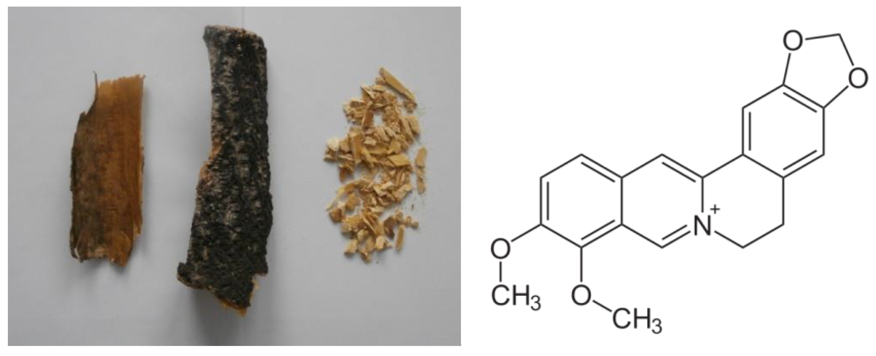
PA bark (
Figure 1) contains mainly isoquinoline alkaloids such as phellodendrine, berberine (
Figure 1), oxyberberine, epiberberine, jatrorrhizine, magnoflorin and palmatine [
2,
3]. This plant is also the source of limonoids (limonin, kihadanin, obacunone [
4]), isovanillin, ferulic acid, ethyl caffeate and other phenolic substances [
5,
6]. These active compounds possess many different biological activities, but they exhibited low toxicity to normal cells and tissues [
7].
The
Phellodendron amurense bark extract is also a potential therapeutic agent to protect cartilage against the osteoarthritic progression [
1,
8,
9]. There are a few preparations with extract from bark of
Phellodendron amurense Rupr. used for the anti-inflammatory and analgesic properties available on the market (e.g., Nexrutine
®, Relora
®) [
10]. It was shown, that total alkaloids obtained in cortex had protective effect on stomach ulcer compared to commercially used omeprazole [
11]. The aqueous extract of
Cortex phellodendri and its main alkaloid berberine are important immunomodulators due to their induction and stimulation of the antiviral state [
12]. Several studies have demonstrated their anticancer property on prostate cancer [
13,
14], pancreatic cancer [
15,
16], breast cancer [
6] and skin cancer [
2,
17]. It was observed that PA inhibits the proliferation of lung and prostate cancer cells by various mechanisms, including the inhibition of cell migration and invasion, induction of apoptosis and cell cycle arrest via a regulation of multiple pathways, such as changes in reactive oxygen species generation, matrix metalloproteinase regulation, p53 activation, NF-kB signal activation and DNA or RNA binding [
18,
19]. Although the mechanism is not fully understood, some of the activity against cancer cells is beginning to be deciphered [
19].
In the initial stage of the carcinogenic process, the nutrition of a tumor tissue depends on the diffusion from the surrounding host tissues. Later, the formation of the vascular system is existentially important for tumor growth. From that time, the research community made significant progress in studying the relationship between angiogenesis and cancer. The number of published papers dealing with cancer and angiogenesis has rising dramatically, which is proof that this topic is constantly of significant interest. Angiogenesis is essential for the progression of tumor growth and metastasis, too. In contrast to normal vascular beds, the new tumor vasculature provides an inefficient blood supply to the tumor, characterized by a highly disorganized structure, increased permeability, abnormal spacing, decreased and abnormal pericyte–endothelial cell adhesions, irregular basement membrane structures and the incorporation of bone-marrow-derived endothelial progenitor cells into the new microvasculature [
20]. The anti-angiogenic therapy can be potentially used for all types of cancer because the tumors need the formation of vasculature in order to grow [
21].
The avian embryos play key role in basic medicinal research. This alternative animal model is cost-effective and easily available, which may be used in the area of testing various materials, for example, plant extracts [
22]. The chick chorioallantoic membrane CAM is a simple and rich vascularized extra-embryonic membrane which surrounds the developing chick embryo [
23]. This Hen’s Egg Test (HET-CAM test) was first reported by Luepke [
24]. It was developed as a rapid, sensitive and inexpensive test the irritancy potential of substances [
25]. The CAM model has been accepted instead of Draize test on rabbits with respect to the 3Rs (Replacement, Reduction, Refinement) [
26]. Chick embryo development takes place outside the mother’s body, and therefore the effect of a substance or biomaterial is observed directly on the embryo without the influence of or detrimental effect on the mother’s metabolism [
27]. The CAM is used for a wider extent of research, including assessing the molecules regulating the formation of blood vessels for use in the treatment of tumors, drugs delivery, angiogenic and anti-angiogenic molecules [
28].
The genus
Artemia from the family Artemiidae is widely used as a model organism for biochemical, physiological, genetic and ecological studies. This genus is able to adapt to wide range of salinity (5–250 g/L) and temperatures (6–35 °C) [
29].
Artemia franciscana is suitable for monitoring toxicity because it is very sensitive to many chemical substances [
30].
The aim of our research was to determine the concentration the secondary metabolites presented in Phellodendron amurense Rupr. and tested their harmless character and its biological activities, such as its anti-angiogenic and antiproliferative potential. To our knowledge, this is the first time the potential inhibition of the formation of blood vessels by Phellodendron amurense is being reported. Our results could contribute to the development of new drugs based on these plant extracts.
2. Materials and Methods
2.1. Preparation of Basic Extracts
The dried trunk bark of Phellodendron amurense Rupr. used in the experiments was obtained from Hlorofitum company, Ukraine. The PA bark was milled for 1 min at 25,000 rpm by electric grinder (Sencor SCG 1050WH, Yiwu, China), and the achieved powder was subsequently sieved by stainless-steel sieve with an aperture of 1000 μm. From milled PA bark, we prepared four extracts according to the principles of pharmacy practice. Decoctum: boiling PA bark (20 g) in water (100 mL) for 30 min; Infusum: boiling PA bark (20 g) in water (100 mL) for 5 min and then to macerate for 45 min at room temperature; Infusum frigidum: maceration PA bark (20 g) in water (100 mL) for 30 min at room temperature; Tinctura: maceration PA bark (20 g) in 60% ethanol (SLAV000003, Labo, Bratislava, Slovakia) (100 mL) for 7 days.
2.2. Determination of Isoquinoline Alkaloids
We mixed 40 mL of the
Tinctura with 1 mL of 26% ammonia (SLAV000036, Labo, Bratislava, Slovakia). This solution was shaken separately three times with 15 mL of chloroform (SLAV000027, Labo, Bratislava, Slovakia). The combined chloroform fractions were dried at temperature not exceeding 50 °C by rotary evaporator (Heidolph, Schwabach, Germany). The dried residue was subsequently dissolved in 50 mL of methanol (SLAV000071, Labo, Bratislava, Slovakia). The absorbance of this solution was measured by spectrophotometer at 348 nm (Beckman DU 530 UV/VIS, Delta Electronics, Pudong Shanghai, China). The concentration of isoquinoline alkaloids in the basic extracts was expressed as berberine (berberine hydrochloride, B3251, Sigma Aldrich, Darmstadt, Germany) by using a calibration graph. The calibration graph (linear correlation) was made by measuring the absorbance of standard berberine solutions in methanol of different concentrations (1 × 10
−3 M, 1 × 10
−4 M, 5 × 10
−4 M, 1 × 10
−5 M, 2.5 × 10
−5 M) at 348 nm [
31].
2.3. UHPLC Analysis
2.3.1. Extraction Procedure for UHPLC Analysis
The tincture was filtered through the 0.22 μm PTFE membrane filter, before the use in the analysis.
2.3.2. Quantitative Analysis
For analysis, the UHPLC Dionex UltiMate 3000 system with DAD detector at a wavelength of 346 nm (Thermo Scientific, Rockford, IL, USA) was used. For separations, the reverse-phase column YMC Meteoric Core C18 Bio (150 × 4.6 mm) with a particle size of 2.7 μm was utilized. The injection volumes were 5 μL. The specificity of the method was studied using standard samples of berberine (purchased from Sigma Aldrich, Germany, B3251). In the range of concentrations between 1.0 and 0.025 mg/mL. For the mobile phase, water (eluent A) and acetonitrile (eluent B) (A/0626/17, Fisher Scientific, Loughborough, Leics, United Kingdom) was used, using gradient elution. In the time of 0–2 min, 80% of eluent A was used with the flow rate range of 1 to 0.1 mL/min, then 50% of eluent A was used using the flow from 0.1 to 1 mL/min. The repeatability of sample measurements was carried out using three replicates of the same sample and was expressed as percentage of relative standard deviation (%RSD). The quantity of berberine was calculated by comparing their peak areas with those of standard solutions.
2.4. Determination of Polyphenols
The concentration of total polyphenols in the basic extracts of PA bark (
decoctum,
infusum,
infusum frigidum,
tinctura) was measured by Singleton method using Folin–Ciocalteu reagent (1.09001, Sigma Aldrich, Darmstadt, Germany) by spectrophotometer (Beckman DU 530 UV/VIS, Delta Electronics, Pudong Shanghai, China) at 760 nm and calculated as concentration of hyperoside [
32].
2.5. MTS Assay
The effect of Phellodendron amurense bark tincture on cell proliferation was determined using the MTS [3-(4,5-dimethylthiazol-2-yl)-5-(3-carboxymethoxyphenyl)-2-(4-sulfophenyl)-2H tetrazolium)] test. Cells (HeLa, HCT, MCF-7, A549 and 3T3) were seeded in 96-well plates at a density of 5 × 10³ cells/well. Then, 24 h after cell seeding, the different concentrations (10–500 µg/mL) of tincture were added, and the plates incubated at 37 °C for an additional 72 h. Vehicle (60% ethanol) final concentration range was tested as internal control. At the end of the treatment period, MTS reagent (Promega, Madison, WI, USA) was added to each well, and the plates were incubated at 37 °C for 4 h in 5% CO2. Cell proliferation was evaluated by measuring the absorbance at 490 nm using an automated CytationTM 3 cell imaging multi-mode reader (Biotek, Winooski, VT, USA). A nonlinear regression method was used to calculate IC50 values (concentrations required to produce 50% growth inhibition).
2.6. Cell Proliferation Analysis
To analyse the proliferation activity of HeLa, HCT116, MCF-7, A549 and 3T3 cells, the CellTraceTM Yellow Cell Proliferation Kit for flow cytometry was used (Thermo Scientific, Rockford, IL, USA). The cells (1 × 10⁶) were resuspended in 1 mL staining solution with final concentration 10 µM and incubated for 20 min at 37 °C in the dark. After incubation, cells were washed in a complete culture medium and incubated again for 5 min at 37 °C. After second incubation, supernatant was removed by centrifugation, and the pellet was resuspended in fresh, pre-warmed complete culture medium and seeded in 6-well plates for 24 h. Afterwards, seeded cancer cells were treated with PA tincture (IC50) and murine 3T3 fibroblasts with the highest IC50 from cancer lines and analysed three different times (24, 48 and 72 h). Cells were also treated with vehicle (60% ethanol) in same v/v %. For analysis of stained cells, a BD FACSCalibur flow cytometer (BD Biosciences, San Jose, CA, USA) was used. A minimum of 1 × 10⁴ events were analysed per analysis and all experiments were performed in triplicates.
2.7. Cell Cycle Analysis
For the cell cycle analyses, HeLa, HCT116, MCF-7, A549 and 3T3 cells were harvested in three different time-points (24, 48 or 72 h) after PA tincture treatment (IC50) or vehicle, washed in cold PBS and fixed in cold ethanol (70%). Afterwards, samples were stored at −20 °C until analysis (minimal 24 h). Prior to each analysis, cells were stained with solution containing 0.5 mg/mL ribonuclease A, 0.2% final concentration Triton X-100 and propidium iodide 0.025 mg/mL in 500 µL PBS (Merck, Darmstadt, Germany) and incubated for 30 min at room temperature in the dark. The BD FACSCalibur flow cytometer (BD Biosciences, San Jose, CA, USA) was used to analyse stained cells. A minimum of 1 × 10⁴ events were analysed, and all experiments were performed in triplicates.
2.8. Artemia Franciscana Test
Screening
Artemia franciscana test was used to determine cytotoxicity of PA. We extracted 1 g of PA bark in 100 mL of 100 °C seawater (NaCl 23.900 g/L, MgCl
2·6H
2O 10.830 g/L, CaCl
2·6H
2O 2.250 g/L, KCl 0.680 g/L, Na
2SO
4·10H
2O 9.060 g/L, NaHCO
3 0.200 g/L, SrCl
2·6H
2O 0.040 g/L, KBr 0.099 g/L, H
3BO
3 0.027 g/L; Ph-8.31) for 10 min to prepare extract. We filled the Petri dishes (diameter 60 mm) with 10 mL of extracts and seawater (as control), and we put the 10 nauplii of the brine shrimp into each of them. In each tested group, 50 nauplii were used divided into the 5 Petri dishes. Petri dishes with nauplii in the tested extract or seawater were put into the thermostat with temperature 20 ± 1 °C. We observed the lethality of
A. franciscana in tested extracts at 24, 48, 72 and 96 h [
33].
2.9. Preparation of Extracts for CAM Assays
The prepared tincture of PA bark was evaporated by a rotary evaporator (Heidolph, Schwabach, Germany) at 50 °C. After the evaporation of solvent, the dry extract of PA was diluted in saline solution (SLAV000004, Labo, Bratislava, Slovakia) to the final concentrations of 0.1, 1.0 and 10.0 mg/mL.
2.10. Preparation of Eggs
Freshly laid fertilized white Leghorn chicken eggs (120 pcs) were purchased from the local hatchery (Parovske Haje, Nitra, Slovakia). The entire exterior of the eggs was cleaned and disinfected by ethanol (70%). They were incubated in a blunt end position in forced-draft incubator (River ET549/A, River Systems, Italy) with automatic continuous rocking mechanisms (rotating of the eggs every 3 h) and with environmental standard conditions of 37.5 °C temperature and 60% relative humidity. On the next day, 2 mL of albumen was removed through the eggshell by a syringe with a needle (20 G) from each egg. Subsequently, the hole in the shell was covered with paraffin. The eggs were returned to incubator with same condition as written above and with daily monitoring to discard dead embryos. On the fourth day of the incubation period, the small part of eggshells from the blunt end of each egg were removed by special scissors, and the inner shell membranes were carefully removed by forceps without destroying vascular chorioallantoic membrane CAM. Windowed CAM assay was performed. On the embryonic day 8, CAM was exposed to working solution and saline solution as control. After 72 h, the changes in CAM were photographed by stereomicroscope Olympus SZ61 (Olympus corporation, Tokyo, Japan), digital camera ARTCAM-300MI (ARTRAY, Tokyo, Japan) and software Quick Photo 2.3 (PROMICRA, Prague, Czech Republic). Then, the chick embryos were humanely killed by decapitation with scissors disposed of safely. The chick embryo is considered as experimental model, as they are exempt from the horizontal legislation on the protection of animals used for scientific purposes (2010/63/EU), as well as applicable laws in the United States, it means that no animal protocol approval for the chick embryo is requested.
2.11. Vasoactivity
The test extracts 0.1, 1.0 and 10.0 mg/mL of PA in saline solution were dropped gently onto the CAM vessels in a volume of 20 µL on the embryonic day 8. Saline solution (20 µL) was used as control. After the application of the test extract, the blood vessels and capillary system of CAM were photographed a stereomicroscope Olympus SZ61 (Olympus corporation, Tokyo, Japan), digital camera ARTCAM-300MI (ARTRAY, Tokyo, Japan) and software Quick Photo 2.3 (PROMICRA, Prague, Czech Republic) after 0.5, 2 and 5 min from application. To minimalized subjective observations, two independent scientists evaluated irritant effects (totally 5 eggs per solutions). As described by Luepke (1985), the numerical time-dependent scores for irritant effect including hyperaemia, haemorrhage and coagulation were summed to give a single numerical value which indicates the irritation assessment [
24].
2.12. Angiogenesis
The PA dissolved in isotonic saline solution (1 mg/mL) was dropped (20 µL) into the ring on the CAM surface. Ten eggs were used for PA extract and control (20 µL saline solution). The holes in the eggshells were immediately covered using an electrical insulation tape and the eggs were returned to the incubator with the same humidity and temperature settings. After 72 h, the blood vessels were observed and photographed to measure the anti-angiogenic effect. The photographs were obtained using a stereomicroscope Olympus SZ61 (Olympus corporation, Tokyo, Japan), digital camera ARTCAM-300MI (ARTRAY, Tokyo, Japan) and software Quick Photo 2.3 (PROMICRA, Prague, Czech Republic). For calculation of anti-angiogenic effect, ImageJ 1.48 software was used (NIH Image, Bethesda, MD, USA). The effect on angiogenesis was evaluated by two methods to minimalize the subjectivity percentage of avascular zone and vessel sprouting. The avascular zone (area without blood vessels) of the chorioallantoic membrane was evaluated by assessing the pixels above a specific threshold [
34]. The percentage inhibition of avascular zone was calculated using the following Formula (1):
Vessels’ sprouting–branching frequency was counted as the number of microvessel branch points in every sample after three days of exposure. Fold induction of vessel sprouting was determined as the ratio between the number of vessels sprouting in sample after incubation to the number of vessels sprouting in the same sample before application of the sample. The enumeration of the vascular area and vessel branching was recorded blindly by two independent observers to minimize subjective mistakes.
2.13. Statistical Analysis
The average score and SEM were calculated and graphically interpreted in graphs. For all the measurements, one-way ANOVA followed by a Dunnet´s post hoc test was used to assess the statistical significance of the difference between each group. A statistically significant difference was considered at the level of p < 0.05 (*), p < 0.01 (**) and p < 0.001 (***) vs. untreated control cells and p < 0.05 (+) and p < 0.01 (++) vs. vehicle-treated cells. Software GraphPad Prism 5 (GraphPad Software, San Diego, CA, USA) was used for statistical analysis.
3. Results
3.1. Phytochemical Analysis
The
Phellodendron amurense bark extract was prepared by different procedures in order to find the most favourable concentration of substances responsible for antiproliferative and antiangiogenic effects. The concentration of total polyphenols and alkaloids differed according the type of extraction procedure. The sequence of extracts from the lowest to the highest content of polyphenols was as follows:
tinctura 0.50 ± 0.012 g/L,
infusum frigidum 0.70 ± 0.017 g/L,
infusum 1.37 ± 0.033 g/L and
decoctum 1.61 ± 0.039 g/L. In case of isoquinoline alkaloids, the highest concentration was observed in ethanolic
tincture 49.4 ± 0.75 mg/L and the lowest in
infusum 2.74 ± 0.05 mg/L (
Table 1).
The content of polyphenols is not in correlation with the concentration of isoquinoline alkaloids in different extracts (R2 = 0.3804). According to these results, the highest amount of isoquinoline alkaloids is in the tincture. Therefore, only alcohol extracts were used to determine the concentration of berberine by HPLC, as well as for further in vitro and in ovo methods.
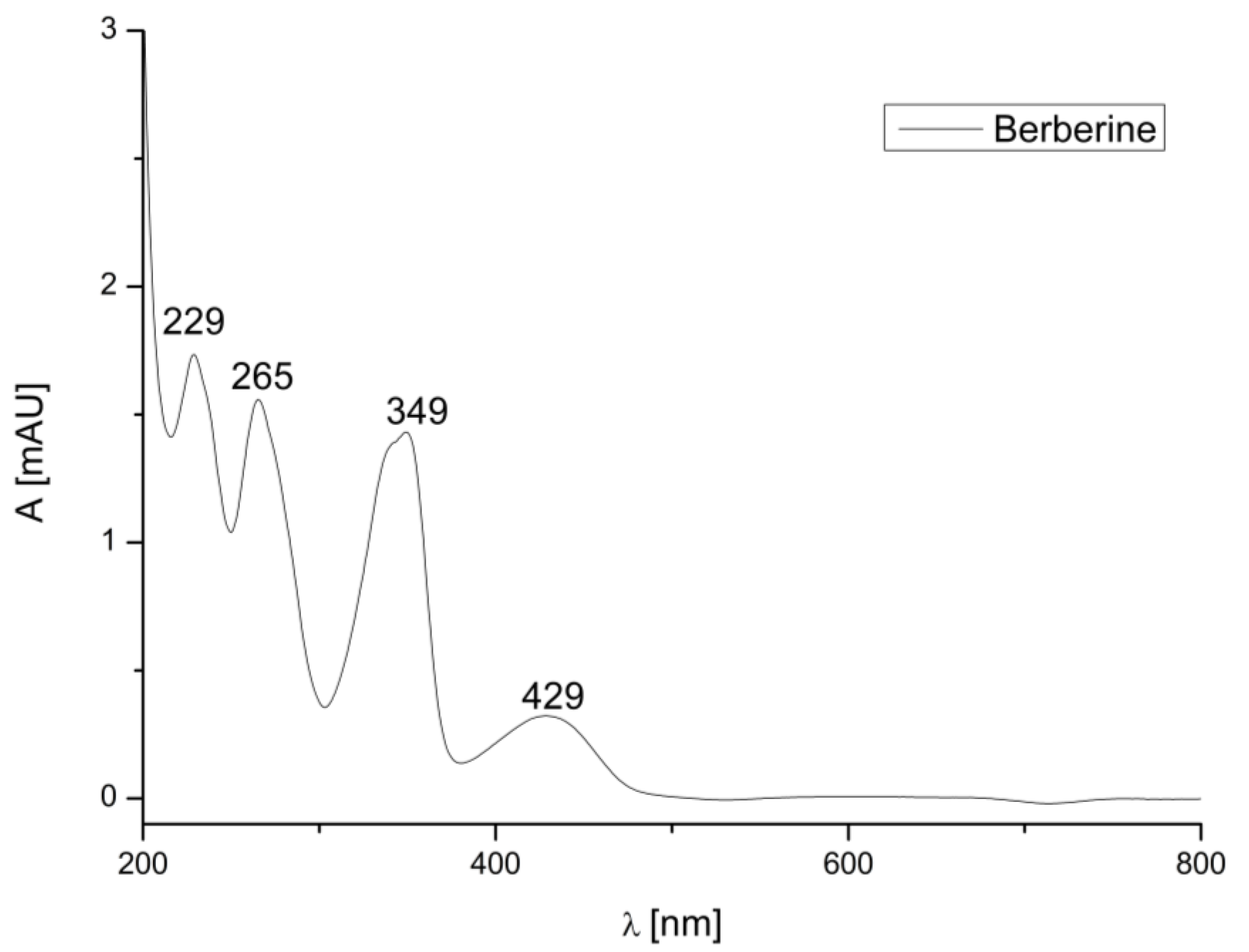
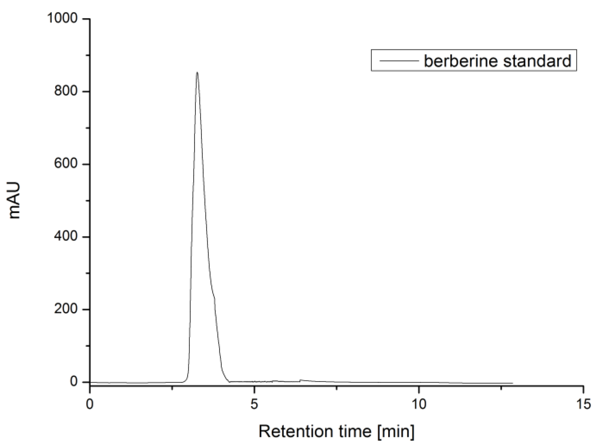
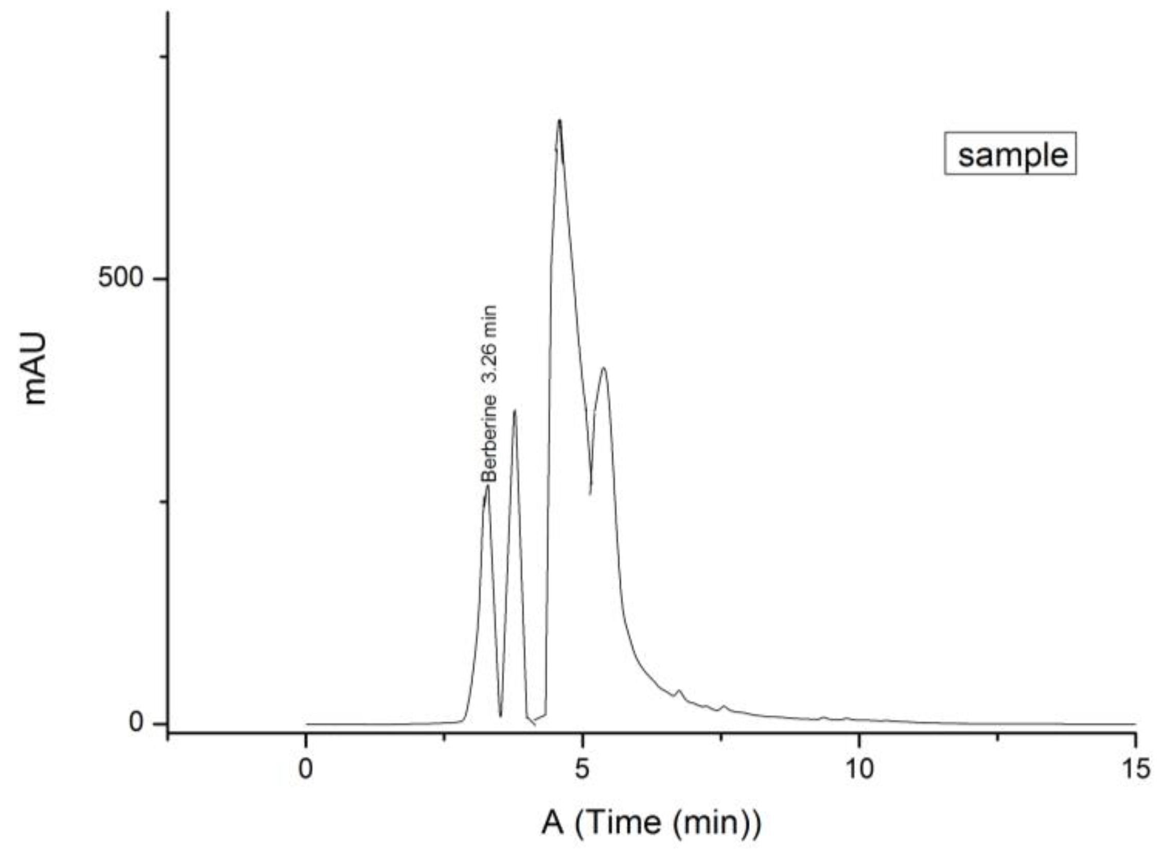
To determine the retention time and peak area, three repeated analyses of standards were carried out under optimal HPLC conditions (
Figure 2,
Figure 3 and
Figure 4). The calibration curve of berberine was linear in a relatively wide concentration range (0.025–1.000 mg/mL), the linear regression was had a high correlation (R
2 = 0.9978) and %RSD was 2.97. LOD and LOQ were defined as the signal-to-noise ratios (S/Ns) of 3 and 10, respectively. According to the calculation, the LOD and LOQ values of berberine were 0.00556 and 0.01684 mg, respectively.
The berberine content of the PA tincture was determined using the calibration curve of standard solutions with known concentrations. The content is expressed as an average value of three analyses ± SD in mg of berberine per kg of dry weight (mg/kg DW), and the values were 32.96 mg/kg DW ± 0.2 and is 28.24% of extractable compounds.
3.2. Effect on Vessels
In the present study, we evaluated the vasogenic potential and safety of application of extract from
Phellodendron amurense Rupr. in the in ovo model (
Table 2). This model is based on the observation of changes in the vessel of the chorioallantoic membrane of fertilized eggs.
The control (saline solution) and test solutions of PA with a concentration of 0.1 mg/mL did not cause a detectable effect after 5 min. On the other side, the administration of the PA extract with a concentration of 1.0 mg/mL may result in a slight irritant effect on vessels after 5 min of the application with the risk of hyperaemia. The extract in a concentration of 10.0 mg/mL caused hyperaemia (after 2 min) and haemorrhage (after 5 min), which is considered moderate irritation. Throughout the examination, none of the tested concentration caused coagulation of the CAM vasculature. Our results show that the increasing concentration of the PA causes the irritation of the CAM blood vessels.
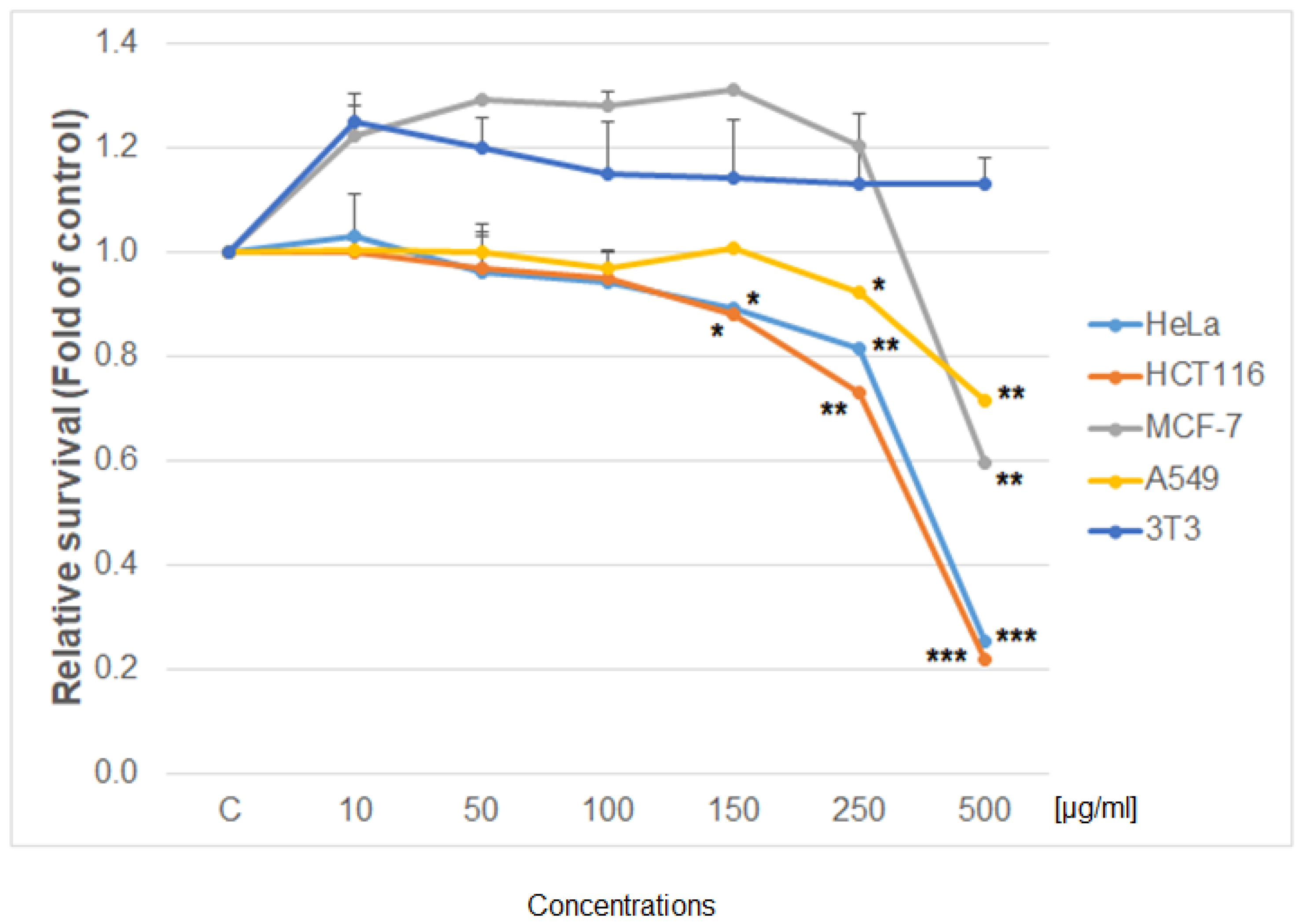
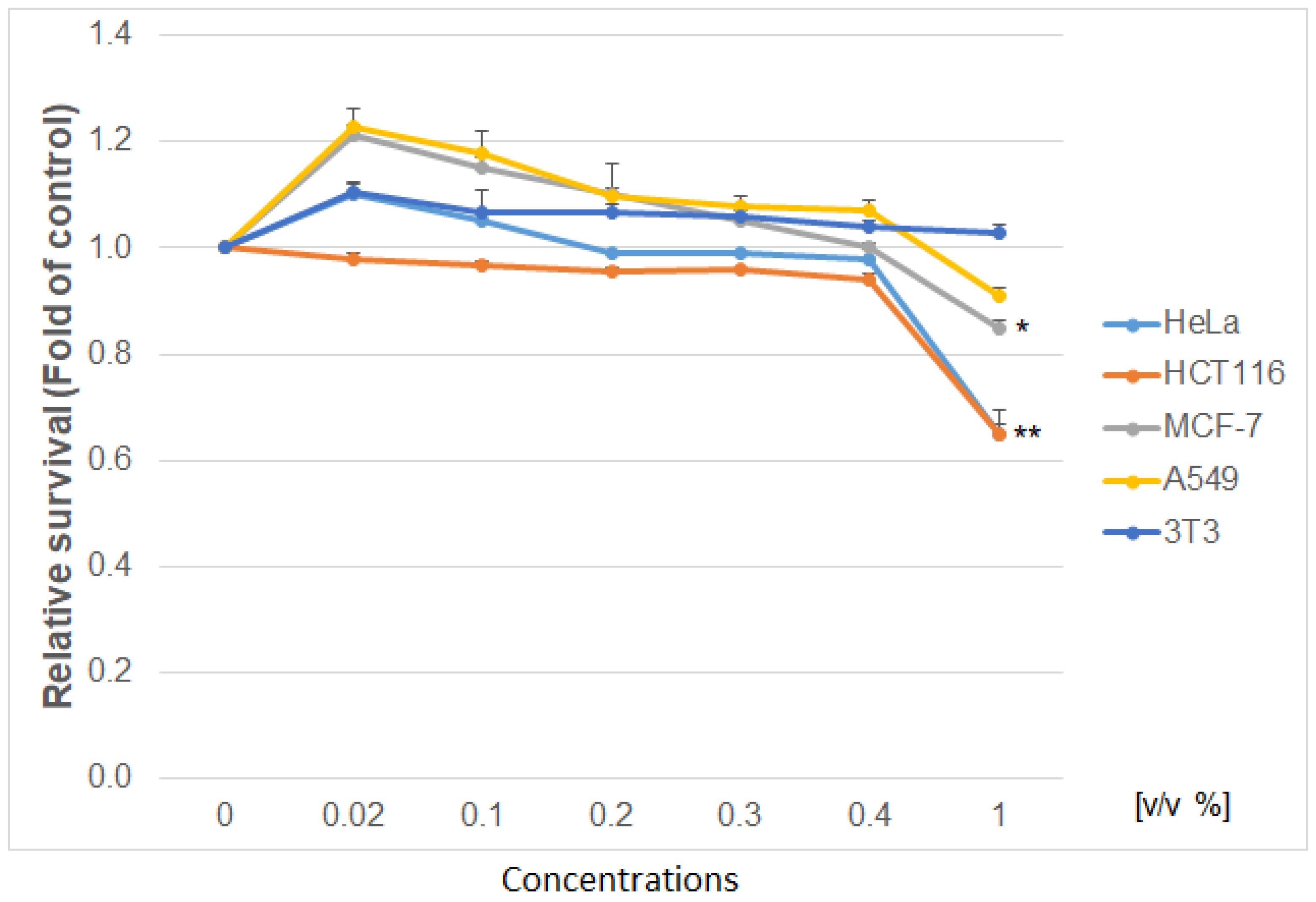
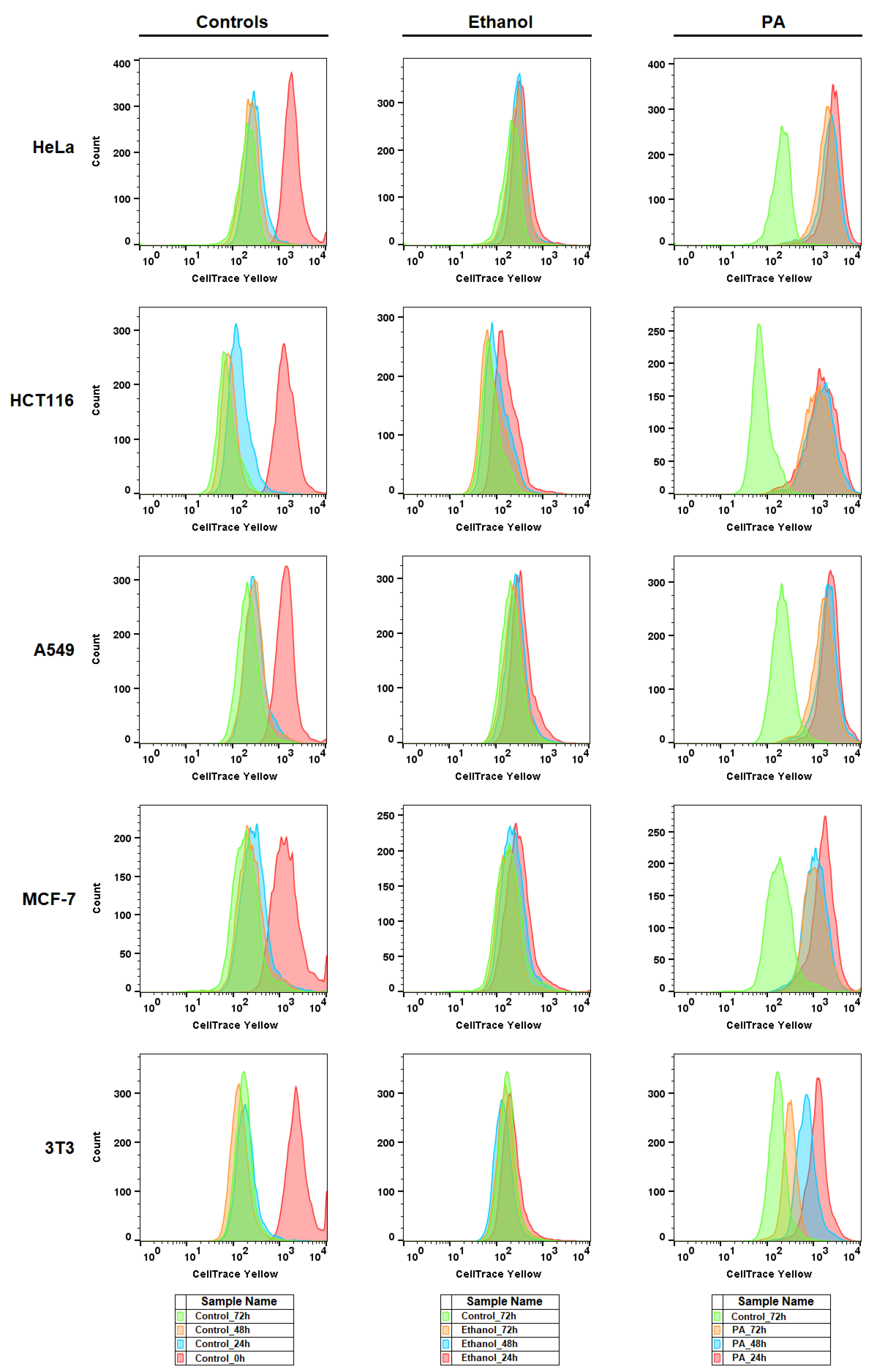
3.3. Antiproliferative Activity
Using the colorimetric, metabolic MTS and Flow cytometry CellTrace
TM Yellow assay, the antiproliferative effect of the studied extract was determined as shown in
Figure 5,
Figure 6 and
Figure 7. The IC50 values of the PA tincture on human cancer (HeLa—cervical carcinoma cells; MCF-7—breast cancer cells; HCT 116—human colorectal carcinoma; A549—lung carcinoma) and non-cancer (3T3—murine fibroblasts) cell lines calculated by MTS test are presented in
Table 3. When comparing the metabolic activity of PA on different cancer cell lines (
Figure 5), the strongest effect was shown on cell proliferation of HCT 116 and HeLa cells followed by MCF-7 and A549 cells. Moreover, the survival of non-cancerous murine fibroblast 3T3 exposed to PA tincture was not affected in the tested concentration range. Vehiculum used for the preparation of the PA tincture (60% ethanol) has no effect on all tested cells (except the highest
v/
v 1%) compared to untreated cells (
Figure 6). Furthermore, the flow cytometry proliferation test (
Figure 7) of the PA tincture IC50 values for every tested cancer cell line showed strong proliferation inhibition in a time-dependent manner (24–72 h) compared to untreated cells and also to vehicle-treated cells. The 3T3 fibroblast after higher IC50 value from A549 cells showed only partial inhibition of proliferation with minimal loss in viability. These findings indicated a high selectivity and specific cell-type-dependent toxicity of PA bark tincture.
3.4. Cell Cycle Analysis
Cell cycle distribution and changes were evaluated after tincture of PA (IC50) treatment in all cancer cell lines and 3T3 fibroblasts. As the result showed (
Table 4,
Table 5,
Table 6,
Table 7 and
Table 8), PA treatment caused time-dependent G1 phase cell cycle arrest in HeLa (24 h), HCT116 (24 h) and A549 (24–48 h) cells. MCF-7 cells showed no cell cycle block. Moreover, PA treatment led to an increase in the sub-G0 population in a time-dependent manner in all cancer cell lines except A549 due the long persistence of the G1 block. The sub-G0 population is evaluated as apoptotic with fragmented DNA. With increased sub-G0 population and G1 arrest, the G1 population consequently decreased, mainly at 72 h after treatment. On the other hand, ethanol treatment as PA solvent caused no significant cell cycle changes in all tested cell lines. Furthermore, the treatment of the 3T3 fibroblast with the tincture of PA bark or 60% ethanol showed no significant changes.
3.5. Artemia Franciscana Test
Screening test of cytotoxicity of PA bark was performed using
A. franciscana (
Table 9). We did not observe signs of toxicity after 24 and also after 48 h. We noted 16 ± 0.5% lethality after 72 h. After 96 hours, lethality increased to 34 ± 0.82% for the
Phellodendron amurense extract. The lethality, which began to appear up to 72 h, shows the negative effect of
Phellodenron amurense on
A. franciscana.
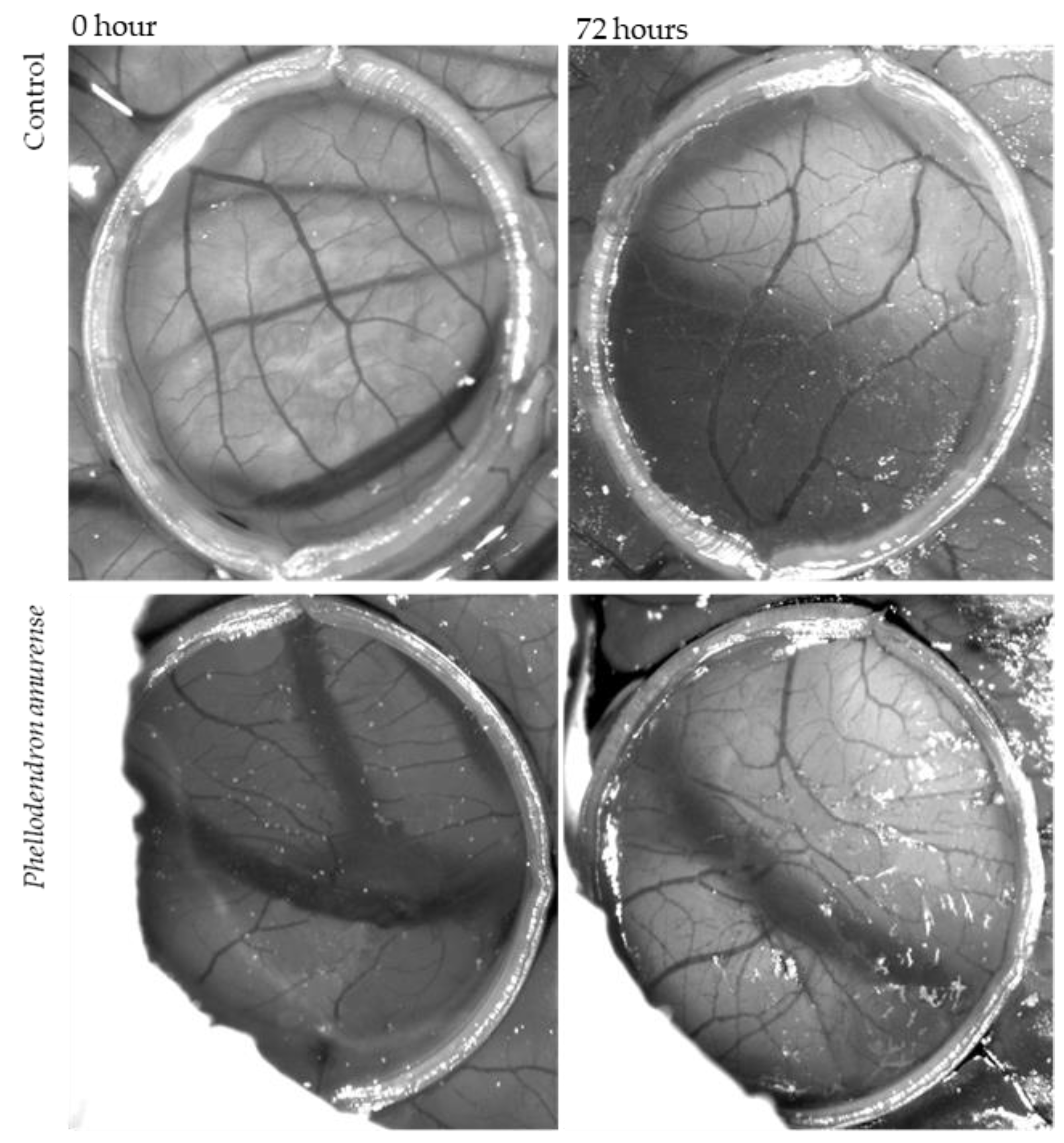
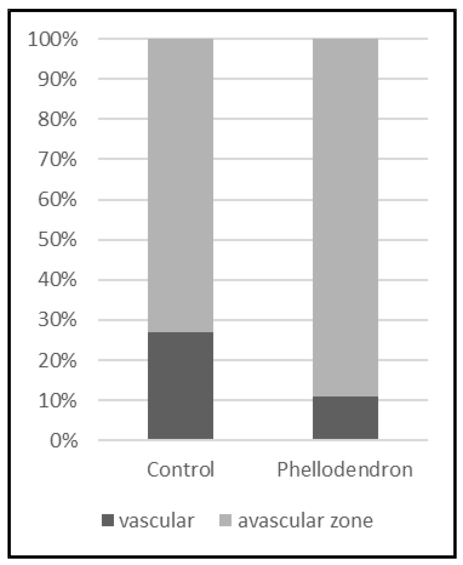
3.6. Antiangiogenic Activity
The CAM assay was used to examine whether Phellodendron amurense has any effect on neovascularization in ovo. The inhibition of angiogenesis was analysed by two methods, which gave us information about total branching points and total vascular zone, which means the vessels’ density.
The anti-angiogenic effect of dry PA residues from tincture dissolved in saline solution (1 mg/mL) on the treated CAM is shown in
Figure 8. It was microscopically observed that tested solutions of PA inhibited angiogenesis in the CAM, causing the reduction in the vascular density compared to the control (saline solution) (
Figure 9). The results can also be interpreted in the percentage of vessels density. Whereas in the case of control 27.5% of the figure can be considered vessels, this value is only 10.9% for the CAM treated with tested solutions of PA bark.
The second method for measuring the antiangiogenic potential of the tested PA solution was based on the enumeration of bifurcation points. An increase in the number of vessels crossing the delimited area was observed in the control (saline solution) (2.4 ± 0.1) and in CAM exposed to PA (1.7 ± 0.4). The present result demonstrated that Phellodendron amurense Rupr. exhibited a significant decrease in the branching of new capillaries from the existing basal vessels compared to the control after 72 h of treatment.
4. Discussion
Although evidence from epidemiological and animal studies suggests that increased consumption of a plant-based diet can reduce the risk of cancer, presently, little information is available concerning the use of botanicals in antiangiogenetic therapy. As a step toward developing botanicals as antiangiogenic agents, we examined the effect of Phellodendron on angiogenesis and on cancer cells.
Polyphenols were presented in prepared extract only in small concentrations. On the other hand, the main component of PA is berberine, which demonstrated different activity and antitumor properties [
19,
35]. The highest concentration of isoquinoline alkaloids was detected by spectrophotometer in
Tinctura (49.4 mg/L ± 0.75 mg/L). It means that ethanol is better for the extraction of berberine and other isoquinoline alkaloids from PA bark than water. UHPLC analysis detected berberine in the concentration of 32.96 mg/kg. In a previous study, the content of berberine was abundant in the Me OH extract of
P. amurense bark compared to other compounds. Its percentage yield was detected as 1.4% [
36].
At first, the low toxicity was verified on vessels of PA by in ovo irritancy test. The present data show no significant signs of the irritancy, such as vasoconstriction, hyperaemia, haemorrhage or coagulation for the concentrations of 0.10 and 1.0 mg/mL of the berberine extract, which indicates its harmless character and the possibility of potential intravenous application. Moderate irritancy was observed only in the highest applied concentration 10 mg/mL. Although we have not recorded vascular irritancy, we evaluated the PA plant as safe and suitable for the production of potential new drugs, which can also be used by intravenous administration. The result of safety application of PA correlate with the observed minimal negative effects of orally administered product with PA in human test subjects [
2,
10,
18]. Only few subjects described gentle stomach discomfort such as nausea [
19]. The safety of application of our tested extract was detected in all tested concentrations; however, with growing concentration, the irritation was growing. A similar trend was observed in the case of administering snake venom [
37]. No symptoms of haemorrhage, coagulation or mortality in embryo were observed after exposure of CAM to extract from lichen
Pseudevernia furfuracea (L.) Zopf and its metabolite, physodic acid [
38].
Recently, the bark of PA and its main component berberine have been demonstrated antitumor properties. We confirmed concentration and time-dependent cytotoxic and antiproliferative potential of PA in several cancer cell lines with different origin, mainly HCT116 (colon), HeLa (cervix), MCF-7 (breast) and A549 (lung). Our results are in accordance with several studies where, in the past decade, the number of reports demonstrating anti-cancer properties of commercial Asian origin Nexrutine
® in melanoma [
17], prostate [
39], pancreatic [
40] and breast [
6] in vitro models increased. Furthermore, we confirmed that proliferation inhibition in all tested cell lines except MCF-7 was caused by G1 cell cycle arrest and apoptosis, similar as described [
6] in SkBr3 and MDA-MB-231 breast cancer model or [
41] in A549 and H1299 lung carcinoma models. Berberine as the main component has stronger activity than PA. It was reported that the IC50 of berberine in HeLa cell was around 0.3 mM after 48 h [
42] (B. Lu et al./Toxicology in Vitro 24 (2010) 1482–1490) and 0.14 mM in A549 after 24 h of incubation Oncol Lett June 2012; 3(6): 1263–1267) [
43]. Moreover, the selectivity index indicated high selectivity for every cancer cell and potential non-toxic effects on normal healthy cells. These finding suggest the great potential of PA in clinical studies where PA could be well-tolerated after treatment of patients. This suggestion confirmed study of Swanson et al., where Nexrutine
® tolerance by prostate cancer patients was high, with no grade 4 toxicity [
18]. For the combined Nexrutine
® and radiation component, no additional patients suffered from grade 3 toxicity.
The present study showed the anti-angiogenic activity tested in ovo of Phellodendron amurense Rupr. for the first time. Both methods of measuring anti-angiogenic effects were compared and showed very similar results. In both cases (vascular zone and vessel sprouting), a reduction in angiogenesis was found. Phellodendron amurense Rupr. was able to significantly induce blood vessel response to stimuli. These results suggested that PA could be particularly attractive for the treatment of cancer according to its antiangiogenic properties. Moreover, the extract of Phellodendron amurense Rupr. did not affect the embryo viability.
To the best of our knowledge, there is no information about the anti-angiogenic efficiency of ethanolic tincture of
Phellodendron amurense Rupr. in vitro or in vivo. The anti-angiogenic effect of hot water extract was observed in vitro HUVECs and ex vivo in the rat aorta [
44]. The anti-angiogenic effect was also tested in combination with other extracts in traditional Chinese herbal medicine, Qing-Luo-Yin (
Sophora flavescens Ait.,
Phellodendron amurense Rupr.,
Sinomenium acutum Rehd. et Wils. and
Dioscorea hypoglauca Palib) for the treatment of rheumatoid arthritis [
45]. The main alkaloid in
Phellodendron amurense Rupr. bark is the isoquinoline alkaloid, berberine. The influence of berberine on angiogenesis was tested by different models, but no CAM analysis was performed in ovo. In vivo, berberine showed antiangiogenic activity on B16F-10 melanoma cells and in C57BL/6 mice [
46]. The main alkaloid from
Phellodendron is berberine, which prevents non-alcoholic steatohepatitis-derived hepatocellular carcinoma by inhibiting inflammation and angiogenesis in mice [
47]. Berberine could reduce metastasis and angiogenesis of cervical cancer cells, so it can be applied as a complementary treatment in metastasis control [
48]. On the other hand, berberine also promotes angiogenesis. It was observed in an animal model that berberine promotes ischemia-induced angiogenesis in mice heart [
49].


 ,
, 


