Preoperative Evaluation through Dermoscopy and Reflectance Confocal Microscopy of the Lateral Excision Margins for Primary Basal Cell Carcinoma

 ,
,  , ,
, ,
Abstract
:1. Introduction
2. Materials and Methods
Statistical Analysis
3. Results
4. Discussion
Author Contributions
Funding
Institutional Review Board Statement
Informed Consent Statement
Data Availability Statement
Acknowledgments
Conflicts of Interest
References
- Carducci, M.; Bozzetti, M.; Foscolo, A.M.; Betti, R. Margin Detection Using Digital Dermatoscopy Improves the Performance of Traditional Surgical Excision of Basal Cell Carcinomas of the Head and Neck. Dermatol. Surg. 2011, 37, 280–285. [Google Scholar] [CrossRef] [PubMed]
- Mun, J.-H.; Jwa, S.-W.; Song, M.; Ko, H.-C.; Kim, B.-S.; Kim, M.-B.; Kim, H.-S. Pitfalls of Using Dermatoscopy in Defining Surgical Margins of Basal Cell Carcinoma. Dermatol. Surg. 2011, 37, 1704–1705. [Google Scholar] [CrossRef] [PubMed]
- Venturini, M.; Gualdi, G.; Zanca, A.; Lorenzi, L.; Pellacani, G.; Calzavara-Pinton, P.G. A new approach for presurgical margin assessment by reflectance confocal microscopy of basal cell carcinoma. Br. J. Dermatol. 2016, 174, 380–385. [Google Scholar] [CrossRef] [PubMed]
- Lupu, M. The Use of In Vivo Reflectance Confocal Microscopy and Dermatoscopy in the Preoperative Determination of Basal Cell Carcinoma Histopathological Subtypes; “Carol Davila” University of Medicine and Pharmacy: Bucharest, Romania, 2019. [Google Scholar]
- Lupu, M.; Popa, I.M.; Voiculescu, V.M.; Boda, D.; Caruntu, C.; Zurac, S.; Giurcaneanu, C. A Retrospective Study of the Diagnostic Accuracy of In Vivo Reflectance Confocal Microscopy for Basal Cell Carcinoma Diagnosis and Subtyping. J. Clin. Med. 2019, 8, 449. [Google Scholar] [CrossRef] [PubMed] [Green Version]
- R Development Core Team. R: A Language and Environment for Statistical Computing; R Foundation for Statistical Computing: Vienna, Austria, 2013. [Google Scholar]
- Bichakjian, C.K.; Olencki, T.; Aasi, S.Z.; Alam, M.; Andersen, J.S.; Berg, D.; Bowen, G.M.; Cheney, R.T.; Daniels, G.A.; Glass, L.F.; et al. Basal Cell Skin Cancer, Version 1.2016, NCCN Clinical Practice Guidelines in Oncology. J. Natl. Compr. Cancer Netw. 2016, 14, 574. [Google Scholar] [CrossRef]
- Lupu, M.; Căruntu, A.; Moraru, L.; Voiculescu, V.M.; Boda, D.; Tănase, C.; Căruntu, C. Non-invasive imaging techniques for early diagnosis of radiation-induced squamous cell carcinoma of the lip. Rom. J. Morphol. Embryol. 2018, 59, 3. [Google Scholar]
- González, S.; Tannous, Z. Real-time, in vivo confocal reflectance microscopy of basal cell carcinoma. J. Am. Acad. Dermatol. 2002, 47, 869–874. [Google Scholar] [CrossRef] [Green Version]
- Nori, S.; Rius-Diaz, F.; Cuevas, J.; Goldgeier, M.; Jaen, P.; Torres, A.; Gonzalez, S. Sensitivity and specificity of reflectance-mode confocal microscopy for in vivo diagnosis of basal cell carcinoma: A multicenter study. J. Am. Acad. Dermatol. 2004, 51, 923–930. [Google Scholar] [CrossRef]
- Curiel-Lewandrowski, C.; Williams, C.M.; Swindells, K.J.; Tahan, S.R.; Astner, S.; Frankenthaler, R.A.; Gonzalez, S. Use of in vivo confocal microscopy in malignant melanoma: An aid in diagnosis and assessment of surgical and nonsurgical therapeutic approaches. Arch. Dermatol. 2004, 140, 1127–1132. [Google Scholar] [CrossRef] [Green Version]
- Rishpon, A.; Kim, N.; Scope, A.; Porges, L.; Oliviero, M.C.; Braun, R.P.; Marghoob, A.A.; Fox, C.A.; Rabinovitz, H.S. Reflectance confocal microscopy criteria for squamous cell carcinomas and actinic keratoses. Arch. Dermatol. 2009, 145, 766–772. [Google Scholar] [CrossRef] [Green Version]
- Couty, E.; Tognetti, L.; Labeille, B.; Douchet, C.; Habougit, C.; Couzan, C.; Biron-Schneider, A.C.; Cambazard, F.; Prade, V.; Rubegni, P. In vivo reflectance confocal microscopy combined with the’spaghetti technique’for the identification of surgical margins of lentigo maligna: Experience in 70 patients. J. Eur. Acad. Dermatol. Venereol. JEADV 2018, 32, e366. [Google Scholar] [CrossRef]
- Cinotti, E.; Fiorani, D.; Labeille, B.; Gonzalez, S.; Debarbieux, S.; Agozzino, M.; Ardigò, M.; Lacarrubba, F.; Farnetani, F.; Carrera, C. The integration of dermoscopy and reflectance confocal microscopy improves the diagnosis of lentigo maligna. J. Eur. Acad. Dermatol. Venereol. 2019, 33, e372–e374. [Google Scholar] [CrossRef] [PubMed]
- Scope, A.; Mahmood, U.; Gareau, D.S.; Kenkre, M.; Lieb, J.A.; Nehal, K.S.; Rajadhyaksha, M. In vivo reflectance confocal microscopy of shave biopsy wounds: Feasibility of intraoperative mapping of cancer margins. Br. J. Dermatol. 2010, 163, 1218–1228. [Google Scholar] [CrossRef] [PubMed]
- Pan, Z.-Y.; Lin, J.-R.; Cheng, T.-T.; Wu, J.-Q.; Wu, W.-Y. In Vivo Reflectance Confocal Microscopy of Basal Cell Carcinoma: Feasibility of Preoperative Mapping of Cancer Margins. Dermatol. Surg. 2012, 38, 1945–1950. [Google Scholar] [CrossRef] [PubMed] [Green Version]
- Guitera, P.; Moloney, F.J.; Menzies, S.W.; Stretch, J.R.; Quinn, M.J.; Hong, A.; Fogarty, G.; Scolyer, R.A. Improving management and patient care in lentigo maligna by mapping with in vivo confocal microscopy. JAMA Dermatol 2013, 149, 692–698. [Google Scholar] [CrossRef] [PubMed] [Green Version]
- Chen, C.S.; Elias, M.; Busam, K.; Rajadhyaksha, M.; Marghoob, A.A. Multimodal in vivo optical imaging, including confocal microscopy, facilitates presurgical margin mapping for clinically complex lentigo maligna melanoma. Br. J. Dermatol. 2005, 153, 1031–1036. [Google Scholar] [CrossRef] [PubMed]
- Maier, T.; Sattler, E.C.; Braun-Falco, M.; Korting, H.C.; Ruzicka, T.; Berking, C. Reflectance confocal microscopy in the diagnosis of partially and completely amelanotic melanoma: Report on seven cases. J. Eur. Acad. Dermatol. Venereol. 2013, 27, e42–e52. [Google Scholar] [CrossRef] [PubMed]
- Pan, Z.Y.; Liang, J.; Zhang, Q.A.; Lin, J.R.; Zheng, Z.Z. In vivo reflectance confocal microscopy of extramammary Paget disease: Diagnostic evaluation and surgical management. J. Am. Acad. Dermatol. 2012, 66, e47–53. [Google Scholar] [CrossRef] [PubMed]
- Navarrete-Dechent, C.; Cordova, M.; Aleissa, S.; Kose, K.; Lee, E.H.; Rossi, A.M.; Nehal, K.S. Use of paper tape to guide reflectance confocal microscopy navigation of large skin lesions. J. Am. Acad. Dermatol. 2020, 82, e199–e201. [Google Scholar] [CrossRef] [PubMed]
- Gualdi, G.; Venturini, M.; Zanca, A.; Calzavara-Pinton, P.; Pellacani, G. Pre-surgical basal cell carcinoma margin definition: The SMART approach. J. Eur. Acad. Dermatol. Venereol. 2016, 30, 474–476. [Google Scholar] [CrossRef]
- Blasdale, C.; Charlton, F.G.; Weatherhead, S.C.; Ormond, P.; Lawrence, C.M. Effect of tissue shrinkage on histological tumour-free margin after excision of basal cell carcinoma. Br. J. Dermatol. 2010, 162, 607–610. [Google Scholar] [CrossRef] [PubMed]
- Dauendorffer, J.N.; Bastuji-Garin, S.; Guero, S.; Brousse, N.; Fraitag, S. Shrinkage of skin excision specimens: Formalin fixation is not the culprit. Br. J. Dermatol. 2009, 160, 810–814. [Google Scholar] [CrossRef] [PubMed]
- Kerns, M.J.; Darst, M.A.; Olsen, T.G.; Fenster, M.; Hall, P.; Grevey, S. Shrinkage of cutaneous specimens: Formalin or other factors involved? J. Cutan. Pathol. 2008, 35, 1093–1096. [Google Scholar] [CrossRef] [PubMed]
- Puig, S.; Cecilia, N.; Malvehy, J. Dermoscopic criteria and basal cell carcinoma. G. Ital. Dermatol. Venereol. 2012, 147, 135–140. [Google Scholar]
- Lupu, M.; Caruntu, C.; Popa, M.I.; Voiculescu, V.M.; Zurac, S.; Boda, D. Vascular patterns in basal cell carcinoma: Dermoscopic, confocal and histopathological perspectives (Review). Oncol. Lett. 2019. [Google Scholar] [CrossRef] [Green Version]
- Pellacani, G.; De Carvalho, N.; Ciardo, S.; Ferrari, B.; Cesinaro, A.M.; Farnetani, F.; Bassoli, S.; Guitera, P.; Star, P.; Rawson, R.; et al. The smart approach: Feasibility of lentigo maligna superficial margin assessment with hand-held reflectance confocal microscopy technology. J. Eur. Acad. Dermatol. Venereol. 2018, 32, 1687–1694. [Google Scholar] [CrossRef]
- Flores, E.S.; Cordova, M.; Kose, K.; Phillips, W.; Rossi, A.; Nehal, K.; Rajadhyaksha, M. Intraoperative imaging during Mohs surgery with reflectance confocal microscopy: Initial clinical experience. J. Biomed. Opt. 2015, 20, 61103. [Google Scholar] [CrossRef] [Green Version]
- Soyer, H.; Prow, T. Reflectance confocal microscopy in the diagnosis of nodular skin lesions. Br. J. Dermatol. 2013, 169, 4. [Google Scholar] [CrossRef]
- De Carvalho, N.; Schuh, S.; Kindermann, N.; Kästle, R.; Holmes, J.; Welzel, J. Optical coherence tomography for margin definition of basal cell carcinoma before micrographic surgery—recommendations regarding the marking and scanning technique. Skin Res. Technol. 2018, 24, 145–151. [Google Scholar] [CrossRef] [Green Version]
- Suppa, M.; Fontaine, M.; Dejonckheere, G.; Cinotti, E.; Yélamos, O.; Diet, G.; Tognetti, L.; Miyamoto, M.; Orte Cano, C.; Perez-Anker, J. Line-field confocal optical coherence tomography of basal cell carcinoma: A descriptive study. J. Eur. Acad. Dermatol. Venereol. 2020. [Google Scholar]
- Tannous, Z.; Torres, A.; Gonzalez, S. In vivo real-time confocal reflectance microscopy: A noninvasive guide for Mohs micrographic surgery facilitated by aluminum chloride, an excellent contrast enhancer. Dermatol. Surg. 2003, 29, 839–846. [Google Scholar] [CrossRef] [PubMed]
- Patel, Y.G.; Nehal, K.S.; Aranda, I.; Li, Y.; Halpern, A.C.; Rajadhyaksha, M. Confocal reflectance mosaicing of basal cell carcinomas in Mohs surgical skin excisions. J. Biomed. Opt. 2007, 12, 034027. [Google Scholar] [CrossRef] [PubMed] [Green Version]


| Histopathological Subtype | Confocal Criterion |
|---|---|
| Nodular BCC | thick collagen bundles surrounding tumor islands |
| increased vascularization | |
| big tumor islands (diameter > 300 µm) | |
| Superficial BCC | cords connected to the epidermis |
| Aggressive BCC | hyporefractile silhouettes |
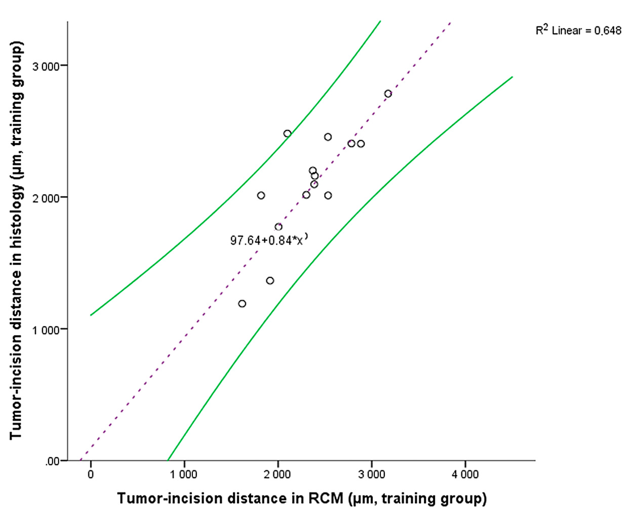
| Case No. | Sex | Age (Years) | Histological Subtype | Tumor Location and Clinical Characteristics | Tumor-Incision Distance, RCM (µm) | Tumor-Incision Distance, Histology (µm) | Tumor-Incision Distance Percentage Reduction between RCM and Histology (%) |
|---|---|---|---|---|---|---|---|
| 1 | M | 77 | nodular | Right zygomatic, 14.4 × 7.42 mm, flat, hypopigmented | 1: 2384.993 2: 2783.069 | 1: 2096.367 2: 2453.325 | 1: 12.1 2: 11.85 |
| 2 | F | 70 | nodular | Scalp, 9.57 × 9.41 mm, elevated, partially pigmented | 1: 2099.000 2: 2523.810 | 1: 2481.481 2: 2240.014 | 1: −18.22 2: 11.24 |
| 3 | F | 82 | nodular | Left pre-auricular, 16.26 × 11.55 mm, nodular, hypopigmented | 1: 3174.603 2: 2333.333 | 1: 2784.000 2: 1272.176 | 1: 12.3 2: 45.48 |
| 4 | F | 64 | nodular | Left zygomatic, 10.59 × 5.71 mm, elevated, partially pigmented | 2783.069 | 2405.983 | 13.55 |
| 5 | M | 78 | superficial | Posterior trunk, 16.64 × 12.79 mm, flat, hypopigmented | 1615.544 | 1190.476 | 26.31 |
| 6 | M | 74 | nodular | Forehead, 8.6 × 7.84 mm, elevated, pigmented | 1: 2274.000 2: 2256.624 | 1: 1703.704 2: 2243.386 | 1: 25.08 2: 0.59 |
| 7 | F | 80 | nodular | Left nazolabial fold, 5.86 × 4 mm, elevated, hypopigmented | 1: 2391.534 2: 3301.021 | 1: 2159.896 2: 3164.021 | 1: 9.69 2: 4.15 |
| 8 | F | 68 | superficial | Posterior trunk, 13.28 × 13.14 mm, flat, hypopigmented | 2552.614 | 2227.513 | 12.74 |
| 9 | M | 61 | nodular | Scalp, 10.65 × 8.2 mm, elevated, pigmented | 4074.866 | 4050.774 | 0.59 |
| 10 | F | 74 | micro-nodular | Right temporal, 11 × 9.59 mm, flat, hypopigmented | 2104.061 | 1851.852 | 11.99 |
| 11 | F | 72 | nodular | Left pre-auricular, 6.95 × 4.61 mm, elevated, partially pigmented | 1: 2300.782 2: 2136.734 | 1: 2015.784 2: 3104.743 | 1: 12.39 2: −45.3 |
| 12 | M | 66 | nodular | Posterior trunk, 6.74 × 6.47 mm, elevated, hypopigmented | 2370 | 2200.321 | 7.16 |
| 13 | F | 65 | nodular | Posterior trunk, 18.53 × 9.06 mm, elevated, hypopigmented | 1817.772 | 2011.811 | −10.67 |
| 14 | F | 81 | infiltrative | Right arm, 4.73 × 3.48 mm, flat, hypopigmented | 2532.065 | 2455.203 | 3.04 |
| 15 | F | 65 | infiltrative | Righ arm, 5.7 × 4.32 mm, elevated, pigmented | 1913.7 | 1365.012 | 28.67 |
| 16 | M | 67 | superficial | Lateral trunk, 12.33 × 6.24 mm, elevated, hypopigmented | 1: 2031 2: 2759.154 | 1: 1666.900 2: 2349.458 | 1: 17.93 2: 14.85 |
| 17 | - | - | superficial | Anterior trunk, 13.78 × 12.03 mm, flat, hypopigmented | 2241.728 | 2100.394 | 6.3 |
| 18 | - | - | superficial | Left arm, 10.77 × 10.02 mm, elevated, hypopigmented | 1: 2883.071 2: 1604.899 | 1: 2403.825 2: 1048.448 | 1: 16.62 2: 34.67 |
| 19 | F | 74 | nodular | Left arm, 12.01 × 8.16 mm, nodular, hypopigmented | 2533.333 | 2011.742 | 20.59 |
| 20 | F | 69 | nodular | Right thigh, 25.13 × 15.63, elevated, hypopigmented | 1: 2004 2: 2171.188 | 1: 1773.070 2: 2141.732 | 1: 11.52 2: 1.36 |
| Histological Positive Margin | Histological Negative Margin | Total | |
|---|---|---|---|
| RCM positive margin | 3 | 1 | 4 |
| RCM negative margin | 5 | 20 | 25 |
| Total | 8 | 21 | 29 |
| Histopathologically Positive Margins | Histopathologically Negative Margins | Total | |
|---|---|---|---|
| RCM positive margins | 4 | 0 | 4 |
| RCM negative margins | 2 | 23 | 25 |
| Total | 6 | 23 | 29 |
Publisher’s Note: MDPI stays neutral with regard to jurisdictional claims in published maps and institutional affiliations. |
© 2021 by the authors. Licensee MDPI, Basel, Switzerland. This article is an open access article distributed under the terms and conditions of the Creative Commons Attribution (CC BY) license (http://creativecommons.org/licenses/by/4.0/).
Share and Cite
Lupu, M.; Voiculescu, V.M.; Caruntu, A.; Tebeica, T.; Caruntu, C. Preoperative Evaluation through Dermoscopy and Reflectance Confocal Microscopy of the Lateral Excision Margins for Primary Basal Cell Carcinoma. Diagnostics 2021, 11, 120. https://0-doi-org.brum.beds.ac.uk/10.3390/diagnostics11010120
Lupu M, Voiculescu VM, Caruntu A, Tebeica T, Caruntu C. Preoperative Evaluation through Dermoscopy and Reflectance Confocal Microscopy of the Lateral Excision Margins for Primary Basal Cell Carcinoma. Diagnostics. 2021; 11(1):120. https://0-doi-org.brum.beds.ac.uk/10.3390/diagnostics11010120
Chicago/Turabian StyleLupu, Mihai, Vlad Mihai Voiculescu, Ana Caruntu, Tiberiu Tebeica, and Constantin Caruntu. 2021. "Preoperative Evaluation through Dermoscopy and Reflectance Confocal Microscopy of the Lateral Excision Margins for Primary Basal Cell Carcinoma" Diagnostics 11, no. 1: 120. https://0-doi-org.brum.beds.ac.uk/10.3390/diagnostics11010120