Review

Depth of Stromal Invasion as the Most Prognostically Relevant Regression System in Locally Advanced Cervical Cancer after Neoadjuvant Treatment: A Systematic Review and Meta-Analysis Grading

by
Gian Franco Zannoni
1,2,*,
Antonio Travaglino
3,
Antonio Raffone
4,
Damiano Arciuolo
1,
Nicoletta D’Alessandris
1,
Giulia Scaglione
1,
Pietro Tralongo
1,
Frediano Inzani
1,
Giuseppe Angelico
1,† and
Angela Santoro
1,†
1
Unità di Ginecopatologia e Patologia Mammaria, Dipartimento Scienze della Salute della Donna, del Bambino e di Sanità Pubblica, Fondazione Policlinico Universitario A. Gemelli, IRCCS, Largo A. Gemelli 8, 00168 Roma, Italy
2
Istituto di Anatomia Patologica, Università Cattolica del Sacro Cuore, Largo A. Gemelli 8, 00168 Roma, Italy
3
Department of Advanced Biomedical Sciences, Pathology Section, School of Medicine, University of Naples “Federico II”, Via Sergio Pansini 5, 80131 Naples, Italy
4
Gynecology and Obstetrics Unit, Department of Neuroscience, Reproductive Sciences and Dentistry, School of Medicine, University of Naples Federico II, 80131 Naples, Italy
*
Author to whom correspondence should be addressed.
These authors have equally contributed.
Submission received: 19 July 2021 / Revised: 20 September 2021 / Accepted: 23 September 2021 / Published: 26 September 2021

Abstract

:
Background: several different criteria have been proposed to categorize the pathological response in cervical cancer after neoadjuvant therapy; although it is unclear what the most prognostically valuable one is. Objective: to assess the prognostic value of pathological criteria for categorizing the response in cervical cancer after neoadjuvant therapy, through a systematic review and meta-analysis. Methods: four electronic databases were searched from January to December 2020 for all studies, assessing the prognostic value of pathological response in cervical cancer after neoadjuvant therapy. Hazard ratio (HR) for overall survival (OS) was calculated with a significant p-value < 0.05. A meta-analysis was performed for each criteria assessed in at least three studies. Results: sixteen studies were included. Criteria for pathological response included (i) residual stromal invasion < vs. >3 mm; (ii) complete response vs. any residual; (iii) proportion of viable cells; (iv) residual tumor diameter; and (v) intracervical vs. extracervical residual. Criteria (i) and (ii) were suitable for meta-analysis. The presence of a residual tumor with stromal invasion > 3 mm showed a HR of 4.604 (95% CI; 3.229–6.565; p < 0.001), while the presence of any residual showed a HR of 1.610 (95% CI; 1.245–2.081; p < 0.001); statistical heterogeneity was absent in both analyses. Conclusions: dichotomizing the pathological response in cervical cancer after neoadjuvant therapy as < vs. >3 mm stromal invasion is more prognostically valuable than dichotomizing as complete response vs. any residual. Further studies are necessary to evaluate other systems.

1. Introduction

Globally, cervical cancer continues to be one of the most common malignancies among females worldwide. In 2018, there were an estimated 569,847 new cases and 311,365 deaths worldwide [1]. Among the numerous clinical and pathological prognostic factors for this neoplasm, the International Federation of Gynecology and Obstetrics (FIGO) stage remains the most important to guide therapeutic strategies, with early cervical cancers treated by surgery alone and more advanced forms dealt with combined modality therapies [2,3]. In particular, locally advanced cervical cancer (LACC) is defined as a great size tumoral mass, including International Federation of Obstetrics and Gynecology (FIGO) stages IB2-IVA. Concurrent neoadjuvant unimodal or multimodal therapies (NACT) followed by surgery have been proposed as possible therapeutical approaches for LACC, with the goals of: (1) down-staging the tumor mass to improve operability; and (2) inhibiting metastasis [4]. The effects of preoperative treatment can be histologically evaluated, and the assessment of neoplastic response to neoadjuvant treatments should be integrated in the pathology reports of resection specimens [5]. In cervical cancers, several tumor regression scoring systems were applied; however, no consensus was observed concerning morphological data, and there was no agreement among the different grading systems. Generally, therapy inducted regressive changes may result in different amounts of residual tumors, up to the complete disappearance of malignant cells, with replacement by fibrous or fibro-inflammatory granulation tissue [6]. Moreover, histopathological determination of tumor regression provides important prognostic information and harbors the potential to guide the clinician to proceed with possible additional therapeutic regimens in the postoperative setting [5]. The aim of the present study was to establish a scoring system, to assess cervical cancer pathological response to neoadjuvant treatment, with the best impact on overall survival (OS) and progression-free survival (PFS).

2. Materials and Methods

A systematic review and a meta-analysis were carried out following previous studies [7,8,9]. Each step of the review process was performed by two independent authors, who sought consultation in the case of a disagreement. This study was reported according to the PRISMA statement [10].

2.1. Search Strategy and Study Selection

Four electronic databases (PubMed, Scopus, Wed of Science, and Google Scholar) were searched from January to December 2020. The following word combination was used: cervical AND (cancer OR carcinoma) AND neoadjuvant AND pathological response. All studies assessing pathological response in cervical cancer after neoadjuvant treatment were included. Inclusion criteria were: extractable hazard ratio (HR) with 95% confidence interval (CI) for the impact of pathological response on overall survival (OS) and progression-free survival (PFS), or available individual data to calculate HR with 95% CI. Exclusion criteria were: <10 patients, studies including less than 10 patients; studies with less than 2 deaths at follow-up; patient cohort already included in previous studies (unless the overlapping studies adopted different pathological criteria to evaluate the response).

2.2. Data Extraction

PICO of our study were: P (population) = women with locally advanced cervical carcinoma; I (intervention, risk factor) = optimal pathological response to neoadjuvant treatment; C (comparator) = suboptimal or no pathological response to neoadjuvant treatment; O (outcome) = OS and PFS [10]. The main data extracted were HR with 95% for the impact of pathological response on OS and PFS when available, the results of the multivariate analysis were used. If HR with 95% was not reported, but individual data were available, pathological response, follow-up time, and status (alive vs. dead or recurred vs. disease-free) were extracted for each patient.

2.3. Risk of Bias Assessment

The risk of bias within studies was assessed by using the QUADAS-2 as a basis, as previously described [11,12]. For the “patient selection” domain, we assessed whether patient selection criteria and period of enrollment were clearly reported; for the “index test” domain, we assessed whether criteria to evaluate pathological response were clearly reported; for the “reference standard” domain, we assessed whether oncologic outcomes (i.e., OS, PFS) were clearly reported; for the “flow and timing” domain, we assessed whether patients were followed for at least 2 years to evaluate oncologic outcomes. The risk of bias was categorized as “low”, “high”, or “unclear” for each domain, as previously described [13].

2.4. Data Analysis

Each criterion to evaluate pathological response was considered suitable for meta-analysis if it was assessed in at least 3 studies. HRs with 95% CI for each study were pooled by using a random effect model; results were reported on forest plots. A p-value < 0.05 was considered significant. Statistical heterogeneity among studies was assessed as previously described [14]. For the studies that reported individual data, HR with 95% CI was calculated by performing a Cox regression survival analysis. Data analysis was performed by using Statistical Package for Social Science (SPSS) 18.0 package (SPSS Inc., Chicago, IL, USA) for analyzing data of individual studies and comprehensive meta-analysis (Biostat, 14 North Dean Street, Englewood, NJ 07631, USA) for pooling results.

3. Results

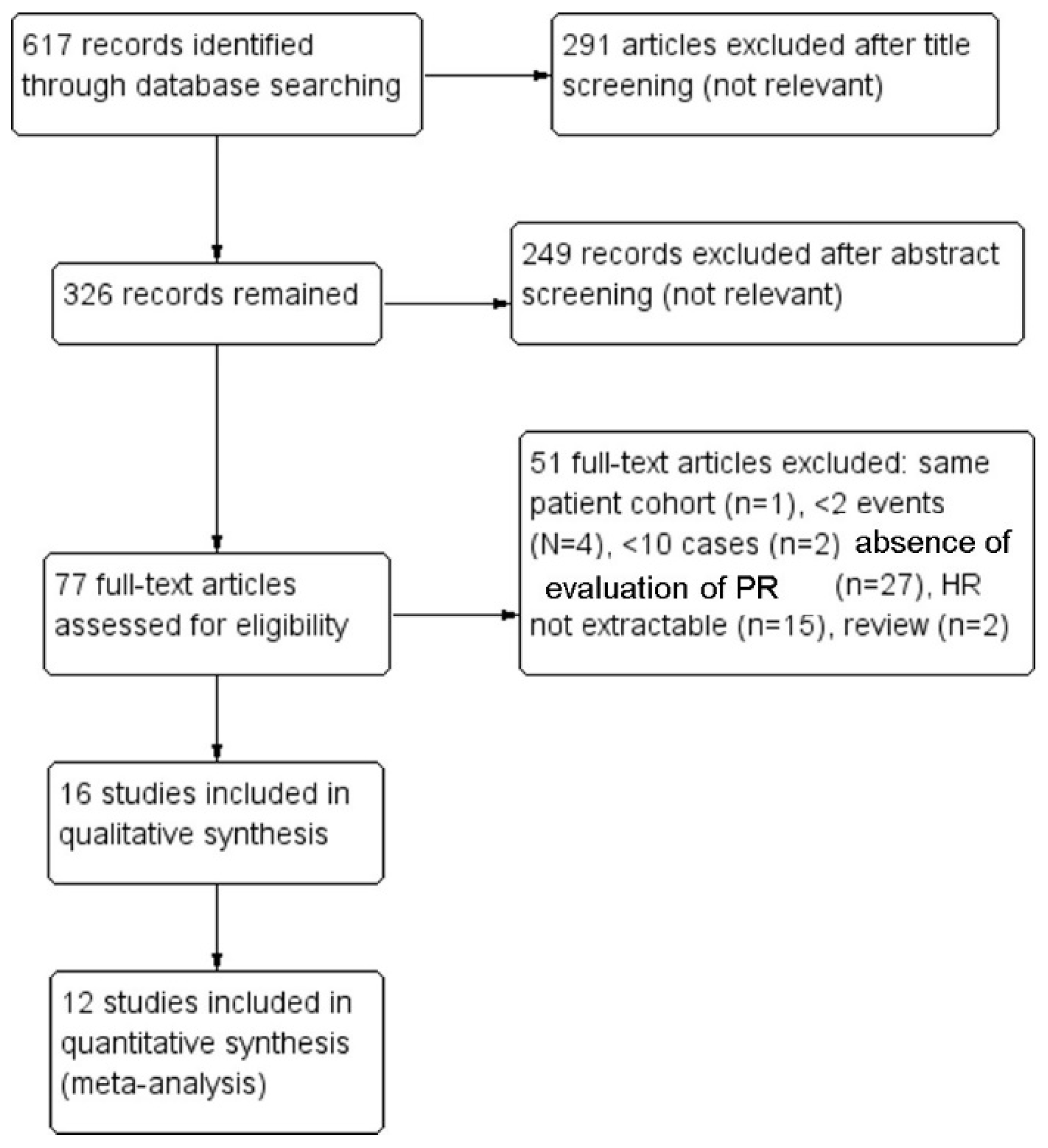
3.1. Study Selection

Sixteen studies were included in the systematic review [15,16,17,18,19,20,21,22,23,24,25,26,27,28,29,30] and 12 studies in the meta-analysis [15,16,17,18,19,20,23,24,25,27,28,29,30]. Fifty-one studies assessed for eligibility were excluded after applying our exclusion criteria, i.e., the same cohort as a study already included across multiple papers (n = 1), <2 events (N = 4), <10 cases (n = 2), evaluation of clinical rather than pathological response (n = 27), HR with 95% CI not extractable (n = 15), review (n = 2). The process of study selection is summarized in Figure 1.

3.2. Study Characteristics

Eight studies (50%) dichotomized pathological response as “optimal” (complete response or residual with <3 mm stromal invasion) vs. “suboptimal” (residual with >3 mm stromal invasion) [17,19,21,23,24,25,26,29]; two studies (12.50%) assessed the proportion of viable cells (<1/3, 1/3-to-2/3 or >2/3) [15,20]; two studies (12.50%) assessed the diameter of the residual tumor (cut-offs at 1 mm or 5 mm) [16,18]; two studies (12.50%) dichotomized pathological response as complete disappearance of the tumor vs. any residual [23,30]; one study (6.25%) dichotomized the residual tumor as intracervical vs. extracervical [27]; the remaining study (6.25%) adopted several systems simultaneously, proposing a combined score [22]. Seven studies (43.75%) assessed OS [15,16,17,18,19,20,30], one (6.25%) assessed PFS [24], and eight (50%) assessed both OS and PFS [21,22,23,24,25,26,27,28,29]. Characteristics of the included studies are reported in Table 1.

3.3. Risk of Bias Assessment

The risk of bias was unclear in two studies (12.50%) for the “patient selection” domain (since they did not report the period of enrollment) [18,23] and in two studies for the “flow and timing domain” (since they did not report the follow-up duration) [23,29]; the other studies were considered at low risk of bias for these domains. For the “index test” and “reference standard” domain, all studies were considered at low risk of bias, since they clearly reported the criteria for pathologic response and the oncologic outcomes, respectively (Figure 2).

3.4. Meta-Analysis

Two different criteria for categorizing the pathological response were suitable for the meta-analysis of OS: the depth of residual stromal invasion (<3 mm vs. >3 mm) and the presence or absence of the residual tumor (complete disappearance vs. any residual). In fact, data regarding a complete disappearance of the tumor were also extractable from three studies (18.75%) that used different criteria (two that assessed the diameter of the residual tumor [16,17,18] and one that assessed the proportion of viable cells [13]. Only the depth of residual stromal invasion was suitable for meta-analysis of PFS, precluding the possibility of comparisons among different systems. The presence of a residual tumor with stromal invasion > 3 mm was significantly associated with decreased OS, with a HR for OS of 4.604 (95% CI, 3.229–6.565; p < 0.001) (Figure 3); statistical heterogeneity among studies was null in both analyses (I2 = 0%). The presence of any residual was significantly associated with decreased OS, with a HR of 1.610 (95% CI, 1.245–2.081; p < 0.001) (Figure 4), with no statistical heterogeneity among studies (I2 = 0%).

4. Discussion

Despite the adopted wide-scale cytological screening programs for early diagnosis based on the Pap and/or HPV test, the introduction of the vaccines against HPV infection, and the increased use of condoms during sexual intercourse—cervix cancer is still a public health problem worldwide [4,31]. For locally advanced neoplasms, concomitant definitive chemoradiotherapy (CCRT) is the gold standard therapy, especially in high-risk patients [32,33]. However, different therapeutical choices in the management of LACC are considered worldwide, with the aim of avoiding a significant increase in morbidity and no evident impact on patient survival related to the combination of radical surgery and postoperative external radiotherapy. In fact, in select cases, in absence of negative risk factors, primary radical hysterectomy with intraoperative assessment of lymph node status or paraaortic lymph node dissection for staging purposes is a possible option; neoadjuvant chemotherapy (NACT) followed by radical surgery and/or adjuvant treatments is a controversial alternative to traditional therapies, although it seems comparable to concurrent CCRT [32,33]. Therefore, the assessment of residual disease is required in the pathological report. Notably, regression grading systems mostly refer to the amount of residual neoplastic cells, the induced fibrosis in relation to the residual tumor, or the estimated percentage of the residual tumor compared to the former tumor site. Moreover, a correct pathological restaging has a huge impact on prognosis, follow-up, and/or additional tailored therapies.
A variety of different histological tumor regression scoring systems has been proposed in literature for cervical cancer, but currently, there is no common standard for processing resection specimens after neoadjuvant treatment and for reporting tumor regression. A four-tiered tumor regression score was proposed (levels 0–3) according to the entity of the cancer cell involvement by treatment-induced tumor degeneration or necrosis [34]. Thus, in this study, a significant association with OS was found in the univariate (but not in the multivariate) analysis [34]. Similarly, Takatori E et al. considered four levels of response (Grade 0–3) according to the entity of the residual viable tumor cells [20]. According to the tumor necrosis rate (TNR), the presence and the extent of microscopic coagulative necrosis should be considered another possible system to categorize the pathological response. However, the TNR failed to be a prognostic indicator for patients affected by cervical carcinoma treated with neoadjuvant therapy [35]. In 2008 a three-tiered tumor pathological regression (pR0-2) system considered three types of pathological response, according to the presence/absence of the residual tumor and its diameter (> or <0.3 cm [6]. This scoring system demonstrated a strong relationship between pathological response and morphological changes. The depth of invasion of the residual seemed to be irrelevant, as a measure of aggressiveness [6]. In fact, in their study, the authors found an isolated group of cells (pR1) deeply infiltrating (>0.5 cm), and pR2 residual tumors infiltrating less than 0.5 cm. On the other hand, parametria infiltration was observed only in pR2 cases [6]. Other authors assessed the pathological response as follows: PCR, pathological complete response, as the complete disappearance of the tumor from the cervix and nodes; PR1, partial response 1, as the residual disease with less than 3 mm stromal invasion, including in situ carcinoma with or without lymphatic metastasis; and PR2, partial response 2, as persistent residual disease with more than 3 mm stromal invasion in the surgical specimen [23]. Studies chose 3 mm as the lowest limit of optimal pathological response (PCR + PR1) because it represents the maximal extension of FIGO stage IA1 cervical cancer [17,19]. Optimal pathological response in the cervix was demonstrated to be related to better survival, so that it may serve as a useful prognostic indicator for cervical cancer patients who received neoadjuvant chemotherapy [23]. Only one study has adopted the Mandard tumor regression grade 5-tiered system for the pathological regression scoring in cervical cancer, referring to the amount of therapy-induced fibrosis in relation to the residual tumor [35]. Grade 1 (complete regression) showed absence of histologically identifiable residual cancer and diffuse fibrosis with or without granuloma. Grade 2 was characterized by the presence of rare residual cancer cells scattered through the fibrosis. Grade 3 induced an increase in the number of residual cancer cells, but fibrosis still predominated. Grade 4 showed residual cancer outgrowing fibrosis. Grade 5 was characterized by the complete absence of regressive changes. However, the authors considered the tumor regression grade as a subjectively poor reproducible criterion not based on an objective measurement, and their results showed no clear prognostic significance. Other studies assessed the diameter of the residual tumor with cut-offs at 1, 3, or 5 mm, without reporting significant prognostic values for these thresholds [16,18,22]. Categorization into intracervical vs. extracervical residual disease was also proposed, showing significant association with both OS and PFS [19]. Finally, many authors simply dichotomized pathological response as a complete disappearance of the tumor vs. any residual. From a clinical perspective, pre-operative clinical FIGO staging as well as the WHO double diameter measurement evaluation and RECIST 1.1 criteria, according to tumor size, assessed by transvaginal ultrasound examination, are considered useful predictive indicators of patient outcomes [36]. In particular, WHO criteria and Response Evaluation Criteria in Solid Tumors (RECIST 1.1) are widely used and based on the presence of clinically measurable lesions. These methods clearly define the terms of complete response (CR), partial remission (PR), progressive disease (PD), and stable disease (SD). Patients with CR or PR were classified as clinical responders, and patients with SD and PD were defined as clinical non responders [34]. However, given the fact that treated cervix cancers did not always have measurable lesions, a pathological evaluation would probably have a better correlation with prognosis than RECIST criteria. Among all of these clinical and pathological tumoral regression scoring methods, it is still a matter of debate which system provides the best interobserver agreement or the most prognostic value. In our study, only two different criteria for categorizing pathological response were suitable for meta-analysis of OS: the depth of residual stromal invasion (<3 mm vs. >3 mm) and the presence or absence of residual tumor (complete disappearance vs. any residual). Only the depth of residual stromal invasion was suitable for meta-analysis of PFS, precluding the possibility of comparisons among different systems. We found that the presence of the residual tumor with stromal invasion >3 mm had a HR for OS of 4.604, while the presence of any residual had an HR of 1.610; the 95% CIs did not overlap, indicating a statistically significant difference between the two. These results support the notion that a criterion of 3 mm of residual stromal invasion might represent an optimal prognostic stratification strategy, better than labeling any residual tumor as a suboptimal response. Our findings show that the pathological evaluation of the residual tumoral invasion depth is highly valuable in stratifying prognosis of LACC after neoadjuvant treatment, suggesting that a residual with <3 mm stromal invasion is prognostically similar to a complete absence of any residual tumor. Such system appears strengthened by the objectivity of the measurement of the depth invasion, which would make it more reproducible than systems based on the proportion of necrotic/degenerative changes. Further studies are necessary to evaluate the prognostic value of other systems to categorize pathological response of LACC to neoadjuvant treatment.

5. Conclusions

Implementing a tumor regression scoring method to cervix cancer appears relevant; neoadjuvant therapy has emerged as a promising step forward in the management of LACC. In line with previous studies, this meta-analysis confirms that evaluating pathological response based on the residual depth of invasion has a high prognostic value in terms of OS. Such an approach appears objective, easily applicable, reproducible, and is based on quantitative measurable parameters. We are aware that further studies should investigate and compare the prognostic impacts of the different clinical and pathological tumor regression scoring systems. Whether complete tumor response indicates a peculiar biological characteristic, as an incapability of developing tumor cell clones resistant to therapy, or is an effect of optimal treatment, remains to be determined in future research, likely at a molecular level.

Author Contributions

Conceptualization, G.A. and A.S.; methodology, A.T. and F.I.; software, A.R. and A.T.; validation, G.S. and N.D.; formal analysis, P.T. and N.D.; investigation, F.I.; resources, A.R. and G.S.; data curation, P.T. and D.A.; writing—original draft preparation, A.T.; writing—review and editing, A.S. and G.A.; visualization, D.A.; supervision, G.F.Z.; project administration, G.F.Z.; funding acquisition, G.F.Z. All authors have read and agreed to the published version of the manuscript.

Funding

This research received no external funding.

Institutional Review Board Statement

Not applicable.

Informed Consent Statement

Not applicable.

Data Availability Statement

All data are available from the corresponding author upon reasonable request.

Acknowledgments

In this section, you can acknowledge any support given which is not covered by the author contribution or funding sections. This may include administrative and technical support, or donations in kind (e.g., materials used for experiments).

Conflicts of Interest

The authors declare no conflict of interest.

References

  1. Bray, F.; Ferlay, J.; Soerjomataram, I.; Siegel, R.L.; Torre, L.A.; Jemal, A. Global cancer statistics 2018: GLOBOCAN estimates of incidence and mortality worldwide for 36 cancers in 185 countries. CA Cancer J. Clin. 2018, 68, 394–424. [Google Scholar] [CrossRef] [Green Version]
  2. McCluggage, W.G.; Judge, M.J.; Alvarado-Cabrero, I.; Duggan, M.A.; Horn, L.C.; Hui, P.; Ordi, J.; Otis, C.N.; Park, K.J.; Plante, M.; et al. Data Set for the Reporting of Carcinomas of the Cervix: Recommendations From the International Collaboration on Cancer Reporting (ICCR). Int. J. Gynecol. Pathol. 2018, 37, 205–228. [Google Scholar] [CrossRef]
  3. Santoro, A.; Angelico, G.; Inzani, F.; Arciuolo, D.; Spadola, S.; Valente, M.; D’Alessandris, N.; Piermattei, A.; Fiorentino, V.; CIanfrini, F.; et al. Standard ultrastaging compared to one-step nucleic acid amplification (OSNA) for the detection of sentinel lymph node metastases in early stage cervical cancer. Int. J. Gynecol. Cancer. 2020, 30, 1871–1877. [Google Scholar] [CrossRef]
  4. Bhatla, N.; Aoki, D.; Sharma, D.N.; Sankaranarayanan, R. Cancer of the cervix uteri. Int. J. Gynecol. Obstet. 2018, 143, 22–36. [Google Scholar]
  5. Santoro, A.; Angelico, G.; Piermattei, A.; Inzani, F.; Valente, M.; Arciuolo, D.; Spadola, S.; Mulè, A.; Zorzato, P.; Fagotti, A.; et al. Pathological Chemotherapy Response Score in Patients Affected by High Grade Serous Ovarian Carcinoma: The Prognostic Role of Omental and Ovarian Residual Disease. Front. Oncol. 2019, 9, 778. [Google Scholar] [CrossRef] [PubMed] [Green Version]
  6. Zannoni, G.F.; Vellone, V.G.; Carbone, A. Morpphological effects of radiochemotherapy on cervical carcinoma: A morphological study of 50 cases of hysterectomy specimens after neoadjuvant treatment. Int. J. Gynecol. Pathol. 2008, 27, 274–281. [Google Scholar]
  7. Travaglino, A.; Raffone, A.; Mascolo, M.; Guida, M.; Insabato, L.; Zannoni, G.F.; Zullo, F. Clear cell endometrial carcinoma and the TCGA classification. Histopathology 2020, 76, 336–338. [Google Scholar] [CrossRef] [PubMed]
  8. Raffone, A.; Travaglino, A.; Santoro, A.; Esposito, I.; Angelico, G.; Spadola, S.; Zannoni, G.F. Accuracy of One-Step Nucleic Acid Amplification in Detecting Lymph Node Metastases in Endometrial Cancer. Pathol. Oncol. Res. 2020, 26, 2049–2056. [Google Scholar] [PubMed]
  9. Travaglino, A.; Raffone, A.; Santoro, A.; Gencarelli, A.; Angelico, G.; Spadola, S.; Marzullo, L.; Zullo, F.; Insabato, L.; Zannoni, G.F. Prognostic significance of atypical mitotic figures in smooth muscle tumors of uncertain malignant potential (STUMP) of the uterus and uterine adnexa. APMIS 2021, 129, 165–169. [Google Scholar] [CrossRef] [PubMed]
  10. Shamseer, L.; Moher, D.; Clarke, M.; Ghersi, D.; Liberati, A.; Petticrew, M.; Shekelle, P.; Stewart, L.A.; PRISMA-P Group. Preferred reporting items for systematic review and meta-analysis protocols (PRISMA-P) 2015: Elaboration and explanation. BMJ 2015, 350, g7647. [Google Scholar] [CrossRef] [PubMed] [Green Version]
  11. Travaglino, A.; Raffone, A.; Mollo, A.; Borrelli, G.; Alfano, P.; Zannoni, G.F.; Insabato, L.; Zullo, F. TCGA molecular subgroups and FIGO grade in endometrial endometrioid carcinoma. Arch. Gynecol. Obstet. 2020, 301, 1117–1125. [Google Scholar] [CrossRef] [PubMed]
  12. Travaglino, A.; Raffone, A.; Gencarelli, A.; Mollo, A.; Guida, M.; Insabato, L.; Santoro, A.; Zannoni, G.F.; Zullo, F. TCGA Classification of Endometrial Cancer: The Place of Carcinosarcoma. Pathol. Oncol. Res. 2020, 26, 2067–2073. [Google Scholar] [CrossRef] [PubMed]
  13. Travaglino, A.; Raffone, A.; Mascolo, M.; Guida, M.; Insabato, L.; Zannoni, G.F.; Zullo, F. TCGA Molecular Subgroups in Endometrial Undifferentiated/Dedifferentiated Carcinoma. Pathol. Oncol. Res. 2020, 26, 1411–1416. [Google Scholar] [CrossRef] [PubMed]
  14. Aoki, Y.; Sato, T.; Watanabe, M.; Sasaki, M.; Tsuneki, I.; Tanaka, K. Neoadjuvant chemotherapy using low-dose consecutive intraarterial infusions of cisplatin combined with 5-fluorouracil for locally advanced cervical adenocarcinoma. Gynecol. Oncol. 2001, 81, 496–499. [Google Scholar] [CrossRef] [PubMed]
  15. Obata, H.; Tabata, T.; Tanaka, K.; Toyoda, N.; Nishiura, K.; Yanoh, K.; Okugawa, T. A pilot study of neoadjuvant chemotherapy with mitomycin C, etoposide, cisplatin, and epirubicin for adenocarcinoma of the cervix. Int. J. Clin. Oncol. 2004, 9, 59–63. [Google Scholar] [CrossRef] [PubMed]
  16. Buda, A.; Fossati, R.; Colombo, N.; Fei, F.; Floriani, I.; Alletti, D.G.; Katsaros, D.; Landoni, F.; Lissoni, A.; Malzoni, C.; et al. Randomized trial of neoadjuvant chemotherapy comparing paclitaxel, ifosfamide, and cisplatin with ifosfamide and cisplatin followed by radical surgery in patients with locally advanced squamous cell cervical carcinoma: The SNAP01 (Studio Neo-Adjuvante Portio) Italian Collaborative Study. J. Clin. Oncol. 2005, 23, 4137–4145. [Google Scholar] [PubMed]
  17. Candelaria, M.; Chanona-Vilchis, J.; Cetina, L.; Flores-Estrada, D.; López-Graniel, C.; González-Enciso, A.; Cantú, D.; Poitevin, A.; Rivera, L.; Hinojosa, J.; et al. Prognostic significance of pathological response after neoadjuvant chemotherapy or chemoradiation for locally advanced cervical carcinoma. Int. Semin. Surg. Oncol. 2006, 3, 1–8. [Google Scholar] [CrossRef] [PubMed] [Green Version]
  18. Gadducci, A.; Sartori, E.; Maggino, T.; Zola, P.; Cosio, S.; Zizioli, V.; Lapresa, M.; Piovano, E.; Landoni, F. Pathological response on surgical samples is an independent prognostic variable for patients with Stage Ib2-IIb cervical cancer treated with neoadjuvant chemotherapy and radical hysterectomy: An Italian multicenter retrospective study (CTF Study). Gynecol. Oncol. 2013, 131, 640–644. [Google Scholar] [CrossRef] [PubMed]
  19. Takatori, E.; Shoji, T.; Omi, H.; Kagabu, M.; Miura, F.; Takeuchi, S.; Kumagai, S.; Yoshizaki, A.; Sato, A.; Sugiyama, T. Analysis of prognostic factors for patients with bulky squamous cell carcinoma of the uterine cervix who underwent neoadjuvant chemotherapy followed by radical hysterectomy. Int. J. Clin. Oncol. 2015, 20, 345–350. [Google Scholar] [CrossRef]
  20. Buda, A.; Lissoni, A.A.; Floriani, I.; Biagioli, E.; Gerardi, C.; Bonazzi, C.; Chiari, S.; Locatelli, L.; Dell’Anna, T.; Signorelli, M.; et al. Long-Term Clinical Benefits of Neoadjuvant Chemotherapy in Women With Locally Advanced Cervical Cancer: Validity of Pathological Response as Surrogate Endpoint of Survival. Int. J. Gynecol. Cancer. 2015, 25, 1468–1475. [Google Scholar] [CrossRef]
  21. Liang, Y.; Lü, B.; Chen, X.; Qin, J.; Cheng, X.; Xie, X.; Lü, W. Prognostic value of pathological response to neoadjuvant chemotherapy in bulky stage Ib2 and IIa cervical squamous cell cancer patients. Virchows Arch. 2016, 468, 329–336. [Google Scholar] [CrossRef] [PubMed]
  22. Huang, K.; Sun, H.; Chen, Z.; Li, X.; Wang, S.; Zhao, X.; Tang, F.; Jia, Y.; Hu, T.; Du, X.; et al. Optimal pathological response indicated better long-term outcome among patients with stage IB2 to IIB cervical cancer submitted to neoadjuvant chemotherapy. Sci. Rep. 2016, 6, 28278. [Google Scholar] [CrossRef] [PubMed] [Green Version]
  23. Li, J.; Wu, M.F.; Lu, H.W.; Zhang, B.Z.; Wang, L.J.; Lin, Z.Q. Impact of Hyperglycemia on Outcomes among Patients Receiving Neoadjuvant Chemotherapy for Bulky Early Stage Cervical Cancer. PLoS ONE 2016, 11, e0166612. [Google Scholar] [CrossRef]
  24. Siesto, G.; Romano, F.; Accardi, A.; Bulletti, C.; Iedà, N.P.; Vitobello, D. Robotic Surgery in Patients With Locally Advanced Cervical Cancer After Neoadjuvant Chemotherapy: Survival Outcomes. Int. J. Gynecol. Cancer. 2016, 26, 521–527. [Google Scholar] [CrossRef] [PubMed]
  25. Bogani, G.; Ditto, A.; Martinelli, F.; Signorelli, M.; Chiappa, V.; Lopez, C.; Indini, A.; Maggiore, U.L.R.; Sabatucci, I.; Lorusso, D.; et al. Impact of Blood Transfusions on Survival of Locally Advanced Cervical Cancer Patients Undergoing Neoadjuvant Chemotherapy Plus Radical Surgery. Int. J. Gynecol. Cancer. 2017, 27, 514–522. [Google Scholar] [CrossRef] [PubMed]
  26. Gadducci, A.; Landoni, F.; Cosio, S.; Zizioli, V.; Zola, P.; Ferrero, A.; Lapresa, M.T.; Maggino, T.; Sartori, E. Neoadjuvant Platinum-based Chemotherapy Followed by Radical Hysterectomy for Stage Ib2-IIb Adenocarcinoma of the Uterine Cervix—An Italian Multicenter Retrospective Study. Anticancer Res. 2018, 38, 3627–3634. [Google Scholar] [CrossRef] [PubMed] [Green Version]
  27. Liang, Y.; Lü, W.; Zhang, X.; Lü, B. Tumor-infiltrating CD8+ and FOXP3+ lymphocytes before and after neoadjuvant chemotherapy in cervical cancer. Diagn. Pathol. 2018, 13, 1–8. [Google Scholar] [CrossRef] [Green Version]
  28. Sun, H.; Huang, K.; Tang, F.; Li, X.; Wang, X.; Long, S.; Zhou, S.; Zhang, J.; Ning, R.; Li, S.; et al. Adjuvant chemotherapy after surgery can improve clinical outcomes for patients with IB2-IIB cervical cancer with neoadjuvant chemotherapy followed by radical surgery. Sci. Rep. 2018, 8, 6443. [Google Scholar] [CrossRef] [Green Version]
  29. Wei, L.-C.; Li, X.; Zhang, Y.; Dang, Y.-Z.; Li, W.-W.; Li, J.-P.; Zhao, L.-N.; Liu, S.-J.; Li, X.; Shi, M. Individualized pelvic lymphadenectomy should follow neoadjuvant concurrent chemoradiotherapy for locally advanced cervical cancer. Medicine 2018, 97, e0331. [Google Scholar] [CrossRef]
  30. Cibula, D.; Pötter, R.; Planchamp, F.; Avall-Lundqvist, E.; Fischerova, D.; Haie Meder, C.; Köhler, C.; Landoni, F.; Lax, S.; Lindegaard, J.C.; et al. The European Society of Gynaecological Oncology/European Society for Radiotherapy and Oncology/European Society of Pathology guidelines for the management of patients with cervical cancer. Radiother Oncol. 2018, 127, 404–416. [Google Scholar] [CrossRef]
  31. de Maria, S.; Santoro, A.; Fuggetta, M.P.; Rocchetti, R.; Cottarelli, A.; Lanzilli, G.; Stiuso, P.; Angelico, G.; Spadola, S.; Franco Zannoni, G.; et al. A possible interplay between HR-HPV and stemness in tumor development: An in vivo investigation of CD133 as a putative marker of cancer stem cell in HPV18-infected KB cell line. APMIS 2020, 128, 637–646. [Google Scholar] [CrossRef] [PubMed]
  32. Al-Mansour, Z.; Verschraegen, C. Locally advanced cervical cancer: What is the standard of care? Curr. Opin. Oncol. 2010, 22, 503–512. [Google Scholar] [CrossRef] [PubMed]
  33. He, Y.; Zhao, Q.; Geng, Y.N.; Yang, S.L.; Li, X.M.; Finas, D.; Yin, C.H.; Wu, Y.M. Analysis of short-term efficacy as defined by RECIST and pathological response of neoadjuvant chemotherapy comprised paclitaxel and cisplatin followed by radical surgery in patients with locally advanced cervical cancer: A prospective observational study. Medicine 2018, 97, e10913. [Google Scholar] [CrossRef] [PubMed]
  34. Lv, Y.; Wang, N.; Liu, Y.; Li, X.; Fan, L.; Li, M.; Wang, L.; Yu, Z.; Yan, Q.; Guo, Y.; et al. Tumor invasion depth is a useful pathologic assessment for predicting outcomes in cervical squamous cell carcinoma after neoadjuvant radiotherapy. Diagn. Pathol. 2015, 10, 200. [Google Scholar] [CrossRef] [Green Version]
  35. Khokher, S.; Qureshi, M.U.; Chaudhry, N.A. Comparison of WHO and RECIST criteria for evaluation of clinical response to chemotherapy in patients with advanced breast cancer. Asian Pac. J. Cancer Prev. 2012, 13, 3213–3218. [Google Scholar] [CrossRef] [Green Version]
  36. Watanabe, H.; Okada, M.; Kaji, Y.; Satouchi, M.; Sato, Y.; Yamabe, Y.; Onaya, H.; Endo, M.; Sone, M.; Arai, Y. New response evaluation criteria in solid tumours-revised RECIST guideline (version 1.1). Gan Kagaku Ryoho. Cancer Chemother. 2009, 36, 2495–2501. [Google Scholar]
Figure 1. Flow diagram of studies identified in the systematic review (Preferred Reporting Item for Systematic Reviews and Meta-analyses (PRISMA) template). PR—pathological response.
Diagnostics 11 01772 g001
Figure 2. Assessment of risk of bias. Summary of risk of bias for each study; plus sign: low risk of bias; minus sign: high risk of bias; question mark: unclear risk of bias.
Diagnostics 11 01772 g002
Figure 3. Forest plot reporting hazard ratio (HR) for overall survival in patients with cervical cancer treated, underwent neoadjuvant therapy, obtained by dichotomizing pathological response as “residual tumor with >3 mm stromal invasion” vs. “residual tumor with <3 mm stromal invasion”.
Diagnostics 11 01772 g003
Figure 4. Forest plot reporting hazard ratio (HR) for overall survival in patients with cervical cancer treated, underwent neoadjuvant therapy, obtained by dichotomizing pathological response as “any residual tumor” vs. “complete disappearance of the tumor”.
Diagnostics 11 01772 g004
Table 1. Characteristics of the included studies. *: Available data for complete disappearance vs. any residual. **: 25th and 75th percentile are reported instead of range.
StudyCountryInstitutionPeriod of EnrollmentSample SizeHistotypeStageEvaluation of Pathological ResponseOncologic Outcome AssessedFollow-up Duration, Mean/Median (Range)
Aoki 2001 [14]JapanNiigata University Hospital1993–199811ACIB-IIIproportion of viable cells
(no vs. <1/3 vs. 1–2/3 vs. >2/3)
OS30 (1–65) m
Tabata 2004 [15]JapanMie University1997–200214ACIB-IIBresidual lesion size
(no vs. <5 mm vs. >5 mm) *
OS47 (14–83) m
Buda 2005 [16]Italy21 Italian centers1997–2000219SCCIB2-IVAresidual stromal invasion depth
(<3 mm vs. >3 mm)
OS43 (31–56) m **
Candelaria 2006 [17]MexicoInstituto Nacional de Cancerologíaunclear178AC, SCCIB2-IIIBresidual lesion size
(no vs. <1 mm vs. >1 mm) *
OS30 (3–66) m
Gadducci 2013 [18]Italy
University of Turin
University of Pisa
University of Brescia
European Institute of Milan
2002–2011
2009–2011
333AC, SCCIB2-IIBresidual stromal invasion depth
(<3 mm vs. >3 mm)
OS, PFS66 (8–212) m
Takatori 2015 [19]Japan
Iwate Medical University
National Hospital Organization Kokura Medical Center
Miyama Hospital, Oshu
Medical Coat Hachinohe West Hospital
2002–201233SCCIB2-IIBproportion of viable cells
(no vs. <1/3 vs. 1–2/3 vs. >2/3) *
OS34 (6–112) m
Buda 2015 [20]ItalySan Gerardo Hospital of Monza1992–2011446AC, SCCIIB-IVAresidual stromal invasion depth
(<3 mm vs. >3 mm)
OS152 (98–193)**
Liang 2015 [21]ChinaWomen’s Hospital, Zhejiang University2003–2012204SCCIB2-IIA-proportion of viable cells
-residual stromal invasion depth
-residual lesion size
OS, PFS64 (26–128) m
Huang 2016 [22]China8 Chinese centersunclear853 (retrospective)
603 (prospective)
AnyIB2-IIBresidual stromal invasion depth
(<3 mm vs. >3 mm)
PFSunclear
Li 2016 [23]ChinaSun Yat-sen Memorial Hospital2005–2010347AC, SCCIB2-IIAany residual lesion
(no vs. yes)
OS, PFS37 (4–65) m
Siesto 2016 [24]ItalyHumanitas Clinical and Research Center, Milan2009–201532AC, SCCIB2-IIBresidual stromal invasion depth
(<3 mm vs. >3 mm)
OS, PFS36 (5–71) m
Bogani 2017 [25]ItalyNational Cancer Institute, Milan1990–2011275AnyIB2-IIBresidual stromal invasion depth
(<3 mm vs. >3 mm)
OS, PFS48 (not reported) m
Gadducci 2018 [26]Italy
University of Turin
University of Pisa
European Institute of Milan
1992–201482ACIB2-IIBresidual disease extent
(intracervical vs. extracervical)
OS, PFS89 (5–208) m
Liang 2018 [27]ChinaWomen’s Hospital, Zhejiang University2007–2014137SCCIB2-IIAresidual stromal invasion depth
(<3 mm vs. >3 mm)
OS, PFS51 (22–117) m
Sun 2018 [28]ChinaHuazhong University of Science and Technology, Wuhan1999–2008393SCCIB2-IIBresidual stromal invasion depth
(<3 mm vs. >3 mm)
OS, PFSunclear
Wei 2018 [29]ChinaThe Fourth Military Medical University2009–2014410AC, SCCIB2-IIIany residual lesion
(no vs. yes)
OS51 (4–97) m
Publisher’s Note: MDPI stays neutral with regard to jurisdictional claims in published maps and institutional affiliations.

