Communication

Whole-Genome Sequencing Analysis of a stx-Negative Escherichia coli O63:H6 Isolate Associated with Hemolytic Uremic Syndrome

1
Department of Laboratory Medicine and Genetics, Samsung Medical Center, Sungkyunkwan University School of Medicine, Seoul 06351, Korea
2
College of Veterinary Medicine, Konkuk University, Seoul 05029, Korea
3
Center for Clinical Medicine, Samsung Biomedical Research Institute, Samsung Medical Center, Seoul 06351, Korea
*
Authors to whom correspondence should be addressed.
These authors contributed equally to this work.
Submission received: 18 August 2021 / Revised: 21 September 2021 / Accepted: 29 September 2021 / Published: 2 October 2021
(This article belongs to the Section Diagnostic Microbiology and Infectious Disease)

Abstract

:
Shiga toxin-encoding genes (stx) of enterohemorrhagic Escherichia coli (EHEC) can be lost during infection or in vitro cultivation, and in clinical practice, it is difficult to distinguish EHEC that have lost stx (EHEC-LST) from enteropathogenic E. coli (EPEC), as both are stx-negative and eae-positive. In this study, we performed whole-genome sequencing (WGS) of a stx-negative, eae-positive E. coli O63:H6 isolate from a child with hemolytic uremic syndrome and compared its genome with those of nine E. coli O63:H6 strains in public databases. Virulence gene profiles were analyzed and core-genome multilocus sequence typing (cgMLST) was conducted. The virulence gene profile of our isolate was consistent with EHEC, except for the absence of stx, and the isolate clustered with seven EHEC strains but was distant from two EPEC strains in cgMLST. In genome alignment, our isolate exhibited a high nucleotide identity with EHEC strain 377323_2f but displayed a gap corresponding to the stx-harboring prophage sequence. Overall, our isolate was genetically closely related to EHEC strains, consistent with this being an EHEC-LST strain. As EHEC-LST may be misdiagnosed as EPEC in routine laboratories, comparative genomic analysis using WGS can be useful to determine whether stx-negative and eae-positive isolates are EHEC-LST or EPEC.

1. Introduction

Shiga toxin-producing Escherichia coli (STEC) can cause human illnesses ranging from uncomplicated diarrhea to life-threatening complications such as hemolytic uremic syndrome (HUS). More than 200 STEC serotypes are known to be associated with human illnesses, among which O157:H7 is considered the most common serotype [1]; however, in recent years, sporadic cases and outbreaks caused by non-O157 STEC serotypes have been increasingly reported [1]. The serogroup/serotype distribution of non-O157 STEC strains varies from country to country. In the United States, six serogroups (O26, O103, O111, O121, O45, and O145) account for the majority of non-O157 STEC cases [2]. In Germany, the Netherlands, and Belgium, a substantial proportion of non-O157 STEC strains belong to the serotype O63:H6 [3,4,5,6], but this serotype has been rarely reported elsewhere.
Shiga toxin (Stx) is the primary virulence factor of STEC involved in the pathogenesis of HUS, and the genes encoding Stx (stx) are located in bacteriophage genomes integrated into the bacterial chromosome [7]. In addition to stx, a subset of STEC strains, termed enterohemorrhagic E. coli (EHEC), possess the locus of enterocyte effacement (LEE) pathogenicity island containing eae, which encodes intimin, a protein required for intimate bacterial attachment to the host intestinal epithelium [7]. Similar to EHEC, enteropathogenic E. coli (EPEC) carries the LEE pathogenicity island, but in contrast to EHEC, EPEC lacks stx and is not associated with HUS [7,8]. EPEC strains are divided into typical EPEC and atypical EPEC based on the presence of the EPEC adherence factor plasmid carrying the bundle-forming pilus (bfp) operon, a cluster of 14 genes that includes bfpA; typical EPEC strains are stx-negative, eae-positive, and bfp-positive, whereas atypical EPEC strains are stx-negative, eae-positive, and bfp-negative [7,9]. However, EHEC strains can lose stx-harboring prophages during infection or in vitro cultivation [10,11,12], and in clinical practice, it is difficult to distinguish EHEC that have lost stx (EHEC-LST) from EPEC, as both are stx-negative and eae-positive.
Recently, we obtained a stx-negative, eae-positive E. coli O63:H6 isolate from a 3-year-old child diagnosed with HUS based on the presence of non-immune hemolytic anemia, thrombocytopenia, and acute kidney injury and performed whole-genome sequencing (WGS) to determine whether this isolate was EHEC-LST or EPEC.

2. Materials and Methods

A stool sample was collected 7 days after the onset of gastrointestinal symptoms (abdominal pain and vomiting) and sent for culture. Species identification was carried out using the Vitek 2 system (bioMérieux, Marcy l’Etoile, France). Serotyping was performed by agglutination test using O and H antisera (Denka Seiken, Tokyo, Japan). The presence of stx1, stx2, stx2f, and eae in both stool sample and E. coli isolate was tested by PCR using the primers described in Supplementary Table S1.
We performed WGS for the molecular characterization of the isolate. Genomic DNA was extracted using the MagAttract HMW DNA kit (Qiagen, Hilden, Germany). To obtain the complete genome of our isolate, long read sequencing was performed using MinION (Oxford Nanopore Technologies, Oxford, UK). To overcome the high error rate of Nanopore long read sequencing, short read sequencing was also performed using HiSeq X Ten (Illumina, San Diego, CA, USA), and the de novo hybrid assembly of Illumina short reads and Nanopore long reads was performed using Unicycler v0.4.7 [13]. The assembled genome was annotated and visualized using Geneious Prime (Biomatters, Auckland, New Zealand) [14], and then the sequence was compared with the genome sequences of nine E. coli O63:H6 strains found in public databases: GenBank 377323_2f; EnteroBase 179620, 77AZUC, 8X4S73, SSI-AC286, SSI-AC307, OLC2140, 190561, and 191861. We identified virulence genes using VirulenceFinder 2.0 [15] and performed in silico multilocus sequence typing (MLST) using MLST 2.0 [16]. Core-genome MLST (cgMLST) was performed using the EnteroBase scheme [17], and a minimum spanning tree was constructed using GrapeTree [18]. Pairwise genome alignment was performed using Mauve v2.4.0 [19] and BLAST Ring Image Generator (BRIG) [20].

3. Results

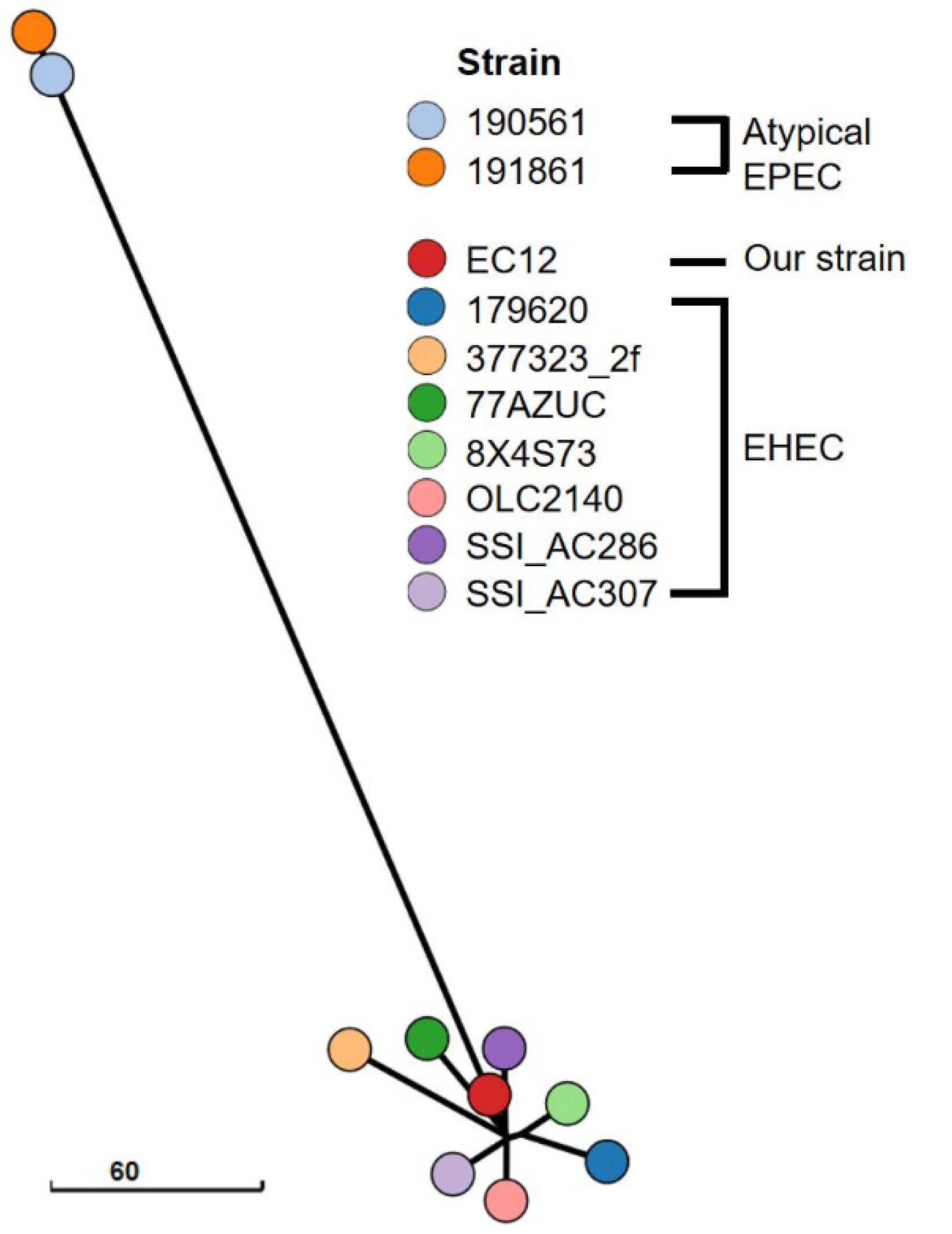
Colorless colonies grew on a sorbitol-MacConkey agar plate, and the isolate (strain EC12) was identified as E. coli O63:H6. Both the stool sample and strain EC12 isolated from the sample tested negative for stx1, stx2, and stx2f but positive for eae. Illumina HiSeq X Ten generated a total of 13,227,764 reads, yielding 403× coverage of the genome. Nanopore MinION generated 324,092 reads, with a mean read length of 6922 bases and a read length N50 of 23,635, resulting in 467× coverage of the genome. The genome sequence of strain EC12 was assembled into three circular contigs: the 4,951,794 bp chromosome; a 163,194 bp IncFIB/FII-type plasmid named pEC12_1; and a 58,414 bp IncI2-type plasmid named pEC12_2 (Figure 1). The chromosome was predicted to contain 4752 protein-coding genes, 22 rRNA genes, and 96 tRNA genes. In the pEC12_1, 201 protein-coding genes and one tRNA gene were predicted. In the pEC12_2, 76 protein-coding genes were predicted. Consistent with the PCR results indicating that it was stx-negative but eae-positive, the chromosome of strain EC12 contained the LEE pathogenicity island but lacked stx-harboring prophages. Plasmid pEC12_1 contained the bfp operon. The virulence genes identified in the genome of strain EC12 and other E. coli O63:H6 strains are compared in Table 1. Seven stx-positive, eae-positive strains (377323_2f, 179620, 77AZUC, 8X4S73, SSI_AC286, SSI_AC307, and OLC2140) and two stx-negative, eae-positive, bfp-negative strains (190561 and 191861) were classified as EHEC and atypical EPEC, respectively. Strain EC12 had the virulence gene profile consistent with EHEC, except for stx. In silico MLST showed that strain EC12 and EHEC strains belong to sequence type 583 (ST583), whereas atypical EPEC strains belong to ST122. The minimum spanning tree based on cgMLST results revealed that strain EC12 was closely related to EHEC strains but distinct from atypical EPEC strains (Figure 2). In genome alignment using Mauve and BRIG, strain EC12 exhibited a high nucleotide identity with EHEC strain 377323_2f but displayed a gap of around 42.5 kbp corresponding to the stx-harboring prophage sequence (Figure 3A,B). Furthermore, plasmid pEC12_1 contained the whole sequence of p377323_2f, a plasmid of strain 377323_2f, in Mauve alignment (Figure 3C). The complete genome sequences of strain EC12 have been deposited in GenBank under accession no. CP070229 (chromosome), CP070230 (pEC12_1), and CP070231 (pEC12_2).

4. Discussion

So far, stx2f has been the only stx subtype found in E. coli O63:H6 strains [3,4,5,6]. STEC strains carrying stx2f are generally associated with mild disease, potentially because these strains carry a relatively small number of virulence genes [5]. However, stx2f-positive STEC strains can cause severe disease such as HUS, albeit rarely [6,21]. Although stx2f-positive STEC strains are frequently found in pigeons, STEC serotypes associated with human diseases, such as O63:H6, are not found in pigeons, suggesting that the stx2f-positive STEC strains responsible for human diseases do not originate from the pigeon reservoir [22].
Infections caused by stx2f-positive STEC strains tend to be underreported, as many molecular assays for laboratory diagnosis of STEC infection are not designed to detect stx2f [5,6]. The addition of stx2f primers to existing PCR assays can be useful for accurate diagnosis of STEC infections, particularly in countries where stx2f-carrying STEC strains are frequently encountered.
Of particular note, strain EC12 was positive for eae but negative for stx. Strain EC12 was initially suspected to be an EHEC-LST strain as it was isolated from an HUS patient. Additionally, as the stool sample of this patient, which was collected 7 days after the onset of gastrointestinal symptoms, was confirmed by PCR to be positive for eae but negative for stx, strain EC12 is presumed to have lost stx in vivo during infection rather than during in vitro culture. The loss of stx is a serotype-related phenomenon and has been found in serotypes O26:H11/NM (nonmotile), O103:H2/NM, O145:H28/NM, and O157:H7/NM [10,11,12]. This study provides the first evidence that stx loss can occur in serotype O63:H6.
When using PCR-based assays targeting only stx and eae, EPEC strains isolated from HUS patients are at risk of being misclassified as EHEC-LST. Whole genome comparison is very useful to determine whether stx-negative, eae-positive E. coli strains isolated from HUS patients are EHEC-LST. A recent WGS study reported that stx-negative, eae-positive E. coli O157:H7/NM strains clustered together with EHEC strains but not with EPEC strains in cgMLST [11]. Furthermore, virulence gene analysis using WGS data revealed that these strains shared a similar virulence gene profile with EHEC strains rather than EPEC strains [11]. Another WGS study demonstrated that a stx-negative, eae-positive E. coli O145:H28 strain had the identical cgMLST and non-stx virulence gene profiles with a stx-positive, eae-positive E. coli O145:H28 strain isolated from the same patient [12]. In pairwise genome alignment, the stx-negative, eae-positive strain was shown to lack the 9 kbp region corresponding to the partial sequence of the stx-harboring prophage [12]. In the present study, phylogenetic analysis using cgMLST showed that strain EC12 clustered together with EHEC strains but not with EPEC strains. Furthermore, this strain shared non-stx virulence genes with EHEC strains and exhibited a very high nucleotide identity with EHEC strain 377323_2f except for the stx-harboring prophage sequence. Taken together, our results indicate that strain EC12 is EHEC-LST, not EPEC. Interestingly, strain EC12 and EHEC strains were nleB-negative and nleC-positive, whereas EPEC strains were nleB-positive and nleC-negative. This finding suggests that, in serotype O63:H6, nleB and nleC may be good molecular markers for differentiating EHEC-LST from EPEC.
In summary, we isolated a stx-negative, eae-positive E. coli O63:H6 strain from a child with HUS. Comparative genomic analysis using WGS data revealed that this strain is genetically more closely related to EHEC than to EPEC, consistent with it being an EHEC-LST strain. Of diagnostic interest, EHEC-LST may be misdiagnosed as EPEC in routine laboratories, and comparative genomic analysis using WGS data can be useful to determine if stx-negative and eae-positive isolates are EHEC-LST or EPEC.

Supplementary Materials

The following are available online at https://0-www-mdpi-com.brum.beds.ac.uk/article/10.3390/diagnostics11101823/s1, Supplementary Table S1: Primers used for detection of stx1, stx2, stx2f, and eae.

Author Contributions

Conceptualization, H.J.H. and S.-W.L.; methodology, S.A.Y. and T.K.; software, T.Y.K. and T.-M.L.; validation, T.Y.K., T.-M.L., H.J.H., S.-W.L. and N.Y.L.; investigation, S.A.Y., T.K., T.Y.K. and T.-M.L.; resources, H.J.H., S.-W.L. and N.Y.L.; data curation, T.Y.K., T.-M.L., H.J.H. and S.-W.L.; writing—original draft preparation, T.Y.K. and T.-M.L.; writing—review and editing, H.J.H., S.-W.L. and N.Y.L. All authors have read and agreed to the published version of the manuscript.

Funding

This research received no external funding.

Institutional Review Board Statement

The study was conducted in accordance with the guidelines of the Declaration of Helsinki and approved by the Institutional Review Board of Samsung Medical Center, Seoul, Korea on 27 February 2021 (IRB no. SMC 2021-02-083).

Informed Consent Statement

Patient consent was waived due to the retrospective nature of the study.

Data Availability Statement

The data presented in this study are available on request from the corresponding author.

Conflicts of Interest

The authors declare no conflict of interest.

References

  1. Valilis, E.; Ramsey, A.; Sidiq, S.; DuPont, H.L. Non-O157 Shiga toxin-producing Escherichia coli—A poorly appreciated enteric pathogen: Systematic review. Int. J. Infect. Dis. IJID Off. Publ. Int. Soc. Infect. Dis. 2018, 76, 82–87. [Google Scholar] [CrossRef] [PubMed] [Green Version]
  2. Brooks, J.T.; Sowers, E.G.; Wells, J.G.; Greene, K.D.; Griffin, P.M.; Hoekstra, R.M.; Strockbine, N.A. Non-O157 Shiga toxin-producing Escherichia coli infections in the United States, 1983–2002. J. Infect. Dis. 2005, 192, 1422–1429. [Google Scholar] [CrossRef] [PubMed] [Green Version]
  3. Prager, R.; Fruth, A.; Siewert, U.; Strutz, U.; Tschäpe, H. Escherichia coli encoding Shiga toxin 2f as an emerging human pathogen. Int. J. Med. Microbiol. IJMM 2009, 299, 343–353. [Google Scholar] [CrossRef] [PubMed]
  4. Buvens, G.; De Gheldre, Y.; Dediste, A.; de Moreau, A.I.; Mascart, G.; Simon, A.; Allemeersch, D.; Scheutz, F.; Lauwers, S.; Piérard, D. Incidence and virulence determinants of verocytotoxin-producing Escherichia coli infections in the Brussels-Capital Region, Belgium, in 2008–2010. J. Clin. Microbiol. 2012, 50, 1336–1345. [Google Scholar] [CrossRef] [PubMed] [Green Version]
  5. Friesema, I.; van der Zwaluw, K.; Schuurman, T.; Kooistra-Smid, M.; Franz, E.; van Duynhoven, Y.; van Pelt, W. Emergence of Escherichia coli encoding Shiga toxin 2f in human Shiga toxin-producing E. coli (STEC) infections in the Netherlands, January 2008 to December 2011. Eurosurveillance 2014, 19, 26–32. [Google Scholar] [CrossRef] [PubMed] [Green Version]
  6. De Rauw, K.; Jacobs, S.; Piérard, D. Twenty-seven years of screening for Shiga toxin-producing Escherichia coli in a university hospital. Brussels, Belgium, 1987–2014. PLoS ONE 2018, 13, e0199968. [Google Scholar] [CrossRef] [PubMed]
  7. Kaper, J.B.; Nataro, J.P.; Mobley, H.L. Pathogenic Escherichia coli. Nat. Rev. Microbiol. 2004, 2, 123–140. [Google Scholar] [CrossRef]
  8. Hartland, E.L.; Leong, J.M. Enteropathogenic and enterohemorrhagic E. coli: Ecology, pathogenesis, and evolution. Front. Cell. Infect. Microbiol. 2013, 3, 15. [Google Scholar] [CrossRef] [PubMed] [Green Version]
  9. Ochoa, T.J.; Contreras, C.A. Enteropathogenic E. coli infection (EPEC) in children. Curr. Opin. Infect. Dis. 2011, 24, 478–483. [Google Scholar] [CrossRef] [Green Version]
  10. Bielaszewska, M.; Köck, R.; Friedrich, A.W.; von Eiff, C.; Zimmerhackl, L.B.; Karch, H.; Mellmann, A. Shiga toxin-mediated hemolytic uremic syndrome: Time to change the diagnostic paradigm? PLoS ONE 2007, 2, e1024. [Google Scholar] [CrossRef]
  11. Ferdous, M.; Zhou, K.; Mellmann, A.; Morabito, S.; Croughs, P.D.; de Boer, R.F.; Kooistra-Smid, A.M.; Rossen, J.W.; Friedrich, A.W. Is Shiga toxin-negative Escherichia coli O157:H7 enteropathogenic or enterohemorrhagic Escherichia coli? Comprehensive molecular analysis using whole-genome sequencing. J. Clin. Microbiol. 2015, 53, 3530–3538. [Google Scholar] [CrossRef] [PubMed] [Green Version]
  12. Senthakumaran, T.; Brandal, L.T.; Lindstedt, B.A.; Jørgensen, S.B.; Charnock, C.; Tunsjø, H.S. Implications of stx loss for clinical diagnostics of Shiga toxin-producing Escherichia coli. Eur. J. Clin. Microbiol. Infect. Dis. Off. Publ. Eur. Soc. Clin. Microbiol. 2018, 37, 2361–2370. [Google Scholar] [CrossRef] [PubMed] [Green Version]
  13. Wick, R.R.; Judd, L.M.; Gorrie, C.L.; Holt, K.E. Unicycler: Resolving bacterial genome assemblies from short and long sequencing reads. PLoS Comput. Biol. 2017, 13, e1005595. [Google Scholar] [CrossRef] [PubMed] [Green Version]
  14. Kearse, M.; Moir, R.; Wilson, A.; Stones-Havas, S.; Cheung, M.; Sturrock, S.; Buxton, S.; Cooper, A.; Markowitz, S.; Duran, C.; et al. Geneious Basic: An integrated and extendable desktop software platform for the organization and analysis of sequence data. Bioinformatics 2012, 28, 1647–1649. [Google Scholar] [CrossRef] [PubMed]
  15. Joensen, K.G.; Scheutz, F.; Lund, O.; Hasman, H.; Kaas, R.S.; Nielsen, E.M.; Aarestrup, F.M. Real-time whole-genome sequencing for routine typing, surveillance, and outbreak detection of verotoxigenic Escherichia coli. J. Clin. Microbiol. 2014, 52, 1501–1510. [Google Scholar] [CrossRef] [Green Version]
  16. Larsen, M.V.; Cosentino, S.; Rasmussen, S.; Friis, C.; Hasman, H.; Marvig, R.L.; Jelsbak, L.; Sicheritz-Pontén, T.; Ussery, D.W.; Aarestrup, F.M.; et al. Multilocus sequence typing of total-genome-sequenced bacteria. J. Clin. Microbiol. 2012, 50, 1355–1361. [Google Scholar] [CrossRef] [Green Version]
  17. Zhou, Z.; Alikhan, N.F.; Mohamed, K.; Fan, Y.; Achtman, M. The EnteroBase user’s guide, with case studies on Salmonella transmissions, Yersinia pestis phylogeny, and Escherichia core genomic diversity. Genome Res. 2020, 30, 138–152. [Google Scholar] [CrossRef] [Green Version]
  18. Zhou, Z.; Alikhan, N.F.; Sergeant, M.J.; Luhmann, N.; Vaz, C.; Francisco, A.P.; Carriço, J.A.; Achtman, M. GrapeTree: Visualization of core genomic relationships among 100,000 bacterial pathogens. Genome Res. 2018, 28, 1395–1404. [Google Scholar] [CrossRef] [Green Version]
  19. Darling, A.C.; Mau, B.; Blattner, F.R.; Perna, N.T. Mauve: Multiple alignment of conserved genomic sequence with rearrangements. Genome Res. 2004, 14, 1394–1403. [Google Scholar] [CrossRef] [Green Version]
  20. Alikhan, N.F.; Petty, N.K.; Ben Zakour, N.L.; Beatson, S.A. BLAST Ring Image Generator (BRIG): Simple prokaryote genome comparisons. BMC Genom. 2011, 12, 402. [Google Scholar] [CrossRef] [Green Version]
  21. Friesema, I.H.; Keijzer-Veen, M.G.; Koppejan, M.; Schipper, H.S.; van Griethuysen, A.J.; Heck, M.E.; van Pelt, W. Hemolytic uremic syndrome associated with Escherichia coli O8:H19 and Shiga toxin 2f gene. Emerg. Infect. Dis. 2015, 21, 168–169. [Google Scholar] [CrossRef] [PubMed]
  22. van Hoek, A.; van Veldhuizen, J.N.J.; Friesema, I.; Coipan, C.; Rossen, J.W.A.; Bergval, I.L.; Franz, E. Comparative genomics reveals a lack of evidence for pigeons as a main source of stx2f-carrying Escherichia coli causing disease in humans and the common existence of hybrid Shiga toxin-producing and enteropathogenic E. coli pathotypes. BMC Genom. 2019, 20, 271. [Google Scholar] [CrossRef] [PubMed] [Green Version]
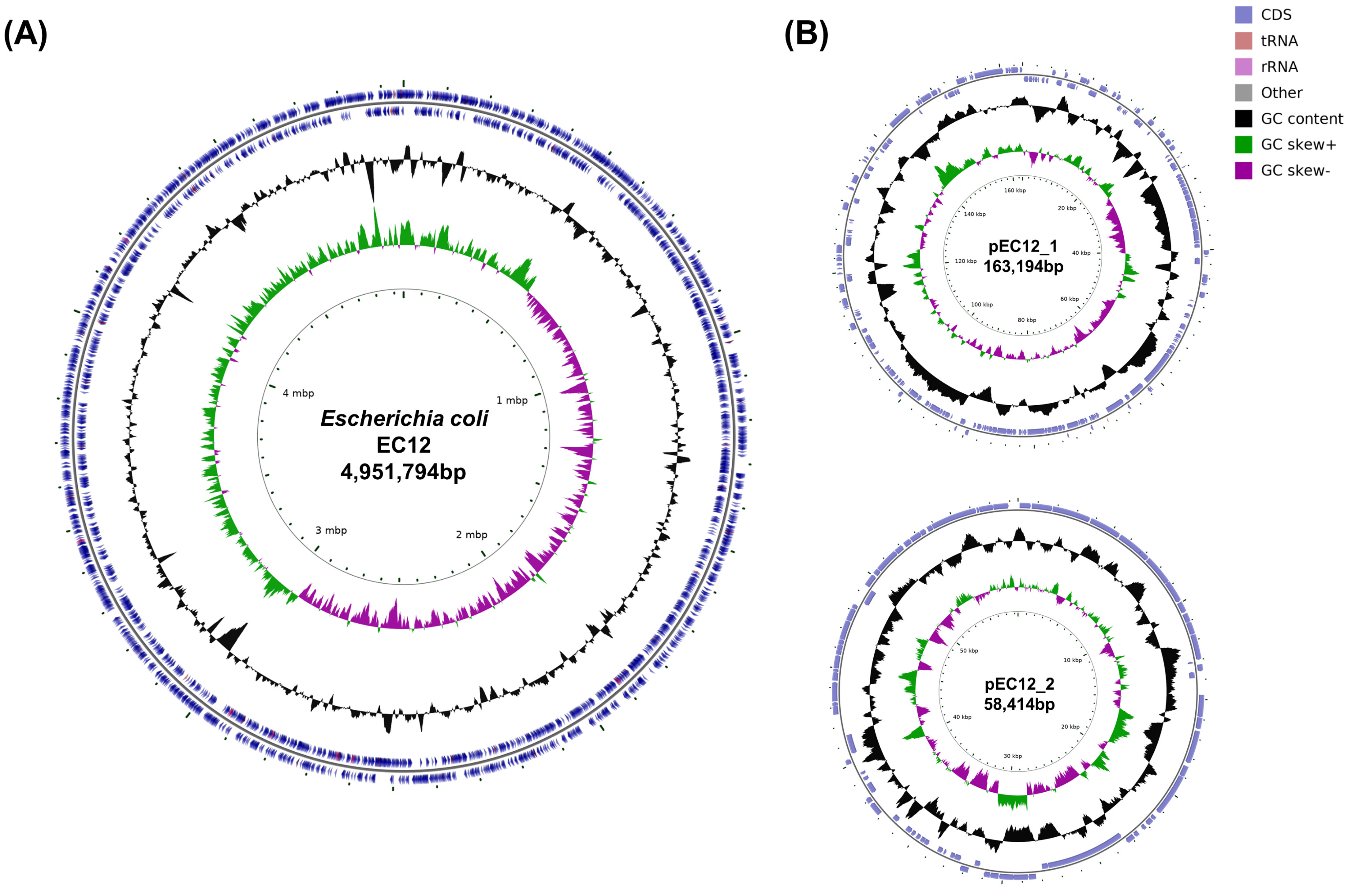
Figure 1. Circular maps of the chromosome (A) and two plasmids (B) of strain EC12. From the outer circle moving inward, circles 1 and 2 represent protein-coding genes (blue), tRNA genes (reddish brown), and rRNA genes (light purple) on the positive and negative strands; circle 3 shows the GC content (black); circle 4 shows the GC skew (positive GC skew, green; negative GC skew, purple); and circle 5 displays the genomic coordinates.
Diagnostics 11 01823 g001
Figure 2. Minimum spanning tree based on cgMLST profiles of 10 E. coli O63:H6 strains included in this study. The branch lengths reflect the number of allelic differences between strains.
Diagnostics 11 01823 g002
Figure 3. Genome comparison of strains EC12 and 377323_2f. (A,B) Chromosome comparisons using Mauve (A) and BRIG (B) demonstrate a gap of around 42.5 kbp in EC12 corresponding to the stx-harboring prophage sequence. (C) Plasmid alignment using Mauve shows that pEC12_1 contains the whole sequence of p377323_2f. In Mauve, identically colored boxes represent homologous regions shared by the aligned genomes.
Diagnostics 11 01823 g003
Table 1. Distribution of virulence genes identified in the genomes of E. coli O63:H6 strains.
StrainastAbfpAchuAcifeaeespAespCespFespJgadibeAnleBnleCompTstxtccPterCtiryfcV
EC12+++++++++++++++++
377323_2f++++++++++++++ *++++
179620++++++++++++++ *+++
77AZUC+++++++++++++ *+++
8X4S73++++++++++++++ *++++
SSI_AC286++++++++++++++ *+++
SSI_AC307+++++++++++++ *+++
OLC2140+++++++++++ *++++
190561++++++++++++++
191861++++++++++++++
* The stx subtype was stx2f.
Publisher’s Note: MDPI stays neutral with regard to jurisdictional claims in published maps and institutional affiliations.