
A Rare Case of Duodenal Pseudomelanosis
by
 , ,
, ,
Marianna D’Ercole
1,2,† ,
,
Gianluca Lopez
1,2,*,†,
Luca Elli
3,
Stefano Ferrero
1,4 and
Giorgio Alberto Croci
1,5 1
Pathology Unit, Fondazione IRCCS Ca’ Granda Ospedale Maggiore Policlinico, 20122 Milan, Italy
2
School of Pathology, University of Milan, 20122 Milan, Italy
3
Gastroenterology Unit, Fondazione IRCCS Ca’ Granda Ospedale Maggiore Policlinico, 20122 Milan, Italy
4
Department of Biomedical, Surgical and Dental Sciences, University of Milan, 20122 Milan, Italy
5
Department of Pathophysiology and Transplantation, University of Milan, 20122 Milan, Italy
*
Author to whom correspondence should be addressed.
†
Equally contributed.
Diagnostics 2021, 11(11), 2152; https://0-doi-org.brum.beds.ac.uk/10.3390/diagnostics11112152
Submission received: 15 November 2021
/
Revised: 17 November 2021
/
Accepted: 18 November 2021
/
Published: 20 November 2021
(This article belongs to the Section Medical Imaging and Theranostics)
Abstract
:A black-spotted duodenal mucosa was observed during endoscopy of a man with several comorbidities including hypertension and end-stage kidney disease. Histopathological examination revealed pigment-laden macrophages in the lamina propria of the duodenal villi, which was consistent with duodenal pseudomelanosis.
Figure 1.
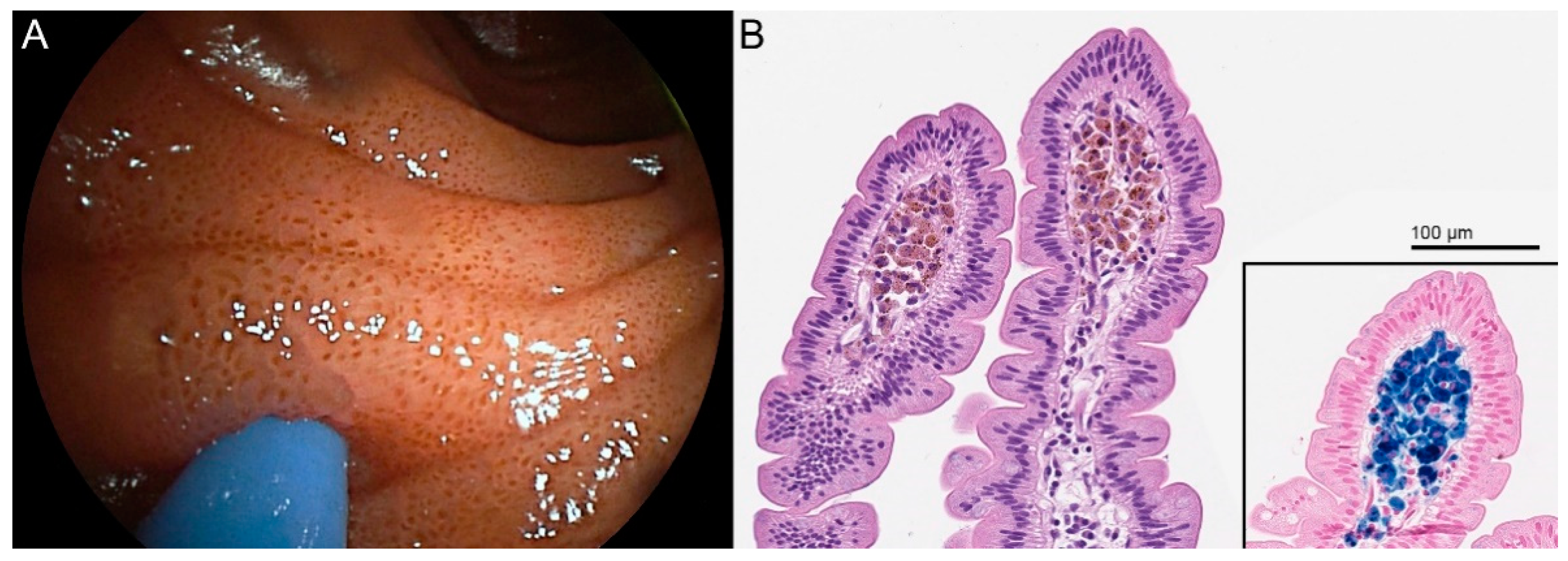
A 75-year-old man underwent endoscopy for obstructive lithiasic cholangitis. His medical history included gastric resection for non-Hodgkin lymphoma, monoclonal gammopathy of undetermined significance (MGUS), HCV infection, hypertension, and stage 4 chronic kidney disease. His medications included furosemide, metoprolol, and amlodipine. During the endoscopy, the duodenal mucosa presented spotted black pigmentation at the tip of the villi (A). Duodenal biopsy samples stained with routine hematoxylin and eosin (B) showed aggregates of pigment-laden macrophages in the lamina propria of the apical portion of the villi, which tested intensely positive with Perl’s stain for iron; enterocytes demonstrated a faint positivity for Perl’s Prussian blue underneath the microvilli (B, inset). These findings were consistent for duodenal pseudomelanosis, a benign condition which harbors no known clinical sequelae [1,2,3,4,5,6,7,8,9,10,11,12,13].
Figure 1.
A 75-year-old man underwent endoscopy for obstructive lithiasic cholangitis. His medical history included gastric resection for non-Hodgkin lymphoma, monoclonal gammopathy of undetermined significance (MGUS), HCV infection, hypertension, and stage 4 chronic kidney disease. His medications included furosemide, metoprolol, and amlodipine. During the endoscopy, the duodenal mucosa presented spotted black pigmentation at the tip of the villi (A). Duodenal biopsy samples stained with routine hematoxylin and eosin (B) showed aggregates of pigment-laden macrophages in the lamina propria of the apical portion of the villi, which tested intensely positive with Perl’s stain for iron; enterocytes demonstrated a faint positivity for Perl’s Prussian blue underneath the microvilli (B, inset). These findings were consistent for duodenal pseudomelanosis, a benign condition which harbors no known clinical sequelae [1,2,3,4,5,6,7,8,9,10,11,12,13].

Author Contributions
M.D. and G.L., histopathologic description, writing—original draft; G.L. and L.E., visualization; L.E. performed endoscopy; G.A.C. and S.F., supervision, writing—review and editing. All authors have read and agreed to the published version of the manuscript.
Funding
This research received no external funding.
Institutional Review Board Statement
Not applicable.
Informed Consent Statement
Written informed consent has been obtained from the patient’s next of kin to publish this paper.
Data Availability Statement
Not applicable.
Conflicts of Interest
The authors declare no conflict of interest.
References
- Giusto, D.; Jakate, S. Pseudomelanosis duodeni: Associated with multiple clinical conditions and unpredictable iron stainability—A case series. Endoscopy 2008, 40, 165–167. [Google Scholar] [CrossRef] [PubMed]
- de Magalhães Costa, M.H.D.M.; Pegado, M.G.F.; Vargas, C.; Castro, M.E.C.; Madi, K.; Nunes, T.; Zaltman, C. Pseudomelanosis duodeni associated with chronic renal failure. World J. Gastroenterol. 2012, 18, 1414–1416. [Google Scholar] [CrossRef] [PubMed]
- Kim, S.Y.; Choung, R.S.; Kwon, B.S.; Hyun, J.J.; Jung, S.W.; Koo, J.S.; Yim, H.J.; Lee, S.W.; Choi, J.H. Small Bowel Pseudomelanosis Associated with Oral Iron Therapy. J. Korean Med. Sci. 2013, 28, 1103–1106. [Google Scholar] [CrossRef] [PubMed]
- Nakanishi, Y.; Jetly-Shridhar, R.; De Felice, K. A Case of Pseudomelanosis Duodeni: Striking Endoscopic Features with Subtle but Characteristic Pathologic Findings. Int. J. Surg. Pathol. 2019, 27, 765–766. [Google Scholar] [CrossRef] [PubMed]
- Tang, S.-J.; Zhang, S.; Grunes, D.E. Gastric and duodenal pseudomelanosis: A new insight into its pathogenesis. VideoGIE 2019, 4, 467–468. [Google Scholar] [CrossRef] [PubMed] [Green Version]
- Shimamura, Y.; Akram, H.; Winer, S.; Marcon, N. Pseudomelanosis Duodeni and Duodenal Polyp. Intern. Med. 2018, 57, 1049–1050. [Google Scholar] [CrossRef] [PubMed] [Green Version]
- Mundi, I.; Pankaj, R.; Chhabra, M.; Banerjee, A.K. Pseudomelanosis Duodeni. Int. J. Surg. Pathol. 2017, 25, 165. [Google Scholar] [CrossRef] [PubMed]
- Abdelwareth, A.; Molyneux, A.; Madhotra, R.; Ishaq, S.; Rostami, K. Small bowel pigmentation. Gastroenterol. Hepatol. Bed Bench 2016, 9, 343–344. [Google Scholar] [PubMed]
- Coelho, R.; Ribeiro, A.; Silva, R.; Rios, E.; Silva, M.; Macedo, G. Pseudomelanosis duodeni: Is there a common denominator? Rev. Española Enferm. Dig. 2016, 108, 658–659. [Google Scholar]
- Kothadia, J.P.; Kaminski, M.; Giashuddin, S. Duodenal siderosis: A rare clinical finding in a patient with duodenal inflammation. Ann. Gastroenterol. 2016, 29, 379. [Google Scholar] [PubMed]
- Sathyamurthy, A.; Chela, H.; Arif, Z.; Holly, J.; Arif, M. Pseudomelanosis Duodeni. ACG Case Rep. J. 2015, 2, 72–73. [Google Scholar] [CrossRef] [PubMed]
- Siderits, R.; Hazra, A.; Mikhail, N.; Chiaffarano, J.; Lou, W.; Fyfe, B. Endoscopically identified pseudomelanosis duodeni: Striking yet harmless. Gastrointest. Endosc. 2014, 80, 508–510. [Google Scholar] [CrossRef] [PubMed]
- Qureshi, N.U.; Younus, M.F.; Alavi, K.; Sheikh, M.Y. Gastric and Duodenal Pseudomelanosis: An Extended Unusual Finding in a Patient with End Stage Kidney Disease. Case Rep. Gastrointest. Med. 2016, 2016, 2861086. [Google Scholar] [CrossRef] [PubMed] [Green Version]
Publisher’s Note: MDPI stays neutral with regard to jurisdictional claims in published maps and institutional affiliations. |
© 2021 by the authors. Licensee MDPI, Basel, Switzerland. This article is an open access article distributed under the terms and conditions of the Creative Commons Attribution (CC BY) license (https://creativecommons.org/licenses/by/4.0/).
Share and Cite
MDPI and ACS Style
D’Ercole, M.; Lopez, G.; Elli, L.; Ferrero, S.; Croci, G.A. A Rare Case of Duodenal Pseudomelanosis. Diagnostics 2021, 11, 2152. https://0-doi-org.brum.beds.ac.uk/10.3390/diagnostics11112152
AMA Style
D’Ercole M, Lopez G, Elli L, Ferrero S, Croci GA. A Rare Case of Duodenal Pseudomelanosis. Diagnostics. 2021; 11(11):2152. https://0-doi-org.brum.beds.ac.uk/10.3390/diagnostics11112152
Chicago/Turabian StyleD’Ercole, Marianna, Gianluca Lopez, Luca Elli, Stefano Ferrero, and Giorgio Alberto Croci. 2021. "A Rare Case of Duodenal Pseudomelanosis" Diagnostics 11, no. 11: 2152. https://0-doi-org.brum.beds.ac.uk/10.3390/diagnostics11112152
Note that from the first issue of 2016, this journal uses article numbers instead of page numbers. See further details here.