
HHV8-Positive Castleman Disease and In Situ Mantle Cell Neoplasia within Dermatopathic Lymphadenitis, in Longstanding Psoriasis

 , , , and
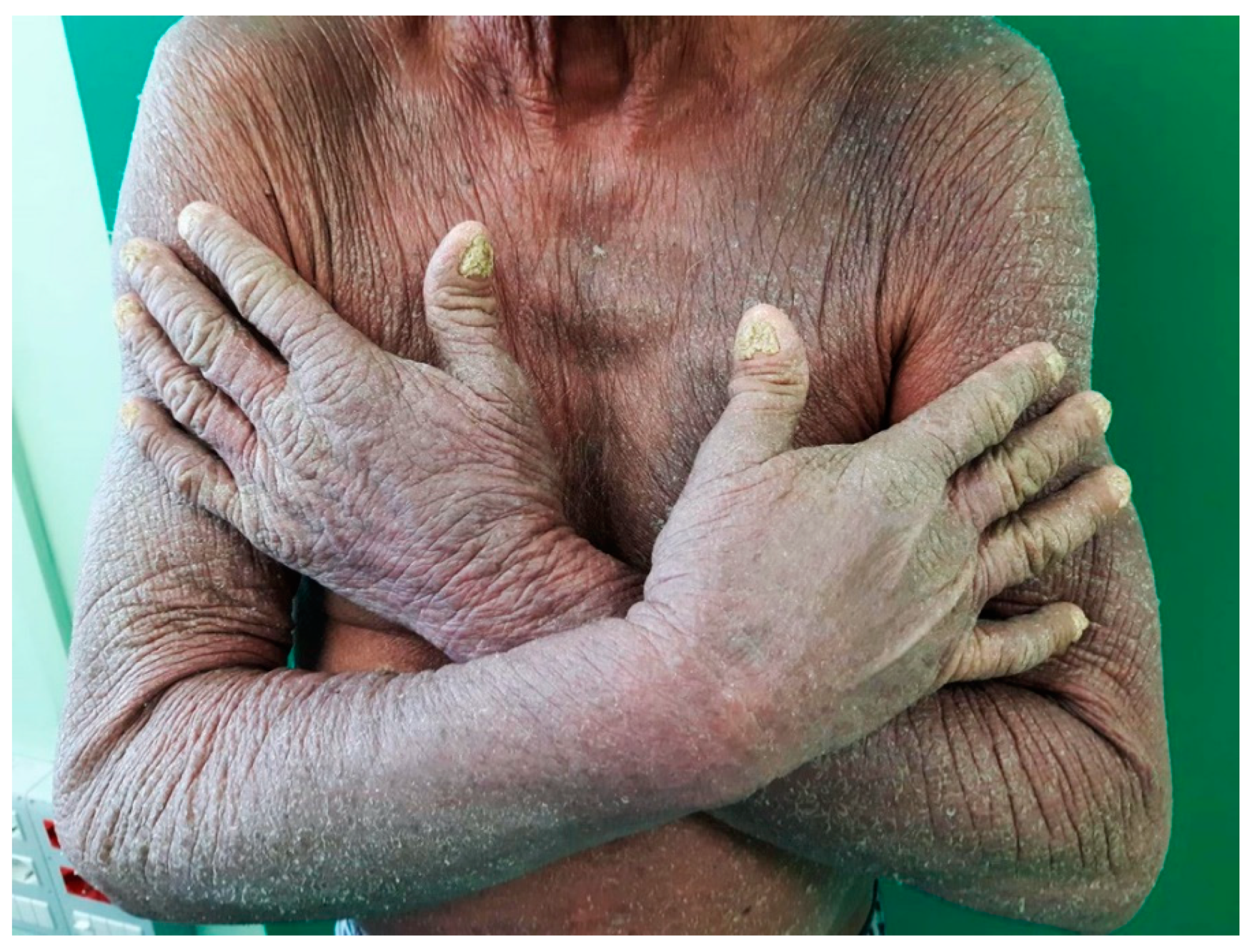
Abstract
:Author Contributions
Funding
Institutional Review Board Statement
Informed Consent Statement
Data Availability Statement
Conflicts of Interest
References
- Said, J.; Isaacson, P.G.; Campo, E.; Harris, N.L. WHO Classification of Tumours of Haematopoietic and Lymphoid Tissues, 4th ed.; WHO Classification of Tumours Editorial Board, Ed.; IARC: Lyon, France, 2017; pp. 325–327. [Google Scholar]
- Zanelli, M.; Sanguedolce, F.; Zizzo, M.; Palicelli, A.; Bassi, M.C.; Santandrea, G.; Martino, G.; Soriano, A.; Caprera, C.; Corsi, M.; et al. Primary effusion lymphoma occurring in the setting of transplanted patients: A systematic review of a rare, life-threatening post-transplantation occurrence. BMC Cancer 2021, 21, 468. [Google Scholar] [CrossRef] [PubMed]
- Zanelli, M.; Zizzo, M.; Bisagni, A.; Froio, E.; De Marco, L.; Valli, R.; Filosa, A.; Luminari, S.; Martino, G.; Massaro, F.; et al. Germinotropic lymphoproliferative disorder: A systematic review. Ann. Hematol. 2020, 99, 2243–2253. [Google Scholar] [CrossRef] [PubMed]
- Zanelli, M.; Fraternali Orcioni, G.; Zizzo, M.; De Marco, L.; Martino, G.; Cerrone, G.; Cabras, A.D.; Ascani, S. HHV-8-and EBV-positive germinotropic lymphoproliferative disorder. Ann. Hematol. 2019, 98, 2439–2441. [Google Scholar] [CrossRef] [PubMed]
- Zanelli, M.; Bertuzzi, C.; Zizzo, M.; Martino, G.; Sabattini, E.; Ascani, S. Extracavitary primary effusion lymphoma in a post-transplantation patient. Br. J. Hematol. 2019, 187, 555. [Google Scholar] [CrossRef] [PubMed]
- Mohagheghi, M.A.; Omranipur, R.; Ensani, F.; Ghannadan, A.; Shahriaran, S.; Samiee, F.; Sedighi, Z. A case of advanced unicentric retroperitoneal Castleman’s disease associated with psoriasis. Acta Med. Iran. 2017, 55, 277–279. [Google Scholar] [PubMed]
- Larroche, C.; Cacoub, P.; Soulier, J.; Oksenhendler, E.; Clauvel, J.P.; Piette, J.C.; Raphael, M. Castleman’s disease and lymphoma: Report of eight cases in HIV-negative patients and literature review. Am. J. Hematol. 2002, 69, 119–126. [Google Scholar] [CrossRef] [PubMed]
- Parente, P.; Zanelli, M.; Zizzo, M.; Covelli, C.; Carosi, I.; Ascani, S.; Graziano, P. Primary effusion lymphoma metachronous to multicentric Castleman disease in an immunocompetent patient. Pathol. Res. Pract. 2020, 216, 153024. [Google Scholar] [CrossRef] [PubMed]
- Yoshida, T.; Sakai, A.; Imagawa, J.; Noda, M.; Takada, S.; Yoshino, T.; Kimura, A. Mantle cell lymphoma superimposed on multicentric Castleman’s disease. J. Clin. Exp. Hematopathol. 2011, 51, 147–150. [Google Scholar] [CrossRef] [Green Version]
- Dobrea, C.; Mihai, M.; Danaila, E.; Gaman, A.; Coriu, D.; Ursuleac, I. “In situ” mantle cell lymphoma associated with hyaline-vascular Castleman disease. Rom. J. Morphol. Embryol. 2011, 52, 1147–1151. [Google Scholar]


Publisher’s Note: MDPI stays neutral with regard to jurisdictional claims in published maps and institutional affiliations. |
© 2021 by the authors. Licensee MDPI, Basel, Switzerland. This article is an open access article distributed under the terms and conditions of the Creative Commons Attribution (CC BY) license (https://creativecommons.org/licenses/by/4.0/).
Share and Cite
Zanelli, M.; Stingeni, L.; Zizzo, M.; Martino, G.; Sanguedolce, F.; Marra, A.; Crescenzi, B.; Pileri, S.A.; Ascani, S. HHV8-Positive Castleman Disease and In Situ Mantle Cell Neoplasia within Dermatopathic Lymphadenitis, in Longstanding Psoriasis. Diagnostics 2021, 11, 1150. https://0-doi-org.brum.beds.ac.uk/10.3390/diagnostics11071150
Zanelli M, Stingeni L, Zizzo M, Martino G, Sanguedolce F, Marra A, Crescenzi B, Pileri SA, Ascani S. HHV8-Positive Castleman Disease and In Situ Mantle Cell Neoplasia within Dermatopathic Lymphadenitis, in Longstanding Psoriasis. Diagnostics. 2021; 11(7):1150. https://0-doi-org.brum.beds.ac.uk/10.3390/diagnostics11071150
Chicago/Turabian StyleZanelli, Magda, Luca Stingeni, Maurizio Zizzo, Giovanni Martino, Francesca Sanguedolce, Andrea Marra, Barbara Crescenzi, Stefano A. Pileri, and Stefano Ascani. 2021. "HHV8-Positive Castleman Disease and In Situ Mantle Cell Neoplasia within Dermatopathic Lymphadenitis, in Longstanding Psoriasis" Diagnostics 11, no. 7: 1150. https://0-doi-org.brum.beds.ac.uk/10.3390/diagnostics11071150