
Cerebral Infarction and Evan’s Ratio on MRI Affect the Severity and Prognosis of Tuberculosis Meningitis Patients


Abstract
:1. Introduction
2. Materials and Methods
2.1. Study Design and Participants
2.2. Evaluation
2.3. Investigations
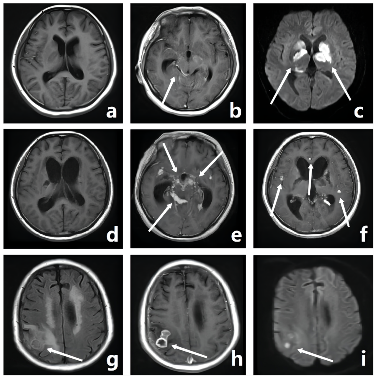
2.4. Image Analysis
2.5. Statistical Analysis
3. Results
3.1. Patients’ Clinical Characteristics
3.2. Relationship between MRI Features and Neurological Deficits
3.3. Relationship between the MRI Features and Prognosis
3.4. Relationship between the Dynamic Changes in MRI Features and Prognosis
4. Discussion
5. Conclusions
Author Contributions
Funding
Institutional Review Board Statement
Informed Consent Statement
Data Availability Statement
Conflicts of Interest
References
- Chan, K.; Cheung, R.; Lee, R.; Mak, W.; Ho, S. Cerebral Infarcts Complicating Tuberculous Meningitis. Cerebrovasc. Dis. 2005, 19, 391–395. [Google Scholar] [CrossRef] [PubMed]
- Wasay, M.; Farooq, S.; Khowaja, Z.A.; Bawa, Z.A.; Ali, S.M.; Awan, S.; Beg, M.A.; Mehndiratta, M.M. Cerebral infarction and tuberculoma in central nervous system tuberculosis: Frequency and prognostic implications. J. Neurol. Neurosurg. Psychiatry 2014, 85, 1260–1264. [Google Scholar] [CrossRef] [PubMed]
- Mai, N.T.; Thwaites, G. Recent advances in the diagnosis and management of tuberculous meningitis. Curr. Opin. Infect. Dis. 2017, 30, 123–128. [Google Scholar] [CrossRef] [PubMed]
- Kalita, J.; Misra, U.K.; Nair, P.P. Predictors of Stroke and Its Significance in the Outcome of Tuberculous Meningitis. J. Stroke Cerebrovasc. Dis. 2009, 18, 251–258. [Google Scholar] [CrossRef]
- Artal, F.J.C. Clinical management of infectious cerebral vasculitides. Expert Rev. Neurother. 2016, 16, 205–221. [Google Scholar] [CrossRef]
- Schoeman, J.F.; Van Zyl, L.E.; Laubscher, J.A.; Donald, P.R. Serial CT Scanning in Childhood Tuberculous Meningitis: Prognostic Features in 198 Cases. J. Child Neurol. 1995, 10, 320–329. [Google Scholar] [CrossRef]
- Sheu, J.-J.; Hsu, C.-Y.; Yuan, R.-Y.; Yang, C.-C. Clinical characteristics and treatment delay of cerebral infarction in tuberculous meningitis. Intern. Med. J. 2012, 42, 294–300. [Google Scholar] [CrossRef]
- Zhang, L.; Zhang, X.; Li, H.; Chen, G.; Zhu, M. Acute ischemic stroke in young adults with tuberculous meningitis. BMC Infect. Dis. 2019, 19, 362. [Google Scholar] [CrossRef]
- Misra, U.K.; Kalita, J.; Maurya, P.K. Stroke in tuberculous meningitis. J. Neurol. Sci. 2011, 303, 22–30. [Google Scholar] [CrossRef]
- Wilkinson, R.J.; on behalf of the Tuberculous Meningitis International Research Consortium; Rohlwink, U.; Misra, U.K.; van Crevel, R.; Mai, N.T.H.; Dooley, K.E.; Caws, M.; Figaji, A.; Savic, R.; et al. Tuberculous meningitis. Nat. Rev. Neurol. 2017, 13, 581–598. [Google Scholar] [CrossRef]
- Pienaar, M.; Andronikou, S.; van Toorn, R. MRI to demonstrate diagnostic features and complications of TBM not seen with CT. Child’s Nerv. Syst. 2009, 25, 941–947. [Google Scholar] [CrossRef] [PubMed]
- Kalita, J.; Prasad, S.; Misra, U.K. Predictors of paradoxical tuberculoma in tuberculous meningitis. Int. J. Tuberc. Lung Dis. 2014, 18, 486–491. [Google Scholar] [CrossRef] [PubMed]
- Davis, A.; Meintjes, G.; Wilkinson, R.J. Treatment of Tuberculous Meningitis and Its Complications in Adults. Curr. Treat. Options Neurol. 2018, 20, 5. [Google Scholar] [CrossRef] [Green Version]
- van Rensburg, P.J.; Andronikou, S.; van Toorn, R.; Pienaar, M. Magnetic resonance imaging of miliary tuberculosis of the central nervous system in children with tuberculous meningitis. Pediatr. Radiol. 2008, 38, 1306–1313. [Google Scholar] [CrossRef]
- Anuradha, H.K.; Garg, R.K.; Sinha, M.K.; Agarwal, A.; Verma, R.; Singh, M.K.; Shukla, R. Intracranial tuberculomas in patients with tuberculous meningitis: Predictors and prognostic significance. Int. J. Tuberc. Lung Dis. 2011, 15, 234–239. [Google Scholar] [PubMed]
- Sun, F.; Ruan, Q.; Wang, J.; Chen, S.; Jin, J.; Shao, L.; Zhang, Y.; Zhang, W. Linezolid Manifests a Rapid and Dramatic Therapeutic Effect for Patients with Life-Threatening Tuberculous Meningitis. Antimicrob. Agents Chemother. 2014, 58, 6297–6301. [Google Scholar] [CrossRef] [PubMed] [Green Version]
- Marshall, G.; Blacklock, J.S.; Cameron, C.; Capon, N.B.; Cruickshank, R.; Gaddum, J.H.; Heaf, F.; Hill, A.; Houghton, L.; Hoyle, J.; et al. HSTREPTOMYCIN treatment of tuberculous meningitis. Lancet 1948, 1, 582–596. [Google Scholar]
- Wasay, M.; Khan, M.; Farooq, S.; Khowaja, Z.A.; Bawa, Z.A.; Ali, S.M.; Awan, S.; Beg, M.A. Frequency and Impact of Cerebral Infarctions in Patients with Tuberculous Meningitis. Stroke 2018, 49, 2288–2293. [Google Scholar] [CrossRef] [PubMed]
- Thwaites, G.E.; van Toorn, R.; Schoeman, J. Tuberculous meningitis: More questions, still too few answers. Lancet Neurol. 2013, 12, 999–1010. [Google Scholar] [CrossRef]
- Thwaites, G.E.; Macmullen-Price, J.; Chau, T.T.H.; Mai, P.P.; Dung, N.T.; Simmons, C.P.; White, N.J.; Hien, T.T.; Summers, D.; Farrar, J. Serial MRI to determine the effect of dexamethasone on the cerebral pathology of tuberculous meningitis: An observational study. Lancet Neurol. 2007, 6, 230–236. [Google Scholar] [CrossRef] [Green Version]
- Wen, L.; Li, M.; Xu, T.; Yu, X.; Wang, L.; Li, K. Clinical features, outcomes and prognostic factors of tuberculous meningitis in adults worldwide: Systematic review and meta-analysis. J. Neurol. 2019, 266, 3009–3021. [Google Scholar] [CrossRef] [PubMed]
- Özateş, M.; Kemaloglu, S.; Gürkan, F.; Ozkan, U.; Hoşoglu, S.; Simsek, M.M.; Kemaloǧlu, S. CT of the brain in tuberculous meningitis. A review of 289 patients. Acta Radiol. 2000, 41, 13–17. [Google Scholar] [CrossRef] [PubMed]
- Lammie, G.A.; Hewlett, R.H.; Schoeman, J.F.; Donald, P.R. Tuberculous cerebrovascular disease: A review. J. Infect. 2009, 59, 156–166. [Google Scholar] [CrossRef] [PubMed]
- Thwaites, G.E.; Bang, N.D.; Dung, N.H.; Quy, H.T.; Oanh, D.T.T.; Thoa, N.T.C.; Hien, N.Q.; Thuc, N.T.; Hai, N.N.; Lan, N.T.N.; et al. Dexamethasone for the Treatment of Tuberculous Meningitis in Adolescents and Adults. N. Engl. J. Med. 2004, 351, 1741–1751. [Google Scholar] [CrossRef] [Green Version]
- Mai, N.T.; Dobbs, N.; Phu, N.H.; Colas, R.A.; Thao, L.T.; Thuong, N.T.; Nghia, H.D.; Hanh, N.H.; Hang, N.T.; Heemskerk, A.D.; et al. A randomised double blind placebo controlled phase 2 trial of adjunctive aspirin for tuberculous meningitis in HIV-uninfected adults. eLife 2018, 7, e33478. [Google Scholar] [CrossRef]
- Soni, N.; Kumar, S.; Shimle, A.; Ora, M.; Bathla, G.; Mishra, P. Cerebrovascular complications in tuberculous meningitis—A magnetic resonance imaging study in 90 patients from a tertiary care hospital. Neuroradiol. J. 2020, 33, 3–16. [Google Scholar] [CrossRef]
- Raut, T.; Garg, R.K.; Jain, A.; Verma, R.; Singh, M.K.; Malhotra, H.S.; Kohli, N.; Parihar, A. Hydrocephalus in tuberculous meningitis: Incidence, its predictive factors and impact on the prognosis. J. Infect. 2013, 66, 330–337. [Google Scholar] [CrossRef]
- Brancusi, F.; Farrar, J.; Heemskerk, D. Tuberculous meningitis in adults: A review of a decade of developments focusing on prognostic factors for outcome. Futur. Microbiol. 2012, 7, 1101–1116. [Google Scholar] [CrossRef] [Green Version]
- Wasay, M.; Moolani, M.K.; Zaheer, J.; Kheleani, B.A.; Smego, R.A.; Sarwari, R.A.; Smego, A.R. Prognostic indicators in patients with intracranial tuberculoma: A review of 102 cases. J. Pak. Med Assoc. 2004, 54, 83–87. [Google Scholar]
- Das, M.; Dooley, K.E.; Gupta, A.; Thakur, K. The Global Neurological Burden of Tuberculosis. Skull Base 2018, 38, 226–237. [Google Scholar] [CrossRef]
- Duc, N.M. The role of diffusion tensor imaging metrics in the discrimination between cerebellar medulloblastoma and brainstem glioma. Pediatr. Blood Cancer 2020, 67, e28468. [Google Scholar] [CrossRef] [PubMed]


| T1WI | T2WI | FLAIR | DWI | Enhanced T1WI | Information to Be Recorded | |
|---|---|---|---|---|---|---|
| Meningeal enhancement | / | / | / | / | Linear enhancement along the meninges | Location: suprasellar cistern, ambient cistern, lateral fissure cistern, interhemispheric fissure cistern, tentorium cerebelli, spinal cord membrane, and other meninges |
| Tuberculoma | Nodules with equal or slightly low signal intensity | The liquefaction part of the nodule shows high signal intensity, and the non-liquefied caseous necrosis part shows low signal intensity | The liquefaction part of the nodule center shows low signal intensity, the non-liquefied caseous necrosis part shows equal signal intensity, and the tumor wall shows high signal intensity | Low signal intensity | Uniform or circular enhancement | Location: frontal lobe, parietal lobe, temporal lobe, occipital lobe, corpus callosum, basal ganglia, cerebellum, brain stem, and meninges Is the lesion diffuse? |
| ACI | Low signal intensity | High signal intensity | High signal intensity | High signal intensity | Enhanced lesion area | Location: frontal lobe, parietal lobe, temporal lobe, and occipital lobe Volume of infarcted brain tissue |
| OCI | Low signal intensity | High signal intensity | Low signal intensity | Low signal intensity | No enhancement in lesion area | Location: frontal lobe, parietal lobe, temporal lobe, and occipital lobe |
| Abscess | The abscess cavity shows low signal intensity, and the abscess wall shows equal signal intensity | The abscess cavity shows high signal intensity, and the abscess wall shows slightly high signal intensity | The abscess cavity shows slightly low signal intensity, and the abscess wall shows slightly high signal intensity | The abscess cavity shows high signal intensity | The abscess wall shows obvious circular enhancement | Does the patient have a brain abscess? |
| Hydrocephalus | Enlargement of the ventricles without widened sulci | Does the patient have hydrocephalus? | ||||
| Lesion size | Including the scope of meningeal enhancement and the size of tuberculoma and abscess | Increase in size was recorded as “1”, unchanged as “0”, and decrease as “−1” | ||||
| Quantity of lesion(s) | Including the number of tuberculoma and abscess | Increase in quantity was recorded as “1”, unchanged as “0”, and decrease as “−1” | ||||
| Severity of hydrocephalus | Reflected by Evan’s ratio | Increase in the Evan’s ratio was recorded as “1”, unchanged as “0”, and decrease as “−1” | ||||
| TBM Diagnosis (n, %) | N = 110 |
|---|---|
| Confirmed | 33 (30.00%) |
| Highly probable | 55 (50.00%) |
| Probable | 22 (20.00%) |
| Age (yrs) | 43.17 ± 16.90 |
| Gender (n, %) | |
| Male | 70 (63.64%) |
| Female | 40 (36.36%) |
| Clinical symptoms(n, %) | |
| Headache | 18 (16.36%) |
| Fever | 39 (35.45%) |
| Focal neurological signs | 35 (31.82%) |
| Altered consciousness | 46 (41.82%) |
| CSF results on admission | |
| Glucose, mmol/L | 0.28 ± 0.10 |
| Protein, mg/L | 2061.00 (1260.00, 3519.00) |
| WBC,×106/L | 120.00 (40.00, 316.00) |
| MRC grading (n, %) N = 109 | |
| I | 31 (28.44%) |
| II | 59 (54.13%) |
| III | 19 (17.43%) |
| Outcome/MRS (n, %) N = 108 | |
| 0 | 59 (54.63%) |
| 1 | 25 (23.15%) |
| 2 | 7 (6.48%) |
| 3 | 6 (5.56%) |
| 4 | 4 (3.70%) |
| 6 | 7 (6.48%) |
| MRC I (n = 31) | MRC II (n = 59) | MRC III (n = 19) | p Value | |
|---|---|---|---|---|
| Meningeal enhancement | 18 (58.06) | 39 (66.10) | 14 (73.68) | 0.52 |
| suprasellar cistern | 8 (25.81) | 16 (27.12) | 11 (57.89) | 0.03 |
| ambient cistern | 14 (45.16) | 25 (42.37) | 10 (52.63) | 0.74 |
| lateral fissure cistern | 7 (22.58) | 19 (32.20) | 10 (52.63) | 0.09 |
| interhemispheric fissure cistern | 3 (9.68) | 15 (25.42) | 5 (26.32) | 0.81 |
| tentorium cerebelli | 5 (16.13) | 17 (28.81) | 7 (36.84) | 0.23 |
| spinal cord membrane | 10 (32.26) | 15 (25.42) | 10 (52.63) | 0.09 |
| other meninges | 5 (16.13) | 20 (33.90) | 9 (47.37) | 0.06 |
| Tuberculoma | 13 (41.94) | 22 (37.29) | 12 (63.16) | 0.14 |
| brain parenchymal tuberculoma | 11 (35.48) | 17 (28.81) | 11 (57.89) | 0.07 |
| frontal lobe | 7 (22.58) | 12 (20.34) | 8 (42.11) | 0.15 |
| parietal lobe | 6 (19.35) | 9 (15.25) | 7 (36.84) | 0.12 |
| temporal lobe | 5 (16.13) | 12 (20.34) | 8 (42.11) | 0.08 |
| occipital lobe | 2 (6.45) | 10 (16.95) | 5 (26.32) | 0.16 |
| corpus callosum | 1 (3.23) | 3 (5.08) | 4 (21.05) | 0.04 |
| basal ganglia | 3 (9.68) | 7 (11.86) | 6 (31.58) | 0.07 |
| cerebellum | 8 (25.81) | 12 (20.34) | 6 (31.58) | 0.58 |
| brain stem | 5 (16.13) | 8 (13.56) | 6 (31.58) | 0.19 |
| meningeal tuberculoma | 8 (25.81) | 14 (23.73) | 4 (21.05) | 0.93 |
| diffuse tuberculoma | 3 (9.68) | 9 (15.25) | 5 (26.32) | 0.29 |
| maximum diameter | 6.30 | 5.35 | 4.80 | 0.38 |
| ACI | 1 (3.23) | 14 (23.73) | 8 (42.11) | 0.00 |
| frontal lobe | 1 (3.23) | 5 (8.47) | 6 (31.58) | 0.01 |
| parietal lobe | 1 (3.23) | 4 (6.78) | 4 (21.05) | 0.07 |
| temporal lobe | 0 (0.00) | 2 (3.39) | 5 (26.32) | 0.00 |
| volume (cm3) | 0.65 | 0.54 | 1.35 | 0.140 |
| OCI | 3 (9.68) | 7 (11.86) | 8 (42.11) | 0.00 |
| frontal lobe | 2 (6.45) | 3 (5.08) | 6 (31.58) | 0.00 |
| parietal lobe | 1 (3.23) | 3 (5.08) | 6 (31.58) | 0.00 |
| temporal lobe | 1 (3.23) | 1 (1.69) | 5 (26.32) | 0.00 |
| Abscess | 2 (6.45) | 3 (5.08) | 2 (10.53) | 0.70 |
| Hydrocephalus | 10 (32.26) | 19 (32.20) | 9 (47.37) | 0.45 |
| Evan’s ratio | 0.26 | 0.27 | 0.29 | 0.00 |
| Rankin 0 (n = 59) | Rankin I (n = 32) | Rankin II (n = 10) | Rankin III (n = 7) | p Value | |
|---|---|---|---|---|---|
| Meningeal enhancement | 37 (62.71) | 21 (65.63) | 7 (70.00) | 5 (71.43) | 0.95 |
| Tuberculoma | 25 (42.37) | 13 (40.63) | 6 (60.00) | 2 (28.57) | 0.60 |
| meningeal tuberculoma | 17 (28.81) | 5 (15.63) | 2 (20.00) | 1 (14.29) | 0.48 |
| brain parenchymal tuberculoma | 19 (32.20) | 11 (34.38) | 5 (50.00) | 2 (28.57) | 0.73 |
| diffuse tuberculoma | 7 (11.86) | 5 (15.63) | 3 (30.00) | 0 (0.00) | 0.31 |
| maximum diameter | 6.10 | 4.50 | 5.50 | 7.65 | 0.41 |
| ACI | 8 (13.56) | 6 (18.75) | 5 (50.00) | 3 (42.86) | 0.03 |
| frontal lobe | 2 (3.39) | 3 (9.38) | 3 (30.00) | 3 (42.86) | 0.00 |
| parietal lobe | 1 (1.69) | 3 (9.38) | 2 (20.00) | 3 (42.86) | 0.00 |
| temporal lobe | 1 (1.69) | 1 (3.13) | 4 (40.00) | 1 (14.29) | 0.00 |
| volume (cm3) | 0.62 | 0.59 | 0.50 | 0.69 | 0.865 |
| OCI | 9 (15.25) | 3 (9.38) | 5 (50.00) | 1 (14.29) | 0.03 |
| Abscess | 3 (5.08) | 2 (6.25) | 2 (20.00) | 0 (0.00) | 0.30 |
| Hydrocephalus | 12 (20.34) | 11 (34.38) | 7 (70.00) | 6 (85.71) | 0.00 |
| Evan’s ratio | 0.26 | 0.27 | 0.29 | 0.29 | 0.00 |
Publisher’s Note: MDPI stays neutral with regard to jurisdictional claims in published maps and institutional affiliations. |
© 2022 by the authors. Licensee MDPI, Basel, Switzerland. This article is an open access article distributed under the terms and conditions of the Creative Commons Attribution (CC BY) license (https://creativecommons.org/licenses/by/4.0/).
Share and Cite
Cao, X.; Yang, Q.; Zhou, X.; Lv, K.; Zhou, Z.; Sun, F.; Ruan, Q.; Zhang, J.; Shao, L.; Geng, D. Cerebral Infarction and Evan’s Ratio on MRI Affect the Severity and Prognosis of Tuberculosis Meningitis Patients. Diagnostics 2022, 12, 1264. https://0-doi-org.brum.beds.ac.uk/10.3390/diagnostics12051264
Cao X, Yang Q, Zhou X, Lv K, Zhou Z, Sun F, Ruan Q, Zhang J, Shao L, Geng D. Cerebral Infarction and Evan’s Ratio on MRI Affect the Severity and Prognosis of Tuberculosis Meningitis Patients. Diagnostics. 2022; 12(5):1264. https://0-doi-org.brum.beds.ac.uk/10.3390/diagnostics12051264
Chicago/Turabian StyleCao, Xin, Qingluan Yang, Xian Zhou, Kun Lv, Zhe Zhou, Feng Sun, Qiaoling Ruan, Jun Zhang, Lingyun Shao, and Daoying Geng. 2022. "Cerebral Infarction and Evan’s Ratio on MRI Affect the Severity and Prognosis of Tuberculosis Meningitis Patients" Diagnostics 12, no. 5: 1264. https://0-doi-org.brum.beds.ac.uk/10.3390/diagnostics12051264