
Superficial Venous Reflux Intervention Guided by Triggered Angiography Non-Contrast-Enhanced Sequence Magnetic Resonance Imaging: Different QFlow Pattern from Health Controls

 ,
,
Abstract
:1. Introduction
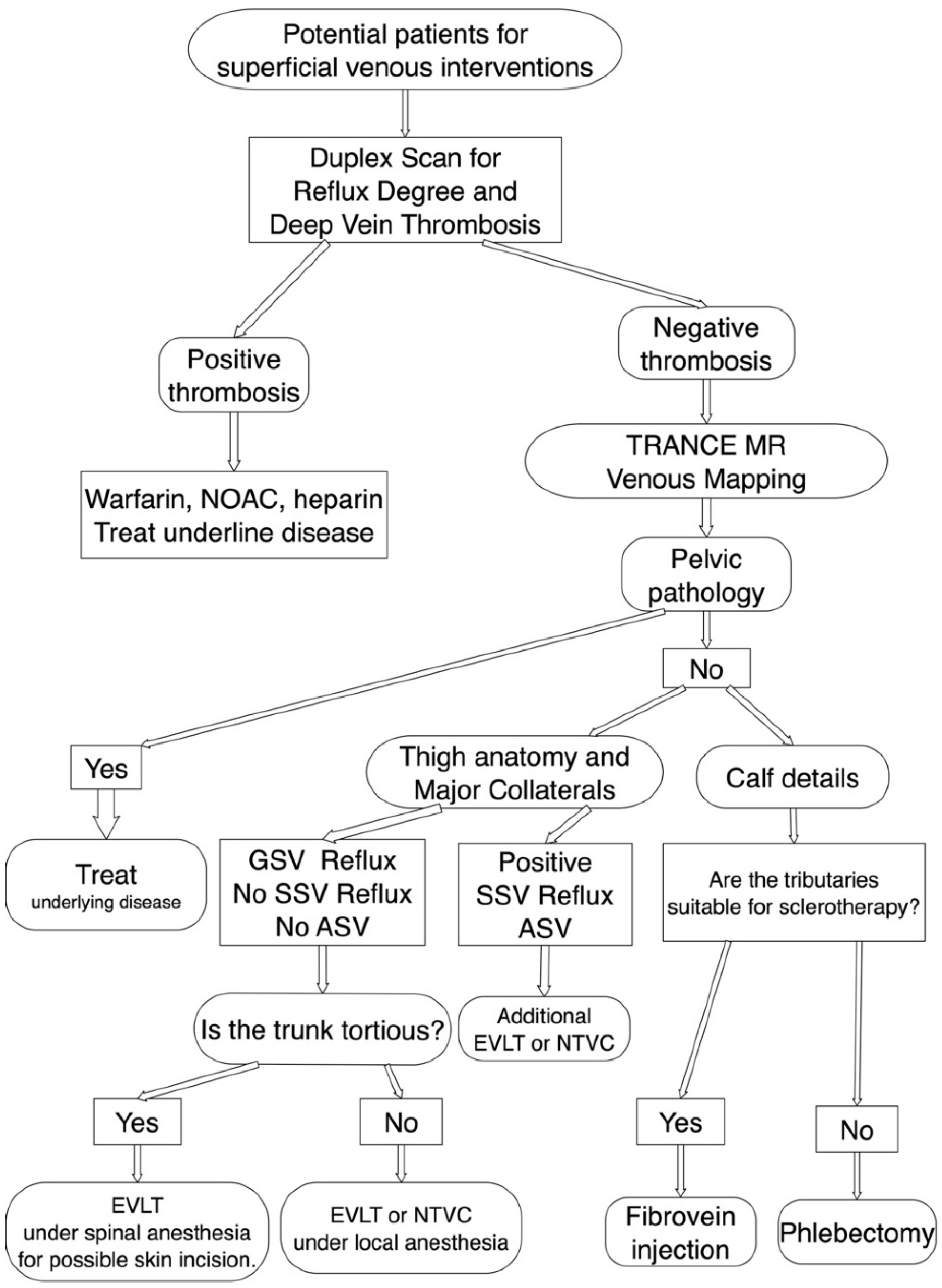
2. Materials and Methods
2.1. Patients
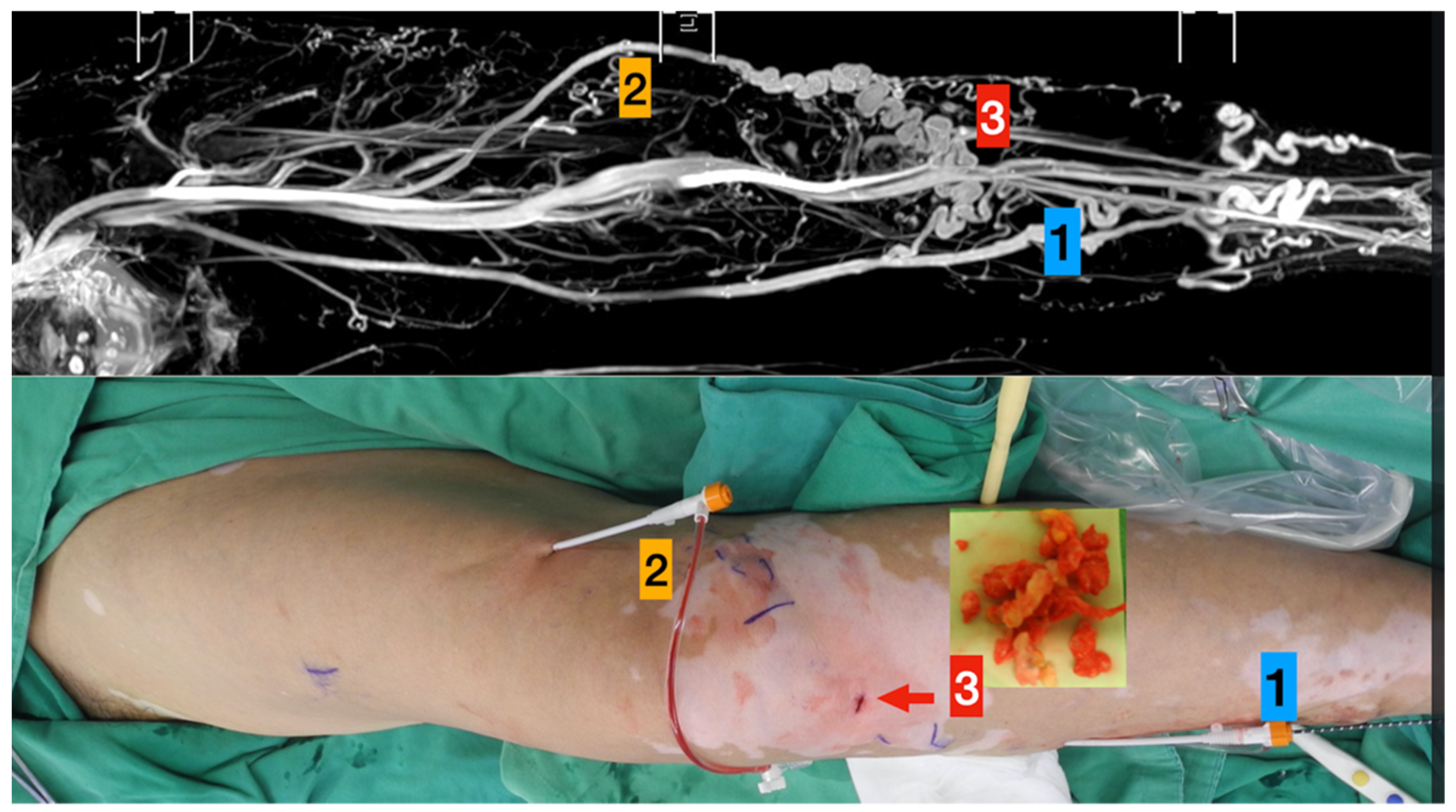
- Left great saphenous vein
- Accessory saphenous vein
- Major communicating tributaries.
- Primary truncal ablation of the great saphenous vein.
- Additional ablation of the accessory saphenous vein.
- Phlebectomy though the small incisions (red arrow).
2.2. MRI Acquisition
2.3. Statistical Analysis
3. Results
3.1. Comparison between Duplex Scanning and TRANCE-MRI Preoperatively
3.2. Comparison of TRANCE-MRI Hemodynamic Parameters between the Morbid Limbs and Healthy Volunteers
4. Discussion
Study Limitations
5. Conclusions
6. Patents
Supplementary Materials
Author Contributions
Funding
Institutional Review Board Statement
Informed Consent Statement
Data Availability Statement
Acknowledgments
Conflicts of Interest
Abbreviations
| 3D | three-dimensional |
| CT | computed tomography |
| CTA | computed tomography angiography |
| DVT | deep venous thrombosis |
| EIV | external iliac vein |
| FFV | forward flow volume |
| FOV | field of view |
| FV | femoral vein |
| GSV | great saphenous vein |
| IR | inversion recovery |
| IRB | institutional review board |
| MF | mean flux |
| MRI | magnetic resonance imaging |
| MRV | magnetic resonance venography |
| MV | mean velocity |
| NSF | nephrogenic systemic fibrosis |
| PV | popliteal vein |
| SD | stroke distance |
| STIR | short tau inversion recovery |
| SV | stroke volume |
| TE | echo time |
| TOF | time-of-flight |
| TR | repetition time |
| TRANCE-MRI | triggered angiography non-contrast-enhanced MRI |
| TSE | turbo spin-echo |
References
- Tassiopoulos, A.K.; Golts, E.; Oh, D.S.; Labropoulos, N. Current concepts in chronic venous ulceration. Eur. J. Vasc. Endovasc. Surg. 2000, 20, 227–232. [Google Scholar] [CrossRef] [PubMed] [Green Version]
- Schleimer, K.; Barbati, M.E.; Grommes, J.; Hoeft, K.; Toonder, I.M.; Wittens, C.H.A.; Jalaie, H. Update on diagnosis and treatment strategies in patients with post-thrombotic syndrome due to chronic venous obstruction and role of endovenous recanalization. J. Vasc. Surg. Venous Lymphat. Disord. 2019, 7, 592–600. [Google Scholar] [CrossRef]
- Millan, S.B.; Gan, R.; Townsend, P.E. Venous Ulcers: Diagnosis and Treatment. Am. Fam. Physician 2019, 100, 298–305. [Google Scholar]
- Lin, B.S.; Chen, C.W.; Zhou, S.K.; Tseng, Y.H.; Wang, S.C.; Huang, Y.K. Evaluation of static ulcer on lower extremities using wireless wearable near-infrared spectroscopy device: Effect of deep venous thrombosis on TRiggered Angiography Non-Contrast-Enhanced sequence magnetic resonance imaging. Phlebology 2020, 35, 814–823. [Google Scholar] [CrossRef]
- Kao, C.C.; Chen, C.W.; Tseng, Y.H.; Tsai, Y.H.; Wang, S.C.; Huang, Y.K. Non-contrast-enhanced magnetic resonance imaging: Objective figures in differentiation between acute and chronic deep venous thrombosis in the lower extremities. Phlebology 2020, 35, 777–783. [Google Scholar] [CrossRef]
- Chen, C.W.; Tseng, Y.H.; Lin, C.C.; Kao, C.C.; Wong, M.Y.; Lin, B.S.; Huang, Y.K. Novel Diagnostic Options without Contrast Media or Radiation: Triggered Angiography Non-Contrast-Enhanced Sequence Magnetic Resonance Imaging in Treating Different Leg Venous Diseases. Diagnostics (Basel) 2020, 10, 355. [Google Scholar] [CrossRef]
- Asciutto, G.; Mumme, A.; Marpe, B.; Koster, O.; Asciutto, K.C.; Geier, B. MR venography in the detection of pelvic venous congestion. Eur. J. Vasc. Endovasc. Surg. 2008, 36, 491–496. [Google Scholar] [CrossRef] [Green Version]
- Lee, Y.L.; Huang, Y.K.; Hsu, L.S.; Chen, P.Y.; Chen, C.W. The use of non-contrast-enhanced MRI to evaluate serial changes in endoleaks after aortic stenting: A case report. BMC Med. Imaging 2019, 19, 82. [Google Scholar] [CrossRef] [PubMed]
- Huang, Y.K.; Tseng, Y.H.; Lin, C.H.; Tsai, Y.H.; Hsu, Y.C.; Wang, S.C.; Chen, C.W. Evaluation of venous pathology of the lower extremities with triggered angiography non-contrast-enhanced magnetic resonance imaging. BMC Med. Imaging 2019, 19, 96. [Google Scholar] [CrossRef] [Green Version]
- Tseng, Y.H.; Chen, C.W.; Wong, M.Y.; Yang, T.Y.; Lin, B.S.; Ting, H.; Huang, Y.K. Discriminating Reflux from Non-Reflux Diseases of Superficial Veins in Legs by Novel Non-Contrast MR with QFlow Technique. J. Pers. Med. 2021, 11, 242. [Google Scholar] [CrossRef]
- Lombardi, P.; Carr, J.C.; Allen, B.D.; Edelman, R.R. Updates in Magnetic Resonance Venous Imaging. Semin. Interv. Radiol. 2021, 38, 202–208. [Google Scholar] [CrossRef]
- Gurel, K.; Gurel, S.; Karavas, E.; Buharalioglu, Y.; Daglar, B. Direct contrast-enhanced MR venography in the diagnosis of May-Thurner syndrome. Eur. J. Radiol. 2011, 80, 533–536. [Google Scholar] [CrossRef]
- Goodman, L.R. Venous thromboembolic disease: CT evaluation. Q. J. Nucl. Med. 2001, 45, 302–310. [Google Scholar]
- Ruehm, S.G.; Zimny, K.; Debatin, J.F. Direct contrast-enhanced 3D MR venography. Eur. Radiol. 2001, 11, 102–112. [Google Scholar] [CrossRef]
- Alfano, G.; Fontana, F.; Ferrari, A.; Solazzo, A.; Perrone, R.; Giaroni, F.; Torricelli, P.; Cappelli, G. Incidence of nephrogenic systemic fibrosis after administration of gadoteric acid in patients on renal replacement treatment. Magn. Reson. Imaging 2020, 70, 1–4. [Google Scholar] [CrossRef]
- Schieda, N.; Maralani, P.J.; Hurrell, C.; Tsampalieros, A.K.; Hiremath, S. Updated Clinical Practice Guideline on Use of Gadolinium-Based Contrast Agents in Kidney Disease Issued by the Canadian Association of Radiologists. Can. Assoc. Radiol. J. 2019, 70, 226–232. [Google Scholar] [CrossRef] [PubMed] [Green Version]
- Ross, M.R.; Pelc, N.J.; Enzmann, D.R. Qualitative phase contrast MRA in the normal and abnormal circle of Willis. AJNR Am. J. Neuroradiol. 1993, 14, 19–25. [Google Scholar] [PubMed]
- Giner, J.F.; Sanz-Requena, R.; Florez, N.; Alberich-Bayarri, A.; Garcia-Marti, G.; Ponz, A.; Marti-Bonmati, L. Quantitative phase-contrast MRI study of cerebrospinal fluid flow: A method for identifying patients with normal-pressure hydrocephalus. Neurologia 2014, 29, 68–75. [Google Scholar] [CrossRef]
- Gutzeit, A.; Sutter, R.; Froehlich, J.M.; Roos, J.E.; Sautter, T.; Schoch, E.; Giger, B.; Wyss, M.; Graf, N.; von Weymarn, C.; et al. ECG-triggered non-contrast-enhanced MR angiography (TRANCE) versus digital subtraction angiography (DSA) in patients with peripheral arterial occlusive disease of the lower extremities. Eur. Radiol. 2011, 21, 1979–1987. [Google Scholar] [CrossRef] [PubMed]
- Suttmeyer, B.; Teichgraber, U.; Rathke, H.; Albrecht, L.; Guttler, F.; Schnackenburg, B.; Hamm, B.; de Bucourt, M. Initial experience with imaging of the lower extremity arteries in an open 1.0 Tesla MRI system using the triggered angiography non-contrast-enhanced sequence (TRANCE) compared to digital subtraction angiography (DSA). Biomed. Tech. (Berl.) 2016, 61, 383–392. [Google Scholar] [CrossRef]
- Radlbauer, R.; Salomonowitz, E.; van der Riet, W.; Stadlbauer, A. Triggered non-contrast enhanced MR angiography of peripheral arteries: Optimization of systolic and diastolic time delays for electrocardiographic triggering. Eur. J. Radiol. 2011, 80, 331–335. [Google Scholar] [CrossRef] [PubMed]
- Ohno, N.; Miyati, T.; Noda, T.; Alperin, N.; Hamaguchi, T.; Ohno, M.; Matsushita, T.; Mase, M.; Gabata, T.; Kobayashi, S. Fast Phase-Contrast Cine MRI for Assessing Intracranial Hemodynamics and Cerebrospinal Fluid Dynamics. Diagnostics (Basel) 2020, 10, 241. [Google Scholar] [CrossRef] [PubMed] [Green Version]
- Altaha, M.A.; Jaskolka, J.D.; Tan, K.; Rick, M.; Schmitt, P.; Menezes, R.J.; Wintersperger, B.J. Non-contrast-enhanced MR angiography in critical limb ischemia: Performance of quiescent-interval single-shot (QISS) and TSE-based subtraction techniques. Eur. Radiol. 2017, 27, 1218–1226. [Google Scholar] [CrossRef]


| No | Age | Sex | Comorbidities | Treating Legs | Symptoms | C in CEAP | E in CEAP | A in CEAP | P in CEAP | Wound Location |
|---|---|---|---|---|---|---|---|---|---|---|
| 1 | 46 | F | Nil | Left | Claudication | C4b | Ep | GSVa, GSVb, ASV | Pr | no |
| 2 | 46 | F | Nil | Left | Claudication | C5 | Ep | GSVa, GSVb | Pr | no |
| 3 | 58 | F | Nil | Left | Claudication | C4a | Ep | GSVa, GSVb | Pr | no |
| 4 | 82 | F | Nil | Left | Claudication | C4c | Ep | GSVa, GSVb, SSV | Pr | no |
| 5 | 59 | F | HTN | Left | Claudication | C4b | Ep | GSVa, GSVb | Pr | no |
| 6 | 84 | F | Severe MR and TR, CHF | Left | calves cramping | C4c | Ep | GSVa, GSVb | Pr | no |
| 7 | 58 | M | Nl | Left | Claudication | C5 | Ep | GSVa, GSVb | Pr | no |
| 8 | 57 | F | Nil | Right | Claudication | C5 | Ep | GSVa, GSVb | Pr | no |
| 9 | 65 | F | HTN | Left | calves cramping | C5 | Ep | GSVa, GSVb, CPV | Pr | no |
| 10 | 53 | F | Nil | Left | calves cramping | C4a | Ep | GSVa, GSVb, CPV | Pr | no |
| 11 | 56 | F | Nil | Right | Wound | C5 | Ep | GSVa, GSVb, SSV, CPV | Pr | medial ankle |
| 12 | 43 | M | Nil | Left | Wound | C5 | Ep | GSVa, GSVb | Pr | no |
| 13 | 59 | F | Nil | Left | Claudication | C4b | Ep | GSVa, GSVb, SSV, Vein of Giacomini | Pr | no |
| 14 | 55 | F | HTN | Left | Claudication | C4a | Ep | GSVa, GSVb, CPV | Pr | no |
| 15 | 69 | F | HTN | Right | Claudication | C4a | Ep | GSVa, GSVb, CPV | Pr | no |
| 16 | 67 | F | nil | Left | Claudication | C4c | Ep | GSVa, GSVb, SSV, CPV | Pr | no |
| 17 | 71 | F | Nil | Left | Claudication | C4b | Ep | GSVa, GSVb, CPV | Pr | no |
| 18 | 38 | F | Nil | Right | Claudication | C4c | Ep | GSVb, SSV, TPV | Pr | no |
| 19 | 59 | F | DM | Left | Wound | C6 | Ep | GSVa, GSVb, SSV, CPV | Pr | gaiter area |
| 20 | 68 | F | Nil | Right | Wound | C6r | Ep | GSVa, GSVb, CPV | Pr | medial ankle |
| 21 | 58 | F | Nil | Right | Wound | C6 | Ep | GSVa, GSVb, CPV | Pr | medial ankle |
| 22 | 40 | M | Nil | Left | Wound | C6 | Ep | GSVa, GSVb | Pr | medial ankle |
| 23 | 43 | M | Obese | Left | Wound | C6r | Ep | GSVa, GSVb, SSV, CPV | Pr | lateral malleola |
| 24 | 53 | F | Nil | Right | Wound | C6 | Ep | GSVa, GSVb, SSV | Pr | medial ankle |
| 25 | 50 | M | Nil | Right | Wound | C6 | Ep | GSVa, GSVb | Pr | medial ankle |
| 26 | 82 | F | Nil | Right | Wound | C6r | Ep | GSVa, GSVb | Pr | medial ankle |
| 27 | 67 | M | CVA, HTN, DM | Left | Wound | C6 | Ep | GSVa, GSVb, SSV | Pr | medial ankle |
| 28 | 58 | F | Nil | Left | Wound | C6r | Ep | GSVa, GSVb | Pr | medial ankle |
| 29 | 54 | F | Nil | Left | calves cramping | C5 | Ep | GSVa, GSVb | Pr | no |
| 30 | 62 | F | Nil | Right | calves cramping | C5 | Ep | GSVa, GSVb, SSV | Pr | no |
| Patient No | Device | Primary Ablation | Secondary Ablation | Thigh Cutdown | Groin Cutdown | Sclerotherapy | Phlebectomy | Tumescent Solution Use | Complication |
|---|---|---|---|---|---|---|---|---|---|
| 1 | VNUS (metronic) | LGSV | ASV | Yes | Nil | Calf (alcohol) | Calf and knee | Yes | Nil |
| 2 | Atoven catheter | LGSV | Nil | Nil | Yes | Calf (Fibrovein) | Calf | Yes | Echymosis |
| 3 | Atoven catheter | LGSV | Nil | Yes | Nil | Nil | Nil | Yes | Nil |
| 4 | Venaseal | LGSV | SSV | Yes | Nil | Nil | Nil | Nil | Nil |
| 5 | Atoven catheter | LGSV | Nil | Yes | Nil | Calf(Fibrovein) | Nil | Yes | Nil |
| 6 | Venaseal | LGSV | Nil | Nil | Nil | Nil | Nil | Nil | Nil |
| 7 | Atoven catheter | LGSV | Nil | Nil | Nil | Calf(Fibrovein) | Nil | Yes | Nil |
| 8 | Venaseal | RGSV | Nil | Yes | Yes | Nil | Nil | Nil | Nil |
| 9 | Atoven catheter | LGSV | LSV | Nil | Nil | Calf(Fibrovein) | Nil | Nil | Nil |
| 10 | A.R.C catheter | LGSV | Nil | Nil | Nil | Calf(Fibrovein) | popliteal fossa | Yes | Nil |
| 11 | Atoven catheter | LGSV | Nil | Nil | Nil | Calf(Fibrovein) | Nil | Yes | Nil |
| 12 | A.R.C catheter | LGSV | Nil | Nil | Nil | Calf(Fibrovein) | Nil | Yes | Nil |
| 13 | A.R.C catheter | LGSV | SSV and PASV | Nil | Nil | Calf(Fibrovein) | Nil | Yes | Nil |
| 14 | Venaseal | LGSV | Nil | Nil | Nil | Nil | Nil | Nil | Nil |
| 15 | Atoven catheter | RGSV | Nil | Nil | Nil | Calf (alcohol) | Nil | Yes | Nil |
| 16 | Venaseal | LGSV | Nil | Nil | Nil | Calf(Fibrovein) | Nil | Nil | Nil |
| 17 | A.R.C catheter | LGSV | bifurcated GSV | Nil | Nil | Nil | Nil | Yes | Nil |
| 18 | A.R.C catheter | RGSV | Nil | Nil | Nil | lateral thigh(Fibrovein) | Nil | Yes | Nil |
| 19 | Atoven catheter | LGSV | Nil | Yes | Nil | Calf(Fibrovein) | Calf and knee | Yes | Nil |
| 20 | Venaseal | RGSV | Nil | Nil | Nil | Nil | Nil | Nil | topical allergy |
| 21 | Atoven catheter | RGSV | bifurcated GSV | Nil | Nil | Calf(Fibrovein) | Nil | Yes | Nil |
| 22 | Venaseal | LGSV | Nil | Nil | Nil | Nil | Nil | Nil | Nil |
| 23 | A.R.C catheter | LGSV | LSSV | Nil | Nil | Calf(Fibrovein) | Nil | Yes | Nil |
| 24 | A.R.C catheter | RGSV | Nil | Nil | Nil | Calf(Fibrovein) | Nil | Yes | Nil |
| 25 | A.R.C catheter | RGSV | Nil | Nil | Nil | Calf(Fibrovein) | Nil | Yes | Nil |
| 26 | Venaseal | RGSV | Nil | Nil | Nil | Nil | Nil | Nil | Nil |
| 27 | A.R.C catheter | LGSV | SSV | Nil | Nil | Calf(Fibrovein) | Nil | Yes | Nil |
| 28 | A.R.C catheter | LGSV | Nil | Nil | Nil | Nil | Nil | Yes | Nil |
| 29 | A.R.C catheter | LGSV | Nil | Nil | Nil | Yes | Nil | ||
| 30 | Atoven catheter | RGSV | Nil | Nil | Nil | Nil | Nil | Yes | Nil |
| No | Dopplex-DVT | Dupplex: SFJ Reflux | Dupplex: Additional Target for Ablation | TRANCE-DVT | TRANCE MR GSV/PV MF QFlow >1 | TRANCE-MTS Like Lesion | TRANCE-Additional Target for Ablation |
|---|---|---|---|---|---|---|---|
| 1 | No | Yes | No | No | Yes | No | Yes (ASV) |
| 2 | No | Yes | No | No | Yes | No | No |
| 3 | No | Yes | No | No | Yes | No | No |
| 4 | No | Yes | No | No | Yes | No | SSV |
| 5 | No | Yes | No | No | Yes | No | No |
| 6 | No | Yes | No | No | Yes | No | No |
| 7 | No | Yes | No | No | Yes | No | No |
| 8 | No | Yes | No | No | Yes | No | No |
| 9 | No | Yes | No | No | Yes | No | LSV |
| 10 | No | Yes | No | No | Yes | No | No |
| 11 | No | Yes | No | No | Yes | No | No |
| 12 | No | Yes | No | No | Yes | No | No |
| 13 | No | Yes | No | No | Yes | No | Yes (SSV and PASV) |
| 14 | No | Yes | No | No | Yes | No | No |
| 15 | No | Yes | No | No | Yes | No | No |
| 16 | No | Yes | No | No | Yes | No | No |
| 17 | No | Yes | No | No | Yes | No | Yes (bifurcated GSV) |
| 18 | No | No | No | No | Yes | No | No |
| 19 | No | Yes | No | No | Yes | No | No |
| 20 | No | Yes | No | No | Yes | No | No |
| 21 | No | Yes | No | No | Yes | No | Yes (bifurcated GSV) |
| 22 | No | Yes | No | No | Yes | No | No |
| 23 | No | Yes | No | No | Yes | Yes | Yes (LSSV) |
| 24 | No | Yes | No | No | Yes | No | No |
| 25 | No | Yes | No | No | Yes | No | No |
| 26 | No | Yes | No | No | Yes | No | No |
| 27 | No | Yes | No | No | Yes | Yes | Yes (LSSV) |
| 28 | No | Yes | No | No | Yes | No | No |
| 29 | No | Yes | No | No | Yes | No | No |
| 30 | No | Yes | No | No | Yes | No | No |
| 0 | Health Volunteers (N = 10) | Planned Superficial Intervention (N = 20) | Power Analysis | ||||||
|---|---|---|---|---|---|---|---|---|---|
| QFlow | Segments | Mean | Standard Deviation | Mean | Standard Deviation | p Value | Power | Effect Size d | Total Sample Size |
| SV (Stroke Volumes) | |||||||||
| IVC | 18.538 | 6.125 | 16.147 | 6.135 | 0.349 | 0.458 | 0.390 | 186 | |
| LEIV | 3.691 | 1.050 | 5.056 | 1.838 | 0.021 * | 0.768 | 0.912 | 36 | |
| LFV | 1.202 | 0.746 | 1.838 | 1.417 | 0.202 | 0.573 | 0.561 | 90 | |
| LGSV | 0.459 | 0.324 | 1.063 | 1.145 | 0.118 | 0.668 | 0.718 | 56 | |
| LPV | 0.643 | 0.332 | 1.112 | 1.324 | 0.285 | 0.524 | |||
| FFV (Foreward Flow Volumes) | |||||||||
| IVC | 18.992 | 6.192 | 16.887 | 6.110 | 0.410 | 0.524 | 0.486 | 120 | |
| LEIV | 3.849 | 1.114 | 5.433 | 2.688 | 0.090 | 0.697 | 0.770 | 50 | |
| LFV | 1.230 | 0.719 | 1.855 | 1.472 | 0.223 | 0.559 | 0.539 | 98 | |
| LGSV | 0.473 | 0.308 | 0.834 | 0.662 | 0.119 | 0.657 | 0.698 | 60 | |
| LPV | 0.654 | 0.317 | 1.144 | 1.409 | 0.293 | 0.519 | 0.479 | 124 | |
| BFV (Backward Flow Volumes) | |||||||||
| IVC | 0.452 | 1.028 | 0.738 | 2.243 | 0.711 | 0.303 | 0.164 | 1038 | |
| LEIV | 0.155 | 0.240 | 0.375 | 1.412 | 0.632 | 0.339 | 0.218 | 590 | |
| LFV | 0.027 | 0.049 | 0.017 | 0.061 | 0.644 | 0.321 | 0.191 | 764 | |
| LGSV | 0.012 | 0.022 | 0.333 | 1.319 | 0.453 | 0.393 | 0.344 | 212 | |
| LPV | 0.009 | 0.020 | 0.030 | 0.095 | 0.502 | 0.370 | 0.306 | 266 | |
| RF (Regurgitant Fraction) | |||||||||
| IVC | 2.186 | 4.892 | 3.963 | 13.140 | 0.688 | 0.426 | 0.344 | 238 | |
| LEIV | 3.749 | 7.477 | 3.230 | 10.679 | 0.888 | 0.233 | 0.056 | 8780 | |
| LFV | 5.206 | 5.852 | 0.629 | 1.877 | 0.168 | 0.826 | 1.053 | 28 | |
| LGSV | 9.650 | 26.954 | 3.924 | 7.372 | 0.296 | 0.389 | 0.290 | 334 | |
| Lt PV | 5.986 | 8.540 | 5.291 | 17.618 | 0.917 | 0.230 | 0.050 | 11048 | |
| ASV (Absolute Stroke Volumes) | |||||||||
| IVC | 19.448 | 6.426 | 17.627 | 6.864 | 0.512 | 0.378 | 0.274 | 374 | |
| LEIV | 4.008 | 1.222 | 5.808 | 3.881 | 0.169 | 0.614 | 0.626 | 74 | |
| LFV | 1.262 | 0.695 | 1.872 | 1.528 | 0.247 | 0.543 | 0.514 | 108 | |
| LGSV | 0.487 | 0.294 | 1.169 | 1.454 | 0.158 | 0.629 | 0.651 | 68 | |
| LPV | 0.665 | 0.303 | 1.174 | 1.495 | 0.301 | 0.514 | 0.472 | 128 | |
| MF (Mean Flux) | |||||||||
| IVC | 21.336 | 6.848 | 18.679 | 7.912 | 0.395 | 0.437 | 0.359 | 218 | |
| LEIV | 3.798 | 0.871 | 5.395 | 2.136 | 0.012 * | 0.797 | 0.979 | 32 | |
| LFV | 1.246 | 0.776 | 1.924 | 1.268 | 0.140 | 0.626 | 0.645 | 70 | |
| LGSV | 0.477 | 0.362 | 1.097 | 1.057 | 0.087 | 0.705 | 0.784 | 48 | |
| LPV | 0.650 | 0.322 | 1.140 | 1.184 | 0.215 | 0.576 | 0.565 | 90 | |
| SD (Stroke Distance) | |||||||||
| IVC | 9.519 | 3.317 | 11.026 | 5.368 | 0.438 | 0.422 | 0.338 | 246 | |
| LEIV | 3.459 | 0.590 | 6.705 | 5.122 | 0.019 * | 0.758 | 0.890 | 38 | |
| LFV | 4.092 | 3.357 | 4.751 | 3.008 | 0.603 | 0.331 | 0.207 | 652 | |
| LGSV | 2.005 | 1.520 | 2.672 | 4.919 | 0.682 | 0.315 | 0.183 | 832 | |
| LPV | 1.408 | 1.124 | 1.384 | 0.883 | 0.952 | 0.214 | 0.024 | 49768 | |
| MV (Mean Velocity) | |||||||||
| IVC | 11.169 | 4.354 | 12.304 | 4.975 | 0.563 | 0.356 | 0.243 | 474 | |
| LEIV | 33.816 | 94.936 | 7.125 | 5.424 | 0.397 | 0.463 | 0.397 | 178 | |
| LFV | 4.347 | 3.893 | 5.079 | 3.249 | 0.604 | 0.330 | 0.204 | 670 | |
| LGSV | 2.033 | 1.554 | 2.954 | 4.973 | 0.577 | 0.361 | 0.250 | 448 | |
| LPV | 1.458 | 1.252 | 1.452 | 0.886 | 0.989 | 0.203 | 0.005 | >10,000 | |
| Health Volunteers (N = 10) | Planned Superficial Intervention (N = 10) | Power Analysis | |||||||
|---|---|---|---|---|---|---|---|---|---|
| QFlow | Segments | Mean | Standard Deviation | Mean | Standard Deviation | p-Value | Power | Effect Size d | Total Sample Size |
| SV (Stroke Volumes) | |||||||||
| IVC | 18.538 | 6.125 | 13.933 | 5.537 | 0.118 | 0.645 | 0.789 | 42 | |
| REIV | 4.303 | 0.872 | 5.456 | 2.725 | 0.282 | 0.527 | 0.570 | 78 | |
| RFV | 1.437 | 0.704 | 1.764 | 0.996 | 0.427 | 0.414 | 0.379 | 174 | |
| RGSV | 0.360 | 0.265 | 0.893 | 0.342 | 0.002 * | 0.934 | 1.742 | 10 | |
| Rt PV | 0.579 | 0.278 | 1.125 | 0.876 | 0.128 | 0.670 | 0.840 | 38 | |
| FFV (Foreward Flow Volumes) | |||||||||
| IVC | 18.992 | 6.192 | 14.594 | 5.395 | 0.133 | 0.670 | 0.840 | 38 | |
| REIV | 4.605 | 1.074 | 5.625 | 2.624 | 0.278 | 0.491 | 0.509 | 98 | |
| RFV | 1.451 | 0.691 | 1.774 | 0.985 | 0.426 | 0.414 | 0.379 | 174 | |
| RGSV | 0.377 | 0.248 | 0.903 | 0.321 | 0.001 * | 0.945 | 1.832 | 10 | |
| RPV | 0.600 | 0.277 | 1.144 | 0.864 | 0.125 | 0.674 | 0.848 | 36 | |
| BFV (Backward Flow Volumes) | |||||||||
| IVC | 0.452 | 1.028 | 0.659 | 1.102 | 0.687 | 0.305 | 0.194 | 660 | |
| REIV | 0.299 | 0.396 | 0.166 | 0.216 | 0.380 | 0.436 | 0.416 | 146 | |
| RFV | 0.012 | 0.025 | 0.009 | 0.025 | 0.788 | 0.267 | 0.128 | 1514 | |
| RGSV | 0.026 | 0.046 | 0.009 | 0.025 | 0.351 | 0.468 | 0.470 | 114 | |
| RPV | 0.018 | 0.030 | 0.016 | 0.024 | 0.896 | ||||
| RF (Regurgitant Fraction) | |||||||||
| IVC | 2.186 | 4.892 | 4.755 | 7.635 | 0.398 | 0.232 | 0.062 | 6416 | |
| REIV | 5.928 | 7.477 | 4.024 | 5.293 | 0.537 | 0.363 | 0.294 | 288 | |
| RFV | 2.317 | 5.852 | 1.321 | 3.178 | 0.672 | 0.315 | 0.211 | 556 | |
| RGSV | 15.705 | 26.954 | 2.731 | 7.725 | 0.176 | 0.574 | 0.654 | 60 | |
| RPV | 4.737 | 8.540 | 4.115 | 6.736 | 0.869 | 0.242 | 0.081 | 3784 | |
| ASV (Absolute Stroke Volumes) | |||||||||
| IVC | 19.448 | 6.426 | 15.258 | 5.474 | 0.162 | 0.600 | 0.702 | 52 | |
| REIV | 4.908 | 1.367 | 5.781 | 2.544 | 0.364 | 0.443 | 0.428 | 138 | |
| RFV | 1.464 | 0.681 | 1.784 | 0.973 | 0.424 | 0.415 | 0.381 | 172 | |
| RGSV | 0.404 | 0.220 | 0.913 | 0.302 | 0.001 | 0.955 | 1.924 | 10 | |
| RPV | 0.620 | 0.278 | 1.164 | 0.852 | 0.120 | 0.678 | 0.858 | 36 | |
| MF (Mean Flux) | |||||||||
| IVC | 21.336 | 6.848 | 16.453 | 7.724 | 0.174 | 0.582 | 0.669 | 58 | |
| REIV | 4.478 | 0.846 | 6.363 | 3.774 | 0.142 | 0.593 | 0.689 | 54 | |
| RFV | 1.471 | 0.753 | 2.048 | 1.293 | 0.253 | 0.512 | 0.545 | 86 | |
| RGSV | 0.372 | 0.289 | 1.013 | 0.400 | 0.001 * | 0.946 | 1.834 | 10 | |
| RPV | 0.584 | 0.268 | 1.348 | 1.200 | 0.118 | 0.688 | 0.878 | 34 | |
| SD (Stroke Distance) | |||||||||
| IVC | 9.519 | 3.317 | 7.770 | 3.355 | 0.285 | 0.500 | 0.524 | 92 | |
| REIV | 3.862 | 1.000 | 5.474 | 1.619 | 0.019 * | 0.814 | 1.198 | 20 | |
| RFV | 4.737 | 3.660 | 3.026 | 1.492 | 0.234 | 0.550 | 0.612 | 68 | |
| RGSV | 1.901 | 1.681 | 3.156 | 2.340 | 0.204 | 0.553 | 0.616 | 68 | |
| Rt PV | 1.055 | 0.450 | 1.413 | 1.089 | 0.357 | 0.444 | 0.429 | 136 | |
| MV ( Mean Velocity) | |||||||||
| IVC | 11.169 | 4.354 | 9.121 | 4.894 | 0.362 | 0.452 | 0.442 | 128 | |
| REIV | 4.086 | 1.246 | 4.995 | 2.719 | 0.359 | 0.444 | 0.430 | 136 | |
| RFV | 4.975 | 4.241 | 3.433 | 1.852 | 0.354 | 0.469 | 0.471 | 114 | |
| RGSV | 1.920 | 1.706 | 3.475 | 2.569 | 0.143 | 0.605 | 0.713 | 52 | |
| RPV | 1.064 | 0.393 | 1.639 | 1.362 | 0.281 | 0.528 | 0.573 | 78 | |
Publisher’s Note: MDPI stays neutral with regard to jurisdictional claims in published maps and institutional affiliations. |
© 2021 by the authors. Licensee MDPI, Basel, Switzerland. This article is an open access article distributed under the terms and conditions of the Creative Commons Attribution (CC BY) license (https://creativecommons.org/licenses/by/4.0/).
Share and Cite
Chen, C.-W.; Tseng, Y.-H.; Fang, Y.-F.; Wong, M.Y.; Lin, Y.-H.; Huang, Y.-K. Superficial Venous Reflux Intervention Guided by Triggered Angiography Non-Contrast-Enhanced Sequence Magnetic Resonance Imaging: Different QFlow Pattern from Health Controls. J. Pers. Med. 2021, 11, 751. https://0-doi-org.brum.beds.ac.uk/10.3390/jpm11080751
Chen C-W, Tseng Y-H, Fang Y-F, Wong MY, Lin Y-H, Huang Y-K. Superficial Venous Reflux Intervention Guided by Triggered Angiography Non-Contrast-Enhanced Sequence Magnetic Resonance Imaging: Different QFlow Pattern from Health Controls. Journal of Personalized Medicine. 2021; 11(8):751. https://0-doi-org.brum.beds.ac.uk/10.3390/jpm11080751
Chicago/Turabian StyleChen, Chien-Wei, Yuan-Hsi Tseng, Yueh-Fu Fang, Min Yi Wong, Yu-Hui Lin, and Yao-Kuang Huang. 2021. "Superficial Venous Reflux Intervention Guided by Triggered Angiography Non-Contrast-Enhanced Sequence Magnetic Resonance Imaging: Different QFlow Pattern from Health Controls" Journal of Personalized Medicine 11, no. 8: 751. https://0-doi-org.brum.beds.ac.uk/10.3390/jpm11080751