
Role of Enhancers in Development and Diseases


Department of Pediatrics, Division of Pediatric Hematology and Oncology, Department of Developmental Biology, School of Medicine, Washington University in St. Louis, 660 South Euclid Avenue, St. Louis, MO 63110, USA
Epigenomes 2021, 5(4), 21; https://0-doi-org.brum.beds.ac.uk/10.3390/epigenomes5040021
Submission received: 28 July 2021
/
Revised: 21 September 2021
/
Accepted: 28 September 2021
/
Published: 4 October 2021
Abstract
:Enhancers are cis-regulatory elements containing short DNA sequences that serve as binding sites for pioneer/regulatory transcription factors, thus orchestrating the regulation of genes critical for lineage determination. The activity of enhancer elements is believed to be determined by transcription factor binding, thus determining the cell state identity during development. Precise spatio-temporal control of the transcriptome during lineage specification requires the coordinated binding of lineage-specific transcription factors to enhancers. Thus, enhancers are the primary determinants of cell identity. Numerous studies have explored the role and mechanism of enhancers during development and disease, and various basic questions related to the functions and mechanisms of enhancers have not yet been fully answered. In this review, we discuss the recently published literature regarding the roles of enhancers, which are critical for various biological processes governing development. Furthermore, we also highlight that altered enhancer landscapes provide an essential context to understand the etiologies and mechanisms behind numerous complex human diseases, providing new avenues for effective enhancer-based therapeutic interventions.
1. Introduction
The term “enhancer” was first coined based on studies of Simian virus 40 (SV40) when, in 1981, Banerji et al. observed for the first time that a viral DNA element from SV40 had the ability to enhance activity towards a T-antigen or β-globin reporter in mammalian cells [1]. Further research has explored endogenous sequences with similar functions in the immunoglobulin heavy chain locus. These preliminary studies have established that enhancers function as short DNA elements that trigger a gene’s transcription from a long distance in an orientation-independent manner. Preliminary studies have determined the exact mechanism of how distal regulatory elements regulate gene transcription through distal enhancers [2]. Enhancers are cis-regulatory elements that carry epigenetic information in DNA sequences through specific histone modifications [3]. Studies assessing the characteristics of enhancers have reported that they can function independently of the orientation and distance to their cognate target genes, at distances sometimes of several hundred kilobases or megabases [2,4,5]. Enhancers can be identified and characterized by various factors, including histone modifications, their transcription into non-coding RNAs, and their epigenetic features [6]. The prominent feature of enhancers is their ability to serve as a docking platform for transcription factor binding, where developmental signaling (intrinsic or extrinsic) cues are interpreted in a highly context-specific manner [7]. The signatures of these enhancers are highly cell type-specific; such cell type-specific use of the epigenetic information has demonstrated the combinatorial function of transcription factors in maintaining cell identity and lineage determination [8]. The cell type-specific enhancer pattern provides a unique cis-regulatory platform, in which transcription factors are activated by developmental cues and modify the transcriptome [9,10,11]. This model revealed that every developmental stage has a cell type-specific transcription factor, which functions by cell type-specific enhancer signatures [12]. Enhancers are actuated in a stage-specific manner, correlated with cell type-specific histone modifications. This combinatorial mechanism of transcription factor binding on cell type-specific enhancers results in the so-called enhancer signature, which serves as a readout to define enhancers in a cell type-specific manner at a global scale [13]. Precise spatial and temporal control of gene expression and the correct interpretation of the enhancer signature are crucial in the development process. Any alterations in the enhancer signature can modify the gene expression pattern and ethe capacity of the enhancer to respond to developmental signals, narrowing cell differentiation and affecting correct lineage formation. Heinz et al. (2010) have shown that lineage-determining transcription factors bind to a genomic region in a cell-specific manner. They showed the genome-wide locations of PU.1 binding patterns in macrophages, B-cells, and diffuse B-cell progenitors [14]. Similarly, Xu et al. (2012) analyzed chromatin state maps, transcription factor occupancy rates, and gene expression profiles during the development of human erythroid cells at the fetal and adult stages, and carried out a comparative analysis to determine the specific procedures of the development stage [15]. Here, it is also important to discuss the study of Choukrallah et al. (2015), who found that the enhancer landscape is dynamically reshaped in each differentiation step. Interesting changes include creating new enhancers and the closing and re-opening of the pre-existing enhancer landscape [16]. The authors also reported that the regulatory signatures of two related types of myeloid leukemia expression fusion proteins (RUNX1-ETO and RUNX1-EV1) display a distinct binding pattern and interact with different transcription factors to impact the epigenome [17]. A study has also reported that MLL-Af9 and MLL-AF4 oncofusion proteins showed distinct binding patterns in the enhancer region and targeted the RUNX1 program in 11q23 acute myeloid leukemia [18]. Enhancers are essential for normal functioning, and the loss of enhancer elements can cause abnormalities; for example, Groschel et al. (2014) showed that the removal of the distal enhancer essential for the GATA2 gene resulted in insufficient Functional GATA2 haploids, which only reduced the expression of the remaining normal alleles [19]. This review focuses on a concise and brief overview of the roles of enhancers in development and disease. We attempt to discuss how enhancers are activated and coordinated with transcription factors, as well as the roles of enhancers in mammalian development. This may comprise the first attempt to compile recently published research on development and disease, focusing on enhancers.
2. Features and Types of Enhancers
Enhancers have been identified in the form of various regulatory domains, primed enhancers, active enhancers, and poised enhancers. Each type of enhancer signature has specific histone modification patterns and can be easily identified by these signatures [13,20,21]. Primed enhancers can be identified by only histone H3K4 mono-methylation, while active enhancers signatures are identified by H3K4 mono-methylation and H3K27ac. Finally, the signatures of poised enhancers are marked with H3K4Me1 with H3K27me3, but not H3K27ac [13,20,21]. Active enhancers are linked to expressed genes, while poised enhancers are always associated to developmental genes, which are inactive in embryonic stem cells or precursor cells and become expressed during different differentiation stages [22]. During differentiation, the poised enhancer’s signature successfully loses the repressive H3K27me3 histone mark, acquires H3K27ac marks, and becomes active. Enhancers are, thus, subject to dynamic change functions, as an on/off switch to tune the target gene expression and changing the cell state from undifferentiated to differentiated phenotype [23]. Hence, the signatures of Poised Enhancers comprise a small set of regulatory signatures in embryonic stem cells that facilitate their timely and stage-specific function, once the correct differentiation signals become available [20]. One term also uses super-enhancers (SEs), which are described as large clusters of active enhancers with robust enrichment for binding transcriptional coactivators [24,25]. These features have also been called stretch enhancers [26], multiple enhancers [27], and enhancer clusters [28], which are similar but not identical between studies (although many of these features overlap). SEs regulate master regulators of pluripotency, such as OCT4, SOX2, and NANOG [24]. It has been reported that the SE signature is often enriched near the oncogene in tumor cells, while an enrichment GWAS has identified SNPs normally associated with several common diseases [29,30].
3. Enhancers and Lineage Determination during Development
Many studies have established the idea that the enhancer signature is intricately orchestrated, in a stage-specific pattern, by several proteins complexes during development [31]. Some enhancer signatures are established early during development in precursor cells, and are modified and activated as cells differentiate in terminal steps along specific lineages [32]. All enhancer marks serve different purposes; for example, poised enhancers serve as signatures for future gene expression [2]. In contrast, active enhancer signatures play a functional role in the current transcriptional state [33]. The chromatin state is mostly invariant across different tissues, whereas histone signature patterns at the enhancer level are highly tissue-specific [34]. Mammals, with about 200 specialized cell types, all have different transcriptional outcomes, reflecting the unique coding pattern and regulatory elements during development [34,35,36]. Indeed, the enhancer repertoire active in a particular lineage, as identified by chromatin marks and transcriptional regulators, represents only a small subset of all genomic regulatory domains [33,37,38]. Studies have suggested that the enhancer repertoire is a pre-established landscape, formed and imposed by the lineage, determining TFs that maintain cell identity. All transcriptional regulation and functional outcomes occur within the differentiated cell under this pre-established enhancer landscape [10]. The study also established that enhancers playing a developmental role are evolutionarily conserved sequences [39,40]. Thus, the pre-established enhancer landscape has a crucial role in lineage determination. Any disturbance in the enhancer landscape affects the lineage, determining the potential of cells. This concept suggests that any external cues that trigger the transitory response cannot functionally change the repertoire of genomic regulatory domains, but act on the pre-established epigenomic landscape. This response is a buffer system that ensures cell identity maintenance, despite the changing environment [10]. A schematic diagram explaining enhancer biology is shown in Figure 1. We also summarize the related studies in Table 1, which have investigated the roles of enhancers in development through different model systems, including evidence in support of early enhancer establishment reported in B-cell and macrophage specification [14,16], T-cell development [41], early hematopoiesis [42], and the commitment of multipotent endoderm cells to liver and pancreas cell fates [43]. Wang et al. (2015) have also inferred the role of the poised enhancer landscape in endoderm development, as well as established a functional link between the gain of poised enhancer chromatin state and the temporal acquisition of competence during developmental progression [44]. Dynamic and coordinated epigenetic regulation has also demonstrated chromatin transition during cardiac lineage commitment [45]. Stage-specific enhancers are synergistically activated in a genome-wide manner during cardiac development by cardiac reprogramming factors [46]. The combinatorial action of pioneer factor and super-enhancer dynamics has also been reported in stem cell plasticity and lineage choice [47]. Further, Enhancer priming by histone methyl-transferase has also been demonstrated to control cell fate transition [48]. The master regulator ‘Scl’ has been reported to bind to pre-established primed enhancer signatures in the mesoderm, as well as regulating hematopoietic and cardiac fate divergence at terminal differentiation steps. Scl uses the pre-established epigenetic landscape during the specification of lineage choices [49]. The pioneer factor FOXA2 is required for enhancer priming during HPSC differentiation in pancreatic lineages [50]. It has also been shown that ERK directly regulates enhancer priming in lineage choice [51]. Developmental stage-specific enhancers drive lineages and control gene expression programs during hematopoiesis [15,52]. Rubin et al. (2017) have reported that lineage-specific dynamic and pre-established enhancer–promoter contacts cooperate in terminal differentiation [53]. Recently, Maurya et al. (2021) [54] have shown that the loss of KMT2C in HPSCs cells significantly reprograms the enhancer landscapes in HPSCs cells, leading to the loss of Hemogenic endothelium during in vitro hematopoietic differentiation. Further, it has also been reported that the deletion of KDM6A in HPSCs cells significantly reprograms the Bivalent chromatin in HPSCs cells, suggesting perturbed development at the terminal developmental steps in particular lineages [55].
4. Enhancer–Promoter Interaction Is the Core Key to Regulating Gene Expression
The precise cell type-specific expression of genes often needs additional cis-regulatory elements, which are physically distanced from the promoter regions. These distal cis-regulatory components harbor TFs that are highly cell type-specific and are expressed in the presence of external developmental cues, such as stage-specific signals during differentiation or proliferation. These regulatory elements set complex gene expression patterns at different developmental stages and time points, by combining other external developmental cues [30]. The enhancer element interacts with a promoter, independent of direction and distance, to activate transcription. The most elegant choreography of the biology of these distal regulatory elements is how enhancers identify their cognate promoters and initiate transcription, in addition to the underlying mechanisms that regulate these preinitiation assemblies. It has been shown that enhancers regulate transcription by a looping mechanism between the enhancers and their cognate promoters. Therefore, a more direct physical contact of cis-regulatory elements is established within the nucleus [60]. This mechanism, underlying the crosstalk between enhancers and promoters, has been considered to explore how long-range interactions form chromatin loops. A strategic enhancer is used to interact with its cognate promoter during the initiation of transcription [60]. This entire cyclic strategy of remote regulatory elements has also been observed for the cytokine locus of t-helper type 2 (TH2) cells [61]. The ability of cis-regulatory elements to communicate with promoters is not restricted to a gene position situated at only cis positions on the same chromosome. Reports are also available which have shown that the olfactory H enhancer elements can communicate with multiple olfactory genes with different chromosomes in epithelial tissues, where these genes are normally expressed [4]. These studies suggest that proper gene expression requires the proper establishment of enhancer–promoter interactions. Enhancers serve as docking sites for TFs, and the activity of enhancers is mostly based on the binding of these TFs [11]. More than a thousand TFs encode the human genome. Open chromatin DNA regions have short (20–30 bp) DNA sequences normally occupied with TFs within the enhancer sequences, known as DNA recognition motifs, which are also characterized by low nucleosome occupancy. Some TFs are cell type-/lineage-specific, while some TFs are bound across different cell types [62,63]. For example, GATA1 is known explicitly for erythroid differentiation, which is required in hematopoietic differentiation, and PU.1 is important for B-cell specification within a hematopoietic lineage [64,65]. Furthermore, enhancers can recruit several additional factors to maintain fine-tuned targeted spatio-temporal expression of genes. The functional roles of epigenetic writers and erasers are also important for enhancers, leading to specific epigenetic modifications in the enhancers. For example, COMPASS complexes (KMT2A, KMT2B, KMT2C, and KMT2D) are histone methyl-transferases that bind cis-regulatory elements mediating the methyl marks on histone [66,67]. These chromatin remodelers and TFs can affect the nucleosome dynamics, thus regulating the enhancer regions. Different TFs show cooperative binding in nucleosome-embedded motifs while, alone, the TFs can bind weakly. TFs can also cooperate with chromatin remodeling factors to accelerate the auxiliary loading mechanism. Although the initial association of a TF with its TFBSc can recruit chromatin modulators near the closed chromatin, it initiates the position and promotes the later binding of secondary TFs and accelerates the positive feedback mechanism [68,69,70]. Several studies have shown that the enhancer epigenetic landscape is intricately orchestrated in a set pattern by several regulatory protein complexes during development [32].
5. Role of Enhancers in Disease Development
The genome-wide sequencing approach has revealed that enhancers are prime targets for genetic or epigenetic alterations that lead to carcinogenesis [5]. The main characteristics of the enhancer signature remain constant for every type of cell (e.g., normal vs. cancer/tumor cells). The difference is that the function of the enhancer signatures differs between normal and cancer cell types; that is, the enhancer’s functional output in tumor/cancer cells differs from that in normal cells [71]. Careful examination of enhancer signatures between normal cells and their counterpart in cancer cells has revealed that cancer cells tend to lose enhancers at positions near cell fate-specifying genes and gain enhancer signatures near growth-associated genes [72,73,74]. Furthermore, it has again been confirmed, after comparing normal colon epithelial crypts and colon cancer lines, that thousands of differentially enriched primed enhancer marks (H3K4Me1) were enriched in cancerous cells known as variant enhancer loci (Veli), and these gained mono-methylation sites were not found in normal counterpart cells. In contrast, the lost sites were relatively specific to crypt cells, indicating that the colon cancer cells acquired a more differentiated cell state [73]. In the post-GWAS era, there is a lot of conceivable evidence that cancer predisposition genomic variants present in the non-coding genomic region lie within these distal regulatory elements [75,76]. This finding is based on the overlap between SNPs associated with diseases and genomic signatures, revealing that SNPs associated with risk phenotypes are frequently enriched in expression quantitative traits and open chromatin regions [60,77]. In cancer cells, risk variants lead to the dysregulation of enhancers, disrupting the fine-tuned target expression of their associated genes and producing a pathological state which leads to abnormal growth. For example, pancreatic-specific differential open regions enriched for non-coding variants (SNPs) have been linked to pancreatic disorders [78]. Correspondingly, monocyte-/macrophage-specific enhancer elements enriched with SNPs associated with ulcerative colitis, celiac disease, Crohn’s disease, or systemic lupus erythematosus have been reported [79]. Studies are also available in which single base-pair point mutations in the distal enhancer of SCNA and PTF1A have been shown to cause sporadic Parkinson’s disease and pancreatic agenesis [80,81]. Altered super-enhancer activity has also been reported in many complex human diseases, such as Alzheimer’s, Type 1 diabetes, and autoimmune disorders [25,28,82,83]. Furthermore, lost super-enhancers and enhancers acquired somatically have been reported to be associated with numerous cancers [25,82]. Meanwhile, changes in histone writers, erasers, and altered activity of DNA modifiers such as methyl and acetyltransferases also reshape the chromatin landscape, which finally leads to abnormal growth and development. Based on known enhancer signatures, several studies (Table 2) have explored the alterations in enhancer activity and histone modification patterns which may be correlated with disease development. Evidence for enhancer alterations in cancer, coming from various studies, is summarized in Table 2.
6. Enhancer Reprogramming
The role of enhancer reprogramming is an emerging area in developmental biology and cancer research. The emerging role of enhancers in fate determination has now been well-established. In cancer research, enhancers have been shown to acclimatize cancer cells to environmental changes encountered during cancer progression and development [127]. Recent findings have highlighted that enhancer reprogramming plays a crucial role in carcinogenesis and metastasis formation, thus playing a role in establishing malignant heterogeneity [127,128]. A cancer genomics study has identified that recurrent genetic mutations mostly occur on genes coding for epigenetic modifiers [129]. In addition to the dysregulation of these trans-epigenetic regulators, genome-wide sequencing of the non-coding genome has evidenced the frequent alterations of cis-regulatory elements, such as enhancers and insulators [71]. Considering that enhancer activity modulation plays a significant role in maintaining and controlling the cell identity and cell adaptation to environmental changes, it is easily conceivable that genetic alternations affecting the epigenetic modifiers and cis-regulatory machinery may alter enhancer activity, thus affecting cell fate determination [130]. Specifically, recent studies have suggested that enhancer function reprogramming could represent a hallmark of carcinogenesis, as it contributes to the deregulated expression of epigenetic modifiers, leading to abnormal cell growth. In this respect, oncogenic enhancer reprogramming, considered as cancer-related alterations, may cause aberrant oncogenic development, leading to altered transcription outputs, thus promoting carcinogenesis. Recent conceptually interesting studies have suggested that carcinogenesis involves transforming the cell to a more primitive stage (that is, a developmentally manipulable differentiation state) driven by major changes in the epigenomic landscape [131]. This work highlighted several thought-provoking questions: First, while enhancers are indispensable for specifying transcriptional outputs that drive cell fate determination, it is interesting that a huge shift of enhancer landscape use is particularly critical for PDA metastasis [131]. It has recently been shown that genetic alterations could induce genetic priming in the cell of origin, imposing the primed cell’s phenotype [132]. This response, thus, indicates a new function for genetic insult. This new transformation mechanism reveals that the first aberrant oncogenic hit imposes a cell differentiation program in cancer-initiating cells which is responsible for the tumor cell phenotype at terminal differentiation steps [132]. It is not necessary that the first oncogenic phenotypes for tumor initiation will be responsible for the altered differentiation program. It has been observed that the oncogene is not at all required in the terminal steps of transformation. This suggests that initial hits are not good targets for therapies, as they do not play an essential role in tumor development [133]. Specific epigenomic profiles can distinguish between different lineages in the hematopoietic differentiation system, as well as in leukemic cells [134]. This epigenomic reprogramming has been observed in some other animal cancer models which resemble human cancer. A recent study by Adelman et al. (2019) showed that age-associated epigenetic reprogramming may form a predisposing condition for the development of age-related AML [135].
7. Concluding Remarks
This review discussed how enhancer signatures function as a critical platform, acting as a receiver for extrinsic and intrinsic developmental signals, as well as conveying the context-specific manner during lineage determination. However, research into the functional annotation of enhancer readout in cancer and the development-specific roles of enhancer biology are still in an initial phase, and several unanswered questions remain poorly explored. Enhancer signatures can potentially be used as signature biomarkers for disease-specific phenotype identification and cancer detection. Although, at present, limited resources are available which correlate enhancer signatures with clinical outcomes, future studies are expected to shed more light on the regulatory mechanisms that modify the chromatin landscape patterns at distal regulatory sequences during the cell’s different stages of development and cell differentiation. Multiomics studies, involving genomics, epigenomics, deep global proteomics, single-cell heterogeneity, and single cell epigenomics in different developmental systems, will provide more concrete information on how histone pre-patterns and chromatin modifiers regulate the chromatin landscape under specific enhancer signatures during the developmental process, as well as their impact on phenotypes. Hopefully, new emerging technologies and genome editing tools will uncover novel insights into the roles of enhancer signatures in regulating developmental processes. Future studies should focus on exploring the roles of enhancers and how cell type-specific gene expression can be pre-established and maintained throughout the terminal steps.
Funding
This research received no external funding.
Acknowledgments
The author thanks Todd Druley and the members of Druley Labs for insightful discussions. The author acknowledges all research groups whose studies were included in this review, and the author apologizes to those research groups whose work we could not add here due to restricted space limitation.
Conflicts of Interest
The authors declare no conflict of interest.
References
- Banerji, J.; Rusconi, S.; Schaffner, W. Expression of a beta-globin gene is enhanced by remote SV40 DNA sequences. Cell 1981, 27, 299–308. [Google Scholar] [CrossRef]
- Cui, K.; Zang, C.; Roh, T.-Y.; Schones, D.E.; Childs, R.W.; Peng, W.; Zhao, K. Chromatin signatures in multipotent human hematopoietic stem cells indicate the fate of bivalent genes during differentiation. Cell Stem Cell 2009, 4, 80–93. [Google Scholar] [CrossRef] [Green Version]
- Geyer, P.K.; Green, M.M.; Corces, V.G. Tissue-specific transcriptional enhancers may act in trans on the gene located in the homologous chromosome: The molecular basis of transvection in Drosophila. EMBO J. 1990, 9, 2247–2256. [Google Scholar] [CrossRef]
- Lomvardas, S.; Barnea, G.; Pisapia, D.J.; Mendelsohn, M.; Kirkland, J.; Axel, R. Interchromosomal interactions and olfactory receptor choice. Cell 2006, 126, 403–413. [Google Scholar] [CrossRef] [Green Version]
- Kron, K.J.; Bailey, S.D.; Lupien, M. Enhancer alterations in cancer: A source for a cell identity crisis. Genome Med. 2014, 6, 77. [Google Scholar] [CrossRef] [PubMed] [Green Version]
- Buecker, C.; Wysocka, J. Enhancers as information integration hubs in development: Lessons from genomics. Trends Genet. 2012, 28, 276–284. [Google Scholar] [CrossRef] [PubMed] [Green Version]
- Natoli, G. Maintaining Cell Identity through Global Control of Genomic Organization. Immunity 2010, 33, 12–24. [Google Scholar] [CrossRef] [PubMed] [Green Version]
- He, A.; Kong, S.W.; Ma, Q.; Pu, W.T. Co-occupancy by multiple cardiac transcription factors identifies transcriptional enhancers active in heart. Proc. Natl. Acad. Sci. USA 2011, 108, 5632–5637. [Google Scholar] [CrossRef] [Green Version]
- Song, L.; Zhang, Z.; Grasfeder, L.L.; Boyle, A.P.; Giresi, P.G.; Lee, B.-K.; Sheffield, N.C.; Gräf, S.; Huss, M.; Keefe, D.; et al. Open chromatin defined by DNaseI and FAIRE identifies regulatory elements that shape cell-type identity. Genome Res. 2011, 21, 1757–1767. [Google Scholar] [CrossRef] [Green Version]
- Ostuni, R.; Piccolo, V.; Barozzi, I.; Polletti, S.; Termanini, A.; Bonifacio, S.; Curina, A.; Prosperini, E.; Ghisletti, S.; Natoli, G. Latent Enhancers Activated by Stimulation in Differentiated Cells. Cell 2013, 152, 157–171. [Google Scholar] [CrossRef] [Green Version]
- Spitz, F.; Furlong, E.E.M. Transcription factors: From enhancer binding to developmental control. Nat. Rev. Genet. 2012, 13, 613–626. [Google Scholar] [CrossRef] [PubMed]
- Herz, H.-M. Enhancer deregulation in cancer and other diseases. Bioessays 2016, 38, 1003–1015. [Google Scholar] [CrossRef]
- Creyghton, M.P.; Cheng, A.W.; Welstead, G.G.; Kooistra, T.; Carey, B.W.; Steine, E.J.; Hanna, J.; Lodato, M.A.; Frampton, G.M.; Sharp, P.A.; et al. Histone H3K27ac separates active from poised enhancers and predicts developmental state. Proc. Natl. Acad. Sci. USA 2010, 107, 21931–21936. [Google Scholar] [CrossRef] [PubMed] [Green Version]
- Heinz, S.; Benner, C.; Spann, N.; Bertolino, E.; Lin, Y.; Laslo, P.; Cheng, J.X.; Murre, C.; Singh, H.; Glass, C.K. Simple combinations of lineage-determining transcription factors prime cis-regulatory elements required for macrophage and B cell identities. Mol. Cell 2010, 38, 576–589. [Google Scholar] [CrossRef] [Green Version]
- Xu, J.; Shao, Z.; Glass, K.; Bauer, D.E.; Pinello, L.; Van Handel, B.; Hou, S.; Stamatoyannopoulos, J.A.; Mikkola, H.K.; Yuan, G.-C.; et al. Combinatorial assembly of developmental stage-specific enhancers controls gene expression programs during human erythropoiesis. Dev. Cell 2012, 23, 796–811. [Google Scholar] [CrossRef] [Green Version]
- Choukrallah, M.-A.; Song, S.; Rolink, A.G.; Burger, L.; Matthias, P. Enhancer repertoires are reshaped independently of early priming and heterochromatin dynamics during B cell differentiation. Nat. Commun. 2015, 6, 8324. [Google Scholar] [CrossRef] [PubMed] [Green Version]
- Loke, J.; Assi, S.A.; Imperato, M.R.; Ptasinska, A.; Cauchy, P.; Grabovska, Y.; Soria, N.M.; Raghavan, M.; Delwel, H.R.; Cockerill, P.; et al. RUNX1-ETO and RUNX1-EVI1 Differentially Reprogram the Chromatin Landscape in t(8;21) and t(3;21) AML. Cell Rep. 2017, 19, 1654–1668. [Google Scholar] [CrossRef] [PubMed] [Green Version]
- Prange, K.; Mandoli, A.; Kuznetsova, T.; Wang, S.-Y.; Sotoca, A.M.; Marneth, A.E.; Van Der Reijden, B.A.; Stunnenberg, H.G.; Martens, J.H.A. MLL-AF9 and MLL-AF4 oncofusion proteins bind a distinct enhancer repertoire and target the RUNX1 program in 11q23 acute myeloid leukemia. Oncogene 2017, 36, 3346–3356. [Google Scholar] [CrossRef] [Green Version]
- Gröschel, S.; Sanders, M.A.; Hoogenboezem, R.; de Wit, E.; Bouwman, B.; Erpelinck, C.; Van Der Velden, V.H.; Havermans, M.; Avellino, R.; Van Lom, K.; et al. A single oncogenic enhancer rearrangement causes concomitant EVI1 and GATA2 deregulation in leukemia. Cell 2014, 157, 369–381. [Google Scholar] [CrossRef] [Green Version]
- Rada-Iglesias, A.; Bajpai, R.; Swigut, T.; Brugmann, S.A.; Flynn, R.A.; Wysocka, J. A unique chromatin signature uncovers early developmental enhancers in humans. Nature 2011, 470, 279–283. [Google Scholar] [CrossRef] [Green Version]
- Zentner, G.E.; Tesar, P.J.; Scacheri, P.C. Epigenetic signatures distinguish multiple classes of enhancers with distinct cellular functions. Genome Res. 2011, 21, 1273–1283. [Google Scholar] [CrossRef] [Green Version]
- Cruz-Molina, S.; Respuela, P.; Tebartz, C.; Kolovos, P.; Nikolić, M.; Fueyo, R.; van Ijcken, W.; Grosveld, F.; Frommolt, P.; Bazzi, H.; et al. PRC2 Facilitates the Regulatory Topology Required for Poised Enhancer Function during Pluripotent Stem Cell Differentiation. Cell Stem Cell 2017, 20, 689–705.e9. [Google Scholar] [CrossRef] [PubMed] [Green Version]
- Karnuta, J.M.; Scacheri, P.C. Enhancers: Bridging the gap between gene control and human disease. Hum. Mol. Genet. 2018, 27, R219–R227. [Google Scholar] [CrossRef]
- Whyte, W.A.; Orlando, D.A.; Hnisz, D.; Abraham, B.; Lin, C.Y.; Kagey, M.H.; Rahl, P.B.; Lee, T.I.; Young, R.A. Master Transcription Factors and Mediator Establish Super-Enhancers at Key Cell Identity Genes. Cell 2013, 153, 307–319. [Google Scholar] [CrossRef] [Green Version]
- Hnisz, D.; Abraham, B.; Lee, T.I.; Lau, A.; Saint-André, V.; Sigova, A.A.; Hoke, H.A.; Young, R.A. Super-enhancers in the control of cell identity and disease. Cell 2013, 155, 934–947. [Google Scholar] [CrossRef] [PubMed] [Green Version]
- Parker, S.C.J.; Stitzel, M.L.; Taylor, L.; Orozco, J.M.; Erdos, M.R.; Akiyama, J.A.; van Bueren, K.L.; Chines, P.S.; Narisu, N.; Black, B.; et al. Chromatin stretch enhancer states drive cell-specific gene regulation and harbor human disease risk variants. Proc. Natl. Acad. Sci. USA 2013, 110, 17921–17926. [Google Scholar] [CrossRef] [Green Version]
- Corradin, O.; Saiakhova, A.; Akhtar-Zaidi, B.; Myeroff, L.; Willis, J.; Dot}Lari, R.C.-S.; Lupien, M.; Markowitz, S.; Scacheri, P.C. Combinatorial effects of multiple enhancer variants in linkage disequilibrium dictate levels of gene expression to confer susceptibility to common traits. Genome Res. 2014, 24, 1–13. [Google Scholar] [CrossRef] [PubMed] [Green Version]
- Pasquali, L.; Gaulton, K.; Rodriguez-Segui, S.A.; Mularoni, L.; Miguel-Escalada, I.; Akerman, I.; Tena, J.J.; Morán, I.; Gómez-Marín, C.; van de Bunt, M.; et al. Pancreatic islet enhancer clusters enriched in type 2 diabetes risk-associated variants. Nat. Genet. 2014, 46, 136–143. [Google Scholar] [CrossRef] [Green Version]
- Lovén, J.; Hoke, H.A.; Lin, C.Y.; Lau, A.; Orlando, D.A.; Vakoc, C.R.; Bradner, J.E.; Lee, T.I.; Young, R.A. Selective Inhibition of Tumor Oncogenes by Disruption of Super-Enhancers. Cell 2013, 153, 320–334. [Google Scholar] [CrossRef] [Green Version]
- Cohen, A.J.; Saiakhova, A.; Corradin, O.; Luppino, J.; Lovrenert, K.; Bartels, C.F.; Morrow, J.J.; Mack, S.C.; Dhillon, G.; Beard, L.; et al. Hotspots of aberrant enhancer activity punctuate the colorectal cancer epigenome. Nat. Commun. 2017, 8, 14400. [Google Scholar] [CrossRef] [Green Version]
- Liber, D.; Domaschenz, R.; Holmqvist, P.-H.; Mazzarella, L.; Georgiou, A.; Leleu, M.; Fisher, A.G.; Labosky, P.; Dillon, N. Epigenetic Priming of a Pre-B Cell-Specific Enhancer through Binding of Sox2 and Foxd3 at the ESC Stage. Cell Stem Cell 2010, 7, 114–126. [Google Scholar] [CrossRef] [PubMed] [Green Version]
- Ong, C.-T.; Corces, V.G. Enhancer function: New insights into the regulation of tissue-specific gene expression. Nat. Rev. Genet. 2011, 12, 283–293. [Google Scholar] [CrossRef] [PubMed] [Green Version]
- Heintzman, N.D.; Hon, G.C.; Hawkins, R.D.; Kheradpour, P.; Stark, A.; Harp, L.F.; Ye, Z.; Lee, L.K.; Stuart, R.; Ching, C.W.; et al. Histone modifications at human enhancers reflect global cell-type-specific gene expression. Nature 2009, 459, 108–112. [Google Scholar] [CrossRef] [PubMed] [Green Version]
- Heintzman, N.D.; Stuart, R.; Hon, G.; Fu, Y.; Ching, C.W.; Hawkins, R.D.; Barrera, L.O.; Van Calcar, S.; Qu, C.; Ching, K.A.; et al. Distinct and predictive chromatin signatures of transcriptional promoters and enhancers in the human genome. Nat. Genet. 2007, 39, 311–318. [Google Scholar] [CrossRef]
- Barski, A.; Cuddapah, S.; Cui, K.; Roh, T.-Y.; Schones, D.E.; Wang, Z.; Wei, G.; Chepelev, I.; Zhao, K. High-resolution profiling of histone methylations in the human genome. Cell 2007, 129, 823–837. [Google Scholar] [CrossRef] [Green Version]
- Wang, Z.; Zang, C.; Rosenfeld, J.A.; Schones, D.E.; Barski, A.; Cuddapah, S.; Cui, K.; Roh, T.-Y.; Peng, W.; Zhang, M.Q.; et al. Combinatorial patterns of histone acetylations and methylations in the human genome. Nat. Genet. 2008, 40, 897–903. [Google Scholar] [CrossRef] [Green Version]
- Ernst, J.; Kheradpour, P.; Mikkelsen, T.S.; Shoresh, N.; Ward, L.; Epstein, C.B.; Zhang, X.; Wang, L.; Issner, R.; Coyne, M.; et al. Mapping and analysis of chromatin state dynamics in nine human cell types. Nature 2011, 473, 43–49. [Google Scholar] [CrossRef]
- Visel, A.; Blow, M.J.; Li, Z.; Zhang, T.; Akiyama, J.A.; Holt, A.; Plajzer-Frick, I.; Shoukry, M.; Wright, C.; Chen, F.; et al. ChIP-seq accurately predicts tissue-specific activity of enhancers. Nature 2009, 457, 854–858. [Google Scholar] [CrossRef] [PubMed] [Green Version]
- Woolfe, A.; Goodson, M.; Goode, D.K.; Snell, P.; McEwen, G.; Vavouri, T.; Smith, S.F.; North, P.; Callaway, H.; Kelly, K.; et al. Highly Conserved Non-Coding Sequences Are Associated with Vertebrate Development. PLOS Biol. 2004, 3, e7. [Google Scholar] [CrossRef]
- Levine, M. Transcriptional Enhancers in Animal Development and Evolution. Curr. Biol. 2010, 20, R754–R763. [Google Scholar] [CrossRef] [Green Version]
- Samstein, R.M.; Arvey, A.; Josefowicz, S.Z.; Peng, X.; Reynolds, A.; Sandstrom, R.; Neph, S.; Sabo, P.; Kim, J.M.; Liao, W.; et al. Foxp3 exploits a pre-existent enhancer landscape for regulatory T cell lineage specification. Cell 2012, 151, 153–166. [Google Scholar] [CrossRef] [Green Version]
- González, A.J.; Setty, M.; Leslie, C.S. Early enhancer establishment and regulatory locus complexity shape transcriptional programs in hematopoietic differentiation. Nat. Genet. 2015, 47, 1249–1259. [Google Scholar] [CrossRef] [Green Version]
- Xu, C.-R.; Cole, P.A.; Meyers, D.J.; Kormish, J.; Dent, S.; Zaret, K.S. Chromatin “Prepattern” and Histone Modifiers in a Fate Choice for Liver and Pancreas. Science 2011, 332, 963–966. [Google Scholar] [CrossRef] [Green Version]
- Wang, A.; Yue, F.; Li, Y.; Xie, R.; Harper, T.; Patel, N.A.; Muth, K.; Palmer, J.; Qiu, Y.; Wang, J.; et al. Epigenetic Priming of Enhancers Predicts Developmental Competence of hESC-Derived Endodermal Lineage Intermediates. Cell Stem Cell 2015, 16, 386–399. [Google Scholar] [CrossRef] [PubMed] [Green Version]
- Wamstad, J.A.; Alexander, J.; Truty, R.M.; Shrikumar, A.; Li, F.; Eilertson, K.; Ding, H.; Wylie, J.N.; Pico, A.; Capra, J.A.; et al. Dynamic and coordinated epigenetic regulation of developmental transitions in the cardiac lineage. Cell 2012, 151, 206–220. [Google Scholar] [CrossRef] [PubMed] [Green Version]
- Paige, S.L.; Thomas, S.; Stoick-Cooper, C.L.; Wang, H.; Maves, L.; Sandstrom, R.; Pabon, L.; Reinecke, H.; Pratt, G.; Keller, G.; et al. A temporal chromatin signature in human embryonic stem cells identifies regulators of cardiac development. Cell 2012, 151, 221–232. [Google Scholar] [CrossRef] [Green Version]
- Adam, R.C.; Yang, H.; Rockowitz, S.; Larsen, S.B.; Nikolova, M.; Oristian, D.S.; Polak, L.; Kadaja, M.; Asare, A.; Zheng, D.; et al. Pioneer Factors Govern Super-Enhancer Dynamics in Stem Cell Plasticity and Lineage Choice. Nature 2015, 521, 366–370. [Google Scholar] [CrossRef] [PubMed] [Green Version]
- Wang, C.; Lee, J.-E.; Lai, B.; Macfarlan, T.S.; Xu, S.; Zhuang, L.; Liu, C.; Peng, W.; Ge, K. Enhancer priming by H3K4 methyltransferase MLL4 controls cell fate transition. Proc. Natl. Acad. Sci. USA 2016, 113, 11871–11876. [Google Scholar] [CrossRef] [Green Version]
- Org, T.; Duan, D.; Ferrari, R.; Montel-Hagen, A.; Van Handel, B.; Kerényi, M.A.; Sasidharan, R.; Rubbi, L.; Fujiwara, Y.; Pellegrini, M.; et al. Scl binds to primed enhancers in mesoderm to regulate hematopoietic and cardiac fate divergence. EMBO J. 2015, 34, 759–777. [Google Scholar] [CrossRef] [Green Version]
- Lee, K.; Cho, H.; Rickert, R.W.; Li, Q.V.; Pulecio, J.; Leslie, C.S.; Huangfu, D. FOXA2 Is Required for Enhancer Priming during Pancreatic Differentiation. Cell Rep. 2019, 28, 382–393.e7. [Google Scholar] [CrossRef]
- Hamilton, W.B.; Mosesson, Y.; Monteiro, R.; Emdal, K.B.; Knudsen, T.; Francavilla, C.; Barkai, N.; Olsen, J.; Brickman, J.M. Dynamic lineage priming is driven via direct enhancer regulation by ERK. Nature 2019, 575, 355–360. [Google Scholar] [CrossRef] [PubMed]
- Mercer, E.M.; Lin, Y.C.; Benner, C.; Jhunjhunwala, S.; Dutkowski, J.; Flores, M.; Sigvardsson, M.; Ideker, T.; Glass, C.K.; Murre, C. Multilineage priming of enhancer repertoires precedes commitment to the B and myeloid cell lineages in hematopoietic progenitors. Immunity 2011, 35, 413–425. [Google Scholar] [CrossRef] [Green Version]
- Rubin, A.J.; Barajas, B.C.; Furlan-Magaril, M.; Lopez-Pajares, V.; Mumbach, M.; Howard, I.; Kim, D.; Boxer, L.; Cairns, J.; Spivakov, M.; et al. Lineage-specific dynamic and pre-established enhancer–promoter contacts cooperate in terminal differentiation. Nat. Genet. 2017, 49, 1522–1528. [Google Scholar] [CrossRef] [PubMed]
- Maurya, S.; Yang, W.; Tamai, M.; Zhang, Q.; Erdmann-Gilmore, P.; Bystry, A.; Rodrigues, F.M.; Valentine, M.C.; Wong, W.H.; Townsend, R.; et al. Loss of KMT2C reprograms the epigenomic landscape in hPSCs resulting in NODAL overexpression and a failure of hemogenic endothelium specification. Epigenetics 2021, 1–19. [Google Scholar] [CrossRef]
- Maurya, S.S.; Yang, W.; Zhang, Q.; Erdmann-Gilmore, P.; Bystry, A.; Townsend, R.; Druley, T.E. KDM6A knockout in human iPSCs alters the genome-wide histone methylation profile at active and poised enhancers, activating expression of ectoderm gene expression pathways. bioRix 2021. [Google Scholar] [CrossRef]
- Stergachis, A.B.; Neph, S.; Reynolds, A.; Humbert, R.; Miller, B.; Paige, S.L.; Vernot, B.; Cheng, J.B.; Thurman, R.E.; Sandstrom, R.; et al. Developmental Fate and Cellular Maturity Encoded in Human Regulatory DNA Landscapes. Cell 2013, 154, 888–903. [Google Scholar] [CrossRef] [Green Version]
- Javierre, B.M.; Burren, O.; Wilder, S.P.; Kreuzhuber, R.; Hill, S.; Sewitz, S.; Cairns, J.; Wingett, S.W.; Várnai, C.; Thiecke, M.J.; et al. Lineage-Specific Genome Architecture Links Enhancers and Non-coding Disease Variants to Target Gene Promoters. Cell 2016, 167, 1369–1384.e19. [Google Scholar] [CrossRef] [Green Version]
- Long, H.K.; Prescott, S.L.; Wysocka, J. Ever-Changing Landscapes: Transcriptional Enhancers in Development and Evolution. Cell 2016, 167, 1170–1187. [Google Scholar] [CrossRef] [Green Version]
- Hashimoto, H.; Wang, Z.; Garry, G.A.; Malladi, V.; Botten, G.A.; Ye, W.; Zhou, H.; Osterwalder, M.; Dickel, D.; Visel, A.; et al. Cardiac Reprogramming Factors Synergistically Activate Genome-wide Cardiogenic Stage-Specific Enhancers. Cell Stem Cell 2019, 25, 69–86.e5. [Google Scholar] [CrossRef] [PubMed]
- Albert, F.W.; Kruglyak, L. The role of regulatory variation in complex traits and disease. Nat. Rev. Genet. 2015, 16, 197–212. [Google Scholar] [CrossRef]
- Spilianakis, C.G.; Flavell, R.A. Long-range intrachromosomal interactions in the T helper type 2 cytokine locus. Nat. Immunol. 2004, 5, 1017–1027. [Google Scholar] [CrossRef] [PubMed]
- Vaquerizas, J.M.; Kummerfeld, S.K.; Teichmann, S.A.; Luscombe, N.M. A census of human transcription factors: Function, expression and evolution. Nat. Rev. Genet. 2009, 10, 252–263. [Google Scholar] [CrossRef] [PubMed]
- Neph, S.; Stergachis, A.B.; Reynolds, A.; Sandstrom, R.; Borenstein, E.; Stamatoyannopoulos, J.A. Circuitry and dynamics of human transcription factor regulatory networks. Cell 2012, 150, 1274–1286. [Google Scholar] [CrossRef] [Green Version]
- Mandel, E.M.; Grosschedl, R. Transcription control of early B cell differentiation. Curr. Opin. Immunol. 2010, 22, 161–167. [Google Scholar] [CrossRef]
- Pevny, L.; Simon, M.C.; Robertson, E.J.; Klein, W.H.; Tsai, S.-F.; Dagati, V.D.; Orkin, S.H.; Costantini, F. Erythroid differentiation in chimaeric mice blocked by a targeted mutation in the gene for transcription factor GATA. Nature 1991, 349, 257–260. [Google Scholar] [CrossRef] [PubMed]
- Hu, D.; Gao, X.; Morgan, M.A.; Herz, H.-M.; Smith, E.R.; Shilatifard, A. The MLL3/MLL4 Branches of the COMPASS Family Function as Major Histone H3K4 Monomethylases at Enhancers. Mol. Cell. Biol. 2013, 33, 4745–4754. [Google Scholar] [CrossRef] [Green Version]
- Kaikkonen, M.; Spann, N.J.; Heinz, S.; Romanoski, C.; Allison, K.A.; Stender, J.D.; Chun, H.B.; Tough, D.F.; Prinjha, R.K.; Benner, C.; et al. Remodeling of the enhancer landscape during macrophage activation is coupled to enhancer transcription. Mol. Cell 2013, 51, 310–325. [Google Scholar] [CrossRef] [Green Version]
- Chen, T.; Dent, S.Y.R. Chromatin modifiers and remodellers: Regulators of cellular differentiation. Nat. Rev. Genet. 2014, 15, 93–106. [Google Scholar] [CrossRef] [Green Version]
- Miller, J.A.; Widom, J. Collaborative competition mechanism for gene activation in vivo. Mol. Cell Biol. 2003, 23, 1623–1632. [Google Scholar] [CrossRef] [Green Version]
- Voss, T.C.; Schiltz, R.L.; Sung, M.-H.; Yen, P.; Stamatoyannopoulos, J.A.; Biddie, S.; Johnson, T.A.; Miranda, T.B.; John, S.; Hager, G.L. Dynamic exchange at regulatory elements during chromatin remodeling underlies assisted loading mechanism. Cell 2011, 146, 544–554. [Google Scholar] [CrossRef] [PubMed] [Green Version]
- Sur, I.; Taipale, J. The role of enhancers in cancer. Nat. Rev. Cancer 2016, 16, 483–493. [Google Scholar] [CrossRef]
- Heyn, H.; Vidal, E.; Ferreira, H.J.; Vizoso, M.; Sayols, S.; Gomez, A.; Moran, S.; Boque-Sastre, R.; Guil, S.; Martinez-Cardus, A.; et al. Epigenomic analysis detects aberrant super-enhancer DNA methylation in human cancer. Genome Biol. 2016, 17, 1–16. [Google Scholar] [CrossRef] [Green Version]
- Akhtar-Zaidi, B.; Cowper-Sal, R.; Corradin, O.; Saiakhova, A.; Bartels, C.F.; Balasubramanian, D.; Myeroff, L.; Lutterbaugh, J.; Jarrar, A.; Kalady, M.F.; et al. Epigenomic enhancer profiling defines a signature of colon cancer. Science 2012, 336, 736–739. [Google Scholar] [CrossRef] [Green Version]
- Aran, D.; Sabato, S.; Hellman, A. DNA methylation of distal regulatory sites characterizes dysregulation of cancer genes. Genome Biol. 2013, 14, R21. [Google Scholar] [CrossRef] [PubMed] [Green Version]
- Schaub, M.A.; Boyle, A.P.; Kundaje, A.; Batzoglou, S.; Snyder, M. Linking disease associations with regulatory information in the human genome. Genome Res. 2012, 22, 1748–1759. [Google Scholar] [CrossRef] [Green Version]
- Maurano, M.T.; Humbert, R.; Rynes, E.; Thurman, R.E.; Haugen, E.; Wang, H.; Reynolds, A.P.; Sandstrom, R.; Qu, H.; Brody, J.; et al. Systematic localization of common disease-associated variation in regulatory DNA. Science 2012, 337, 1190–1195. [Google Scholar] [CrossRef] [Green Version]
- Nicolae, D.L.; Gamazon, E.; Zhang, W.; Duan, S.; Dolan, M.E.; Cox, N.J. Trait-associated SNPs are more likely to be eQTLs: Annotation to enhance discovery from GWAS. PLoS Genet. 2010, 6, e1000888. [Google Scholar] [CrossRef] [PubMed]
- Arda, H.E.; Tsai, J.; Rosli, Y.R.; Giresi, P.; Bottino, R.; Greenleaf, W.J.; Chang, H.Y.; Kim, S.K. A Chromatin Basis for Cell Lineage and Disease Risk in the Human Pancreas. Cell Syst. 2018, 7, 310–322.e4. [Google Scholar] [CrossRef] [Green Version]
- Pham, T.-H.; Benner, C.; Lichtinger, M.; Schwarzfischer, L.; Hu, Y.; Andreesen, R.; Chen, W.; Rehli, M. Dynamic epigenetic enhancer signatures reveal key transcription factors associated with monocytic differentiation states. Blood 2012, 119, e161–e171. [Google Scholar] [CrossRef] [PubMed]
- Soldner, F.; Stelzer, Y.; Shivalila, C.S.; Abraham, B.; Latourelle, J.C.; Barrasa, M.I.; Goldmann, J.; Myers, R.H.; Young, R.A.; Jaenisch, R. Parkinson-associated risk variant in distal enhancer of α-synuclein modulates target gene expression. Nature 2016, 533, 95–99. [Google Scholar] [CrossRef] [PubMed] [Green Version]
- Weedon, M.; International Pancreatic Agenesis Consortium; Cebola, I.D.S.; Patch, A.-M.; Flanagan, S.; De Franco, E.; Caswell, R.; Rodriguez-Segui, S.A.; Shaw-Smith, C.; Cho, C.H.-H.; et al. Recessive mutations in a distal PTF1A enhancer cause isolated pancreatic agenesis. Nat. Genet. 2014, 46, 61–64. [Google Scholar] [CrossRef] [PubMed] [Green Version]
- Ooi, W.F.; Xing, M.; Xu, C.; Yao, X.; Ramlee, M.K.; Lim, M.C.; Cao, F.; Lim, K.; Babu, D.; Poon, L.-F.; et al. Epigenomic profiling of primary gastric adenocarcinoma reveals super-enhancer heterogeneity. Nat. Commun. 2016, 7, 12983. [Google Scholar] [CrossRef] [PubMed]
- Vahedi, G.; Kanno, Y.; Furumoto, Y.; Jiang, K.; Parker, S.C.J.; Erdos, M.R.; Davis, S.; Roychoudhuri, R.; Restifo, N.P.; Gadina, M.; et al. Super-enhancers delineate disease-associated regulatory nodes in T cells. Nature 2015, 520, 558–562. [Google Scholar] [CrossRef] [Green Version]
- Taub, R.; Kirsch, I.; Morton, C.; Lenoir, G.; Swan, D.; Tronick, S.; Aaronson, S.; Leder, P. Translocation of the c-myc gene into the immunoglobulin heavy chain locus in human Burkitt lymphoma and murine plasmacytoma cells. Proc. Natl. Acad. Sci. USA. 1982, 79, 7837–7841. [Google Scholar] [CrossRef] [PubMed] [Green Version]
- Lettice, L.A.; Heaney, S.J.; Purdie, L.A.; Li, L.; De Beer, P.; Oostra, B.A.; Goode, D.; Elgar, G.; Hill, R.E.; De Graaff, E. A long-range Shh enhancer regulates expression in the developing limb and fin and is associated with preaxial polydactyly. Hum. Mol. Genet. 2003, 12, 1725–1735. [Google Scholar] [CrossRef]
- Loots, G.G.; Kneissel, M.; Keller, H.; Baptist, M.; Chang, J.; Collette, N.; Ovcharenko, D.; Plajzer-Frick, I.; Rubin, E.M. Genomic deletion of a long-range bone enhancer misregulates sclerostin in Van Buchem disease. Genome Res. 2005, 15, 928–935. [Google Scholar] [CrossRef] [Green Version]
- Emison, E.S.; McCallion, A.S.; Kashuk, C.S.; Bush, R.T.; Grice, E.; Lin, S.; Portnoy, M.E.; Cutler, D.J.; Green, E.D.; Chakravarti, A. A common sex-dependent mutation in a RET enhancer underlies Hirschsprung disease risk. Nature 2005, 434, 857–863. [Google Scholar] [CrossRef]
- Rahimov, F.; Program, N.C.S.; Marazita, M.L.; Visel, A.; Cooper, M.E.; Hitchler, M.J.; Rubini, M.; Domann, F.; Govil, M.; Christensen, K.; et al. Disruption of an AP-2 alpha binding site in an IRF6 enhancer is associated with cleft lip. Nat. Genet. 2008, 40, 1341–1347. [Google Scholar] [CrossRef] [PubMed] [Green Version]
- Jia, L.; Landan, G.; Pomerantz, M.; Jaschek, R.; Herman, P.; Reich, D.; Yan, C.; Khalid, O.; Kantoff, P.; Oh, W.; et al. Functional enhancers at the gene-poor 8q24 cancer-linked locus. PLoS Genet. 2009, 5, e1000597. [Google Scholar] [CrossRef]
- Pomerantz, M.M.; Ahmadiyeh, N.; Jia, L.; Herman, P.; Verzi, M.P.; Doddapaneni, H.; Beckwith, C.A.; Chan, J.A.; Hills, A.; Davis, M.; et al. The 8q24 cancer risk variant rs6983267 shows long-range interaction with MYC in colorectal cancer. Nat. Genet. 2009, 41, 882–884. [Google Scholar] [CrossRef] [Green Version]
- Tuupanen, S.; Turunen, M.; Lehtonen, R.J.; Hallikas, O.; Vanharanta, S.; Kivioja, T.; Bjorklund, M.; Wei, G.-H.; Yan, J.; Niittymäki, I.; et al. The common colorectal cancer predisposition SNP rs6983267 at chromosome 8q24 confers potential to enhanced Wnt signaling. Nat. Genet. 2009, 41, 885–890. [Google Scholar] [CrossRef] [Green Version]
- Sotelo, J.; Esposito, D.; Duhagon, M.A.; Banfield, K.; Mehalko, J.; Liao, H.; Stephens, R.M.; Harris, T.J.R.; Munroe, D.J.; Wu, X. Long-range enhancers on 8q24 regulate c-Myc. Proc. Natl. Acad. Sci. USA. 2010, 107, 3001–3005. [Google Scholar] [CrossRef] [Green Version]
- Wasserman, N.F.; Aneas, I.; Nobrega, M.A. An 8q24 gene desert variant associated with prostate cancer risk confers differential in vivo activity to a MYC enhancer. Genome Res. 2010, 20, 1191–1197. [Google Scholar] [CrossRef] [PubMed] [Green Version]
- Lettice, L.A.; Daniels, S.; Sweeney, E.; Venkataraman, S.; Devenney, P.S.; Gautier, P.; Morrison, H.; Fantes, J.; Hill, R.E.; Fitzpatrick, D.R. Enhancer-adoption as a mechanism of human developmental disease. Hum. Mutat. 2011, 32, 1492–1499. [Google Scholar] [CrossRef]
- Sur, I.K.; Hallikas, O.; Vähärautio, A.; Yan, J.; Turunen, M.; Enge, M.; Taipale, M.; Karhu, A.; Aaltonen, L.A.; Taipale, J. Mice Lacking a Myc Enhancer That Includes Human SNP rs6983267 Are Resistant to Intestinal Tumors. Science 2012, 338, 1360–1363. [Google Scholar] [CrossRef] [PubMed]
- Laurell, T.; VanderMeer, J.E.; Wenger, A.M.; Grigelioniene, G.; Nordenskjöld, A.; Arner, M.; Ekblom, A.G.; Bejerano, G.; Ahituv, N.; Nordgren, A. A novel 13 base pair insertion in the sonic hedgehog ZRS limb enhancer (ZRS/LMBR1) causes preaxial polydactyly with triphalangeal thumb. Hum. Mutat. 2012, 33, 1063–1066. [Google Scholar] [CrossRef] [Green Version]
- Smemo, S.; Campos, L.C.; Moskowitz, I.P.; Krieger, J.E.; Pereira, A.C.; Nobrega, M.A. Regulatory variation in a TBX5 enhancer leads to isolated congenital heart disease. Hum. Mol. Genet. 2012, 21, 3255–3263. [Google Scholar] [CrossRef] [PubMed] [Green Version]
- Aran, D.; Hellman, A. DNA methylation of transcriptional enhancers and cancer predisposition. Cell 2013, 154, 11–13. [Google Scholar] [CrossRef] [Green Version]
- Chapuy, B.; McKeown, M.R.; Lin, C.Y.; Monti, S.; Roemer, M.G.; Qi, J.; Rahl, P.B.; Sun, H.H.; Yeda, K.T.; Doench, J.G.; et al. Discovery and characterization of super-enhancer-associated dependencies in diffuse large B cell lymphoma. Cancer Cell 2013, 24, 777–790. [Google Scholar] [CrossRef] [PubMed] [Green Version]
- Shi, J.; Whyte, W.A.; Zepeda-Mendoza, C.J.; Milazzo, J.P.; Shen, C.; Roe, J.-S.; Minder, J.L.; Mercan, F.; Wang, E.; Eckersley-Maslin, M.; et al. Role of SWI/SNF in acute leukemia maintenance and enhancer-mediated Myc regulation. Genes Dev. 2013, 27, 2648–2662. [Google Scholar] [CrossRef] [PubMed] [Green Version]
- Bauer, D.E.; Kamran, S.; Lessard, S.; Xu, J.; Fujiwara, Y.; Lin, C.; Shao, Z.; Canver, M.; Smith, E.C.; Pinello, L.; et al. An erythroid enhancer of BCL11A subject to genetic variation determines fetal hemoglobin level. Science 2013, 342, 253–257. [Google Scholar] [CrossRef] [PubMed] [Green Version]
- Bhatia, S.; Bengani, H.; Fish, M.; Brown, A.; Divizia, M.T.; De Marco, R.; Damante, G.; Grainger, R.; Van Heyningen, V.; Kleinjan, D.A. Disruption of autoregulatory feedback by a mutation in a remote, ultraconserved PAX6 enhancer causes aniridia. Am. J. Hum. Genet. 2013, 93, 1126–1134. [Google Scholar] [CrossRef] [PubMed] [Green Version]
- Weinhold, N.; Jacobsen, A.; Schultz, N.; Sander, C.; Lee, W. Genome-wide analysis of noncoding regulatory mutations in cancer. Nat. Genet. 2014, 46, 1160–1165. [Google Scholar] [CrossRef]
- Herranz, D.; Ambesi-Impiombato, A.; Palomero, T.; Schnell, S.A.; Belver, L.; Wendorff, A.A.; Xu, L.; Castillo-Martin, M.; Llobet-Navás, D.; Cordon-Cardo, C.; et al. A NOTCH1-driven MYC enhancer promotes T cell development, transformation and acute lymphoblastic leukemia. Nat. Med. 2014, 20, 1130–1137. [Google Scholar] [CrossRef] [PubMed] [Green Version]
- Northcott, P.A.; Lee, C.; Zichner, T.; Stütz, A.M.; Erkek, S.; Kawauchi, D.; Shih, D.J.H.; Hovestadt, V.; Zapatka, M.; Sturm, D.; et al. Enhancer hijacking activates GFI1 family oncogenes in medulloblastoma. Nature 2014, 511, 428–434. [Google Scholar] [CrossRef] [PubMed]
- Lohan, S.; Spielmann, M.; Doelken, S.; Flottmann, R.; Muhammad, F.; Baig, S.; Wajid, M.; Hülsemann, W.; Habenicht, R.; Kjaer, K.; et al. Microduplications encompassing the Sonic hedgehog limb enhancer ZRS are associated with Haas-type polysyndactyly and Laurin-Sandrow syndrome. Clin. Genet. 2014, 86, 318–325. [Google Scholar] [CrossRef]
- Seumois, G.; Chavez, L.; Gerasimova, A.; Lienhard, M.; Omran, N.; Kalinke, L.; Vedanayagam, M.; Ganesan, A.P.V.; Chawla, A.; Djukanović, R.; et al. Epigenomic analysis of primary human T cells reveals enhancers associated with T H 2 memory cell differentiation and asthma susceptibility. Nat. Immunol. 2014, 15, 777–788. [Google Scholar] [CrossRef]
- Mansour, M.; Abraham, B.; Anders, L.; Berezovskaya, A.; Gutierrez, A.; Durbin, A.; Etchin, J.; Lawton, L.; Sallan, S.E.; Silverman, L.B.; et al. An oncogenic super-enhancer formed through somatic mutation of a noncoding intergenic element. Science 2014, 346, 1373–1377. [Google Scholar] [CrossRef] [Green Version]
- Zhou, H.Y.; Katsman, Y.; Dhaliwal, N.K.; Davidson, S.; Macpherson, N.N.; Sakthidevi, M.; Collura, F.; Mitchell, J.A. A Sox2 distal enhancer cluster regulates embryonic stem cell differentiation potential. Genes Dev. 2014, 28, 2699–2711. [Google Scholar] [CrossRef] [Green Version]
- Fortini, B.K.; Tring, S.; Plummer, S.J.; Edlund, C.K.; Moreno, V.; Bresalier, R.; Barry, E.L.; Church, T.R.; Figueiredo, J.C.; Casey, G. Multiple Functional Risk Variants in a SMAD7 Enhancer Implicate a Colorectal Cancer Risk Haplotype. PLoS ONE 2014, 9, e111914. [Google Scholar] [CrossRef] [Green Version]
- Yamazaki, H.; Suzuki, M.; Otsuki, A.; Shimizu, R.; Bresnick, E.H.; Engel, J.D.; Yamamoto, M. A remote GATA2 hematopoietic enhancer drives leukemogenesis in inv(3)(q21;q26) by activating EVI1 expression. Cancer Cell 2014, 25, 415–427. [Google Scholar] [CrossRef] [Green Version]
- Yashiro-Ohtani, Y.; Wang, H.; Zang, C.; Arnett, K.L.; Bailis, W.; Ho, Y.; Knoechel, B.; Lanauze, C.; Louis, L.; Forsyth, K.S.; et al. Long-range enhancer activity determines Myc sensitivity to Notch inhibitors in T cell leukemia. Proc. Natl. Acad. Sci. USA 2014, 111, E4946–E4953. [Google Scholar] [CrossRef] [Green Version]
- Oldridge, D.A.; Wood, A.C.; Weichert-Leahey, N.; Crimmins, I.; Sussman, R.; Winter, C.; McDaniel, L.D.; Diamond, M.; Hart, L.S.; Zhu, S.; et al. Genetic predisposition to neuroblastoma mediated by a LMO1 super-enhancer polymorphism. Nature 2015, 528, 418–421. [Google Scholar] [CrossRef] [Green Version]
- Delgado, S.; Velinov, M. 7q21.3 Deletion involving enhancer sequences within the gene DYNC1I1 presents with intellectual disability and split hand-split foot malformation with decreased penetrance. Mol. Cytogenet 2015, 8, 37. [Google Scholar] [CrossRef] [PubMed] [Green Version]
- Giorgio, E.; Robyr, D.; Spielmann, M.; Ferrero, E.; Di Gregorio, E.; Imperiale, D.; Vaula, G.; Stamoulis, G.; Santoni, F.; Atzori, C.; et al. A large genomic deletion leads to enhancer adoption by the lamin B1 gene: A second path to autosomal dominant adult-onset demyelinating leukodystrophy (ADLD). Hum. Mol. Genet. 2015, 24, 3143–3154. [Google Scholar] [CrossRef] [Green Version]
- He, H.; Li, W.; Liyanarachchi, S.; Srinivas, M.; Wang, Y.; Akagi, K.; Wang, Y.; Wu, D.; Wang, Q.; Jin, V.; et al. Multiple functional variants in long-range enhancer elements contribute to the risk of SNP rs965513 in thyroid cancer. Proc. Natl. Acad. Sci. USA 2015, 112, 6128–6133. [Google Scholar] [CrossRef] [PubMed] [Green Version]
- Rasmussen, K.D.; Jia, G.; Johansen, J.V.; Pedersen, M.T.; Rapin, N.; Bagger, F.O.; Porse, B.T.; Bernard, O.A.; Christensen, J.; Helin, K. Loss of TET2 in hematopoietic cells leads to DNA hypermethylation of active enhancers and induction of leukemogenesis. Genes Dev. 2015, 29, 910–922. [Google Scholar] [CrossRef] [PubMed] [Green Version]
- Wang, E.; Kawaoka, S.; Roe, J.-S.; Shi, J.; Hohmann, A.F.; Xu, Y.; Bhagwat, A.S.; Suzuki, Y.; Kinney, J.B.; Vakoc, C.R. The transcriptional cofactor TRIM33 prevents apoptosis in B lymphoblastic leukemia by deactivating a single enhancer. eLife 2015, 4, e06377. [Google Scholar] [CrossRef] [PubMed]
- Zhang, X.; Choi, P.; Francis, J.M.; Imielinski, M.; Watanabe, H.; Cherniack, A.D.; Meyerson, M. Identification of focally amplified lineage-specific super-enhancers in human epithelial cancers. Nat. Genet. 2016, 48, 176–182. [Google Scholar] [CrossRef]
- Khurana, E.; Fu, Y.; Chakravarty, D.; Demichelis, F.; Rubin, M.A.; Gerstein, M. Role of non-coding sequence variants in cancer. Nat. Rev. Genet. 2016, 17, 93–108. [Google Scholar] [CrossRef]
- Kandaswamy, R.; Sava, G.; Speedy, H.E.; Beà, S.; Martín-Subero, J.I.; Studd, J.B.; Migliorini, G.; Law, P.; Suarez-Puente, X.; Garcia, D.M.; et al. Genetic Predisposition to Chronic Lymphocytic Leukemia Is Mediated by a BMF Super-Enhancer Polymorphism. Cell Rep. 2016, 16, 2061–2067. [Google Scholar] [CrossRef] [PubMed] [Green Version]
- Yang, L.; Rodriguez, B.; Mayle, A.; Park, H.J.; Lin, X.; Luo, M.; Jeong, M.; Curry, C.V.; Kim, S.-B.; Ruau, D.; et al. DNMT3A Loss Drives Enhancer Hypomethylation in FLT3-ITD-Associated Leukemias. Cancer Cell 2016, 29, 922–934. [Google Scholar] [CrossRef] [Green Version]
- Will, A.J.; Cova, G.; Osterwalder, M.; Chan, W.-L.; Wittler, L.; Brieske, N.; Heinrich, V.; de Villartay, J.; Vingron, M.; Klopocki, E.; et al. Composition and dosage of a multipartite enhancer cluster control developmental expression of Ihh (Indian hedgehog). Nat. Genet. 2017, 49, 1539–1545. [Google Scholar] [CrossRef]
- McKeown, M.R.; Corces, M.R.; Eaton, M.L.; Fiore, C.; Lee, E.; Lopez, J.T.; Chen, M.W.; Smith, D.; Chan, S.M.; Koenig, J.L.; et al. Superenhancer Analysis Defines Novel Epigenomic Subtypes of Non-APL AML, Including an RARα Dependency Targetable by SY-1425, a Potent and Selective RARα Agonist. Cancer Discov. 2017, 7, 1136–1153. [Google Scholar] [CrossRef] [PubMed] [Green Version]
- Wong, R.W.J.; Ngoc, P.C.T.; Leong, W.Z.; Yam, A.W.Y.; Zhang, T.; Asamitsu, K.; Iida, S.; Okamoto, T.; Ueda, R.; Gray, N.S.; et al. Enhancer profiling identifies critical cancer genes and characterizes cell identity in adult T-cell leukemia. Blood 2017, 130, 2326–2338. [Google Scholar] [CrossRef] [Green Version]
- Li, Z.; Abraham, B.; Berezovskaya, A.; Farah, N.; Liu, Y.; Leon, T.E.; Fielding, A.; Tan, S.H.; Sanda, T.; Weintraub, A.S.; et al. APOBEC signature mutation generates an oncogenic enhancer that drives LMO1 expression in T-ALL. Leukemia 2017, 31, 2057–2064. [Google Scholar] [CrossRef] [Green Version]
- Santos, F.; Dean, W. Epigenetic reprogramming during early development in mammals. Reproduction 2004, 127, 643–651. [Google Scholar] [CrossRef] [PubMed] [Green Version]
- McDonald, O.G.; Li, X.; Saunders, T.; Tryggvadottir, R.; Mentch, S.J.; Warmoes, M.; Word, A.; Carrer, A.; Salz, T.H.; Natsume, S.; et al. Epigenomic reprogramming during pancreatic cancer progression links anabolic glucose metabolism to distant metastasis. Nat. Genet. 2017, 49, 367–376. [Google Scholar] [CrossRef] [PubMed]
- Fujimoto, A.; Totoki, Y.; Abe, T.; Boroevich, K.; Hosoda, F.; Nguyen, H.H.; Aoki, M.; Hosono, N.; Kubo, M.; Miya, F.; et al. Whole-genome sequencing of liver cancers identifies etiological influences on mutation patterns and recurrent mutations in chromatin regulators. Nat. Genet. 2012, 44, 760–764. [Google Scholar] [CrossRef]
- Vogelstein, B.; Papadopoulos, N.; Velculescu, V.E.; Zhou, S.; Diaz, L.A.; Kinzler, K.W. Cancer Genome Landscapes. Science 2013, 339, 1546–1558. [Google Scholar] [CrossRef]
- Mostoslavsky, R.; Bardeesy, N. Reprogramming Enhancers to Drive Metastasis. Cell 2017, 170, 823–825. [Google Scholar] [CrossRef] [PubMed]
- Raboso-Gallego, J.; Casado-García, A.; Isidro-Hernández, M.; Vicente-Dueñas, C. Epigenetic Priming in Childhood Acute Lymphoblastic Leukemia. Front. Cell Dev. Biol. 2019, 7, 137. [Google Scholar] [CrossRef] [Green Version]
- Vicente-Dueñas, C.; Hauer, J.; Cobaleda, C.; Borkhardt, A.; Sánchez-García, I. Epigenetic Priming in Cancer Initiation. Trends Cancer 2018, 4, 408–417. [Google Scholar] [CrossRef] [PubMed]
- Hu, D.; Shilatifard, A. Epigenetics of hematopoiesis and hematological malignancies. Genes Dev. 2016, 30, 2021–2041. [Google Scholar] [CrossRef] [PubMed]
- Adelman, E.R.; Huang, H.-T.; Roisman, A.; Olsson, A.; Colaprico, A.; Qin, T.; Lindsley, R.C.; Bejar, R.; Salomonis, N.; Grimes, H.L.; et al. Aging Human Hematopoietic Stem Cells Manifest Profound Epigenetic Reprogramming of Enhancers That May Predispose to Leukemia. Cancer Discov. 2019, 9, 1080–1101. [Google Scholar] [CrossRef]
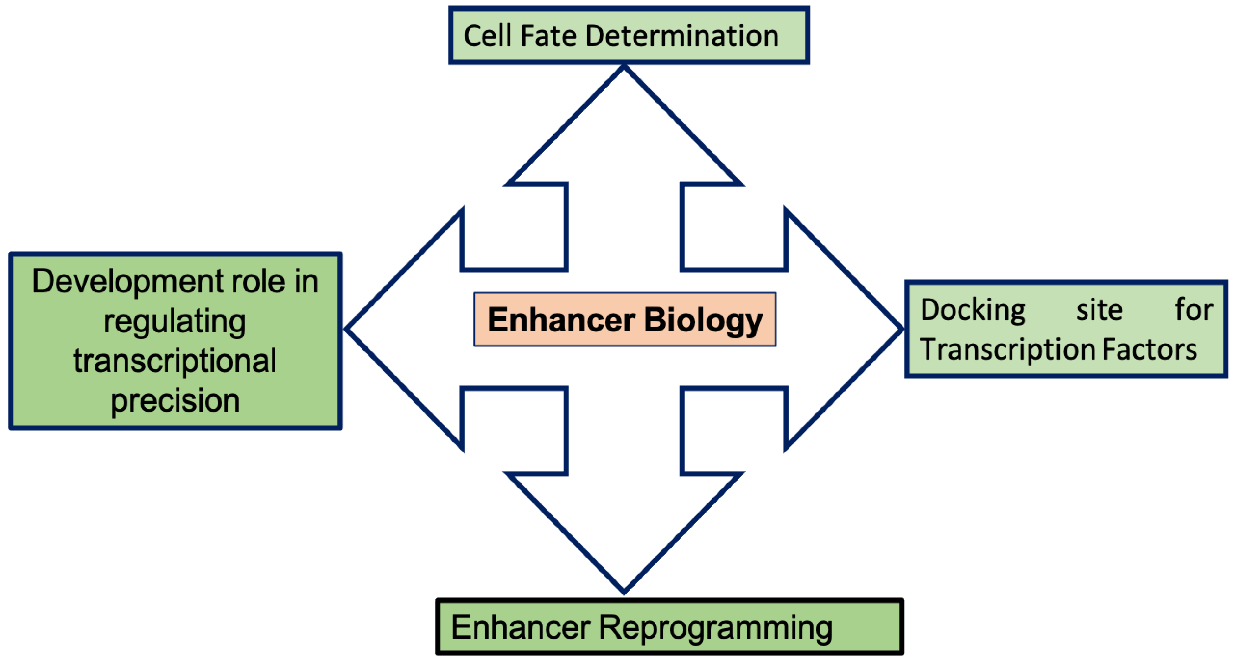
Figure 1.
Schematic diagram representing enhancer biology.


Table 1.
Studies investigating the roles of enhancers in development.
| Number | Title | Author | Reference |
|---|---|---|---|
| 1 | Highly conserved non-coding sequences are associated with vertebrate development | Woolfe et al., 2005 | [39] |
| 2 | Simple Combinations of Lineage-Determining Transcription Factors Prime cis-Regulatory Elements Required for Macrophage and B Cell identities | Heinz et al., 2010 | [14] |
| 3 | Transcriptional enhancers in animal development and evolution | Levine Mike, 2010 | [40] |
| 4 | A unique chromatin signature uncovers early development enhancers in humans | Rada-Iglesias et al., 2011 | [20] |
| 5 | Multilineage Priming of Enhancer Repertoires Precedes Commitment to the B and Myeloid Cell Lineages in Hematopoietic Progenitors | Mercer et al., 2011 | [52] |
| 6 | Chromatin “Prepattern” and Histone Modifiers in a Fate Choice for Liver and Pancreas | Xu et al., 2011 | [43] |
| 7 | Foxp3 Exploits a Pre-Existent Enhancer Landscapes for Regulatory T Cell Lineages Specification | Samstein et al., 2012 | [41] |
| 8 | A Temporal Chromatin Signature in Human Embryonic Stem Cells Identifies Regulators of Cardiac Development | Paige et al., 2012 | [46] |
| 9 | Combinatorial Assembly of Development Stage-Specific Enhancers Controls Gene Expression Programs during Human Erythropoiesis | Xu, J. et al., 2012 | [15] |
| 10 | Dynamic and Coordinated Epigenetic Regulation of Developmental Transitions in the Cardiac Lineage | Wamstad et al., 2012 | [45] |
| 11 | Enhancers as information integration hubs in development: lesson from genomics | Buecker & Wysocka, 2012 | [6] |
| 12 | Developmental Fate and Cellular Maturity Encoded in Human Regulatory DNA Landscapes | Stergachis et al., 2013 | [56] |
| 13 | Latent Enhancers Activated by Stimulation in Differentiated Cells | Ostuni et al., 2013 | [10] |
| 14 | Epigenetic Priming of Enhancers Predicts Developmental Competence of hESC-Derived Endodermal Lineage Intermediates | Wang et al., 2015 | [44] |
| 15 | Scl binds to primed enhancers in mesoderm to regulate hematopoietic and cardiac fate divergence | Org et al., 2015 | [49] |
| 16 | Pioneer factors govern super-enhancer dynamics in stem cell plasticity and lineage choice | Rc et al., 2015 | [47] |
| 17 | Early enhancer establishment and regulatory locus complexity shape transcriptional programs in hematopoietic differentiation | Gonzalez et al., 2015 | [42] |
| 18 | Enhancer repertoires are reshaped independently of early priming and heterochromatin dynamics during B cell differentiation | Choukrallah et al., 2015 | [16] |
| 19 | Lineage-Specific Genome Architecture Links Enhancer and Non-coding Disease Variants to Target Gene Promoters | Javierre et al., 2016 | [57] |
| 20 | Enhancer priming by H3K4 methyltransferase MLL4 controls cell fate transition | Wang et al., 2016 | [48] |
| 21 | Ever Changing landscape: transcriptional enhancers in development and evolution | Long et al., 2016 | [58] |
| 22 | Lineage-specific dynamic and pre-established enhancer-promoter contacts cooperate in terminal differentiation | Rubin, J.A. et al., 2017 | [53] |
| 23 | Dynamic lineage priming is driven via direct enhancer regulation by ERK | Hamilton et al., 2019 | [51] |
| 24 | FOXA2 is Required for Enhancer Priming during Pancreatic Differentiation | Lee et al., 2019 | [50] |
| 25 | Cardiac Reprogramming Factors Synergistically Activate Genome-wide Cardiogenic Stage-Specific Enhancers | Hashimoto et al., 2019 | [59] |
Table 2.
Studies investigating the roles of Enhancers in disease development.
| Number | Title | Author | Reference |
|---|---|---|---|
| 1 | Translocation of the c-myc gene into the immunoglobulin heavy chain locus in human Burkitt lymphoma and murine plasmacytoma cells | Taub et al., 1982 | [84] |
| 2 | A long-range Shh enhancer regulates expression in the developing limb and fin and is associated with preaxial polydactyly | Lettice, L.A. et al., 2003 | [85] |
| 3 | Genomic deletion of a long-range bone enhancer misregulates in Van Buchem disease | Loots, G.G. et al., 2005 | [86] |
| 4 | A common sex dependent mutations in a RET enhancer underlies Hirschsprung disease risk | Emison, E.S. et al., 2005 | [87] |
| 5 | Disruption of an AP2-alpha binding site in an IRF6 enhancer is associated with cleft lip | Rahimov et al., 2008 | [88] |
| 6 | Functional enhancers at the gene-poor 8q24 cancer linked locus | Jia, L. et al., 2009 | [89] |
| 7 | The 8q24 cancer risk variant rs6983267 shows long-range interaction with MYC in colorectal cancer | Pomeratz, M.M. et al., 2009 | [90] |
| 8 | The common colorectal cancer predisposition SNP rs6983267 at chromosome 8q24 confers potential to enhanced WNT signaling | Tuupanen, S. et al., 2009 | [91] |
| 9 | Long-range enhancers on 8q24 regulate c-Myc | Sotelo et al., 2010 | [92] |
| 10 | An 8q24 gene desert variant associated with prostate cancer risk confers differential in vivo activity to a MYC enhancer. | Wasserman, N.F. et al., 2010 | [93] |
| 11 | Enhancer-adoption as a mechanism of human developmental disease | Lettice, L.A. et al., 2011 | [94] |
| 12 | Systematic localization of common disease associated variation in regulatory DNA | Maurano et al., 2012 | [76] |
| 13 | Epigenomic enhancer profiling defines a signature of colon cancer | Akhtar-Zaidi, B. et al., 2012 | [73] |
| 14 | Mice lacking a Myc enhancer that includes human SNP rs6983267 are resistant to intestinal tumors | Sur et al., 2012 | [95] |
| 15 | A novel 13 base pair insertion in the sonic hedgehog ZRS limb enhancer (LMBR1) causes preaxial polydactyly with triphalangeal thumb | Laurell, T. et al., 2012 | [96] |
| 16 | Regulatory variation in a TBX5 enhancer leads to isolated congenital heart diseases | Smemo, S. et al., 2012 | [97] |
| 17 | DNA methylation of transcriptional enhancers and cancer predisposition | Aran and Hallman et al., 2013 | [98] |
| 18 | Discovery and characterization of super-enhancer associated dependencies in diffuse large B cell lymphoma | Chapuy, B. et al., 2013 | [99] |
| 19 | Chromatin stretch enhancer states drive cell specific gene regulation and harbor human disease risk variants | Parker, S.C. et al., 2013 | [26] |
| 20 | Role of SWI/SNF in acute leukemia maintenance and enhancer-mediated Myc regulation | Shi, J. et al., 2013 | [100] |
| 21 | Selective inhibition of tumor oncogenes by disruption of super-enhancers | Loven, J. et al., 2013 | [29] |
| 22 | An erythroid enhancer of BCL11A subject to genetic variation determines fetal hemoglobin level | Bauer, D.E. et al., 2013 | [101] |
| 23 | Disruption of autoregulatory feedback by a mutation in a remote, ultraconserved PAX6 enhancer causes aniridia | Bhatia, S. et al., 2013 | [102] |
| 24 | Genome-wide analysis of noncoding regulatory mutations in cancer | Weinhold, N. et al., 2014 | [103] |
| 25 | A NOTCH driven MYC enhancer promotes T cell development, transformation and acute lymphoblastic leukemia | Herranz, D. et al., 2014 | [104] |
| 26 | Combinatorial effects of multiple enhancer variants linkage disequilibrium dictate levels of gene expression to confer susceptibility to common traits | Corradin, O. et al., 2014 | [27] |
| 27 | Enhancer hijacking activates GFI1 family oncogenes in medulloblastoma | Northcott, P.A. et al., 2014 | [105] |
| 28 | Microduplications encompassing the sonic hedgehog limb enhancer ZRS are associated with Hass-type polysyndactyly and Laurin Sandrow syndrome | Lohan, S. et al., 2014 | [106] |
| 29 | Epigenomic analysis of primary human T cells reveals enhancers associated with TH2 memory cell differentiation and asthma susceptibility | Seumois, G. et al., 2014 | [107] |
| 30 | Oncogenic regulation. An oncogenic Super enhancer formed through somatic mutation of a noncoding intergenic element. | Mansour, M.R. et al., 2014 | [108] |
| 31 | A Sox2 distal enhancer cluster regulates embryonic stem cell differentiation potential | Zhou, H.Y. et al., 2014 | [109] |
| 32 | Multiple functional risk variants in a SMAD7 enhancer implicate a colorectal cancer risk haplotype | Fortini, B.L. et al., 2014 | [110] |
| 33 | A remote GATA2 hematopoietic enhancer drives leukemogenesis in inv(3) (q21;q26) by activating EVI1 expression | Yamazaki et al., 2014 | [111] |
| 34 | Long range enhancer activity determines Myc sensitivity to Notch inhibitors in T cell leukemia | Yashiro-Ohtani et al., 2014 | [112] |
| 35 | Recessive mutations in a distal PTF1A enhancer cause isolated pancreatic agenesis | Weedon et al., 2014 | [81] |
| 36 | A single oncogenic enhancer rearrangement causes concomitant EV1 and GATA2 deregulation in leukemia | Groschel et al., 2014 | [19] |
| 37 | Genetic predisposition to neuroblastoma mediated by a LMO1 super enhancer polymorphisms | Oldridge, D.A. et al., 2015 | [113] |
| 38 | 7q21.3 Deletion involving enhancer sequences within the gene DYNC1I1 presents with intellectual disability and split hand-split foot malformation with decreased penetrance | Delgado, S. and Velinov, M., 2015 | [114] |
| 39 | A large genomic deletion leads to enhancer adoption by the lamin B1 gene: a second path to autosomal dominant adult-onset demyelinating leukodystrophy (ADLD) | Giorgio, E. et al., 2015 | [115] |
| 40 | Multiple functional variants in long-range enhancer contribute to the risk of SNP rs965513 in thyroid cancer | He, H. et al., 2015 | [116] |
| 41 | Loss of TET2 in hematopoietic cells leads to DNA hypermethylation of active enhancers and induction of leukemogenesis | Rasmussen et al., 2015 | [117] |
| 42 | The Transcriptional cofactor TRIM33 prevents apoptosis B lymphoblastic leukemia by deactivating a single enhancer | Wang et al., 2015 | [118] |
| 43 | Super-enhancers delineate disease-associated regulatory nodes in T cells | Vahedi et al., 2015 | [83] |
| 44 | Identification of focally amplified lineage-specific super-enhancers in human epithelial cancer | Zhang, X. et al., 2016 | [119] |
| 45 | Role of non-coding sequence variants in cancer | Khurana, E. et al., 2016 | [120] |
| 46 | Ever-changing landscapes: transcriptional enhancers in development and evolution | Long, H.K. et al., 2016 | [58] |
| 47 | Genetic Predisposition to Chronic Lymphocytic Leukemia is mediated by a BMF Super-Enhancer Polymorphisms | Kandaswamy et al., 2016 | [121] |
| 48 | DNMT3A Loss drives Enhancer Hypomethylation in FLT3-ITD-Associated Leukemias | Yang et al., 2016 | [122] |
| 49 | Epigenomic profiling of primary gastric adenocarcinoma reveals super-enhancer heterogeneity | Ooi et al., 2016 | [82] |
| 50 | Parkinson associated risk variants in distal enhancers of a-syncuclein modulates target gene expression | Soldner et al., 2016 | [80] |
| 51 | Hotspots of aberrant enhancer activity punctuate the colorectal cancer epigenome | Cohen, A.J. et al., 2017 | [30] |
| 52 | Composition and dosage of a multipartite enhancer cluster control developmental expression of Ihh (Indian hedgehog) | Will, A.J. et al., 2017 | [123] |
| 53 | Superenhancer Analysis Defines Novel Epigenomic Subtypes of Non-APL AML, including an RARaalpha Dependency Targetable by SY-1425, a Potent and Selective RARalpha Agonist | MCKeown, M.R. et al., 2017 | [124] |
| 54 | Enhancer profiling identifies critical cancer genes and characterize cell identity in adult T-cell leukemia | Wong et al., 2017 | [125] |
| 55 | APOBEC signature mutation generate an oncogenic enhancer that drives LMO1 expression in T-ALL Leukemia | Li et al., 2017 | [126] |
Publisher’s Note: MDPI stays neutral with regard to jurisdictional claims in published maps and institutional affiliations. |
© 2021 by the author. Licensee MDPI, Basel, Switzerland. This article is an open access article distributed under the terms and conditions of the Creative Commons Attribution (CC BY) license (https://creativecommons.org/licenses/by/4.0/).
Share and Cite
MDPI and ACS Style
Maurya, S.S. Role of Enhancers in Development and Diseases. Epigenomes 2021, 5, 21. https://0-doi-org.brum.beds.ac.uk/10.3390/epigenomes5040021
AMA Style
Maurya SS. Role of Enhancers in Development and Diseases. Epigenomes. 2021; 5(4):21. https://0-doi-org.brum.beds.ac.uk/10.3390/epigenomes5040021
Chicago/Turabian StyleMaurya, Shailendra S. 2021. "Role of Enhancers in Development and Diseases" Epigenomes 5, no. 4: 21. https://0-doi-org.brum.beds.ac.uk/10.3390/epigenomes5040021