Article

Fungal Species Causing Maize Leaf Blight in Different Agro-Ecologies in India

1
Department of Botany and Plant Physiology, Chaudhary Charan Singh Haryana Agricultural University Regional Research Station, Karnal 132001, India
2
Sustainable Agricultural Systems Laboratory, USDA-ARS, Beltsville, MD 20705, USA
3
ICAR-Indian Institute of Maize Research (Delhi Unit), Pusa Campus, New Delhi 110012, India
4
Department of Mycology and Plant Pathology, Rajiv Gandhi South Campus, Banaras Hindu University, Mirzapur 231001, India
5
Germplasm Evaluation Division, National Bureau of Plant Genetic Resources, New Delhi 110012, India
*
Authors to whom correspondence should be addressed.
Submission received: 9 November 2021 / Revised: 2 December 2021 / Accepted: 8 December 2021 / Published: 14 December 2021
(This article belongs to the Section Fungal Pathogens)

Abstract

:
Foliar diseases of maize cause severe economic losses in India and around the world. The increasing severity of maize leaf blight (MLB) over the past ten years necessitates rigorous identification and characterization of MLB-causing pathogens from different maize production zones to ensure the success of resistance breeding programs and the selection of appropriate disease management strategies. Although Bipolaris maydis is the primary pathogen causing MLB in India, other related genera such as Curvularia, Drechslera, and Exserohilum, and a taxonomically distant genus, Alternaria, are known to infect maize in other countries. To investigate the diversity of pathogens associated with MLB in India, 350 symptomatic leaf samples were collected between 2016 and 2018, from 20 MLB hotspots in nine states representing six ecological zones where maize is grown in India. Twenty representative fungal isolates causing MLB symptoms were characterized based on cultural, pathogenic, and molecular variability. Internal Transcribed Spacer (ITS) and glyceraldehyde-3-phosphate dehydrogenase (GADPH) gene sequence-based phylogenies showed that the majority of isolates (13/20) were Bipolaris maydis. There were also two Curvularia papendorfii isolates, and one isolate each of Bipolaris zeicola, Curvularia siddiquii, Curvularia sporobolicola, an unknown Curvularia sp. isolate phylogenetically close to C. graminicola, and an Alternaria sp. isolate. The B. zeicola, the aforesaid four Curvularia species, and the Alternaria sp. are the first reports of these fungi causing MLB in India. Pathogenicity tests on maize plants showed that isolates identified as Curvularia spp. and Alternaria sp. generally caused more severe MLB symptoms than those identified as Bipolaris spp. The diversity of fungi causing MLB, types of lesions, and variation in disease severity by different isolates described in this study provide baseline information for further investigations on MLB disease distribution, diagnosis, and management in India.

1. Introduction

Maize (Zea mays L.) is an important cereal crop in India and ranks third in production after wheat and rice [1]. Maize has wide adaptability and is gaining popularity as evidenced by its rising production and productivity [2]. Most of the produce in India is consumed as food for humans, fodder for animals, and feed for poultry, apart from applications as industrial raw materials. Among the 35 diseases affecting crop health, viz. seed rots and seedling blights, root and stalk rots, foliar diseases, and ear rots, maize leaf blight (MLB) caused by Bipolaris maydis [(Nisikado& Miyake) Shoem] is one of the major diseases of maize [1,2]. This disease has been detected in almost all maize growing areas of India [3] and is a constraint on crop improvement programs. In India, MLB occurs in the states of Punjab, Haryana, Delhi, Uttar Pradesh, Bihar, Madhya Pradesh, Gujarat, Jammu and Kashmir, Sikkim, Meghalaya, Rajasthan, Andhra Pradesh, and Maharashtra [3].
Bipolaris and related genera such as Curvularia, Dreschslera, and Exserohilum are ascomyceteous fungi known to infect maize [4], and belong to the Class Dothideomycetes, Order Pleosporales, and Family Pleosporaceae. Although the name Cochliobolus (1934) has been in the literature longer than the name Bipolaris (1959), it is not frequently used in disease reports, and Bipolaris is widely applied in taxonomy [5]. The association of the genus Bipolaris with plants from the family Poaceae is very common; however, species of Bipolaris have been reported to infect 60 other host genera, either as saprophytes or phytopathogens [6,7]. The spread of phytopathogenic species of the genus Bipolaris may have occurred due to international trade [8].
Based on molecular phylogenetic analysis with the ITS1-5.8S-ITS2 region of rDNA and partial sequence of the GAPDH (glyceraldehyde-3-phosphate dehydrogenase) gene [9], Drechslera, Bipolaris, and Curvularia are distinct genera even though they share many morphological similarities [4,9,10]. In addition, Bipolaris and Curvularia both have sexual morphs in Cochliobolus [11]. In molecular analyses of ITS and GAPDH gene data, two major clades, Cochliobolus Group-1 and Group-2 were clustered [12]. Similar results were obtained from a combined analysis of ITS, GAPDH, TEF (translation elongation factor-1 alpha gene), and LSU (28S nrRNA gene) sequence data [13]. Group-1 included the genus Bipolaris (type species B. maydis), and Group-2 included the genus Curvularia, (type species C. lunata).
The fungus B. maydis ((Teleomorph: Cochliobolus heterostrophus (Drechsler) Drechsler) exists as four different races infecting maize across the world, viz. race ‘T’, ‘O’ ‘C’ ‘S’ [14]. Race ‘T’ of B. maydis is highly virulent and was reported to cause the devastating ‘Southern corn leaf blight’ epidemic in the USA during the 1970s, resulting in huge losses due to the extremely susceptible response of Texas Male Sterile maize lines [6,15,16,17,18]. Although race ‘T’ has been reported on seeds of Phaseolus mungo, Vigna sinensis, and Paspalum scrobiculatum from India [19], B. maydis race ‘O’ is the prevalent pathogen of maize in India and elsewhere in the world [4,20,21]. Drechslera maydis (synonym of B. maydis) on maize was reported for the first time in Punjab [22]. The occurrence of a pathotype resembling race ‘T’ in India on maize hybrids 2310 and 2420 from Ludhiana was reported for the first time in 1978 [23]. Race C is the most virulent in maize cytoplasmic male sterile line C (c-cms), currently reported only in China [20,24].
For over a decade, the severity of MLB on maize in the Indian subcontinent has been increasing and the disease has spread to areas where it was not previously reported. A remarkable variability in the range of symptoms has also been recorded. Due to this variability in symptoms, it is very important to document the species of Bipolaris and other associated genera infecting maize, and to precisely characterize the infecting species complex. The rising incidence of Curvularia sp. on maize is also a concern for crop improvement programs [25]. The objectives of this study were (1) to examine the diversity of Bipolaris species and associated fungal genera causing MLB across different agro-ecological zones of India, (2) to provide correct morphological descriptions and identification of the species/isolates for reference in order to device identification and management strategies, and (3) to assess the virulence spectrum of common MLB pathogenic species. Knowledge gained from this study will provide benchmark information necessary to accurately identify MLB pathogens, breed for resistant cultivars, and improve disease management practices in India.

2. Results

2.1. Survey, Collection, and Maintenance of Fungal Isolates

A total of 350 symptomatic maize leaf samples showing maize leaf blight (MLB)-like symptoms were collected from 20 hotspots representing six agro-ecological zones under maize production during survey and surveillance visits during the Kharif seasons (June–October) in 2016–2017 and 2017–2018 (Supplementary Material: Figure S1). The six hotspots, viz. Godhra, Anand, Sukheri, Lakhawali, Mandya, and Mysore showed variability in morphological and pathogenic profiles. However, in view of the sample size and constraints of the resources available for this study, one candidate isolate was selected from each of the 20 hotspots, viz. Pantnagar, Bajaura, Kangra, Patelnagar, Ludhiana, Pichola, Banswara, Kota, Chittorgarh, Dungarpur, Amberi, Karnal, Dholi, Samastipur, Godhra, Anand, Sukheri, Lakhawali, Mandya, and Mysore for phylogenetic analysis (Table 1). Isolates were examined for typical characteristics of B. maydis which is the prevalent species infecting maize in India, as reported by earlier workers [23,26,27,28]. Variability in colony growth (slow, medium, or fast), color (light grey, dark grey, light black, or black), texture (rough raised, rough appressed, smooth raised, smooth appressed with or without zonation, and regular or irregular margins), conidial dimensions (7.6 µm to 19.3 µm × 3.1 µm to 6.1 µm), pathogenicity (mild to high) and disease severity (30 to 90%) revealed that out of 20 hotspots surveyed, fungal isolates from 14 hotspots (Table 2) showed typical morphological and pathogenic characteristics of Cochliobolus heterostrophus (Drechs.) Drechs. (anamorph = Bipolaris maydis (Nisi-kado) Shoemaker; synonym = Helminthosporium maydis Nisikado).

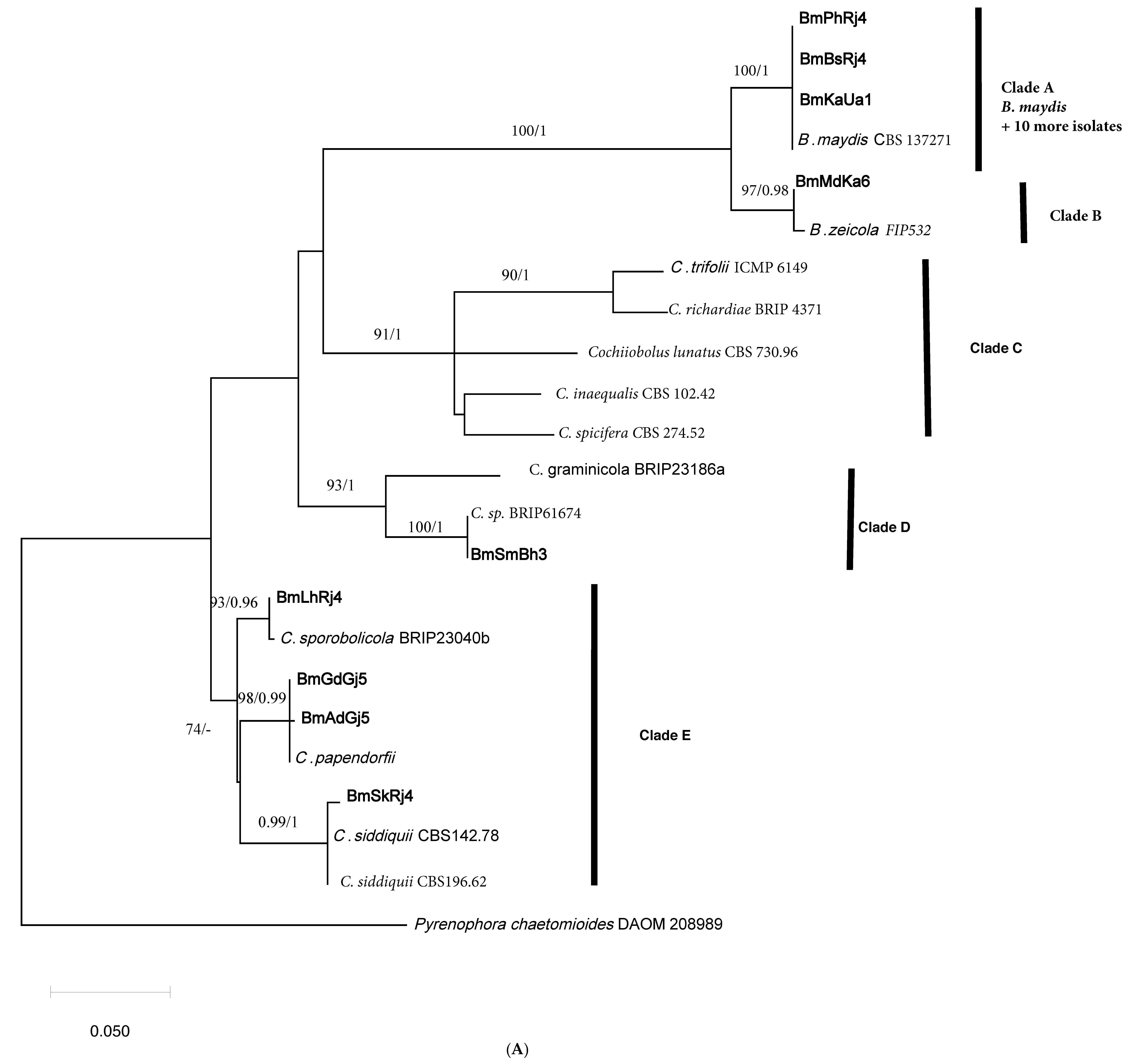
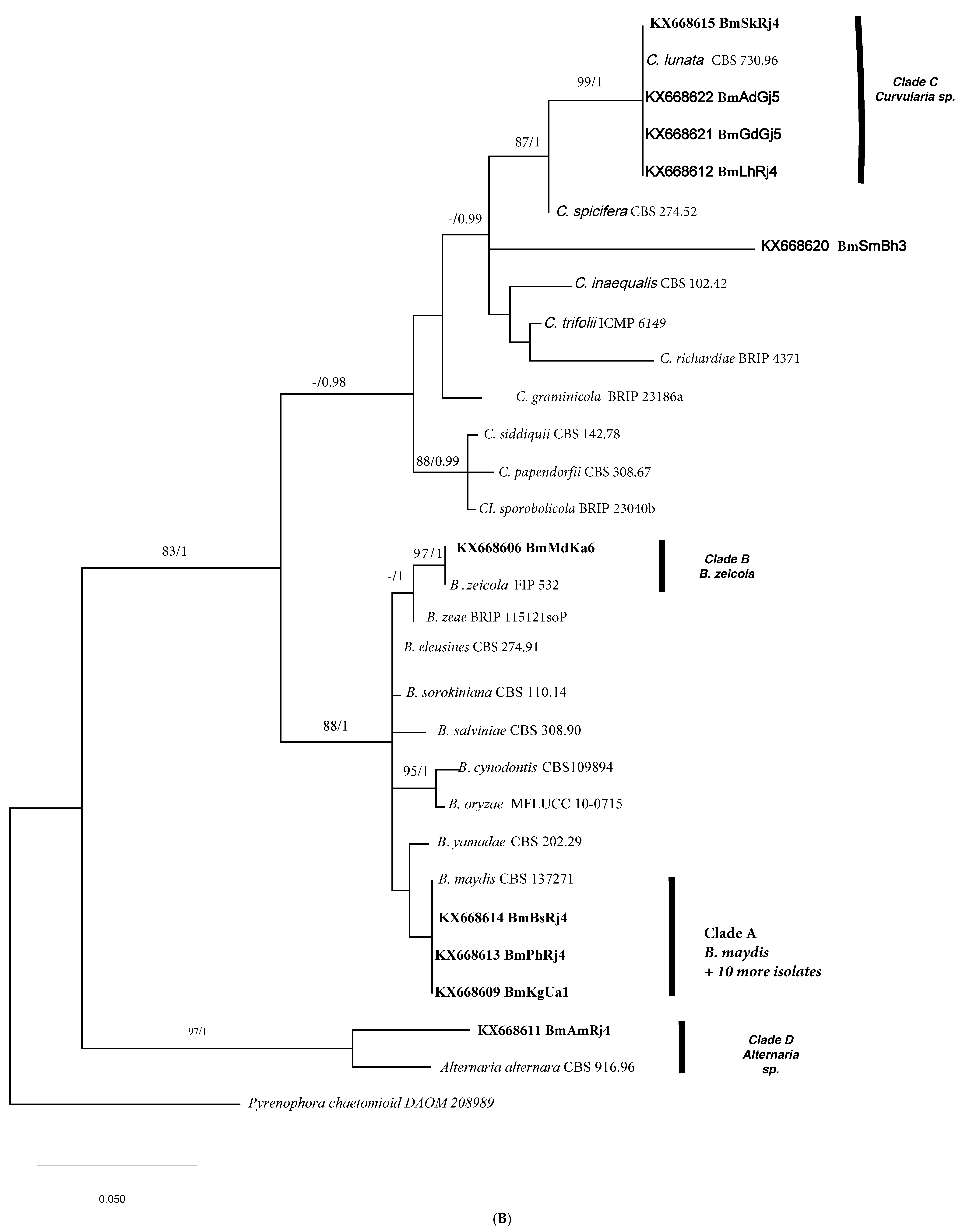
2.2. Phylogenetic Analyses

The final ITS alignment file had thirty taxa and 483 characters of which 84 were parsimony informative representing 17% of the total characters. The GADPH file had 23 taxa and 512 characters of which 118 were parsimony informative representing 23% of the total characters. For both files, reference sequences representing mostly sequences of ex-type cultures of Bipolaris or Curvularia species were obtained from GenBank and were included in Supplementary Material (Table S1). The phylogenetic trees obtained with Bayseian analyses had essentially identical topology to the trees obtained by maximum likelihood (ML). Therefore, for each locus, only one ML tree was selected for this publication, but the posterior probability (PP) support for the branches from the Bayesian trees is reflected on the maximum likelihood tree branches. The maximum likelihood tree shown in Figure 1A based on GAPDH sequence data reveals that three taxa under study formed a highly supported subclade (A) with a sequence of ex-type culture B. maydis. As a matter of fact, there were 10 additional isolates with identical sequences to taxa in clade A which were not included in the analysis due to the tree size. In other words, 13 of 20 isolates were identified as B. maydis. The tree based on ITS sequence data (Figure 1B) was congruent with the GAPDH tree and revealed the same clade (A) within a large all Bipolaris species clade. One isolate (BmMdKa6) had identical sequence to the ex-type culture of B. zeicola and they together formed a highly supported (BS = 97%; PP = 0.98) subclade B (Figure 1A). This subclade was sister to the B. maydis subclade with BS support of 100% and PP of 1. Similar to subclade A, isolate BmMdKa6 was also identified as B. zeicola in the ITS tree (subclade B; Figure 1B). In brief, among the 20 isolates identified morphologically and pathologically as fungi causing MLB, 13 isolates (viz. BmPhRj4, BmBjUa1, BmBsRj4, BmDhBh3, BmCgRj4, BmPnDl2, BmDnRj4, BmKgUa1, BmKrHr2, BmKtRj4, BmLdPj2, BmPtUa1, and BmMyKa6) representing 13 hotspots were identified as B. maydis and one isolate (BmMdKa6) was identified as B. zeicola, based on phylogenetic trees of two loci, GAPDH, and ITS.
The remaining taxa in the tree were Curvularia sp. isolates and they formed three main supported clades C, D, and E. (Figure 1A). Clades C, D, and E had a polytomy relationship to each other and to Bipolaris clades B and A. (Figure 1A). Within clade E, isolates BmAdGj5 and BmGdGj5 and ex-type culture of C. papendorfii had identical sequences, and together they formed a highly supported subclade (BS = 98% and PP = 0.99 subclade). Isolate BmLhRj4 had an identical sequence to an ex-type culture of C. sporobolicola, and together they formed a highly supported subclade. Isolate BmSkRj4 formed a subclade with two isolates of C. siddiquii, with high support (BS = 0.99 and PP = 1). In the ITS tree, however, the isolates that fell into clade E (BmLhRj4, BmGdGj5, BmAdGj5, and BmSkRj4) formed a highly supported clade with C. lunata and they all had identical ITS sequences. Clearly, phylogeny based on ITS sequence data appears to be unsuccessful in resolving Curvularia isolates to the species level like the GAPDH tree. Isolate BmSmBh3 (Figure 1A) formed a clade with the sequence of the unidentified species of Curvularia (Accession No. KU552166). The species C. graminicola was shown to be the closest relative to them (Clade D, Figure 1A). Therefore, this unidentified species remained ambiguous, needs additional markers-based phylogeny for clarity, and was named C. graminicola-like fungus for this report. In the ITS tree, isolate BmSmBh3 fell into the large Curvularia clade but did not form a clade with a sequence of any known species of Curvularia.
The isolate BmAmRj4 did not fall into the Bipolaris or Curvularia clades but formed a clade with the ex-type culture of Alternaria alternata in the ITS tree (Figure 1B). The two sequences had 100% homology. However, we could not obtain the GAPDH sequence for this isolate, and thus it was excluded from the phylogenetic analyses based on GAPDH. In summary, in addition to the 14 isolates of B. maydis and B. zeicola that were causative agents for MLB, we identified two isolates of C. papendorfii, and one isolate each of C. siddiquii, C. sporobolicola, a C. graminicola-like fungus, and an Alternaria sp. as MLB causing fungi in India.

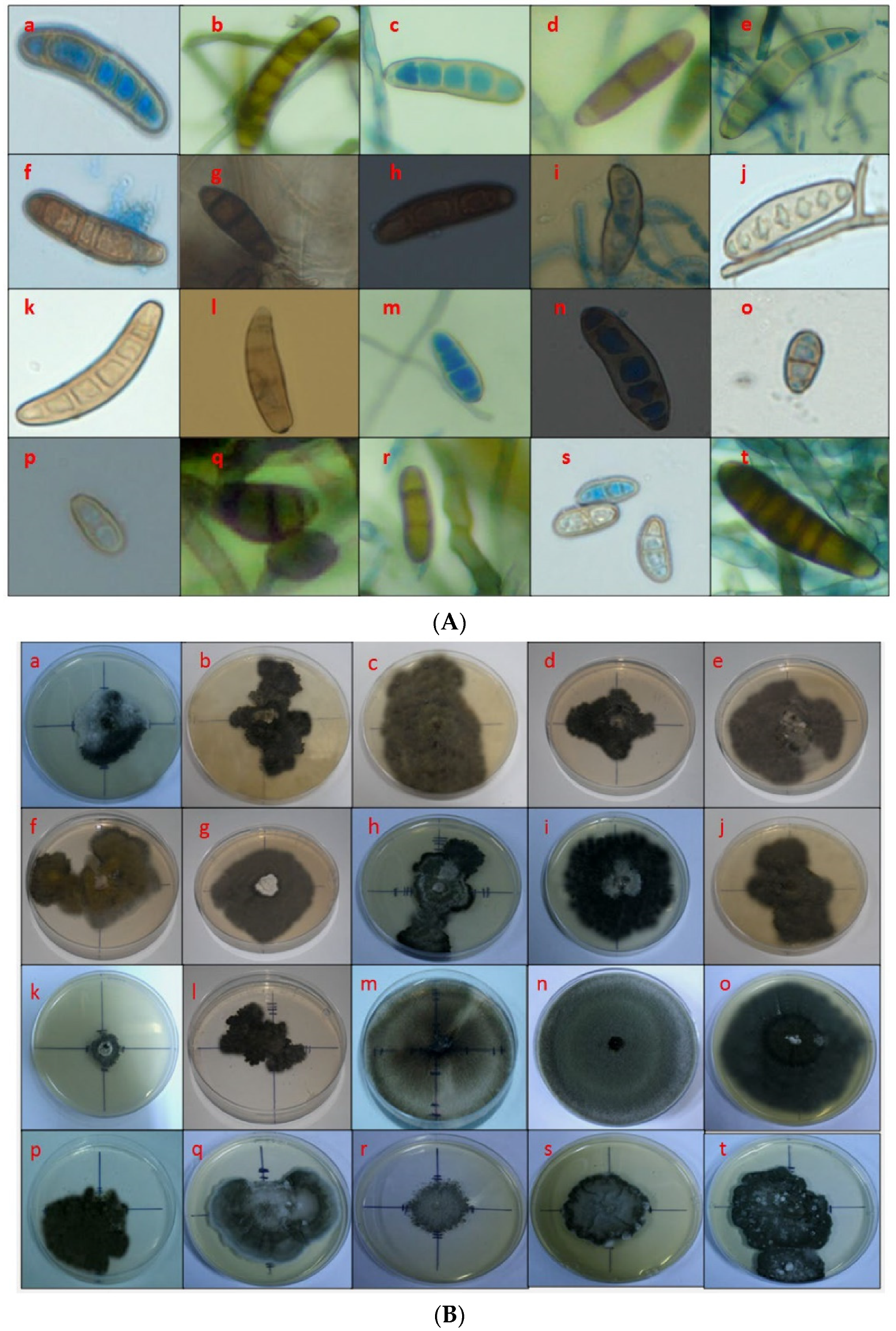
2.3. Morphological Characterization of Fungal sp. Causing Maize Leaf Blight

The 20 candidate fungal isolates maintained on Potato Dextrose Agar (PDA) plates showed striking variability in culture morphology and conidial dimensions (Table 2; Figure 2A). The length and width of conidia of different isolates varied between 7.6 µm (BmgGdGj5, Godhra) to 19.3 µm (BmPnDl12, Patel Nagar) and 6.1 µm to 3.1 µm (BmSmBh3, Samastipur and BmgGdGj5, Godhra or BmSkRj4, Sukher), respectively. The isolate BmPnDl12 (Patel Nagar) had the largest sized conidia with mean length of 19.3 µm and width of 5.0 µm and BmgGdGj5 (Godhra) had the smallest conidia, 7.6 µm × 3.1 µm. The conidial dimensions of the rest of the 18 isolates were in the order BmAmRj4 (Amberi) > BmLhrj5 (Lakhawali) > BmBjUa1 (Bajaura) > BmSkRj4 (Sukher) > BmKrHr2 (Karnal) > BmCgRj4 (Chittorgarh) > BmPtUa1 (Pantnagar) > BmDhBh3 (Dholi) > BmLdPj2 (Ludhiana) > BmMyKa6 (Mysore) > BmKtRj4 (Kota) > BmBsRj4 (Banswara) > BmAdGj5 (Anand) > BmMdKa6 (Mandya) > BmDnRj4 (Dungarpur) > BmKgUa1 (Kangra) > BmPhRj4 (Pichola) > BmSmBh3 (Samastipur) (Table 2).
The number of septa of various isolates ranged between one to three (BmgGdGj5, Godhra isolate) to five to eight (BmBjUa1, Bajaura isolate) (Table 2; Figure 2A). For the rest of the isolates, it was in the order BmAmRj4 (Amberi) = BmLhRj4 (Lakhawali) > BmKrHr2 (Karnal) > BmMdKa6 (Mandya) > BmPnDl12 (Patel Nagar) > BmBsRj4 (Banswara) = BmLdPj2 (Ludhiana) > BmDhBh3 (Dholi) > BmPtUa1 (Pantnagar) = BmMyKa6 (Mysore) > BmCgRj4 (Chittorgarh) > BmAdGj5 (Anand) > BmSkRj4 (Sukher) > BmSmBh3 (Samastipur) > BmKtRj4 (Kota) = BmDnRj4 (Dungarpur) = BmPhRj4 (Pichola) (Table 2).
Various cultural characteristics like radial growth, colony texture, color, and diameter revealed that the isolates from Banswara, Chittorgarh, Patel Nagar, Dungarpur, Kangra, Karnal, Godhra, Mandya, and Mysore had fast radial growth on PDA, whereas isolates from Pichola, Bajaura, Dholi, Kota, Pantnagar, Amberi, Lakhawali, and Samastipur were medium growing and isolates from Ludhiana and Sukher were slow growing. The isolate BmMdKa6 (Mandya) had the fastest radial growth, whereas BmLdPj2 had the slowest growth. Colony colors were light black (BmPhRj4 and BCgRj4); light grey (BmDnRj4, BmMdKa6, BmMyKa6); dark grey (BmKgUA1, BmKrHr2, BmBmLdPj2, BmPtUa1, BmLhRj4, BmSkRj4, BmSmBh3) and black (BmBjUa1, BmBsRj4, BmDhBh3, BmPnDl12, BmKtRj4, BmGdGj5, BmAmRj4, BmAdGj5) (Figure 2B).
The culture morphology of C. papendorfii showed a rough surface with irregular margins and no zonation, conidial dimensions 7.6 µm × 3.1 µm to 16.9 µm × 4.1 µm, and one or more septa (Table 2) which falls into Group-2 (Table S2); whereas B. zeicola showed smooth appressed surface with regular margins and zonation, conidial dimensions 7.8 µm × 6.1 µm to 13.1 µm × 3.8 µm slightly larger than C. papendorfii, and 3 to 4 septa (Table 2) formed Group-5 (Table S2). The C. papendorfii isolate from Godhra showed a rough raised surface while the Anand isolate showed a rough appressed surface with no zonation and smooth margins. The conidial size was also very small, with 1 to 2 septa, and ranged from 7.6 to 12.1 µm × 3.1 to 4.3 µm. Ecologically both B. zeicola and C. papendorfii prevailed in different parts of the plateau and hill regions which are at high altitudes with cool and moist climates close to the sea (C. papendorfii from the western part having sandy loam soil and B. zeicola from the southern part of the plateau and hill region having laterite soils). Isolates from Lakhawali, Sukher, and Anand showed variable morpho–pathogenic profiles and represented different groups being different species in the genus Curvularia. The conidia of the C. sporobolicola (Lakhawali) isolate were slightly curved with 2 to 3 septa, conidial size ranging from 17.2 µm × 3.7 µm, forming a rough colony with zonation and irregular margins. However, the C. siddiquii (Sukher) isolate had conidial sizes smaller than the C. sporobolicola isolate (i.e., 16.8 µm × 3.1 µm in size) with a slight curvature, 3 to 4 septa, smooth appressed colony, no zonation, and rough margins. The conidial morphology of C. papendorfii was oval or obpyriform with 1–3 septa. The C. graminicola–like sp. (Samastipur) isolate produced sickle-shaped conidia with 2 to 3 septa, ranging in size from 7.8 to 6.1 µm and 2.6 µm average diameter. Alternaria sp. was also isolated from maize in some regions from the Amberi hotspot in Rajasthan.

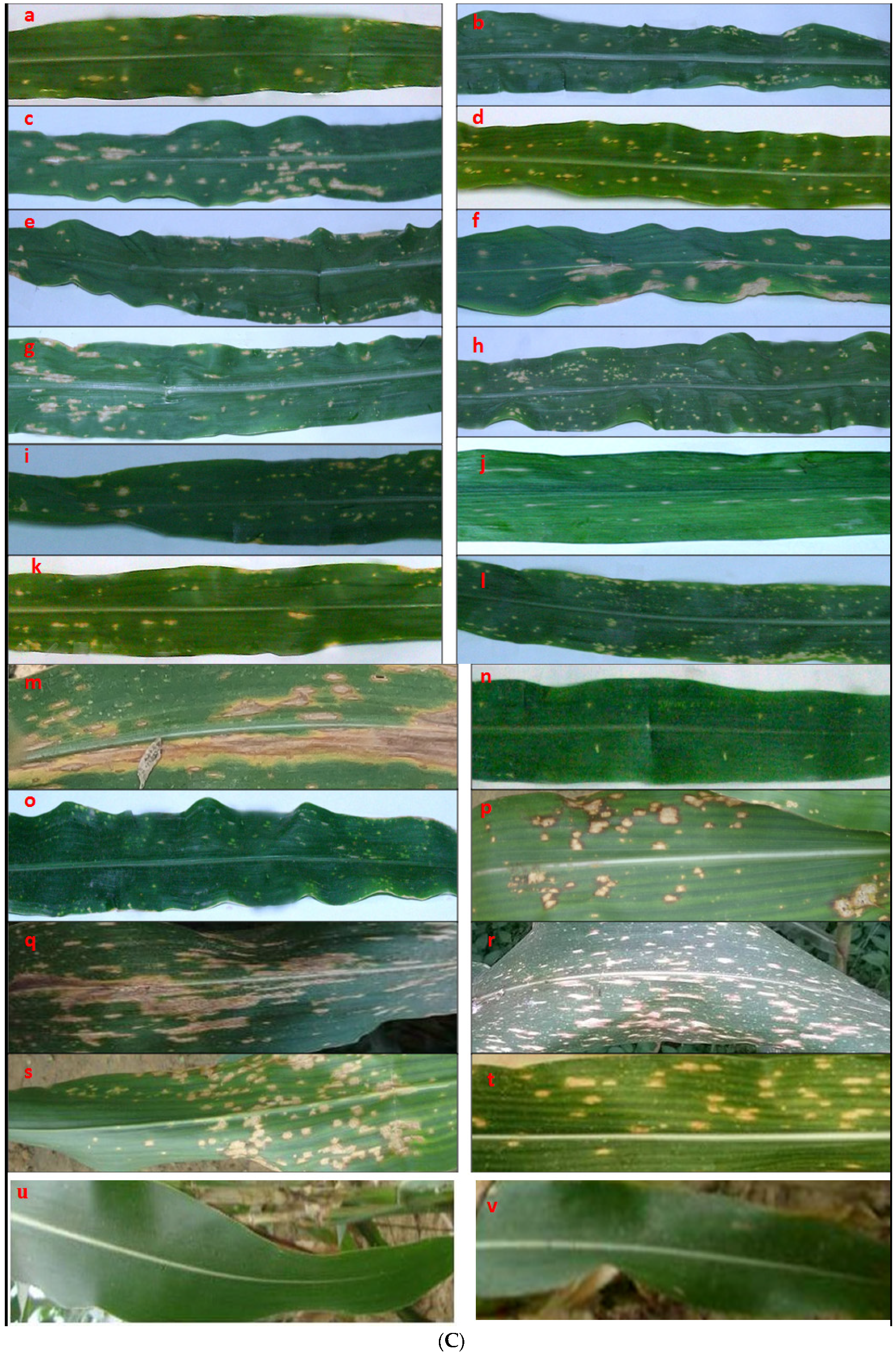
2.4. Pathogenic Variability of Fungal Isolates on Zea mays c.v. DHM 117

Koch’s postulates experiments were performed for all 20 isolates, and all were positive for causing MLB disease. The MLB symptoms appeared as small yellowish necrotic spots 3 to 4 days after inoculation of the test plants, Zea mays c.v. DHM 117. Necrotic spots coalesced with the progression of the disease resulting in blight symptoms. The severity of the disease differed between the isolates (Figure 2C). In general, isolates identified molecularly as Curvularia spp. (SN 15 to 19, Table 2) and Alternaria sp. (SN20, Table 2) caused more severe disease with scores of 3.3 to 4.5 and an average score of 3.91, than isolates identified as B. maydis (SN 1 to 13, Table 2) or B. zeicola (SN 14, Table 2), with scores of 1.5 to 4.3 and an average score of 2.65. The type of lesions on the leaves differed, and thus, we categorized them into Types I to VI (Table 2). The type of lesions was overlapping between the isolates identified as Bipolaris spp. and those identified as Curvularia spp. The isolates BmPhRj4, BmDhBh3, BmKrHr2, BmGdGj5, and BmMdKa6 produced Type-I symptoms but mild and moderate to severe virulence within 48 to 72 h after inoculation. The isolates BmBjUa1, BmCgRj4, BmDnRj4, BmKgUa1, BmLdPj2, and BmPtUa1 showed Type-II symptoms with 48 to 76 h incubation period and moderate to severe virulence. Type-III symptoms were shown by isolates BmBsRj4, BmPnDl2, BmKtRj4, and BmLhRj4 with 48 to 96 h incubation period and mild and moderate to severe virulence. Type-IV symptoms were observed in isolates BmAmRj4 and BmMyKa6 with an incubation period of 72 h and severe virulence. Isolates BmSkRj4 and BmSmBh3 showed Type-V symptoms with a 72 to 76 h incubation period and severe virulence. Type-VI symptoms were expressed by isolate BmAdGj5 with a 72 h incubation period and severe virulence.
The colony texture of isolates correlated with their pathogenic characteristics resulting in the categorization of the isolates into six groups (Table S1; Figure S3). Group-1, represented 64 samples (18.28%) of the total population examined, and had rough colonies with no zonation and irregular margins as shown by isolates BmPhRj4, BmAmRj4, and BmLdPj2. The isolates of this group had mild to moderate virulence, an incubation period of 72 h on the host, and disease severity of 1.5 to 4.3 on the rating scale; the highest virulence being recorded was for BmLdPj2. Group-2 with 71 samples (20.28%), had rough colonies with no zonation and regular margins, as shown by isolates BmBjUa1, BmCgRj4, BmPtUa1, and BmGdGj5, had moderate virulence, an incubation period of 48 to 72 h and disease severity of 2.6 to 3.3. Group-3 with 26 samples (7.42%) had rough colonies with zonation and irregular margins, as shown by BmPnDl12 and BmLhRj4, which had moderate to high virulence, an incubation period of 72 h, and disease severity of 2.7 to 3.7. Group-4 with 110 samples (31.42%), had smooth appressed colonies with no zonation and regular margins, as shown by isolates BmBsRj4, BmDnRj4, BmKgUa1, BmKrHr2, BmKtRj4, and BmSmBh3, had mild to high virulence, an incubation period of 48 to 96 h and disease severity of 1.9 to 3.6. Group-5 with 40 samples (11.42%) had smooth appressed colonies with zonation and regular margins, as shown by isolates BmMdKa6, BmMyKa6, and BmSmBh3 had mild to high virulence, an incubation period of 72 to76 h, and disease severity of 1.7 to 4.3. Finally, Group-6 with 39 samples (11.14%) had rough appressed colonies with no zonation and regular margins and high virulence, a 72 h incubation period, and disease rating of 3.7 to 4.5.
Based on the expression of symptoms (Type-I to -VI, Figure 2C), the isolates were categorized into six groups. The morphological and pathological variations of B. maydis isolates were distributed among all six groups, whereas Curvularia isolates were distributed from Group-2 to Group-6, and Alternaria sp. came under Group-1 (Table S1). B. maydis was the dominant pathogenic species out of the total population of MLB collected across 20 hotspots in six maize production zones being identified in 72.85% of the 350 disease samples collected, followed by Curvularia species being identified in 20.28% of the samples. A few instances of B. zeicola (3.71%), C. papendorfii (3.42%), and Alternaria sp. (3.14%) were also detected from the MLB samples. Mixed infection of B. maydis, C. papendorfii, and Alternaria sp. were noticed from the same leaf in Mysore (Karnataka state), Amberi (Rajasthan state), and Anand (Gujarat state) (data not shown).
Visually the symptoms of B. maydis could be differentiated from B. zeicola in having oval lesions (4 to 5 mm × 7 to 9 mm) which later elongated and coalesced into larger irregular necrotic lesions along the mid rib (9 to 12 cm), whereas B. zeicola produced circular to oval dot–like yellowish necrotic lesions (2 mm × 3 mm) which did not coalesce with the progression of the disease (Figure 2C). The lesions increased in circumference but remained distinct, increasing in size up to 1.2 cm × 2.5 cm. However, for both the B. maydis and B. zeicola isolates, no lesions could be observed on husks and leaf sheaths, and no wilting was observed in the diseased plants in field observations as well as in the greenhouse inoculation experiments. This may serve as a preliminary indication that the isolates of B. maydis observed in our survey belong to race type “O” (20). B. maydis showed an extensive distribution across all maize cropping zones.
The C. papendorfii isolates formed very minute dot-like, yellowish necrotic spots more profuse than symptoms caused by B. zeicola the lesions showing irregular purplish brown margins (3 mm × 5 mm) which remained discrete and showed no coalescence with progression in size of spots, whereas C. sporobolicola and C. siddiquii formed tan colored streaks on the leaves. While C. sporobolicola showed coalescence of streaks to form large necrotic streaks on the leaf (Figure 2A, plate q), the C. siddiquii isolate formed elongated mosaic-like streaks which remained discrete and did not coalesce. Symptoms of C. graminicola were close to C. papendorfii; however, the lesions were smaller than those formed by C. papendorfii with no purplish margins, and coalescence with the progression of the symptoms was observed. The C. papendorfii and B. zeicola isolates were weakly to moderately pathogenic on maize in the plateau and hill regions of India. Another pathogen, Alternaria sp. was detected to cause MLB in India. Symptoms of Alternaria were visible as discrete oval to irregularly elongated, yellow necrotic spots (1.3 cm × 2.7 cm to 2.5 cm × 3.7 cm), discrete and larger in size than with B. maydis, C. papendorfii, and B. zeicola. With disease progression, lesions coalesced up to 2.7 cm × 3.4 cm in size, but not as large irregular elongations. Symptoms of Curvularia sp. generally were scattered 3 to 5 mm × 6 to 7 mm lesions, visible as mosaic patterns on the leaf lamina, which coalesced to form necrotic regions on leaves.

3. Discussion

Maize leaf blight (MLB) is listed as a major biotic stress on maize in India, and every year monitoring visits are undertaken to survey the disease-prone areas to examine yield losses due to the disease [1,2]. Precise characterization of the species infecting the maize crop is needed for developing effective strategies to manage the economic losses. In the global climate change scenario, it is very important to examine the trends in yield losses and the severity of MLB on the crop within different cropping zones. In the current investigation, for molecular identification of fungi obtained from MLB samples, we depended primarily on the section of the GAPDH gene, which has been regarded as the best single marker for delineating species of the genus Bipolaris [29]. Additionally, we used phylogeny based on ITS to further support of the results. B. maydis was identified in 13 of 20 hotspots surveyed. This observation supports B. maydis as the dominant MLB pathogen in India [3]. However, B. zeicola was present in one hotspot. These two species were the only Bioplaris species detected in our relatively small sample size for characterizations. Our results are in agreement with the survey in China where they found that these two species accounted for about 97% of Bipolaris species causing diseases in maize [20]. In Yunnan, Sichuan, and Shaanxi Provinces of China, B. zeicola isolates were reported to produce long, narrow linear lesions [8,20,30,31]. However, the pathogenicity of the Indian isolate of B. zeicola is slightly different from the Chinese isolates. Isolates of B. maydis from different hotspots across six maize production zones of India showed mainly Type-I, -II, and -III symptoms (Table 2; Figure 2C). Therefore, elongated, necrotic lesions were the typical symptoms caused by B. maydis, as reported in previous studies [3,28]. Although the association of B. zeicola (synonym B. carbonum) in healthy maize seeds [32] and on healthy rice leaves [33] in India were noted earlier, the reports lacked pathogenicity data of the respective organisms. B. zeicola is also reported to be a pathogen of rice and maize in China [34] and a pathogen of rice in Pakistan [35].
Of the four reported races of B. maydis viz. O, T, C, and S [14,36], lesions of race O were tan in color with buff to brown borders. They began as small, diamond-shaped lesions and sometimes elongated within the veins to become larger and rectangular. However, race O lesions were contained within the leaves. Lesion size ranges from 2 to 6 mm × 2 to 22 mm. Lesions produced by race T were oval and larger than those produced by race O and isolates of race T commonly affected husks and leaf sheaths. Lesions caused by race C were necrotic and found to be about 5 mm long. They also tended to cause wilt [14]. While it is beyond the scope of this study, based on symptomatology we conclude that the B. maydis isolates observed in our survey belong to race O. Race O was also found to be the predominant pathogen of maize in India by other workers [27,37]. Studies on the virulence of B. maydis and B. zeicola isolates revealed that both are adapted to distinct ecological conditions [31,38,39]. It was reported that race 3 of B. zeicola with narrow linear lesions on the leaves of mature maize plants may have been a mountain ecotype, favoring high humidity and cool temperatures at high elevations for infection. However, in our study B. zeicola produced dot-like, spherical lesions.
In recent years variations in MLB symptoms on maize have been noticed in India. This points towards the likely establishment of other species/races also infecting maize. In substantiation, we report for the first time that along with B. zeicola, four Curvularia pathogens (C. papendorfii, C. sporobolicola, C. siddiquii, C. graminicola–like fungus), and Alternaria sp. are pathogens causing MLB disease in India. Earlier, a leaf spot disease of maize caused by C. clavata and maize leaf spot caused by C. geniculata were recorded in India [25,40]. The presence of C. papendorfii in rice soil [41] has been documented from India but the report lacked any pathogenicity data for the organism. However, the presence of four Curvularia species in five out of 20 molecularly identified pathogens raises concern that Curvularia is an emerging threat to maize in India. C. sporobolicola was shown to be a pathogen of the grass Sporobolus australasicus in Australia [42] and C. siddiquii a pathogen of Pennisetum americanum in India [43], C. graminicola was isolated from Aristida ingrata (Poaceae) in Australia [44], and a taxonomically close relative of the fungus (Curvularia sp. BRIP 61674) was found to be a pathogen of Oryza spp. in Australia [45]. Furthermore, C. papendorfii (synonym: B. papendorfii) was shown to be a pathogen of maize in China [46]. Alternaria species including A. tenuissima, A. alternata, and A. burnsii were shown to cause disease in maize [47,48]. Migration of these pathogenic fungal species to maize may have happened from adjacent sugarcane or rice fields or due to secondary inoculum developed on previous crops in the rotation schedule, which necessitates further investigations.
Previous investigations on the characterization of maize pathogens from MLB disease prone maize production zones indicated the presence of the disease caused by B. maydis in all the maize production zones, especially in Kharif maize [3,28]. C. papendorfii and B. zeicola were characterized by Godhra and Mandya hotspots which were in the plateau and hill region. In addition to the widely distributed B. maydis and minor reports of B. zeicola, some other Bipolaris species such as B. sorokiniana have also been reported in other countries as harmful pathogens of maize [4,49,50]. Interestingly, B. sorokiniana that causes wheat root rot and leaf spot was the dominant species infecting wheat in India [51]. Therefore, the chance establishment of this species on maize, particularly in traditional wheat–maize rotations is probable. B. sorokiniana has been reported from maize fields under wheat–maize rotation in Sichuan [20]. Here it is worth mentioning that reports of B. sorokiniana as a dominant species infecting wheat in India can be an emerging threat to maize because the crop cycle of spring maize has some overlap with the wheat season. Similarly, B. sacchari a pathogen of sugarcane in India [43], was also reported as a pathogen of maize in China [52]. Recently, a new sheath spot disease of maize caused by Waitea circinata var. prodigus has been reported from eastern India [53]. Therefore, regular monitoring in maize fields for the possible presence of new or emerging pathogens along with B. maydis, B. zeicola, Curvularia spp., and Alternaria sp. (this report) are necessary to document various fungi causing MLB in India.
Our studies on pathogenicity supported previous reports, however, severity varied for different isolates. Although isolates of B. zeicola, C. papendorfii, C. sporobolicola, C. siddiquii, and C. graminicola–like pathogens showed weak, moderate to severe virulence on maize, their occurrence is a concern for the Western and Southern Plateau and Hill region. Their incidence may rise in the future with changing environments. Hence, it is essential to test the virulence on maize lines and to establish the lines showing severe infection for advisories to avoid huge maize losses. We suggest a more rigorous screening of maize germplasm under simulating epiphytotic conditions to examine the pathogenicity of species of Bipolaris, Curvularia, and Alternaria at high altitudes and cold regimes. The race diversity of B. maydis, B. zeicola, Curvularia spp., and Alternaria sp. on maize also needs to be investigated to avoid complications of possible mixed infection.
Taken together, we explored the species diversity of fungi causing MLB in maize production zones of India based on cultural morphology and symptoms on the host. We found that B. maydis was the dominant species infecting maize in all geographical locations surveyed. The isolates of B. maydis also showed a variation in symptoms on Zea mays c.v. DHM 117. The B. zeicola, C. papendorfii, C. sporobolicola, C. siddiquii, C. graminicola–like fungus, and Alternaria sp. are six new species we identified to cause MLB in India. Of these C. sporobolicola, C. siddiquii, C. graminicola–like fungus are probably the first worldwide reported as pathogens of maize. The symptoms of the four Carvularia species and the Alternaria sp. recorded on maize in India were more severe than the dominant species B. maydis probably due to more rigorous crop management strategies against the target species. These species recorded in the study reported here were occasionally able to cause mixed infections in the field but were distinguished by pure culture isolations and symptom expressions on test plants. These findings may contribute greatly to the understanding of the species diversity in the maize production zones of India and aid in the diagnosis of MLB pathogens and management.

4. Materials and Methods

4.1. Collection and Maintenance of Isolates

Maize leaves showing characteristic maize leaf blight (MLB)–like symptoms (n = 350) were collected from maize fields across the different agro-climatic zones of India covering the states of Uttaranchal, Himachal Pradesh, Karnataka, Haryana, Delhi, Rajasthan, Bihar, Gujarat, and Punjab (Figure S1). Surveys were undertaken in disease prone areas to collect different fungal isolates from maize showing leaf spots and blights (Table 1). Symptomatic samples were thoroughly washed in sterile water and 1–2 mm bits of infected leaf tissue showing lesions were cut and surface sterilized using 2% sodium hypochlorite for 5 min, washed with sterile water, and blotted dry. The sterilized bits were then transferred aseptically into Petri plates containing PDA These plates were incubated at 25 ± 1 °C in a BOD Incubator (REMI Cl-10, Mumbai, India). Pure cultures of isolates were established by single spore isolation and examined under a light microscope (Olympus BX-53, Tokyo, Japan) to study characteristic features. The fungal isolates were maintained at standard storage conditions on PDA slants for further studies. From a total of 350 samples analyzed, 20 fungal isolates representing candidate isolates for each location surveyed were further examined to assess the MLB causing fungal species diversity.

4.2. Fungal DNA Extraction, Amplification, Sequencing and Phylogenetic Analysis

Growth plugs (10 mm diameter) from actively growing 7-day-old cultures of 20 fungal isolates (Table 1) maintained on PDA were inoculated into 100 mL of potato dextrose broth media in Erlenmeyer flasks and incubated at 25 °C in a BOD incubator, with shaking at 100 rpm. Mycelia were harvested with a sterile Whatman No. 4 filter disk and Buchner funnel attached to a vacuum flask. Then, mycelia were washed with sterile distilled water, blotted dry between layers of tissue paper, wrapped in aluminum foil, frozen in liquid nitrogen, and stored at −80 °C until needed.
Genomic DNA was extracted using the Cetyltrimethylammonium bromide (CTAB) method [54]. DNA concentrations were determined using a Pico-drop Spectrophotometer (Pico-drop Ltd., Cambridge, UK), and the concentration adjusted to 200 ng/µL. DNA solutions were stored at −80 °C until used.
PCR was performed to amplify the internal transcribed region of the rRNA gene using primers ITS1 and ITS4 [55]. The PCR mixture (50 µL) contained 10 µL of 5× PCR Go TaqFlexi buffer (Promega Corp., supplied by Pragati Biomedical, New Delhi, India), 0.2 mM dNTPs, 0.2 µM of each primer, and 1.25 units of Taq Polymerase GoTaq Flexi (Promega Corp., Pvt. Ltd., Mumbai, India). Reaction mixtures in PCR vials were placed in a thermocycler (CT-100 Bio-Rad, Gurugram, India). The program used for amplification consisted of an initial 2 min denaturation at 94 °C, followed by 35 cycles of 1 min denaturation at 94 °C, 1 min annealing at 55 °C, and 1 min extension at 72 °C. The program was concluded with a 10 min extension at 72 °C. PCR amplification was checked by gel electrophoresis on 1% agarose gels stained with ethidium bromide. PCR products of 439 to 587 bp were excised from the gel and cleaned using the Wizard® SV Gel and PCR Clean–Up System (Promega Corp., Madison, WI, USA). Cleaned PCR products were sequenced using an ABI 3730 XL Sequencer (Xcleris Labs Pvt. Ltd., Ahmedabad, India).and the BigDye Terminator cycle sequencing kit (Applied Biosystems, Foster City, CA, USA). Products were analyzed directly on a 3730 XL DNA sequencer (Applied Biosystems). Both DNA strands were sequenced with primers ITS1 and ITS4 in separate reactions. PCR for amplification of GAPDH was performed as described for ITS above except that the annealing temperature used was 52 °C and the primers were GPD-1 and GPD-2 (12). All sequences were submitted to GenBank (Table 1) and subjected to the Basic Local Alignment Search Tool (BLAST) available at http://blast.ncbi.nlm.nih.gov/Blast.cgi (accessed on 12 October 2021).

4.3. Phylogenetic Analysis

ITS sequences for 20 isolates under study plus additional sequences of reference strains obtained from GenBank (Table S1) were aligned using MEGA X [56] and then manually adjusted if needed in Mesquite [57]. Ends of the alignment were cut to make the analysis on common regions for all the taxa. Phylogeny trees derived from ITS sequences were constructed using maximum likelihood with substitution model K2 + g obtained by MEGA. Support for the branches was obtained with bootstrap, 500 replicates. Phylogeny trees were also obtained with Mrbayes (3.2.7a) [58]. The Bayesian analysis used the DNA substitution model of Kimara 2 parameter (K2 + G, nst = 2) with gamma distribution determined using MEGA X. Four chains and 1,000,000 Markov chain–Monte Carlo generations were run, and the current tree was saved to a file every 1000 generations. The stability of likelihood scores was confirmed with the plot of likelihood score versus generation number in Microsoft Excel. 25% of the initial trees were discarded as the burn–in phase. Posterior probabilities of above 95 were considered significant support for the clades. The maximum likelihood and the Bayesian trees were rooted to Pyrenophora chaetomioides.
GADPH trees of the twelve GenBank accessions (Table S1) and the 19 isolates under investigation were also obtained with two methods, maximum likelihood with MEGA X and Bayesian, as described for ITS. The models used for both trees were K2 + G, obtained with MEGA X. Character status of the data was obtained with MEGA X.

4.4. Cultural and Morphological Variation

Single-spore-purified fungal cultures from maize leaf spots were maintained on PDA in 100 mm × 15 mm sterile polystyrene Petri plates (Fisher Scientific, Thane, India). For observations of morphological variability, 5 mm plugs of the seven-day-old culture of the isolates were placed in the center of the PDA plates and incubated at 27 ± 1 °C in a BOD incubator with alternate light and dark for 12 h daily. Observations were recorded in triplicate. Morphometric variations in the size of conidia and number of septa were examined at 100× magnification with the BX-53 microscope fitted with a camera and imaging software. The length and width of conidia as well as the number of septa were observed microscopically and compared with the identification key of Manamgoda et al., 2014 [4]; measurements being taken using Biovis Image Plus Software with advanced image analysis and image processing tools (developed by Expert Vision Labs, Mumbai, India). Averages of 10 conidia in a microscopic field are presented in Table 2. The observations on colony color and texture were recorded for 10 days after inoculation (DAI) from the top and bottom sides of the culture plates. The isolates were designated to different groups based on cultural characteristics. Observations on radial growth patterns were recorded at 24 h intervals and the final growth at 10 DAI is presented in Table 2. Average radial growth was recorded, and the cultures were assigned as fast (+++), medium (++), and slow (+) groups (Table 2).

4.5. Pathogenic Variability

Large-scale growth of inoculum was done on sorghum grains [59]. Sorghum grains were thoroughly washed in tap water, sterilized in 2 % sodium hypochlorite for 5 min, and soaked overnight in distilled water. Excess water from the soaked grains was drained off through several layers of cheesecloth, the grains dispensed into aliquots of 100 g each in 250 mL Erlenmeyer flasks, and autoclaved at 120 °C for 40 min. The sorghum grains were inoculated with bits (5 mm diameter) of actively growing culture of each of the fungal isolates. Cultures were shaken after every two days and incubated for 10 to 15 days at 28 °C. Colonized sorghum grains were dried under shade for 7 to 10 days and powdered. Simultaneously, seeds were planted in 20 inch-diameter pots in a sterilized soil mixture containing vermiculite, coco peat, and sand (2:2:1) in four replications of six pots each.
The Koch’s Postulates test [60] for pathogenicity of the 20 fungal isolates was conducted on the susceptible maize inbred line DHM117 in the greenhouse under controlled conditions (28 ± 2 °C temperature, relative humidity 85%, and 16 h photoperiod). Inoculation of maize seedlings was done using powdered sorghum grains. Twelve seedlings in six pots were inoculated twice viz., at the seven–leaf stage and eight days after the first inoculation, with a small pinch of inoculum (about 100 mg) applied in the leaf whorl as per standard techniques for disease resistance screening [61] (Figure S2). Twelve control plants were treated with sterilized healthy sorghum seed powder only. Pots were covered with polythene bags to maintain the desired humidity. The plants were observed daily, and disease scoring was done up to 20 days after inoculation. The inoculation experiment was performed a total of three times. Disease scoring was done using the rating scale of Payak and Sharma [58], which is based upon the severity of the infected leaves after 20 days of inoculation (1 to 5 rating scale) (Figure S2B).
  • Very mild infection, as 1 to 2 or more scattered lesions on lower leaves of the host.
  • Moderate infection showing few lesions on lower leaves only of the host.
  • Moderate infection, with abundant lesions on lower leaves, spreading up to middle leaves and extending to upper leaves of the host.
  • Severe infection showing abundant lesions on lower and middle leaves, extending to upper leaves of the host.
  • Intense severity with abundant lesions on almost all the leaves showing premature drying or necrosis of infected leaf tissue.
Pathogens from the diseased leaf spots following inoculations were reisolated and observed to have similar morphology of the respective fungal inoculum.

4.6. Statistical Analysis

The data from cultural, morphological, and pathogenic variability was analyzed statistically to derive significance by SAS Ver 10.0. (SAS Institute, Cary, NC, USA), with desired statistic estimates such as Means, Standard Error (SE), Standard Deviation (SD), and Coefficient of Variation (CV). Percent Disease Index (PDI) was calculated based on an average of 10 replications using the formula:
PDI = (Sum of all the ratings/maximum disease rating) × 100.
PDI > 70–100%—highly virulent (HV); PDI > 50–69%—Moderate virulence (MoV); PDI > 20–49%—Mild virulence (MV).

Supplementary Materials

The following are available online at https://0-www-mdpi-com.brum.beds.ac.uk/article/10.3390/pathogens10121621/s1, Figure S1, Hotspot locations covered under survey and surveillance for monitoring maize leaf blight symptoms, Figure S2A, Inoculation of fungal isolates to the leaf whorl in test plants, Zea mays at 7–leaf stage to examine the symptoms and pathogenicity; Figure S2B, Disease rating scale of Payak and Sharma [59], Figure S3, Distribution of morphological and pathogenic variability in the total population of fungal species sampled from 6 maize production zones of India; Table S1: Description of fungal species, strain numbers, hosts, counties, and GenBank accessions of ITS and GAPDH used as references in the study. Table S2, Distribution of morphological and pathogenic variability in the total population of maize leaf blight isolates sampled from six maize production zones of India.

Author Contributions

Conceptualization, V.S.; methodology, V.S.; software, A.I.; validation, A.I.; formal analysis, D.K.L.; investigation, V.S.; resources, K.S.H.; data curation, A.A. and S.K.; writing—original draft preparation, V.S.; writing—review and editing, D.P.R.; supervision, D.K.L.; funding acquisition, K.S.H. All authors have read and agreed to the published version of the manuscript.

Funding

This research was supported by DST/KIRAN, India, Grant Number: SR/WOS-A/LS-221/2018(G), and USDA-ARS base fund project 8042 21220 181 000 D.

Data Availability Statement

The datasets generated during and/or analyzed during the current study can be find in the main text and the Supplementary Materials.

Acknowledgments

We are grateful to ICAR-Indian Institute of Maize Research, Delhi/Ludhiana (Punjab) Unit for extending facilities for this work and CCS HAU Regional Research Station, Karnal (Haryana) for supporting pathogenicity tests. We are also grateful to all of the other Regional Research Stations at hotspots of the disease for supporting our research.

Conflicts of Interest

The authors declare no conflict of interest in this work. All forms of financial support are duly acknowledged in the contribution. This work does not involve any human participants or animals. All authors have offered their consent to submission.

References

  1. Yadav, O.P.; Hossain, F.; Karjagi, C.G.; Kumar, B.; Zaidi, P.H.; Jat, S.L.; Chawla, J.S.; Kaul, J.; Hooda, K.S.; Kumar, P.; et al. Genetic improvement of maize in India: Retrospect and prospects. Agric. Res. 2015, 4, 325–338. [Google Scholar] [CrossRef]
  2. ICAR-IIMR Annual Report. ICAR-Indian Institute of Maize Research, Punjab Agricultural University Campus, Ludhiana–141004; ICAR-IIMR: Telangana, Indian. Available online: https://iimr.icar.gov.in/publications-category/annual-reports/ (accessed on 9 November 2021).
  3. Kaur, H.; Hooda, K.S.; Khokhar, M.K. Maydis leaf blight of maize: Historical perspective, impact and present status. Maize J. 2014, 3, 1–8. [Google Scholar]
  4. Manamgoda, D.S.; Rossman, A.Y.; Castlebury, L.A.; Crous, P.W.; Madrid, H.; Chukeatirote, E.; Hyde, K.D. The genus Bipolaris. Stud. Mycol. 2014, 79, 221–288. [Google Scholar] [CrossRef] [Green Version]
  5. Rossman, A.Y.; Manamgoda, D.S.; Hyde, K.D. Proposal to conserve the name Bipolaris against Cochliobolus (Ascomycota: Pleosporales: Pleosporaceae). Taxon 2013, 62, 1331–1332. [Google Scholar] [CrossRef]
  6. Manamgoda, D.; Cai, L.; Bahkali, A.; Chukeatirote, E.; Hyde, K.D. Cochliobolus: An overview and current status of species. Fung. Diver. 2011, 51, 3–42. [Google Scholar] [CrossRef]
  7. Farr, D.F.; Rossman, A.Y. Fungal Databases, Systematic Mycology and Microbiology Laboratory; ARS, USDA: Beltsville, MD, USA, 2013. Available online: http://nt.arsgrin.gov/fungaldatabases (accessed on 9 November 2021).
  8. Zhang, X.F.; Cui, L.N.; Li, X.; Zou, C.J.; Yang, X.R. Biological characters and the resistance study of corn leaf spot caused by Bipolaris zeicola in southwest China. J. Maize Sci. 2013, 21, 128–133. [Google Scholar]
  9. Berbee, M.L.; Carmean, D.A.; Winka, K. Ribosomal DNA and resolution of branching order among the ascomycota: How many nucleotides are enough? Mol. Phylogenet. Evol. 2000, 17, 337–344. [Google Scholar] [CrossRef]
  10. Hernandez-Restrepo, M.; Madrid, H.; Tan, Y.P.; Da Cunha, K.C.; Gene, J.; Guarro, J.; Crous, P.W. Multi-locus phylogeny and taxonomy of Exserohilum. Persoonia 2018, 41, 71–108. [Google Scholar] [CrossRef] [PubMed]
  11. Boedijn, K.B. Ueber einige phragmosporen Dematiazeen. Ann. Jard. Bot. Buitenzorg 1933, 13, 123. [Google Scholar]
  12. Berbee, M.L.; Pirseyedi, M.; Hubbard, S. Cochliobolus phylogenetics and the origin of known, highly virulent pathogens, inferred from ITS and glyceraldehyde-3-phosphate dehydrogenase gene sequences. Mycologia 1999, 91, 964–977. [Google Scholar] [CrossRef]
  13. Manamgoda, D.S.; Cai, L.; McKenzie, E.H.C.; Crous, P.W.; Madrid, H.; Chukeatirote, E.; Shivas, R.G.; Tan, Y.P.; Hyde, K.D. A phylogenetic and taxonomic re-evaluation of the Bipolaris–Cochliobolus–Curvularia complex. Fung. Diver. 2012, 56, 131–144. [Google Scholar] [CrossRef]
  14. Zhang, L.X.; Dong, M.; Yang, L.M.; Wang, J.H.; Tan, G.J. Identification of physiological races of Bipolaris maydis and their sensitivities to diniconazole in Anhui Province. Acta Phytopathol. Sin. 2011, 41, 441–444. [Google Scholar]
  15. Ullstrup, A.J. The impacts of the southern corn leaf blight epidemics of 1970–1971. Annu. Rev. Phytopathol. 1972, 10, 37–50. [Google Scholar] [CrossRef]
  16. Misra, A.P. Variability, physiologic specialization and genetics of pathogenicity in graminicolous Helminthosporia affecting cereal crops. Indian Phytopath. 1979, 32, 1–22. [Google Scholar]
  17. Carson, M.L. Aggressiveness and perennation of isolates of Cochliobolus heterostrophus from North Carolina. Plant Dis. 1998, 82, 1043–1047. [Google Scholar] [CrossRef] [PubMed] [Green Version]
  18. Lev, S.; Sharon, A.; Hadar, R.; Ma, H.; Horwitz, B.A. A mitogen-activated protein kinase of the corn leaf pathogen Cochliobolus heterostrophus is involved in conidiation, appressorium formation, and pathogenicity: Diverse roles for mitogen activated protein kinase homologs in foliar pathogens. Proc. Nat. Acad. Sci. USA 1999, 96, 13542–13547. [Google Scholar] [CrossRef] [Green Version]
  19. Fatima, R.; Mathur, S.B.; Paul, N. Importance of Drechslera maydis on seed crops other than maize. Seed Sci. Technol. 1974, 2, 371–383. [Google Scholar]
  20. Sun, X.; Qi, X.; Wang, W.; Liu, X.; Zhao, H.; Wu, C.; Chang, X.; Zhang, M.; Chen, H.; Gong, G. Etiology and symptoms of maize leaf spot caused by Bipolaris spp. in Sichuan, China. Pathogens 2020, 9, 229. [Google Scholar] [CrossRef] [PubMed] [Green Version]
  21. Gogoi, R.; Singh, S.; Singh, P.K.; Kulanthaivel, S.; Rai, S.N. Genetic variability in the isolates of Bipolaris maydis causing maydis leaf blight of maize. Afr. J. Agric. Res. 2014, 9, 1906–1913. [Google Scholar] [CrossRef]
  22. Mitra, M. India: New diseases reported during the year 1931. Int. Bull. Pl. Prot. 1931, 7, 103–104. [Google Scholar]
  23. Sharma, R.C.; Lilaramani, J.; Payak, M.M. Outbreak of a new pathotype of Helminthosporium maydis on maize in India. Indian Phytopath. 1978, 31, 112–113. [Google Scholar]
  24. Wei, J.K.; Liu, K.M.; Chen, J.P.; Luo, P.C.; Stadelmann, O.Y.L. Pathological and physiological identification of race C of Bipolaris maydis in China. Phytopathology 1988, 78, 550–554. [Google Scholar] [CrossRef]
  25. Mandokhot, A.M.; Chaudhary, B.K.C. A new leaf spot of maize incited by Curvularia clavata. Neth J. Plant Pathol. 1972, 78, 65–68. [Google Scholar] [CrossRef]
  26. Gopi, R. Pathogenic and Molecular Variability in Bipolaris maydis Incitant of Maydis Leaf Blight in Maize. Ph.D. Thesis, P.G. School, Indian Agricultural Research Institute, New Delhi, India, 2008. [Google Scholar]
  27. Yadav, R.L.; Bunker, R.N.; Sharma, S.S. Morphological and pathogenic variability among isolates of Bipolaris maydis causing maydis leaf blight of maize. J. Mycol. Plant Pathol. 2013, 43, 401–408. [Google Scholar]
  28. Pal, I.; Singh, V.; Gogoi, R.; Hooda, K.S.; Bedi, N. Characterization of Bipolaris maydis isolates of different maize cropping zones of India. Indian Phytopath. 2015, 68, 63–66. [Google Scholar] [CrossRef]
  29. Bhunjun, C.S.; Dong, Y.; Jayawardena, R.; Jeewon, R.; Phukhamsakda, C.; Bundhun, D.; Hyde, K.D.; Sheng, J. A polyphasic approach to delineate species in Bipolaris. Fungal Divers. 2020, 102, 225–256. [Google Scholar] [CrossRef]
  30. Lu, C.H.; Wu, Y.X.; Ho, H.H.; Mao, Z.C.; He, Y.Q. Identification of races and mating types of Cochliobolus carbonum from corn in the Yunnan Province in China. J. Phytopathol. 2014, 162, 313–321. [Google Scholar] [CrossRef]
  31. Zhang, R.; Wang, Q.; Sun, G.Y.; Mao, Q.; Gleason, M.L. First report of race 3 of Bipolaris zeicola on corn in China. Plant Dis. 2007, 91, 1360. [Google Scholar] [CrossRef]
  32. Kumar, M.; Agarwal, V.K. Location of seed borne fungi associated with discoloured maize seeds. Indian Phytopathol. 1998, 51, 247–250. [Google Scholar]
  33. Viswanathan, R.; Narayanasamy, P. Nature of rice phylloplane microorganisms and their sensitivity to fungicides. Acta Botan. Indic. 1991, 19, 236–238. [Google Scholar]
  34. Xiao, J.Z.; Tsuge, T.; Doke, N.; Nakatsuka, S.; Tsuda, M.; Nishimura, S. Rice-specific toxins produced by Bipolaris zeicola, race 3; Evidence for role as pathogenicity factors for rice and maize plants. Physiol. Mol. Plant Pathol. 1991, 38, 67–82. [Google Scholar] [CrossRef]
  35. Aslam, H.M.U.; Naveed, K.; Hussain, S.I.; Shakeel, Q.; Ashraf, W.; Anwaar, H.A.; Raza, M.M.; Sarfraz, S.; Tariq, I. First report of brown leaf spot of rice caused by Bipolaris zeicola in Pakistan. Plant Dis. 2020, 105, 212. [Google Scholar] [CrossRef]
  36. Wang, M.; Wang, S.Q.; Ma, J.; Yu, C.J.; Gao, J.X.; Chen, J. Detection of Cochliobolus heterostrophus races in South China. J. Phytopathol. 2017, 165, 681–691. [Google Scholar] [CrossRef]
  37. Sharma, R.C.; Rai, S.N. Evaluation of maize inbred lines for resistance to maydis leaf blight. Indian Phytopath. 2005, 58, 339–340. [Google Scholar]
  38. Gan, L.; Dai, Y.L.; Ruan, H.C.; Shi, N.N.; Du, Y.X.; Chen, F.R.; Yang, X.J. Pathotype and its population structure of Cochliobolus heterostrophus in Fujian Province. Chin. Agric. Sci. Bull. 2018, 34, 147–151. [Google Scholar]
  39. Tsukiboshi, T.; Kimigafukuro, T.; Sato, T. Identification of races of Bipolaris zeicola, the causal fungus of Helminthosporium leaf spot on corn in Japan. Ann. Phytopathol. Soc. Jpn. 1987, 53, 647–649. [Google Scholar] [CrossRef]
  40. Manzar, N.; Kashyap, A.S.; Sharma, P.K.; Saxena, A.K. First report of leaf spot of maize caused by Curvularia geniculata in India. Plant Dis. 2021. [Google Scholar] [CrossRef]
  41. Giridharan, P.; Verekar, S.A.; Gohil, A.R.; Mishra, P.D.; Khanna, A.; Deshmukh, S.K. Antiprolif-erative activity of hamigerone and radicinol isolated from Bipolaris papendorfii. BioMed Res. Int. 2014, 2014, 890904. [Google Scholar] [CrossRef]
  42. Marin-Felix, Y.; Hernández-Restrepo, M.; Crous, P.W. Multi-locus phylogeny of the ge-nus Curvularia and description of ten new species. Mycol Progress 2020, 19, 559–588. [Google Scholar] [CrossRef]
  43. Sivanesan, A. Graminicolous Species of Bipolaris, Curvularia, Drechslera, Exserohilum and tTheir Teleomorphs; Mycological Papers No. 158; C.A.B. International: Wallingford, UK, 1987; pp. 1–261. [Google Scholar]
  44. Cui, W.L.; Lu, X.Q.; Bian, J.Y.; Qi, X.L.; Li, D.W.; Huang, L. Curvularia spicifera and Curvularia muehlenbeckiae causing leaf blight on Cunninghamia lanceolata. Plant Pathol. 2020, 69, 1139–1147. [Google Scholar] [CrossRef]
  45. Khemmuk, W.; Shivas, R.G.; Henry, R.J.; Geering, A.D. Fungi associated with foliar diseases of wild and cultivated rice (Oryza spp.) in northern Queensland. Australasian Plant Pathol. 2016, 45, 297–308. [Google Scholar] [CrossRef]
  46. Li, P.P.; Cao, Z.Y.; Dong, J.G.; Zhang, L.H.; Jia, H.; Liu, N.; Li, S.H.; Hao, Z.M.; Gu, S.Q.; Wang, X.Y. First report of Bipolaris papendorfii causing corn leaf spot in China. Plant Dis. 2013, 97, 1506–1507. [Google Scholar] [CrossRef]
  47. Trainor, M.J.; Martinson, C.J. Epidemiology of Alternaria leaf blight of maize (Abstr.). Phytopathology 1981, 71, 262. [Google Scholar]
  48. Xu, X.; Zhang, L.; Yang, X.; Cao, H.; Li, J.; Cao, P.; Guo, L.; Wang, X.; Zhao, J.; Xiang, W. Alternaria spp. associated with leaf blight of maize in Heilongjiang Province, China. Plant Dis. 2021. [Google Scholar] [CrossRef] [PubMed]
  49. Guo, Y.J.; Niu, Y.C.; Deng, H. Bipolaris and Curvularia species associated with corn leaf spot in northern China. Plant Prot. 2016, 42, 39–46. [Google Scholar]
  50. Guo, N.; Ni, X.; Shi, J.; Ma, J.Y.; Xue, C.S.; Chen, J. The occurrence and pathogen identification of leaf spot on maize. Acta Phytopathol. Sin. 2017, 47, 1–8. [Google Scholar]
  51. Acharya, K.; Dutta, A.K.; Pradhan, P. Bipolaris sorokiniana (Sacc.) Shoem.: The most destructive wheat fungal pathogen in the warmer areas. Aust. J. Crop. Sci. 2011, 5, 1064–1071. [Google Scholar]
  52. Farr, D.F.; Rossman, A.Y. Fungal Databases, Systematic Mycology and Microbiology Laboratory; ARS, USDA: Beltsville, MD, USA, 2020. Available online: https://nt.arsgrin.gov/fungaldatabases (accessed on 9 November 2021).
  53. Singh, V.; Lakshman, D.K.; Roberts, D.P.; Ismaiel, A.; Hooda, K.S.; Gogoi, R. Morphopathological and molecular morphometric characterization of Waitea circinata var. prodigus causing a novel sheath spot disease of maize in India. Plant Dis. 2021. [Google Scholar] [CrossRef]
  54. Murray, M.G.; Thompson, W.F. Rapid isolation of high molecular weight plant DNA. Nucl. Acids Res. 1980, 8, 4321–4325. [Google Scholar] [CrossRef] [Green Version]
  55. White, T.F.; Bruns, T.; Lee, S.; Taylor, J. Amplification and direct sequencing of fungal ribosomal RNA genes for phylogenetics. In PCR Protocol: A Guide to Methods and Applications; Innis, M.A., Gelfand, D.H., Sninsky, F.S., White, T.T., Eds.; Academic Press: San Diego, CA, USA, 1990; pp. 315–322. [Google Scholar]
  56. Kumar, S.; Stecher, G.; Li, M.; Knyaz, C.; Tamura, K. MEGA X: Molecular evolutionary genetics analysis across computing platforms. Mol. Biol. Evol. 2018, 35, 1547–1549. [Google Scholar] [CrossRef]
  57. Maddison, W.P.; Maddison, D.R. Mesquite: A modular system for evolutionary analysis, Version 3.70. Evolution 2021. Available online: https://www.mesquiteproject.org (accessed on 9 November 2021).
  58. Huelsenbeck, J.P.; Ronquist, F. MRBAYES: Bayesian inference of phylogeny. Bioinformatics 2001, 17, 754–755. [Google Scholar] [CrossRef] [PubMed] [Green Version]
  59. Payak, M.M.; Sharma, R.C. Disease rating scales in maize in India. In Techniques of Scoring for Resistance to Important Diseases of Maize; All India Coordinated Maize Improvement Project; IARI: New Delhi, India, 1983; pp. 1–4. [Google Scholar]
  60. Mullen, J.M. Plant Disease Diagnosis. In Plant Pathology Concepts and Laboratory Exercises, 2nd ed.; Trigiano, R.N., Windham, M.T., Windham, A.S., Eds.; CRC Press: Boca Raton, FL, USA, 2008. [Google Scholar]
  61. Hooda, K.S.; Bagaria, P.K.; Khokhar, M.; Kaur, H.; Rakshit, S. Mass Screening Techniques for Resistance to Maize Diseases; ICAR-Indian Institute of Maize Research, PAU Campus: Ludhiana, India, 2018. [Google Scholar]
Figure 1. (A). Maximum likelihood tree obtained by MEGA X derived from glyceraldehyde 3-phosphate dehydrogenase (GAPDH) sequence data of 20 fungal isolates causing maize leaf blight (MLB) within reference sequences obtained from GenBank. The bootstrap values ≥ 70% and posterior probabilities ≥ 0.95 from Bayesian analysis are indicated above the branches, respectively. The scale bar refers to the number of nucleotide substitutions per site. The tree is rooted to Pyrenophora chaetomiodes. The leaf names in bold letters refer to isolates used in this investigation. (B). Maximum likelihood tree obtained by MEGA X derived from internal transcribed spacer (ITS) sequence data of 20 fungal isolates causing maize leaf blight (MLB) within reference sequences obtained from GenBank. The bootstrap values ≥ 70% and posterior probabilities ≥ 0.95 from Bayesian analysis are indicated above the branches, respectively. The scale bar refers to number of nucleotide substitutions per site. The tree is rooted to Pyrenophora chaetomiodes. The leaf names in bold letters refer to isolates used in this investigation.
Pathogens 10 01621 g001aPathogens 10 01621 g001b
Figure 2. (A). Conidial morphology of various fungi. Panels a to m, culture of Bipolaris maydis isolates (Bar = 20 µm) (BmPhRj4, BmBjUa1, BmBsRj4, BmDhBh3, BmCgRj4, BmPnDl2, BmDnRj4, BmKgUa1, BmKrHr2, BmKtRj4, BmLdPj2, BmPtUa1, and BmMyKa6); panel n, B. zeicola (BmMdKa6); panels o & p, Curvularia papendorfii (BmGdGj5 and BmAdGj5); panel q, C. sporobolicola (BmLhRj4); panel r, C. siddiquii (BmSkRj4); panel s, C. graminicola–like (BmSmBh3); panel t, Alternaria sp. (BmAmRj4). (B). Cultural variations among various fungi. Panels a to m, culture of Bipolaris maydis isolates (BmPhRj4, BmBjUa1, BmBsRj4, BmDhBh3, BmCgRj4, BmPnDl2, BmDnRj4, BmKgUa1, BmKrHr2, BmKtRj4, BmLdPj2, BmPtUa1, and BmMyKa6); panel n, B. zeicola (BmMdKa6); panels o & p, Curvularia papendorfii (BmGdGj5 and BmAdGj5); panel q, C. sporobolicola (BmLhRj4); panel r, C. siddiquii (BmSkRj4); panel s, C. graminicola–like (BmSmBh3); panel t, Alternaria sp. (BmAmRj4). (C). Symptoms of maize leaf blight (MLB) caused by various fungi. Panels a to m, symptoms caused by Bipolaris maydis isolates (BmPhRj4, BmBjUa1, BmBsRj4, BmDhBh3, BmCgRj4, BmPnDl2, BmDnRj4, BmKgUa1, BmKrHr2, BmKtRj4, BmLdPj2, BmPtUa1, and BmMyKa6); panel n, B. zeicola (BmMdKa6); panels o & p, Curvularia papendorfii (BmGdGj5 and BmAdGj5); panel q, C. sporobolicola (BmLhRj4); panel r, C. siddiquii (BmSkRj4); panel s, C. graminicola–like (BmSmBh3); panel t, Alternaria sp. (BmAmRj4); u & v are mock–inoculated control and uninoculated samples, respectively.
Pathogens 10 01621 g002aPathogens 10 01621 g002b
Table 1. Details of maize leaf blight causing isolates from Zea mays collected from different agro-ecological zones under maize production.
S. No. $New CodeLocationAEZ *StateLatitudeLongitudeSoil TypeNo. of Samples CollectedAccession No.
ITSGAPDH
1BmPhRj4Pichola (Udaipur)WDR Rajasthan24°36′48.00″ N73°40′48.00″ ESandy/clay loam to dessert loam15KX668613OL519604
2BmBjUa1BajauraWHR Uttaranchal31°50′54.2483″ N 77°9′51.6013″ ESandy to clay loam23KX668605OL519605
3BmBsRj4Banswara (Udaipur)WDR Rajasthan23°32′48.3252″ N74°26′1.7880″ ESandy/clay loam to dessert loam11KX668614OL519606
4BmDhBh3DholiMGP Bihar25°51′25.9951″ N85°46′85.5895″ EDeep loamy/silt/clay loam27KX668619OL519607
5BmCgRj4Chittorgarh (Udaipur)WDR Rajasthan24°54′16.5716″ N74°42′29.558″ ESandy/clay loam to dessert loam18KX668617OL519608
6BmPnDl2Patel Nagar TGP Delhi28°39′8.7966″ N77°11′29.9389″ EAlluvium15KX668610OL519609
7BmDnRj4Dungarpur (Udaipur)WDR Rajasthan23°50′16.5716″ N73°50′29.558″ ESandy/clay loam to dessert loam19KX668616OL519610
8BmKgUa1KangraWHR Uttaranchal32°5′59.2944″ N76°16′8.7744″ EShallow to deep loam21KX668609OL519611
9BmKrHr2Karnal TGP Haryana29°41′8.4944″ N76°59′25.737″ ESandy clay20KX668608OL519612
10BmKtRj4Kota (Udaipur)WDR Rajasthan24°10′16.5716″ N75°52′29.558″ ESandy/clay loam to dessert loam23KX668618OL519614
11BmLdPj2LudhianaTGP Punjab30°54′3.474″ N75°51′26.1929″ EDeep loamy/sandy/clay loam34KX668623OL519613
12BmPtUa1PantnagarWHR Uttaranchal29°1′15.74″ N79°29′23.06″ ESandy clay18KX668604OL519615
13BmMyKa6MysoreSPHR Karnataka12°31′25.4316″ N76°53′40.8624″ ELight red sandy loam9OK576634OL519616
14BmMdKa6MandyaSPHR Karnataka12°31′25.4316″ N76°53′40.8624″ ELight red sandy loam13KX668606OL502169
15BmGdGj5GodhraGPHR Gujarat22°46′24.9456″ N73°36′49.9824″ ESandy loam12KX668621OL502170
16BmAdGj5AnandGPHR Gujarat22°33′14.5044″ N72°56′56.1696″ ESandy loam12KX668622OL502171
17BmLhRj4Lakhawali (Udaipur)WDR Rajasthan24°34′16.5716″ N73°41′29.558″ ESandy/clay loam to dessert loam11KX668612OL502172
18BmSkRj4Sukher (Udaipur)WDR Rajasthan24°34′16.5716″ N73°41′29.558″ ESandy/clay loam to dessert loam16KX668615OL519603
19BmSmBh3SamastipurMGPBihar25°51′46.6848″ N85°46′51.7044″ EDeep loamy/silt/clay loam 18KX668620OL519617
20BmAmRj4Amberi (Udaipur)WDR Rajasthan26°55′16.5716″ N73°50′29.558″ ESandy/clay loam to dessert loam15KX668611**
* AEZ = Agro-ecological zone; WHR = Western Himalayan Region; TGP = Transgangetic Plains; MGP = Middle Gangetic Plains; SPHR = Southern Plateau and Hill Region; WDR = Western Dry Region. ** GAPDH sequencing not done. $ Based on phylogeny, serial numbers (SN) 1 to 13 were identified as Bipolaris maydis, SN 14 as B. zeicola, SN 15 and 16 as Curvularia papendorfii, SN 17 as C. sporobolicola, SN 18 as C. siddiquii, SN 19 as Curvularia sp. (C. graminicola-like), and SN 20 as Alternaria sp.
Table 2. Cultural characteristics and pathogenic profiles of maize leaf blight (MLB)-causing isolates from Zea mays collected from different agro-ecological zones under maize production on PDA at 27 ± 1 °C.
S N $IsolateSize of Conidia (µm) and Septations *Radial Growth (in mm) **Disease Score ***Incubation Period (in h) Colony Texture #Colour of Colony **Disease Index β (PDI)
Length (µm)Width (µm)No. of Septa
1BmPhRj49.8 (6.54–17.86)3.4 (2.25–4.63)2.7 (2–5)47.5 (++)1.572 (Type-I)Rr/Nz/IrmLight black30 (MV)
2BmBjUa116.9 (10.68–20.84)4.1 (3.72–4.45)7.1 (5–8)58.5 (++)2.672 (Type-II)Rr/Nz/RmBlack52 (MoV)
3BmBsRj412.7 (7.79–22.78)4.9 (4.36–5.26)4.7 (3–6)71.4 (+++)2.348 (Type-III)Sap/Nz/RmBlack46 (MV)
4BmDhBh314.4 (11.91–16.66)4.5 (3.91–5.43)3.7 (3–5)51.1 (++)3.772 (Type-I)Rap/Nz/RmBlack74 (HV)
5BmCgRj414.8 (9.26–20.96)4.3 (2.48–4.78)4.2 (2–9)72.3 (+++)2.948 (Type-II)Rr/Nz/RmLight black58 (MoV)
6BmPnDl219.3 (13.27–21.36)5.0 (3.91–5.73)4.3 (3–7)68.8 (++)2.772 (Type-III)Rr/Z/IrmBlack54 (MoV)
7BmDnRj411.1 (6.52–14.94)4.7 (3.63–5.51)3.0 (2–5)69.0 (++)1.948 (Type-II)Sap/Nz/RmLight grey38 (MV)
8BmKgUa110.1 (7.34–15.43)4.5 (3.89–5.46)3.1 (2–6)63.4 (++)2.476 (Type-II)Sap/Nz/RmDark grey48 (MV)
9BmKrHr215.1 (19.25–11.03)4.4 (4.19–4.93)6.1 (4–7)74.3 (+++)3.548 (Type-I)Sap/Nz/RmDark grey70 (HV)
10BmKtRj413.0 (8.92–19.4)3.9 (2.54–5.22)3.7 (2–5)54.3 (++)1.796 (Type-III)Sap/Nz/RmBlack34 (MV)
11BmLdPj214.3 (8.78–23.57)4.4 (2.55–5.77)5.1 (3–6)37.4 (+)4.372 (Type-II)Rr/Nz/IrmDark grey86 (HV)
12BmPtUa114.7 (11.89–17.74)4.9 (4.23–5.43)4.1 (3–4)45.5 (++)3.272 (Type-II)Rr/Nz/RmDark grey64 (MoV)
13BmMyKa613.1 (10.77–17.03)3.8 (2.46–4.78)4.1 (3–4)78.9 (+++)2.772 (Type-IV)Sap/Z/RmLight grey54 (MoV)
14BmMdKa611.6 (6.88–15.32)3.6 (4.12–5.36)3.8 (4–2)81.8 (+++)1.772 (Type-I)Sap/Z/RmLight grey34 (MV)
15BmGdGj57.6 (6.83–9.77)3.1 (3.61–4.52)1.0 (1–3)79.3 (+++)3.348 (Type-I)Rr/Nz/RmBlack 66 (MoV)
16BmAdGj512.1 (8.23–15.98)4.3 (3.84–5.86)3.0 (2–9)51.6 (++)4.572 (Type-VI)Rap/Nz/RmBlack90 (HV)
17BmLhRj417.2 (10.33–23.13)3.7 (2.44–4.57)5.0 (4–8)59.3 (++)3.772 (Type-III)Rr/Z/IrmDark grey74 (HV)
18BmSkRj416.8 (9.78–20.11)3.1 (2.98–4.91)3.0 (2–8)37.7 (+)3.672 (Type-V)Sap/Nz/RmDark grey72 (HV)
19BmSmBh37.8 (6.12–10.11)6.1 (4.21–7.93)2.0 (2–6)42.6 (++)4.376 (Type-V)Sap/Z/RmDark grey86 (HV)
20BmAmRj418.6 (7.33–21.67)5.8 (3.22–8.44)6.0 (4–8)67.3 (++)4.172 (Type-IV)Rr/Nz/IrmBlack82 (HV)
CD at 5%6.21.22.01.281.1
* Range is given in parentheses. ** Radial growth was recorded 10 days after inoculation (DAI) and expressed as an average of 3 replications; (+) = Slow growth (30–40 mm), (++) = Medium growth (40–70 mm), (+++) = Fast growth (70–90 mm). # Rr = rough raised, Z = Zonation, Nz = no zonation, Rm = regular margin, Rap = rough appressed, Irm = irregular margin; Sap = smooth appressed, Sr = smooth raised. *** Disease score expressed as an average of 10 replications using a rating scale of 1–5 (Payak and Sharma, 1983). β PDI (Percent Disease Index) expressed as an average of 10 replications using the formula PDI = sum of all ratings/maximum disease rating × 100; PDI > 70–100%—highly virulent (HV), PDI > 50–69%—Moderate virulence (MoV), PDI > 20–49%—Mild virulence (MV). Symptoms: Type-I, Small, dot–like yellowish necrotic lesions scattered away from midrib; Type-II, Dot–like, tan–colored lesions scattered profusely on the leaf surface; Type-III, Elongated, nearly long strip, tan–colored lesions restricted by veins; Type-IV, Long, narrow tan–colored linear lesions; Type-V: Circular lesions that were larger than the Type I lesions with purplish margins; Type-VI: Very large necrotic lesions along the length of leaf margins or parallel to midrib. $ Based on phylogeny, serial numbers (SN) 1 to 13 were identified as Bipolaris maydis, SN 14 as B. zeicola, SN 15 and 16 as Curvularia papendorfiii, SN 17 as C. sporobolicola, SN 18 as C. siddiquii, SN 19 as Curvularia sp. (C. graminicola–like), and SN 20 as Alternaria sp.
Publisher’s Note: MDPI stays neutral with regard to jurisdictional claims in published maps and institutional affiliations.

