Article

New Insights into Gastrointestinal and Pulmonary Parasitofauna of Wild Eurasian lynx (Lynx lynx) in the Harz Mountains of Germany

1
Institute of Parasitology, Faculty of Veterinary Medicine, Justus Liebig University Giessen, 35392 Giessen, Germany
2
Harz National Park, 38855 Wernigerode, Germany
3
Institute of Epidemiology, Friedrich-Loeffler-Institut, Federal Research Institute for Animal Health, 17493 Greifswald-Insel Riems, Germany
*
Author to whom correspondence should be addressed.
Submission received: 9 November 2021 / Revised: 16 December 2021 / Accepted: 17 December 2021 / Published: 20 December 2021
(This article belongs to the Special Issue Parasitic Diseases of Domestic, Wild, and Exotic Animals)

Abstract

:
The Eurasian lynx (Lynx lynx) represents an endangered wild felid species. In Germany, it currently occurs in three isolated populations in and around the Harz Mountains, the Palatinate Forest and the Bavarian Forest. Lynx parasitic infections affect animal health and might have an influence on population performance. Therefore, we investigated the protozoan and helminth fauna of free-ranging Eurasian lynx of the Harz population with emphasis on zoonotic parasites. Individual scat samples (n = 24) were collected from wild animals between 2019 and 2021 in the Harz National Park and surrounding areas. In total, 15 taxa of endoparasites were detected, including seven nematodes (i.e., Aelurostrongylus abstrusus, Angiostrongylus spp., Uncinaria stenocephala, Toxascaris leonina, Toxocara cati, Cylicospirura spp. and Capillaria spp.), one cestode (Diphyllobothriidae) and one trematode (Heterophylidae) as well as six protozoans (i.e., Cystoisospora rivolta, Cystoisospora felis, Toxoplasma gondii/Hammondia spp., Sarcocystis spp., Giardia intestinalis and Cryptosporidium spp.). Moreover, first-stage larvae (L1) of spurious lungworm, Protostrongylus pulmonalis, originating from lagomorph preys were identified. This work represents the first report on patent A. abstrusus and Angiostrongylus spp. infections in wild German Eurasian lynxes. Some of the identified parasites represent relevant pathogens for lynxes, circulating between these carnivorous definitive hosts and a variety of mammalian and invertebrate intermediate hosts, e.g., Sarcocystis spp., T. gondii/Hammondia spp., T. cati, T. leonina, A. abstrusus and Angiostrongylus spp., while others are considered exclusively pathogenic for wild felids (e.g., Cylicospirura spp., C. rivolta, C. felis). This study provides insights in the occurrence of zooanthroponotically relevant metazoan (i.e., T. cati and U. stenocephala) and protozoan (i.e., G. intestinalis) species in free-ranging lynx. The present work should be considered as a baseline study for future monitoring surveys on endoparasites circulating in wild Eurasian lynx for appropriate management practices in lynx conservation strategies in Europe.

1. Introduction

The Eurasian lynx (Lynx lynx) is the largest wild felid species in Europe. It was once widespread in Central Europe. Subsequently, it became extinct in this geographic region due to the fact of human persecution and to increased habitat losses [1]. After almost 200 years of absence, the Eurasian lynx has recently been reintroduced in some geographic regions in Germany and other central European countries, owing to several successful conservation projects [1]. Thus, this large feline carnivore species is currently strictly protected and well monitored in Germany (Figure 1). Since 2020, the Eurasian lynx has been listed as critically endangered on the Red List of the German Federal Agency for Nature Conservation [2].
In Germany, there are three isolated Eurasian lynx populations: (i) in the Bavarian Forest, (ii) in the Harz Mountains (HM) and (iii) in the Palatinate Forest [3,4]. In the monitoring year 2019/20, the HM population consisted of at least 71 independent adult lynxes and 38 juveniles [5], inhabiting an area in and around the HM (2200 km2), of which 247 km2 belonged to the Harz National Park (HNP) [1,6,7]. Eurasian lynx endoparasite fauna has been investigated in some European countries, such as Poland, Finland, Latvia, Estonia, Bosnia–Herzegovina, Slovakia, Sweden and Switzerland, where autochthonous and reintroduced lynx populations exist [8,9,10,11,12,13,14,15,16,17,18] (Table 1). Mostly, these studies focused on helminth parasites and analysed necropsied animals [9,14,19]. Consequently, reports on analyses of faecal samples in free-ranging Eurasian lynxes are still scarce [12,20]. Moreover, data on ecto- and endoparasites of wild lynx populations in Germany are still missing. The same holds true for data on the potential role of wild Eurasian lynx as apex predators in the epidemiology of zoonotic ecto- and endoparasitoses or the potential danger of parasitic transmission from wild cats and domestic/feral cats into lynx population habitats, e.g., for species of emerging and neglected feline gastropod-borne diseases of domestic pets such as aelurostrongylosis, angiostrongylosis, crenosomosis and troglostrongylosis [21,22,23] or for hookworms, where transmission from domestic cats to endangered Iberian lynx (Lynx pardinus) is reported [24].
Furthermore, as apex predators within a complex food chain [30], European lynxes are predisposed to acquire food-borne parasitoses (e.g., sarcocystosis, toxoplasmosis, taeniosis, mesocestoidosis, opisthorchiosis and trichinellosis), water-borne parasitoses (e.g., giardiasis and cryptosporidiosis), soil-borne parasitoses (toxocarosis and uncinariosis) and can be ecological indicators for biome health as recently reported for close related L. pardinus in Europe [31]. Some endoparasite infections (e.g., toxocarosis) affecting lynx health at the population level are still not fully understood and, therefore, controversially discussed [13,14]. Some ecto- and endoparasite infestations/infections can be fatal for lynx, and they represent at least additional risk factors to the fitness of these large wild felids as previously demonstrated elsewhere [32,33,34,35].
Therefore, epidemiological research on parasitic infections of wild felids in Germany, such as the Eurasian lynx and the European wildcat (Felis silvestris silvestris), is important for two reasons: firstly, in terms of species conservation, as parasitic infections are known to contribute to impairing lynx/wildcat populations’ health [8,14,36]; secondly, in terms of a One Health perspective, as relevant bioindicators for the ecological status of natural lynx habitats, involving many other terrestrial wild/domestic mammals, birds, amphibians, reptiles, fish, invertebrates (e.g., gastropods, insects and crustaceans), humans and plants. Thus, constant monitoring of the presence of zooanthroponotic parasitoses as well as assessment of possible transmission routes within their natural environment are highly recommended [24,31,37].
The aim of this study was thus to investigate the prevalence and diversity of endoparasites in the free-ranging HM lynx population in Central Germany, with particular emphasis on zoonotic parasitoses, by sampling living animals with non-invasive and non-disturbing methods [38,39,40], i.e., via scat sample collection from natural lynx habitats to be as close as possible to the in vivo situation.

2. Results

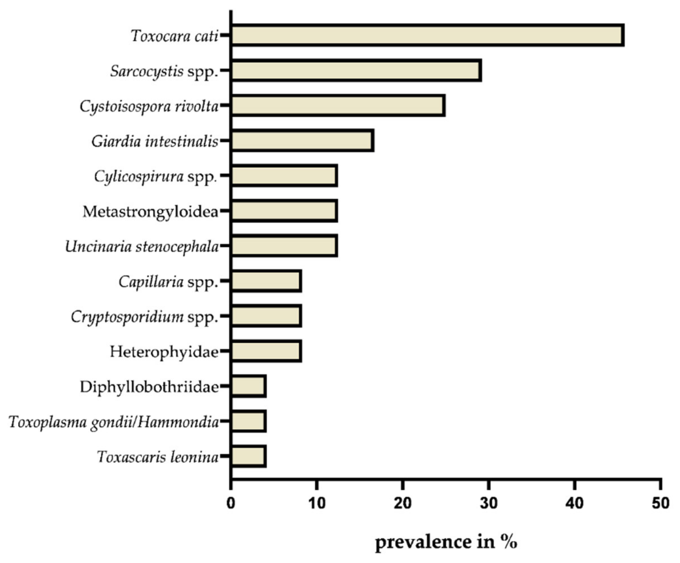
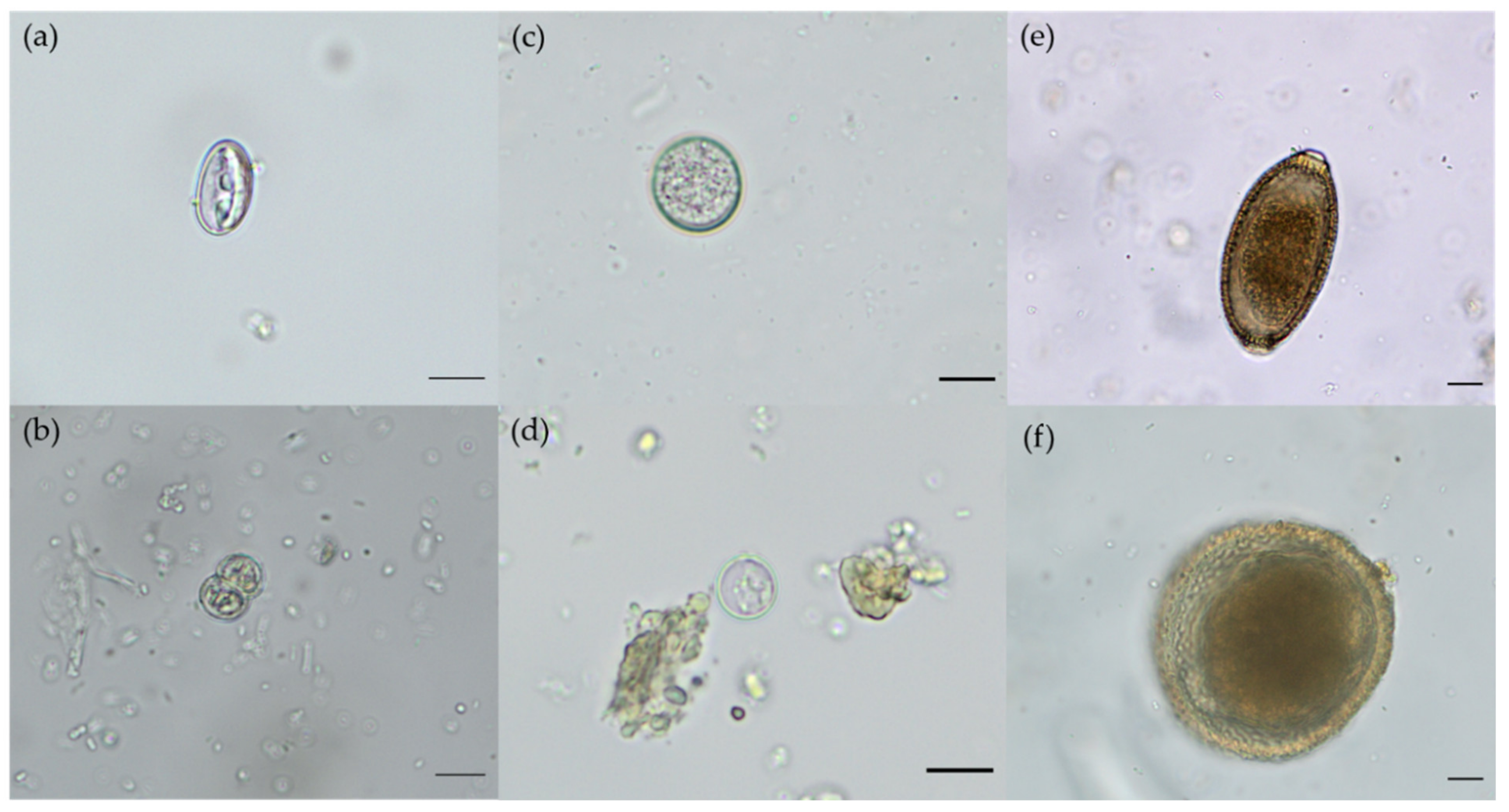
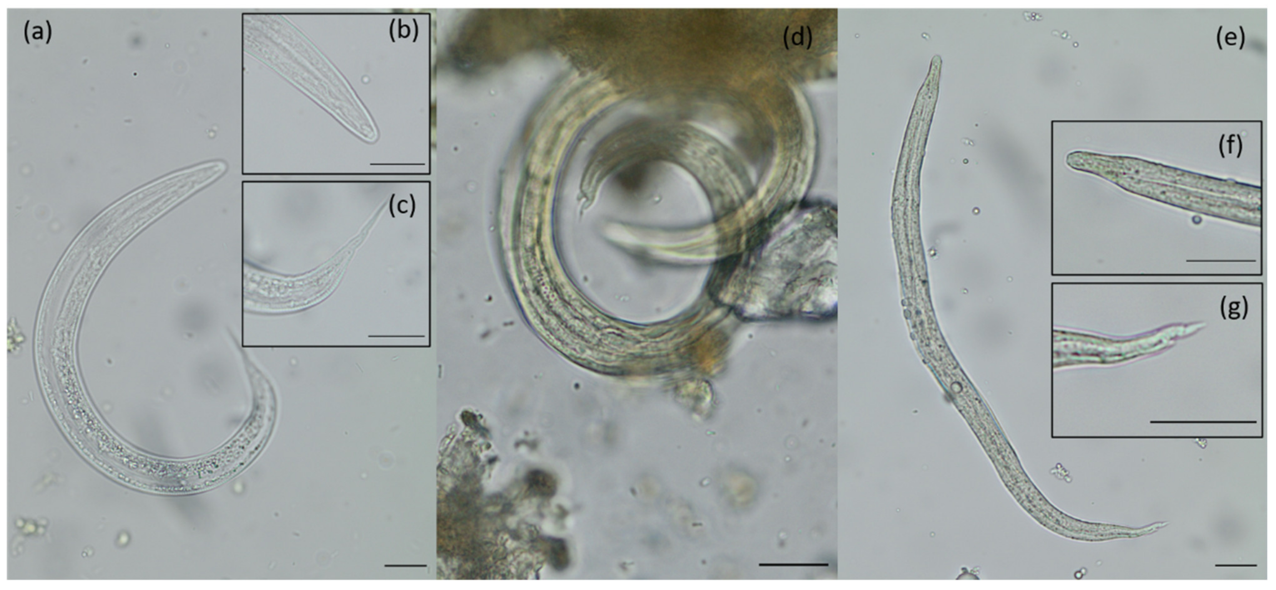
2.1. Endoparasite Diversity and Prevalences

The parasitological evaluation of Eurasian lynx faeces through standardised coprological methods simultaneously unveiled a wide variety of parasite species and stages. In total 15 taxa were diagnosed including metazoan and protozoan parasites. Eight metazoan parasite genera (i.e., Uncinaria, Aelurostrongylus, Angiostrongylus, Protostrongylus, Toxascaris, Toxocara, Cylicospirura and Capillaria) belonged to the class Nematoda, one (Diphyllobothriidae) to the class Cestoda and one (Heterophyidae) to the class Trematoda. Four identified intestinal protozoan genera (i.e., Cystoisospora, Toxoplasma/Hammondia, Sarcocystis and Cryptosporidium) belonged to the phylum Alveolata (subphylum Apicomplexa) and one (Giardia) to the phylum Metamonada (Figure 2).
A rather large proportion, namely, 71% (17/24) of analysed lynx faecal samples contained parasitic stages (i.e., cysts, oocysts, sporocysts, eggs and larvae). The mean metazoan species richness reached 1.88/lynx. Five of the 24 analysed samples (21%) contained four or more different parasite species, indicating a wide diversity of endoparasite fauna in the examined Eurasian lynxes.
In total, 50% (12/24) of faecal samples tested microscopically positive for protozoan stages and mean protozoan species richness reached 0.79/lynx. In 54% (13/24) of the analysed lynx scat samples, nematode stages (i.e., eggs and larvae) were found and a mean nematode species richness of 0.96/lynx was calculated. Trematodes and cestodes were less frequently found with a species richness of 0.08/lynx and 0.04/lynx, respectively.
Concerning helminth species richness, nematodes were the richest group with a total of seven species (i.e., Uncinaria stenocephala, A. abstrusus, Angiostrongylus spp., Toxascaris leonina, Toxocara cati, Cylicospirura spp. and Capillaria spp.), followed by one cestode (family Diphyllobothriidae) and one trematode species (family Heterophyidae). With regards to protozoan species richness, apicomplexans with at least five species (i.e., T. gondii/Hammondia spp., Cystoisospora rivolta, Cystoisospora felis, Sarcocystis spp. and Cryptosporidium spp.) were the richest group, followed by one metamonad parasite species (Giardia intestinalis). Overall, T. cati was the most prevalent parasite with eleven out of 24 (45.8%) scat samples that tested positive for this nematode, followed by the protozoan taxa Sarcocystis spp. and C. rivolta, with 29.2% (7/24) and 25% (6/24), respectively (for microscopic images see Figure 3).
In total, 16.7% (4/24) of the tested lynx samples were positive for G. intestinalis and in 12.5% (3/24) of the samples metastrongyloid first-stage larvae (L1) were detected.
The found larvae, belonging to the superfamily Metastrongyloidea, were identified as A. abstrusus-like L1, Angiostrongylus spp.-like L1 and a spurious lynx parasite, i.e., P. pulmonalis of lagomorph prey animals (see Figure 4).
Moreover, in 12.5% (3/24) of the samples, Cylicospirura spp. and U. stenocephala eggs were found. Capillaria spp. eggs, Cryptosporidium spp. oocysts, and trematode eggs were only observed in 8.3% (2/24) of the samples. The lowest prevalence, with 4.2% (1/24) was calculated for Diphyllobothriid eggs, T. gondii/Hammondia spp. oocysts and T. leonina eggs.

2.2. Molecular Analyses

2.2.1. Protozoa

Two out of the four coproantigen Giardia spp. ELISA (ProSpecT®, Oxoid, Wesel, Germany) positive findings were qPCR confirmed as G. intestinalis. Among Giardia spp.-ELISA negative samples (n = 20), a single sample tested qPCR positive for G. intestinalis. Within all samples (n = 24), a single sample tested qPCR positive for Cryptosporidium. This sample was among those which had tested microscopically negative (n = 22). Two samples positive by microscopy tested negative by qPCR.
In six out of seven samples, microscopically positive for Sarcocystis spp., a nested Sarcocystis spp. PCR revealed amplicons of an expected size. The same was true in 14 out of 17 samples, negative during microscopy. However, in none of the Sarcocystis spp. positive nested PCR findings, sequencing of cloned amplicons revealed a sequence related to Sarcocystis spp. In one case, sequencing allowed to identify a C. felis-related sequence. The obtained consensus sequence can be found in the GenBank under accession number: OL689225. BLAST search revealed an identity of 99.82% to the best match with the accession number KT184364 (C. felis found in F. silvestris catus, Canada). The respective sample had tested microscopically positive for Cystoisospora spp.

2.2.2. Nematodes

The T. cati-specific qPCR confirmed n = 5/11 samples positive by microscopy. None of the samples that tested microscopically negative were positive in the Toxocara spp. qPCR. No inhibitions of the qPCRs were observed.
DNA obtained from two of three microscopically lungworm larvae positive samples, could be successfully amplified by nematode specific PCRs, using the primers NC1/NC2 as reported elsewhere [41]. GenBank analyses of the obtained sequence, with the accession number MZ801778, showed a 98.87 identity in the BLAST search tool with the best match MT345056, U. stenocephala, found in a wild boar, France. The other sequence OK480966 showed 100% identity with the best match to KJ450994 (a sequence reported for P. pulmonalis, found in Lepus europaeus, France).
Unfortunately, none of the samples showed effective amplification on performed metastronglyloid specific qPCRs.

3. Discussion

The helminthofauna of the Eurasian lynx in Europe consists so far of approximately 16 nematode species, ten cestode species and one trematode species (for an overview please refer to Table 1) [9,14,22]. In line, we detected 15 taxa of parasites so far, of which five endoparasites (Cryptosporidium spp., G. intestinalis, Sarcocystis spp., U. stenocephala and Angiostrongylus spp.) never have been described in wild Eurasian lynx before. In related European studies, the most prevalent parasites seemed to be T. cati and Taenia spp. with the species T. krabbei and T. lynciscapreoli [9,11,13,14]. As such, the prevalence for T. cati infections in wild lynxes from Latvia reached up to 77% [9], in Finland and Estonia 68% [11,13], and in Switzerland 40% [8]. In the current study, in 45.8% of German lynx scat samples, T. cati eggs were detected, which showed similar prevalences as in other European countries. Regardless of the fact that T. cati is a common lynx parasite, this nematode can cause enteritis with severe clinical signs leading to cachexia and a reduced general condition in juvenile wild felids, making these young animals more receptive to other diseases [32]. There are also some reports of fatal cases of toxocarosis in juveniles [8,32]. Moreover, toxocarosis can affect humans and belongs to the most commonly reported zoonotic helminth infections worldwide, which can cause, among others, visceral larva migrans (VLM) and ocular larva migrans (OLM) syndromes [42].
In contrast to T. cati eggs, no Taenia spp. eggs were found in current copromicroscopic analyses. However, in other related studies, the methodology was mostly different, as necropsied animals were investigated and, therefore, adult Taenia spp. cestodes were identified. Taeniid cestodes, such as T. krabbei and T. lynciscapreoli, are known to pass their eggs mostly intermittently and inside mature proglottids into the environment. This biological feature could explain the discrepancy in the prevalences of Taenia spp.-infections previously reported, as the estimates may depend on the diagnostic performance of the examination method used [43]. To obtain more clarity on taeniid cestode fauna circulating in German lynxes, further studies on lynx carcasses from naturally deceased animals of the HM population, as well as analyses of lynx prey in this habitat, are highly desirable. They can also help in order to clarify another discrepancy of the current work, namely, differences between microscopic and molecular typing results. We hypothesise that the fact that we tested faecal samples with methods that vary widely in their sensitivity may explain this variance. Furthermore, lynx scat samples showed a wide endoparasite diversity with quite variable types of different parasitic stages (i.e., cysts, sporocysts, oocysts, eggs and larvae) per sample. As faeces do not represent a homogenous organic material and since we further took one portion from each investigated sample for copromicroscopic and another part for molecular analysis, the results for each method might diverge. Moreover, molecular analyses on faecal samples collected from the field proved to be challenging and possibly more specific assays need to be developed. However, despite the weakness of copromicroscopic analysis, there are still crucial advantages when compared to other parasitological diagnostic methods. One obvious advantage is, for example, the ability to detect parasite stages that other diagnostic methods (e.g., molecular analysis, muscle digestion or macroscopic pathological examination) will miss. More importantly, copromicroscopic analysis provides evidence on the patency and also detect infective stages for definitive, intermediate or paratenic hosts.
The second most frequently occurring parasitic stages microscopically diagnosed were apicomplexan Sarcocystis spp. oocysts/sporocysts (i.e., infective stages for various mammalian prey animals) and C. rivolta oocysts (infective stages for lynxes or other closely related wild felids (F. silvestris silvestris)), both well-known coccidian parasites of felines, belonging to the family Sarcocystidae. However, molecular analyses to amplify Sarcocystis spp. 18 S rDNA using a nested PCR resulted in amplicons of an expected size but cloning and sequencing did not reveal Sarcocystis sp. sequences. Thus, microscopical observations have to be regarded as non-confirmed. This zoonotic apicomplexan parasite genus is characterised by a heteroxenous life cycle, with a clear prey–predator relationship for transmission [44,45]. The fact that the carnivorous species do not only act as a final host but sometimes also as an intermediate host makes the role of the carnivore unusual [45,46,47,48]. Some Sarcocystis species occurring in wild animals, such as S. nesbitti, can be even pathogenic for humans, acting as aberrant intermediate hosts [49,50] while others are infectious for domestic and wild animals [46,50]. The typical clinical sign of sarcocystosis in predator definitive hosts is diarrhoea due to gamogony-induced enteritis after ingestion of infected intermediate hosts containing muscular Sarcocystis-cysts with thousands of bradyzoites [46]. In contrast, clinical signs of sarcocystosis in intermediate hosts include myositis, vasculitis, myalgia, haemorrhages, neurological signs (e.g., ataxia, paralyses), cachexia, anorexia, weight loss and abortion [44], corresponding well to clinical manifestations of S. nesbitti-infected human patients [49,50]. As Sarcocystis spp. infections led to encephalitis in Canada lynxes (Lynx canadensis) [51], and this parasite was the second most prevalent in the current study, we recommend investigations involving muscle and cerebral analyses of lynx, as well as prey animals (e.g., cervids, hares, mice), and molecular reprocessing of Sarcocystis spp.-positive samples for identification of species and for testing more alternative intermediate hosts as well as final hosts in the lynx range area within Germany.
After Sarcocystis spp., the second most prevalent apicomplexan parasite found in this study was C. rivolta. As for toxocarosis, cystoisosporosis was mainly observed in juvenile felids and, depending on the age, immune status and primary infective dose can lead to enteritis with catharralic diarrhoea. Unfortunately, little is known about C. rivolta-derived pathogenesis in young Eurasian lynxes. As stated above, in domestic cats (F. silvestris catus) it can cause clinical manifested cystoisosporosis with diarrhoea, dehydration and weight losses, especially in juveniles [52]. In one of the samples analysed here, the presence of C. felis DNA was confirmed by cloning and sequencing. Recently it was shown that C. felis oocysts obtained from closely related bobcats (Lynx rufus) in North America, can also infect domestic cats via consumption of experimentally infected mice [53]. Therefore, transmission of feline Cystoisospora species between free-ranging Eurasian lynxes and wild cat (F. silvestris silvestris) populations might also be possible to occur in the HM, but this assumption needs further clarification. Moreover, domestic cats (F. silvestris catus), either as feral cats or as cats kept outdoors (but owned as pets) might be identified as natural sources of highly resistant C. rivolta and/or C. felis oocysts, and perpetuating the life cycle in lynx habitats such as the HM.
Furthermore, metastrongyloid lungworm species were detected microscopically, i.e., A. abstrusus- and Angiostrongylus spp.-like larvae. These parasites are known to affect companion animals’ health status, mainly the respiratory and cardiovascular system. Molecular analysis revealed P. pulmonalis, which represents a lungworm-infecting lagomorphs (i.e., hares and rabbits), indicating that studies by copromicroscopy can help to reveal aspects of the trophic chain for this apex predator detecting spurious parasites of prey animals and, therefore, generating valuable data concerning wild animal diets and biome health. Beside spurious P. pulmonalis lungworm, so far three feline specific metastrongyloid lungworm species in European lynx populations have been described, namely, A. abstrusus [8,20], Troglostrongylus brevior [25] and recently Crenosoma vismani [22]. The last species (i.e., C. vismani (Crenosomatidae)) represents a newly described metastrongyloid nematode of the European lynx [22]. By morphological and morphometric analyses, A. abstrusus-like L1 were detected in collected lynx faeces. Unfortunately, the molecular analysis failed for A. abstrusus, most probably either due to the insufficient DNA quality or bacteria-mediated DNA degradation. The same holds true for observed Angiostrongylus spp.-like L1, where molecular analysis also failed, but classical tail morphology as well as morphometry (see Figure 4) indicated similarities to the genus Angiostrongylus. We hypothesise, considering the known literature of metastrongyloid nematodes infecting wild felids and morphological observations of the detected larvae, that either T. brevior or Angiostrongylus chabaudi are the most probable species circulating in the HM lynx population, which has so far been isolated from other lynx occurrences. Among them, A. chabaudi has not been described so far to infect the Eurasian lynx; however, it is recognised to infect European wildcats (F. silvestris silvestris) [54,55], an endemic feline species in the HM lynx range habitats. We also cannot exclude the possibility that observed Angiostrongylus spp.-like L1 belong to the morphological similar species T. brevior (Crenosomatidae), which is rarely reported in Germany [56]. Prevalences for lungworms in F. silvestris silvestris in Germany reached 17% [56] and A. chabaudi was molecularly detected in a German wildcat by Hirzmann et al. (2018) (GenBank accession number: MH899656). Furthermore, wildcats are suggested to be a sylvatic reservoir for feline lungworms [57,58].
New approaches for better DNA isolation/preservation of lungworm larvae obtained from freshly passed samples by Baermann funnel technique and ongoing molecular analyses will hopefully clarify species spectrum of metastrongyloid species in the near future. As metastrongyloid lungworm infections seem to spread within Europe [41,59] and South America [60,61], new species have been identified and prevalences seem to be rising, thus further research in neglected gastropod-borne diseases is valuable [22,62,63,64,65,66,67].
In line with these results, we detected not only gastropod-borne nematode and trematode (Heterophyidae) infections but also zoonotic fish-borne cestode infections (dibothriocephalosis (former diphyllobothriosis) or spirometrosis (sparganosis)). The former is an interesting finding, considering that HM lynxes were not yet observed to consume fish [30]. Pseudophyllidean infections with neglected zoonotic-relevant species such as Dibothriocephalus latus, Spirometra janickii and Spirometra erinaceieuropae were already described in Eurasian lynx [10,12] and, thus, demanding improved surveillance.
Furthermore, the findings of Cylicospirura spp. eggs in the present study add novel data on the helminth fauna of L. lynx. In Europe, cylicospirurosis is rarely reported in wildcats (F. silvestris silvestris) and the life cycle of Cylicospirura, which involves insects as intermediate hosts and small vertebrates as paratenic hosts, remains in many parts uncovered [68,69,70]. Pathology of neglected cylicospirurosis, which affects mainly wild felids, among them also lynxes (L. canadensis and L. rufus), is associated with stomach nodules due to severe granuloma formation [68,71,72].
In addition to the abovementioned parasites, this study detected for the first time two protozoan genera (i.e., Giardia and Cryptosporidium) in Eurasian lynxes by coproantigen ELISAs, qPCR analyses and carbolfuchsin-stained faecal smears. As there is a lack of studies in intestinal protozoan parasite prevalence in feline wildlife [36], our results highlight that further research is necessary in this field. Evaluating the results further, it stands out, that two of the four G. intestinalis-positive samples were collected from lynx family groups with juveniles. In these juvenile scat samples, a much higher signal intensity in performed coproantigen ELISAs was observed, when compared to positive ones from adults. We hypothesise, that similar to other mammalian host species, juveniles are more frequently infected thereby shedding more infective G. intestinalis cysts into environment as compared to partially or fully immunocompetent adults thereby contributing significantly to habitat contamination. In addition, a potentially zoonotic helminth species (i.e., U. stenocephala) was molecularly confirmed in lynxes. In general, this hookworm species is described to infect more frequently canids, but rarely also found in felids [73,74]. To the best of our knowledge, U. stenocephala has not yet been described in Eurasian lynx populations in Germany. Nevertheless, some studies identified Uncinaria sp. infections in Eurasian lynxes in Switzerland [8] and in a closely related Canada lynx (L. canadensis), U. stenocephala was detected as well [75]. As domestic cats are reported to act as reservoir for hookworms affecting the Iberian lynx (L. pardinus), companion animals could play a similar role in the epidemiology of Uncinaria sp. in Eurasian lynx [24,35,76].
Overall, the spectrum of protozoa and helminth parasites with respective prevalences reported in this study are in accordance to previous reports on parasite fauna of free-living lynxes from other European countries, but also added new species to the list of L. lynx parasites [8,9,11,12,13,14]. Findings on aelurostrongylosis, angiostrongylosis, dibothriocephalosis as well as sarcocystosis and cylicospirurosis, call for more research activities, not only on definitive hosts and intermediate hosts but also on paratenic hosts to generate novel insights into the epidemiology of neglected parasitoses circulating in Eurasian lynx populations.

4. Materials and Methods

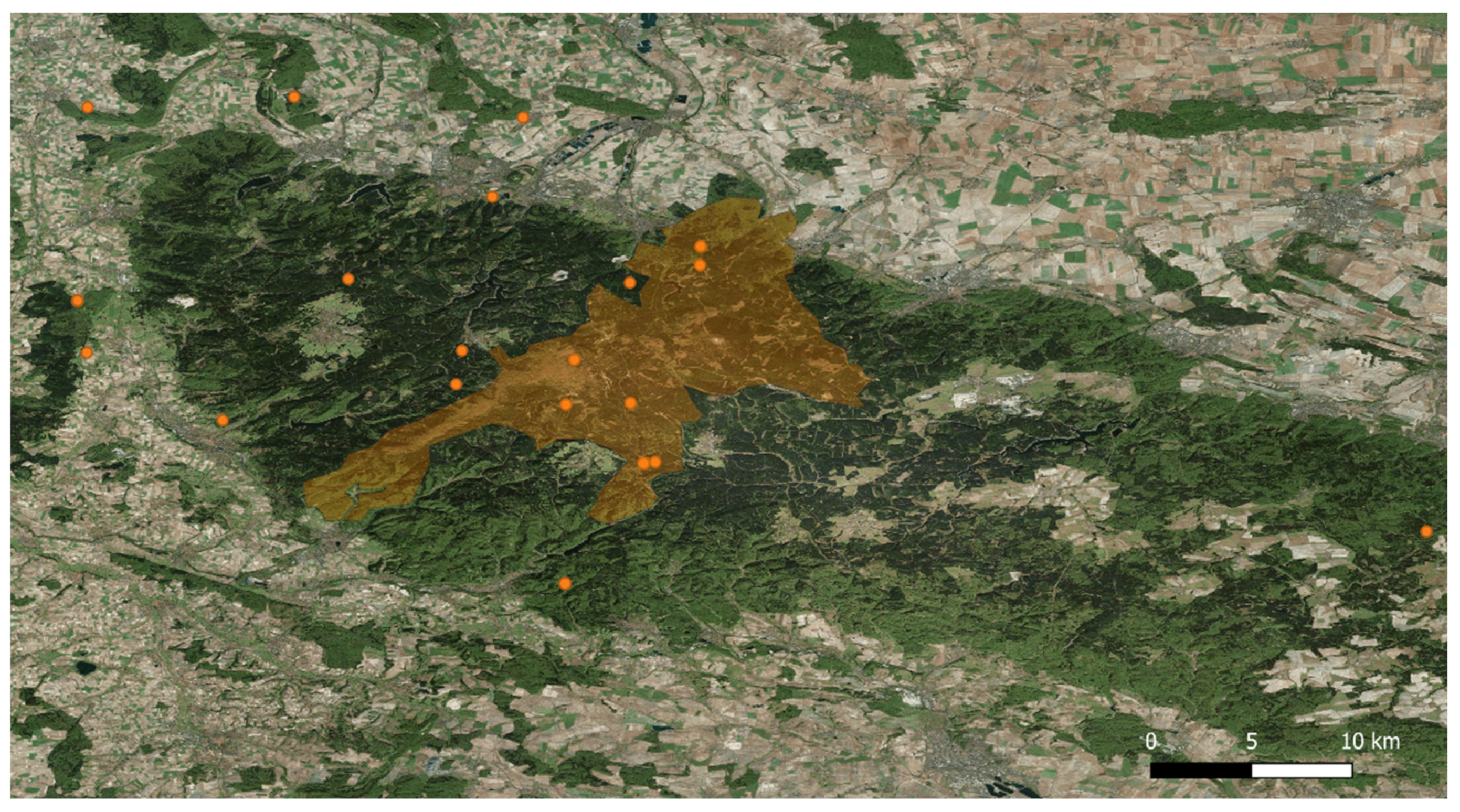
4.1. Study Area and Sampling Collection

Scat collection was performed in the range of the Harz lynx population in Germany (see Figure 5). This range includes an area of 2200 km2 forested midland mountains, the HM which, in turn, contains the approximately 247 km2 protected area of the HNP (51°47′0″ N, 10°34′0″ E), which is part of the European nature conservation network of Natura 2000. The altitude in the HM varies from less than 300 m at the northern and southern borders, to 1141 m above sea level on the summit of the Brocken Mountain [6]. Around 75% of the HM are forested, with variable types of mainly anthropogenic spruce forest. Beech, which is actually dominant here, has been pushed back by human influence into rather small areas of the low mountain range.
Some animals living in the HM, including mammals, birds, invertebrates, amphibians, reptiles, and fish, belong to endangered species. By scat analysis, nine prey animals of lynx could be assigned to the species level. Those include roe deer, red deer, red fox, European water vole (Arvicola terrestris), bank vole (Myodes glareolus), common vole, red squirrel (Scirus vulgaris), brown rat (Rattus norvegicus) and black woodpecker (Dryocopus martius) [30]. However roe deer and red deer are the main prey species in the Harz lynx population [30,77]. In 2000, the reintroduction programme of L. lynx into the HM started. As the lynx range is wider than the National Park borders (Figure 5), these wild felids are also inhabiting surrounding areas, which are dominated by agriculture. In the western and southern foreland of the HM the forest cover reaches a maximum of about 25% whereas north and east of the area, forest is scarce due to the fertile soils allowing profitable agricultural production [33]. Beside the Eurasian lynx, there is another wild feline predator living in this area, namely, the European wildcat (F. silvestris silvestris).
During the general lynx monitoring of the HNP, a total of 24 faecal samples of Eurasian lynxes were collected at 20 different collection sites between 2019 and 2021. The coordinates of these collection sites were recorded, with seven locations within the HNP and 13 at surrounding areas (see Figure 5). Lynx faeces were mostly located next to killed prey, e.g., red deer (C. elaphus) and roe deer (C. capreolus) (Figure 1). In some cases, more than one scat was found at a killing site. The allocation of the faecal samples to specific lynx individuals was possible because the animals wore collar transmitters or were genetically sampled (e.g., saliva sample from a prey animal close to where the faecal sample was found). In addition, prey killing sites were often monitored with camera trap images, so that female lynxes with offspring could be identified. Collected samples were immediately fixed in 80% ethanol until further investigation. Thereafter, the scat samples were transferred to the Institute of Parasitology at Justus Liebig University Giessen (JLU), Giessen, Germany, for further analyses.
According to Council Regulation (EC) No 338/97 of 9 December 1996 on the protection of species of wild fauna and flora by regulating trade therein, faeces are defined as natural excretions and, thus, not considered as products of wild animals; therefore, no official permit was necessary to perform this copromicroscopic study. Furthermore, official employees of the HNP were exclusively involved in scat sample collections.

4.2. Coprological Analyses

4.2.1. Microscopical Analyses

The faecal samples were copromicroscopically analysed by using the standard sodium acetate acetic acid formaldehyde (SAF) technique previously used for wild felids and domestic animals [43,78,79]. SAF stock solution was prepared with 15 g sodium acetate, 20 mL acetic acid, 40 mL formaldehyde (37%) and 925 mL tap water. Lynx faeces (4 g) was filled in a sampling tube with 8 mL SAF solution and suspended. After filtering the sample through a layer of gauze into a conical centrifuge tube, it was centrifuged at 2000 rpm for 1 min. The supernatant was discarded and 7 mL of 0.9% sodium chloride solution and 2 mL of acetic acid ethyl ester was added and stirred up. Then, a final centrifugation step followed at 600× g for 3 min, and the sediment was examined.
The light microscopy-based identification of parasitic stages such as eggs, larvae, cysts, sporocysts and oocysts was based on morphological/morphometric characteristics. Moreover, carbolfuchsin-stained faecal smears according to [80] were performed for the detection of Cryptosporidium spp. oocysts.
Metastrongyloid lungworm first-stage larvae (L1) were morphologically identified by body length and width, oesophagus form (non-rhabditiform), ratio of oesophagus length to body length (1:3–1:2) as well as typical tail morphology as reported by other authors [25,58,66,81] (see Figure 4).

4.2.2. Coproantigen-ELISAs

Additionally, coproantigen-ELISAs (ProSpecT®, Oxoid, Wesel, Germany) were carried out for the detection of C. parvum and G. intestinalis antigens in faecal samples of mammalian species.

4.2.3. Molecular Analyses

Protozoa

Faecal samples (n = 24), preserved in 80% ethanol, were washed two times in distilled water. DNA was extracted from the final faecal suspension (200 µL) using a commercial kit, the Quick-DNA Fecal/Soil Microbe Miniprep Kit (Zymo Research Europe, Freiburg, Germany).
The following real-time PCR was used to detect G. intestinalis and Cryptosporidium spp. using previously publishes primers and probes (Table 2).
For G. intestinalis we used Gd-80F 5′-GACGGCTCAGGACAACGGTT-3′, Gd-127R 5′-TTGCCAGCGGTGTCCG-3′ as primers and as a probe Gd-FT 5′-FAM-CCCGCGGCGGTCCCTGCTAG-DDQ1-3′ [82]. For Cryptosporidium spp. primers Cp-583F 5′-CAAATTGATACCGTTTGTCCTTCTG-3′ and Cp-733R 5′-GGCATGTCGATTCTAATTCAGCT-3′ as well as the probe Cp-TRT 5′-Texas Red-TGCCATACATTGTTGTCCTGACAAATTGAAT-DDQ2-3′ were used [82].
The qPCR reactions (25 µL final volume) consisted of 12.5 µL of QuantiTect Multiplex PCR NoROX Master Mix (Qiagen, Hilden, Germany), 3 mmol/L MgCl2, 0.4 µmol/L of G. intestinalis primers and 0.12 µmol/L of the G. intestinalis probe, 1 µmol/L Cryptosporidium spp. primers and 0.5 µmol/L of the Cryptosporidium spp. probe and 3 µL of a 1:5 diluted template DNA.
To monitor the inhibition of the real-time PCRs, a heterologous plasmid with DNA sequences resembling the enhanced green fluorescent protein (EGFP) gene was added to the reaction mix in all real-time PCRs. The internal control PCR included the primers EGFP1-F, EGFP2-R and the probe EGFP1 as previously reported [83].
Cycling condition for qPCR was as follows. In case of G. intestinalis and Cryptosporidium spp. the cycling conditions consisted of 15 min at 95 °C denaturation and 45 cycles of 60 s at 95 °C denaturation, 30 s at 55 °C annealing and elongation for 30 s at 72 °C with a ramping of 3.3 °C/s. The measurement of the fluorescence was taken at the end of the annealing.
Sarcocystis spp. 18S rDNA was detected by nested PCR using 1 µL of extracted template DNA. The external Sarcocystis spp.-specific PCR, amplified fragments of a size of around 900 bp using the primers SarcoFext and SarcoRext [85]. For the internal PCR, 1 µL of the external reaction was used, employing the primer pair SarcoFint/SarcoRint [85]. The external as well as the internal PCR was performed in a final reaction volume of 25 µL with 1 unit of Taq DNA polymerase per reaction (Stratec molecular, Germany) under the following conditions (final concentrations): 20 µg/mL BSA, 1 × reaction buffer supplied with the polymerase, 1.5 mM MgCl2, 250 µM dNTPs, 0.5 µM of each primer. The following cycler program was used: 94 °C (4 min), 40 cycles of 94 °C (40 s), 58 °C (1 min), 72 °C (1 min) and a final extension at 72 °C (5 min). Five µL of each product were examined in 1.5% agarose gels stained with ethidium bromide.
For sequencing of amplicons, bands of the expected size were excised from agarose gels and purified with a commercial kit (NucleoSpin® Gel and PCR Clean-up, Macherey-Nagel, Düren, Germany), following the manufacturer’s instructions. Purified amplification products were cloned into a commercially available vector (pGEM®-T Easy Vector System I, Promega, Mannheim, Germany) and used to transform chemically competent Escherichia coli (OneShot TOP10, Thermo Fisher Scientific, Langenselbold, Germany). The transformed E. coli were cultivated and the plasmid DNA was subsequently purified using a commercial kit (QIAprep Spin Miniprep Kit, Qiagen, Hilden, Germany) according to the manufacturer’s instructions. Sequencing was performed using the BigDye Terminator v1.1 Cycle Seq. Kit (Thermo Fisher Scientific, Langenselbold, Germany) and passed through NucleoSEQ Columns (Macherey-Nagel, Düren, Germany) for cleaning up nucleic acids, in an ABI 3130 capillary sequencer (Thermo Fisher Scientific, Langenselbold, Germany).
The forward and reverse sequences were aligned, if necessary trimmed based on primer sequence information and the consensus sequences for the individual cloned amplification products compared to sequences stored in GenBank, EMBL, DDBJ, or RefSeq using BLASTn with standard conditions.

Nematodes

All samples were analysed by Toxocara spp. multiplex qPCR, using Tcanis-F 5′-GCGCCAATTTATGGAATGTGAT-3′ and Tcati-F 5′-ACGCGTACGTATGGAATGTGCT-3′ as forward primers and TCC-R 5′-GAGCAAACGACAGCSATTTCTT-3′ as the reverse primer [84]. As probes, for T. canis Tcanis-S 5′-FAM-CCATTACCACACCAGCATAGCTCACCGA-3′-BHQ1 and for T. cati Tcati-S 5′-Cy5-TCTTTCGCAACGTGCATTCGGTGA-3′-BHQ3 were used [84]. Including 12.5 µL of Master Mix, 0.3 µmol/L of each primer, 0.1 µmol/L of each probe and 2.5 µL of a 1:5 diluted template DNA were used. The cycling conditions consisted of 15 min at 95 °C denaturation and 45 cycles of 60 s at 94 °C denaturation and 60 s at 60 °C annealing and elongation with measurement at the end.
The samples being microscopically positive for metastrongyloid L1 (n = 3) were further analysed by PCR and sequencing, in order to identify them to species level. DNA was extracted using a commercial kit (QIAamp Fast DNA Stool Mini Kit®, Qiagen, Hilden, Germany). For the first PCR the universal nematode primers NC1 5′-ACGTCTGGTTCAGGGTTGTT-3′ and NC2 5′-TTAGTTTCTTTTCCTCCGCT-3′ [86] were used in a reaction volume of 50 μL, HOT FIREPol® Blend Master Mix (Solis BioDyne, Tartu, Estonia) and 5 μL of DNA template with following conditions: denaturation 95 °C 15 min, 35 cycles of denaturation 95 °C 20 s, annealing 52 °C 30 s and extension 72 °C 30 s, followed by a final elongation of 72 °C 5 min. PCR products were analysed by gel electrophoresis and used as templates for specific nested PCRs. For identification of A. abstrusus and T. brevior, a duplex three step PCR was performed, using the forward primers TrogloF 5′-GCACTTGAAATCTTCGACA-3′, AeluroF 5′-GCATTTATGCTAGTGATATC-3′ and the single reverse primer MetR 5′-CCGCTAAATGATATGCTTA-3′ as described elsewhere [87]. The final reaction volume of 20 μL consisted of 4 μL 5 × HOT FIREPol® Evagreen® qPCR Mix Plus (Solis BioDyne, Tartu, Estonia), 250 nM of each primer, 13.5 mL sterile water and 1 μL template. Cycling protocol was 95 °C 15 min, 35 cycles of denaturation 95 °C 20 s, annealing 59 °C 20 s, and extension 72 °C 30 s.
As these metastrongyloid real-time PCRs were negative or inconclusive and the quantity of the amplicon-DNA from the first PCR was low, a second nested conventional PCR was performed, using the primers NC1/MetR. Two samples were purified and sent to a commercial sequencing service (LGC Genomics, Berlin, Germany). The obtained sequences were analysed by BLAST search (http://0-www-ncbi-nlm-nih-gov.brum.beds.ac.uk/BLAST/; accessed on 15 December 2021) and then submitted to the GenBank.

5. Conclusions

We investigated the endoparasite fauna of free-ranging lynxes of the Harz lynx population and report for the first time on the occurrence of feline aelurostrongylosis and angiostrongylosis. Many parasite species found in Eurasian lynx can also be found in wild cats and even in domestic cats that live in direct contact with humans. Therefore, a One Health approach is requested in order to identify parasite transmission routes. Relevant zooanthroponotic parasites, such as water-borne (i.e., G. intestinalis) as well as soil-borne (i.e., T. cati and U. stenocephala) infections were detected. These results may help to improve our understanding of the impact of these parasites on the health status of wild lynx and also on obligate intermediate or paratenic hosts within the range of the ecological habitat.

Author Contributions

Conceptualisation, C.H.; data curation, L.S.; formal analysis, L.S.; funding acquisition, A.T., F.J.C.; investigation, L.S., D.T.W., G.S. and C.H.; methodology, A.T. and C.H.; project administration, O.A., T.L.M., A.T. and C.H.; resources, O.A. and T.L.M.; supervision, A.T. and C.H.; validation, L.S.; visualisation, L.S.; writing—original draft, L.S.; writing—review and editing, L.S., O.A., T.L.M., P.M., G.S., F.J.C., D.T.W., A.T. and C.H. All authors have read and agreed to the published version of the manuscript.

Funding

This research was funded by the Institute of Parasitology, Justus Liebig University Giessen, Germany and Friedrich-Loeffler-Institut, Federal Research Institute for Animal Health, Institute of Epidemiology, Greifswald-Insel Riems, Germany. D.T.W. was funded by the German Federal Ministry of Agriculture and Food (SHIP-NEXT Modul ONE HEALTH, FKZ: 28N203202).

Institutional Review Board Statement

Ethical review and approval were waived for this study, as faecal sampling is a non-invasive and non-disturbing method. Considering the current Council Regulation (EC) No 338/97 of 9 December 1996 on the protection of species of wild fauna and flora by regulating trade therein, faeces are defined as natural excretions and, thus, not considered as products of wild animals; therefore, no official permit was necessary to obtain to perform this coprological study. Furthermore, official employees of the Harz Mountain National Park, Germany, were exclusively involved in scat sample collections.

Informed Consent Statement

Not applicable.

Data Availability Statement

The putative P. pulmonaris, C. felis and U. stenocephala sequences obtained from L. lynx were deposited in the GenBank database (National Center for Biotechnology Information, HIH, Bethesda, USA) and are available under accession numbers: MZ801778, OL689225 and OK480966.

Acknowledgments

We are grateful to Christine Henrich, Juan Vélez, and Iván Conejeros (Institute of Parasitology, Justus Liebig University Giessen, Giessen, Germany); Mareen Tuschy (Friedrich-Loeffler-Institut, Riems, Germany); Jörg Hirzmann (Veterinary Laboratory Freiburg, Germany) for their excellent help in processing samples, helping to perform molecular analyses and their precious advice.

Conflicts of Interest

The authors declare no conflict of interest. The funders had no role in the design of the study; in the collection, analyses, or interpretation of data; in the writing of the manuscript, or in the decision to publish the results.

References

  1. Port, M.; Henkelmann, A.; Schröder, F.; Waltert, M.; Middelhoff, L.; Anders, O.; Jokisch, S. Rise and Fall of a Eurasian Lynx (Lynx lynx) Stepping-Stone Population in Central Germany. Mammal Res. 2020, 66, 45–55. [Google Scholar] [CrossRef]
  2. Meinig, H.; Boye, P.; Dähne, M.; Hutterer, R.; Lang, J. Rote Liste und Gesamtartenliste der Säugetiere (Mammalia) Deutschlands; Bundesamt für Naturschutz: Bonn-Bad Godesberg, Germany, 2020. [Google Scholar]
  3. Reinhardt, I.; Kaczensky, P.; Knauer, F. Monitoring von Wolf, Luchs und Bär in Deutschland; BfN-Skripten; Bundesamt für Naturschutz: Bonn-Bad Godesberg, Germany, 2015; ISBN 978-3-89624-148-1. [Google Scholar]
  4. Bull, J.K.; Heurich, M.; Saveljev, A.P.; Schmidt, K.; Fickel, J.; Förster, D.W. The Effect of Reintroductions on the Genetic Variability in Eurasian Lynx Populations: The Cases of Bohemian–Bavarian and Vosges–Palatinian Populations. Conserv. Genet. 2016, 17, 1229–1234. [Google Scholar] [CrossRef] [Green Version]
  5. Luchsvorkommen in Deutschland im Monitoringjahr 2019/2020 (1.5.2019–30.04.2020). 2021. Available online: https://www.bfn.de/sites/default/files/2021-04/2021_02_25_BfN_Luchs_kommentierte_Vorkommenskarte_bf.pdf (accessed on 15 December 2021).
  6. Middelhoff, T.L.; Anders, O. Abundanz und Dichte des Luchses Im Östlichen Harz. Fotofallenmonitoring 2017; Nationalpark Harz: Wernigerode, Germany, 2018. [Google Scholar]
  7. Mueller, S.A.; Reiners, T.E.; Middelhoff, T.L.; Anders, O.; Kasperkiewicz, A.; Nowak, C. The Rise of a Large Carnivore Population in Central Europe: Genetic Evaluation of Lynx Reintroduction in the Harz Mountains. Conserv. Genet. 2020, 21, 577–587. [Google Scholar] [CrossRef] [Green Version]
  8. Schmidt-Posthaus, H.; Breitenmoser-Würsten, C.; Posthaus, H.; Bacciarini, L.; Breitenmoser, U. Causes of Mortality in Reintroduced Eurasian Lynx in Switzerland. J. Wildl. Dis. 2002, 38, 84–92. [Google Scholar] [CrossRef] [Green Version]
  9. Bagrade, G.; Vismanis, K.; Kirjušina, M.; Ozoliņš, J. Preliminary Results on the Helminthofauna of the Eurasian Lynx (Lynx lynx) in Latvia. Acta Zool. Litu. 2003, 13, 3–7. [Google Scholar] [CrossRef]
  10. Bagrade, G.; Králová-Hromadová, I.; Bazsalovicsová, E.; Radačovská, A.; Kołodziej-Sobocińska, M. The First Records of Spirometra erinaceieuropaei (Cestoda: Diphyllobothriidae), a Causative Agent of Human Sparganosis, in Latvian Wildlife. Parasitol. Res. 2020, 120, 365–371. [Google Scholar] [CrossRef] [PubMed]
  11. Valdmann, H.; Moks, E.; Talvik, H. Helminth Fauna of Eurasian Lynx (Lynx lynx) in Estonia. J. Wildl. Dis. 2004, 40, 356–360. [Google Scholar] [CrossRef] [Green Version]
  12. Szczęsna, J.; Popiołek, M.; Schmidt, K.; Kowalczyk, R. Coprological Study on Helminth Fauna in Eurasian Lynx (Lynx lynx) From the Białowieża Primeval Forest in Eastern Poland. J. Parasitol. 2008, 94, 981–984. [Google Scholar] [CrossRef]
  13. Deksne, G.; Laakkonen, J.; Näreaho, A.; Jokelainen, P.; Holmala, K.; Kojola, I.; Sukura, A. Endoparasites of the Eurasian Lynx (Lynx lynx) in Finland. J. Parasitol. 2013, 99, 229–234. [Google Scholar] [CrossRef] [PubMed]
  14. Kołodziej-Sobocińska, M.; Yakovlev, Y.; Schmidt, K.; Hurníková, Z.; Ruczyńska, I.; Bednarski, M.; Tokarska, M. Update of the Helminth Fauna in Eurasian Lynx (Lynx lynx) in Poland. Parasitol. Res. 2018, 117, 2613–2621. [Google Scholar] [CrossRef] [PubMed] [Green Version]
  15. Kärssin, A.; Häkkinen, L.; Vilem, A.; Jokelainen, P.; Lassen, B. Trichinella spp. in Wild Boars (Sus scrofa), Brown Bears (Ursus arctos), Eurasian Lynxes (Lynx lynx) and Badgers (Meles meles) in Estonia, 2007–2014. Animals 2021, 11, 183. [Google Scholar] [CrossRef]
  16. Górski, P.; Zalewski, A.; Lakomy, M. Parasites of Carnivorous Mammals in Białowieza Primeval Forest. Wiad. Parazytol. 2006, 52, 49–53. [Google Scholar]
  17. Ryser-Degiorgis, M.-P.; Jakubek, E.-B.; Hård af Segerstad, C.; Bröjer, C.; Mörner, T.; Jansson, D.S.; Lundén, A.; Uggla, A. Serological Survey of Toxoplasma gondii Infection in Free-Ranging Eurasian Lynx (Lynx lynx) from Sweden. J. Wildl. Dis. 2006, 42, 182–187. [Google Scholar] [CrossRef] [Green Version]
  18. Hurnikova, Z.; Miterpakova, M.; Chovancová, B. The Important Zoonoses in the Protected Areas of the Tatra National Park (TANAP). Wiad. Parazytol. 2009, 55, 395–398. [Google Scholar] [PubMed]
  19. Ozoliņa, Z.; Bagrade, G.; Deksne, G. First Confirmed Case of Alaria alata Mesocercaria in Eurasian Lynx (Lynx lynx) Hunted in Latvia. Parasitol. Res. 2020, 119, 759–762. [Google Scholar] [CrossRef] [PubMed]
  20. Szczęsna, J.; Popiołek, M.; Schmidt, K.; Kowalczyk, R. The First Record of Aelurostrongylus abstrusus (Angistrongylidae: Nematoda) in Eurasian Lynx (Lynx lynx L.) from Poland Based on Fecal Analysis. Wiad. Parazytol. 2006, 52, 321–322. [Google Scholar] [PubMed]
  21. Crisi, P.E.; Di Cesare, A.; Boari, A. Feline Troglostrongylosis: Current Epizootiology, Clinical Features, and Therapeutic Options. Front. Vet. Sci. 2018, 5, 126. [Google Scholar] [CrossRef]
  22. Stunžėnas, V.; Binkienė, R. Description of Crenosoma vismani n. sp., Parasitic in the Lungs of Lynx lynx (L.) (Carnivora: Felidae), with Identification Key to the Species of the Genus Crenosoma Molin, 1861 (Nematoda: Crenosomatidae). Syst. Parasitol. 2020, 98, 73–83. [Google Scholar] [CrossRef]
  23. Traversa, D.; Morelli, S.; Di Cesare, A.; Diakou, A. Felid Cardiopulmonary Nematodes: Dilemmas Solved and New Questions Posed. Pathogens 2021, 10, 30. [Google Scholar] [CrossRef]
  24. Millán, J.; Blasco-Costa, I. Molecular Evidence of Shared Hookworm Ancylostoma tubaeforme Haplotypes between the Critically Endangered Iberian Lynx and Sympatric Domestic Cats. Vet. Parasitol. 2012, 186, 518–522. [Google Scholar] [CrossRef] [PubMed]
  25. Alić, A.; Traversa, D.; Duscher, G.G.; Kadrić, M.; Di Cesare, A.; Hodžić, A. Troglostrongylus brevior in an Eurasian Lynx (Lynx lynx) from Bosnia and Herzegovina. Parasit. Vectors 2015, 8, 653. [Google Scholar] [CrossRef] [PubMed] [Green Version]
  26. Deksne, G.; Segliņa, Z.; Jahundoviča, I.; Esīte, Z.; Bakasejevs, E.; Bagrade, G.; Keidāne, D.; Interisano, M.; Marucci, G.; Tonanzi, D.; et al. High Prevalence of Trichinella spp. in Sylvatic Carnivore Mammals of Latvia. Vet. Parasitol. 2016, 231, 118–123. [Google Scholar] [CrossRef] [PubMed]
  27. Frey, C.F.; Schuppers, M.E.; Müller, N.; Ryser-Degiorgis, M.P.; Gottstein, B. Assessment of the Prevalence of Trichinella spp. in Red Foxes and Eurasian Lynxes from Switzerland. Vet. Parasitol. 2009, 159, 295–299. [Google Scholar] [CrossRef]
  28. Bajer, A.; Alsarraf, M.; Dwużnik, D.; Mierzejewska, E.J.; Kołodziej-Sobocińska, M.; Behnke-Borowczyk, J.; Banasiak, Ł.; Grzybek, M.; Tołkacz, K.; Kartawik, N.; et al. Rodents as Intermediate Hosts of Cestode Parasites of Mammalian Carnivores and Birds of Prey in Poland, with the First Data on the Life-Cycle of Mesocestoides melesi. Parasit. Vectors 2020, 13, 95. [Google Scholar] [CrossRef]
  29. Jokelainen, P.; Deksne, G.; Holmala, K.; Näreaho, A.; Laakkonen, J.; Kojola, I.; Sukura, A. Free-Ranging Eurasian Lynx (Lynx lynx) as Host of Toxoplasma gondii in Finland. J. Wildl. Dis. 2013, 49, 527–534. [Google Scholar] [CrossRef] [PubMed]
  30. Schulte, M. Feeding Ecology of Eurasian Lynx (Lynx lynx) in the Harz Mountains. Master’s Thesis, Faculty of Biology, Bielefeld University, Bielefeld, Germany, 2017. [Google Scholar]
  31. Figueiredo, A.M.; de Carvalho, L.M.; González, M.J.P.; Torres, R.T.; Pla, S.; Núñez-Arjona, J.C.; Rueda, C.; Vallverdú-Coll, N.; Silvestre, F.; Peña, J.; et al. Parasites of the Reintroduced Iberian Lynx (Lynx pardinus) and Sympatric Mesocarnivores in Extremadura, Spain. Pathogens 2021, 10, 274. [Google Scholar] [CrossRef] [PubMed]
  32. Ryser-Degiorgis, M.-P. Todesursachen und Krankheiten beim Luchs–Eine Übersicht; KORA Bericht Nr. 8; KORA: Bern, Switzerland, 2001. [Google Scholar]
  33. Anders, O.; Middelhoff, T.L. The Development of the Harz Lynx Population. CATnews 2021, 14, 24–28. [Google Scholar]
  34. Acosta, L.; Leon-Quinto, T.; Bornay-Llinares, F.; Simon, M.; Esteban, J. Helminth Parasites in Faecal Samples from the Endangered Iberian Lynx (Lynx pardinus). Vet. Parasitol. 2011, 179, 175–179. [Google Scholar] [CrossRef] [PubMed]
  35. Vicente, J.; Palomares, F.; Ruiz de Ibañez, R.; Ortiz, J. Epidemiology of Ancylostoma spp. in the Endangered Iberian Lynx (Lynx pardinus) in the Doñana National Park, South-West Spain. J. Helminthol. 2004, 78, 179–183. [Google Scholar] [CrossRef] [Green Version]
  36. Millán, J.; Naranjo, V.; Rodríguez, A.; de la Lastra, J.M.P.; Mangold, A.J.; de la Fuente, J. Prevalence of Infection and 18S RRNA Gene Sequences of Cytauxzoon Species in Iberian Lynx (Lynx pardinus) in Spain. Parasitology 2007, 134, 995–1001. [Google Scholar] [CrossRef] [PubMed] [Green Version]
  37. Mateo, M.; de Mingo, M.H.; de Lucio, A.; Morales, L.; Balseiro, A.; Espí, A.; Barral, M.; Lima Barbero, J.F.; Habela, M.Á.; Fernández-García, J.L.; et al. Occurrence and Molecular Genotyping of Giardia duodenalis and Cryptosporidium spp. in Wild Mesocarnivores in Spain. Vet. Parasitol. 2017, 235, 86–93. [Google Scholar] [CrossRef]
  38. Kelly, M.; Weckworth, J.; Wultsch, C.; Mesa, B.; Mills, L. Non-Invasive Sampling for Carnivores. In Carnivore Ecology and Conservation: A Handbook of Techniques; Oxford University Press: Oxford, UK, 2012; pp. 47–69. ISBN 978-0-19-955852-0. [Google Scholar]
  39. Ebmer, D.; Navarrete, M.J.; Muñoz, P.; Flores, L.M.; Gärtner, U.; Brabec, J.; Poppert, S.; Taubert, A.; Hermosilla, C. Anthropozoonotic Parasites Circulating in Synanthropic and Pacific Colonies of South American Sea Lions (Otaria flavescens): Non-Invasive Techniques Data and a Review of the Literature. Front. Mar. Sci. 2020, 7, 847. [Google Scholar] [CrossRef]
  40. Dib, L.V.; Palmer, J.P.S.; de Souza Carvalho Class, C.; Pinheiro, J.L.; Ramos, R.C.F.; dos Santos, C.R.; Fonseca, A.B.M.; Rodríguez-Castro, K.G.; Gonçalves, C.F.; Galetti, P.M.; et al. Non-Invasive Sampling in Itatiaia National Park, Brazil: Wild Mammal Parasite Detection. BMC Vet. Res. 2020, 16, 295. [Google Scholar] [CrossRef] [PubMed]
  41. Segeritz, L.; Cardona, A.; Taubert, A.; Hermosilla, C.; Ruiz, A. Autochthonous Angiostrongylus cantonensis, Angiostrongylus vasorum and Aelurostrongylus abstrusus Infections in Native Terrestrial Gastropods from the Macaronesian Archipelago of Spain. Parasitol. Res. 2021, 120, 2671–2680. [Google Scholar] [CrossRef]
  42. Ma, G.; Holland, C.V.; Wang, T.; Hofmann, A.; Fan, C.-K.; Maizels, R.M.; Hotez, P.J.; Gasser, R.B. Human Toxocariasis. Lancet Infect. Dis. 2018, 18, e14–e24. [Google Scholar] [CrossRef]
  43. Uribe, M.; Payán, E.; Brabec, J.; Vélez, J.; Taubert, A.; Chaparro-Gutiérrez, J.J.; Hermosilla, C. Intestinal Parasites of Neotropical Wild Jaguars, Pumas, Ocelots, and Jaguarundis in Colombia: Old Friends Brought Back from Oblivion and New Insights. Pathogens 2021, 10, 822. [Google Scholar] [CrossRef] [PubMed]
  44. Dubey, J.P.; Speer, C.A.; Fayer, R. Sarcocystosis of Animals and Man. In Sarcocystosis of Animals and Humans; CRC Press: Boca Raton, FL, USA, 1989. [Google Scholar]
  45. Kirillova, V.; Prakas, P.; Calero-Bernal, R.; Gavarāne, I.; Fernández-García, J.L.; Martínez-González, M.; Rudaitytė-Lukošienė, E.; Martínez-Estéllez, M.Á.H.; Butkauskas, D.; Kirjušina, M. Identification and Genetic Characterization of Sarcocystis arctica and Sarcocystis lutrae in Red Foxes (Vulpes vulpes) from Baltic States and Spain. Parasit. Vectors 2018, 11, 173. [Google Scholar] [CrossRef] [Green Version]
  46. Dubey, J.P.; Rosenthal, B.M.; Sundar, N.; Velmurugan, G.V.; Beckmen, K.B. Sarcocystis arctosi sp. nov. (Apicomplexa, Sarcocystidae) from the Brown Bear (Ursus arctos), and Its Genetic Similarity to Schizonts of Sarcocystis canis-like Parasite Associated with Fatal Hepatitis in Polar Bears (Ursus maritimus). Acta Parasitol. 2007, 52, 299–304. [Google Scholar] [CrossRef]
  47. Gjerde, B.; Schulze, J. Muscular Sarcocystosis in Two Arctic Foxes (Vulpes lagopus) Due to Sarcocystis arctica n. sp.: Sarcocyst Morphology, Molecular Characteristics and Phylogeny. Parasitol. Res. 2014, 113, 811–821. [Google Scholar] [CrossRef]
  48. Ye, Y.; Liang, Y.; Hu, J.; Huang, Z.; Zhang, Y. First Isolation of Sarcocystis caninum Sarcocysts from Two Domestic Dogs (Canis familiaris) from China. Parasitol. Res. 2018, 117, 3613–3618. [Google Scholar] [CrossRef] [PubMed]
  49. Lau, Y.L.; Chang, P.Y.; Tan, C.T.; Fong, M.Y.; Mahmud, R.; Wong, K.T. Sarcocystis nesbitti Infection in Human Skeletal Muscle: Possible Transmission from Snakes. Am. J. Trop. Med. Hyg. 2014, 90, 361–364. [Google Scholar] [CrossRef] [Green Version]
  50. Fayer, R.; Esposito, D.H.; Dubey, J.P. Human Infections with Sarcocystis Species. Clin. Microbiol. Rev. 2015, 28, 295–311. [Google Scholar] [CrossRef] [PubMed] [Green Version]
  51. Forest, T.W.; Abou-Madi, N.; Summers, B.A.; Tornquist, S.J.; Cooper, B.J. Sarcocystis neurona-like Encephalitis in a Canada Lynx (Felis lynx canadensis). J. Zoo Wildl. Med. Off. Publ. Am. Assoc. Zoo Vet. 2000, 31, 383–387. [Google Scholar] [CrossRef]
  52. Dubey, J.P. A Review of Cystoisospora felis and C. rivolta-Induced Coccidiosis in Cats. Vet. Parasitol. 2018, 263, 34–48. [Google Scholar] [CrossRef] [PubMed]
  53. Dubey, J.P.; Houk, A.E.; Verma, S.K.; Calero-Bernal, R.; Humphreys, J.G.; Lindsay, D.S. Experimental Transmission of Cystoisospora felis-like Coccidium from Bobcat (Lynx rufus) to the Domestic Cat (Felis catus). Vet. Parasitol. 2015, 211, 35–39. [Google Scholar] [CrossRef] [PubMed]
  54. Diakou, A.; Psalla, D.; Migli, D.; Di Cesare, A.; Youlatos, D.; Marcer, F.; Traversa, D. First Evidence of the European Wildcat (Felis silvestris silvestris) as Definitive Host of Angiostrongylus chabaudi. Parasitol. Res. 2016, 115, 1235–1244. [Google Scholar] [CrossRef]
  55. Stevanović, O.; Diakou, A.; Morelli, S.; Paras, S.; Trbojević, I.; Nedic, D.; Sladojević, Ž.; Kasagić, D.; Di Cesare, A. Severe Verminous Pneumonia Caused by Natural Mixed Infection with Aelurostrongylus abstrusus and Angiostrongylus chabaudi in a European Wildcat from Western Balkan Area. Acta Parasitol. 2019, 64, 411–417. [Google Scholar] [CrossRef]
  56. Steeb, S.; Hirzmann, J.; Eskens, U.; Volmer, K.; Bauer, C. Lungenwurm-Infektionen bei der Europäischen Wildkatze. Kompakt Vet. 2014, 3, 9. [Google Scholar]
  57. Falsone, L.; Brianti, E.; Gaglio, G.; Napoli, E.; Anile, S.; Mallia, E.; Giannelli, A.; Poglayen, G.; Giannetto, S.; Otranto, D. The European Wildcats (Felis silvestris silvestris) as Reservoir Hosts of Troglostrongylus brevior (Strongylida: Crenosomatidae) Lungworms. Vet. Parasitol. 2014, 205, 193–198. [Google Scholar] [CrossRef] [PubMed]
  58. Giannelli, A.; Kirkova, Z.; Abramo, F.; Latrofa, M.S.; Campbell, B.; Zizzo, N.; Cantacessi, C.; Dantas-Torres, F.; Otranto, D. Angiostrongylus chabaudi in Felids: New Findings and a Review of the Literature. Vet. Parasitol. 2016, 228, 188–192. [Google Scholar] [CrossRef]
  59. Maksimov, P.; Hermosilla, C.; Taubert, A.; Staubach, C.; Sauter-Louis, C.; Conraths, F.J.; Vrhovec, M.G.; Pantchev, N. GIS-Supported Epidemiological Analysis on Canine Angiostrongylus vasorum and Crenosoma vulpis Infections in Germany. Parasit. Vectors 2017, 10, 108. [Google Scholar] [CrossRef] [PubMed] [Green Version]
  60. Penagos-Tabares, F.; Lange, M.K.; Vélez, J.; Hirzmann, J.; Gutiérrez-Arboleda, J.; Taubert, A.; Hermosilla, C.; Chaparro Gutiérrez, J.J. The Invasive Giant African Snail Lissachatina fulica as Natural Intermediate Host of Aelurostrongylus abstrusus, Angiostrongylus vasorum, Troglostrongylus brevior, and Crenosoma vulpis in Colombia. PLoS Negl. Trop. Dis. 2019, 13, e0007277. [Google Scholar] [CrossRef] [PubMed]
  61. Lopez-Osorio, S.; Navarro-Ruiz, J.L.; Rave, A.; Taubert, A.; Hermosilla, C.; Chaparro-Gutierrez, J.J. Aelurostrongylus abstrusus Infections in Domestic Cats (Felis silvestris catus) from Antioquia, Colombia. Pathogens 2021, 10, 337. [Google Scholar] [CrossRef] [PubMed]
  62. Jefferies, R.; Shaw, S.E.; Willesen, J.; Viney, M.E.; Morgan, E.R. Elucidating the Spread of the Emerging Canid Nematode Angiostrongylus vasorum between Palaearctic and Nearctic Ecozones. Infect. Genet. Evol. 2010, 10, 561–568. [Google Scholar] [CrossRef] [PubMed]
  63. Deak, G.; Gherman, C.M.; Ionică, A.M.; Vezendan, A.D.; D’Amico, G.; Matei, I.A.; Daskalaki, A.A.; Marian, I.; Damian, A.; Cozma, V.; et al. Angiostrongylus vasorum in Romania: An Extensive Survey in Red Foxes, Vulpes vulpes. Parasit. Vectors 2017, 10, 330. [Google Scholar] [CrossRef] [Green Version]
  64. Deak, G.; Ionică, A.M.; Mihalca, A.D.; Gherman, C.M. Troglostrongylus brevior: A New Parasite for Romania. Parasit. Vectors 2017, 10, 599. [Google Scholar] [CrossRef] [Green Version]
  65. Schnyder, M.; Bilbrough, G.; Hafner, C.; Schaper, R. Angiostrongylus Vasorum, “The French Heartworm”: A Serological Survey in Dogs from France Introduced by a Brief Historical Review. Parasitol. Res. 2017, 116, 31–40. [Google Scholar] [CrossRef] [Green Version]
  66. Lange, M.K.; Penagos-Tabares, F.; Hirzmann, J.; Failing, K.; Schaper, R.; Van Bourgonie, Y.R.; Backeljau, T.; Hermosilla, C.; Taubert, A. Prevalence of Angiostrongylus vasorum, Aelurostrongylus abstrusus and Crenosoma vulpis Larvae in Native Slug Populations in Germany. Vet. Parasitol. 2018, 254, 120–130. [Google Scholar] [CrossRef]
  67. Penagos-Tabares, F.; Groß, K.M.; Hirzmann, J.; Hoos, C.; Lange, M.K.; Taubert, A.; Hermosilla, C. Occurrence of Canine and Feline Lungworms in Arion vulgaris in a Park of Vienna: First Report of Autochthonous Angiostrongylus vasorum, Aelurostrongylus abstrusus and Troglostrongylus brevior in Austria. Parasitol. Res. 2020, 119, 327–331. [Google Scholar] [CrossRef] [PubMed]
  68. Diakou, A.; Migli, D.; Dimzas, D.; Morelli, S.; Di Cesare, A.; Youlatos, D.; Lymberakis, P.; Traversa, D. Endoparasites of European Wildcats (Felis silvestris) in Greece. Pathogens 2021, 10, 594. [Google Scholar] [CrossRef]
  69. Krone, O.; Guminsky, O.; Meinig, H.; Herrmann, M.; Trinzen, M.; Wibbelt, G. Endoparasite Spectrum of Wild Cats (Felis silvestris Schreber, 1777) and Domestic Cats (Felis catus L.) from the Eifel, Pfalz Region and Saarland, Germany. Eur. J. Wildl. Res. 2008, 54, 95–100. [Google Scholar] [CrossRef]
  70. Napoli, E.; Anile, S.; Arrabito, C.; Scornavacca, D.; Mazzamuto, M.V.; Gaglio, G.; Otranto, D.; Giannetto, S.; Brianti, E. Survey on Parasitic Infections in Wildcat (Felis silvestris silvestris Schreber, 1777) by Scat Collection. Parasitol. Res. 2016, 115, 255–261. [Google Scholar] [CrossRef]
  71. Pence, D.B.; Samoil, H.P.; Stone, J.E. Spirocercid Stomach Worms (Nematoda: Spirocercidae) from Wild Felids in North America. Can. J. Zool. 1978, 56, 1032–1042. [Google Scholar] [CrossRef] [PubMed]
  72. Ferguson, J.A.; Woodberry, K.; Gillin, C.M.; Jackson, D.H.; Sanders, J.L.; Madigan, W.; Bildfell, R.J.; Kent, M.L. Cylicospirura Species (Nematoda: Spirocercidae) and Stomach Nodules in Cougars (Puma concolor) and Bobcats (Lynx rufus) in Oregon. J. Wildl. Dis. 2011, 47, 140–153. [Google Scholar] [CrossRef] [PubMed] [Green Version]
  73. Seguel, M.; Gottdenker, N. The Diversity and Impact of Hookworm Infections in Wildlife. Int. J. Parasitol. Parasites Wildl. 2017, 6, 177–194. [Google Scholar] [CrossRef]
  74. Otranto, D.; Deplazes, P. Zoonotic Nematodes of Wild Carnivores. Int. J. Parasitol. Parasites Wildl. 2019, 9, 370–383. [Google Scholar] [CrossRef] [PubMed]
  75. Smith, J.; Addison, E.; Joachim, D.; Smith, L.; Quinn, N. Helminth Parasites of Canada Lynx (Felis Canadensis) from Northern Ontario. Can. J. Zool. 2011, 64, 358–364. [Google Scholar] [CrossRef]
  76. Millán, J.; Casanova, J.C. Helminth Parasites of the Endangered Iberian Lynx (Lynx pardinus) and Sympatric Carnivores. J. Helminthol. 2007, 81, 377–380. [Google Scholar] [CrossRef] [Green Version]
  77. Anders, O.; Kaphegyi, T.; Kubik, F. Untersuchungen Zum Dispersionsverhalten Eines Männlichen Luchses (Lynx lynx) Im Dreiländereck Zwischen Thüringen, Niedersachsen Und Hessen. Säugetierkd. Inf. 2012, 8, 455–462. [Google Scholar]
  78. Yang, J.; Scholten, T. A Fixative for Intestinal Parasites Permitting the Use of Concentration and Permanent Staining Procedures. Am. J. Clin. Pathol. 1977, 67, 300–304. [Google Scholar] [CrossRef]
  79. Young, K.H.; Bullock, S.L.; Melvin, D.M.; Spruill, C.L. Ethyl Acetate as a Substitute for Diethyl Ether in the Formalin-Ether Sedimentation Technique. J. Clin. Microbiol. 1979, 10, 852–853. [Google Scholar] [CrossRef] [PubMed] [Green Version]
  80. Heine, J. Eine Einfache Nachweismethode Für Kryptosporidien Im Kot. Zent. Vet. 1982, 29, 324–327. [Google Scholar] [CrossRef] [PubMed]
  81. Guilhon, J.; Cens, B. Angiostrongylus vasorum (Baillet, 1866). Etude biologique et morphologique. Ann. Parasitol. Hum. Comp. 1973, 48, 567–596. [Google Scholar] [CrossRef] [Green Version]
  82. Haque, R.; Roy, S.; Siddique, A.; Mondal, U.; Rahman, S.M.M.; Mondal, D.; Houpt, E.; Petri, W.A. Multiplex Real-Time PCR Assay for Detection of Entamoeba histolytica, Giardia intestinalis, and Cryptosporidium spp. Am. J. Trop. Med. Hyg. 2007, 76, 713–717. [Google Scholar] [CrossRef] [PubMed] [Green Version]
  83. Schares, G.; Joeres, M.; Rachel, F.; Tuschy, M.; Czirják, G.Á.; Maksimov, P.; Conraths, F.J.; Wachter, B. Molecular Analysis Suggests That Namibian Cheetahs (Acinonyx jubatus) Are Definitive Hosts of a so Far Undescribed Besnoitia Species. Parasit. Vectors 2021, 14, 201. [Google Scholar] [CrossRef]
  84. Durant, J.-F.; Irenge, L.M.; Fogt-Wyrwas, R.; Dumont, C.; Doucet, J.-P.; Mignon, B.; Losson, B.; Gala, J.-L. Duplex Quantitative Real-Time PCR Assay for the Detection and Discrimination of the Eggs of Toxocara canis and Toxocara cati (Nematoda, Ascaridoidea) in Soil and Fecal Samples. Parasit. Vectors 2012, 5, 288. [Google Scholar] [CrossRef] [Green Version]
  85. Moré, G.; Schares, S.; Maksimov, A.; Conraths, F.J.; Venturini, M.C.; Schares, G. Development of a Multiplex Real Time PCR to Differentiate Sarcocystis spp. Affecting Cattle. Vet. Parasitol. 2013, 197, 85–94. [Google Scholar] [CrossRef] [PubMed]
  86. Gasser, R.B.; Chilton, N.B.; Hoste, H.; Beveridge, I. Rapid Sequencing of RDNA from Single Worms and Eggs of Parasitic Helminths. Nucleic Acids Res. 1993, 21, 2525–2526. [Google Scholar] [CrossRef] [PubMed] [Green Version]
  87. Annoscia, G.; Latrofa, M.S.; Campbell, B.E.; Giannelli, A.; Ramos, R.A.N.; Dantas-Torres, F.; Brianti, E.; Otranto, D. Simultaneous Detection of the Feline Lungworms Troglostrongylus brevior and Aelurostrongylus abstrusus by a Newly Developed Duplex-PCR. Vet. Parasitol. 2014, 199, 172–178. [Google Scholar] [CrossRef] [PubMed]
Figure 1. (a) Camera trap-images of monitored free-ranging Eurasian lynxes (Lynx lynx) in the Harz Mountains, Germany; (b) killed red deer (Cervus elaphus); (c) GPS collar transmitter-carrying Eurasian lynx monitored within protected Harz National Park.
Pathogens 10 01650 g001
Figure 2. Detected helminth and protozoan endoparasite species with respective prevalence estimates, based on microscopic, Giardia/Cryptosporidium-coproantigen ELISA (ProSpecT®) and molecular analyses of Eurasian lynx (Lynx lynx) scat samples (n = 24) from the Harz Mountain population, Germany.
Pathogens 10 01650 g002
Figure 3. Endoparasite stages in Eurasian lynx scat samples: (a) Sarcocystis spp. oocyst; (b) Sarcocystis spp. sporocyst; (c) Cystoisospora rivolta oocyst; (d) Toxoplasma/Hammondia spp. oocyst; (e) Capillaria spp. egg; (f) Toxocara cati egg; scale bars: 10 µm.
Pathogens 10 01650 g003
Figure 4. Morphological and morphometric characteristics of detected metastrongyloid first-stage larvae (L1) in Eurasian lynx scat samples: (a) spurious lynx parasite: Protostrongylus pulmonalis, 328 µm length, 21 µm width, 126 µm oesophagus length; (b) detail of the anterior extremity: oesophagus non-rhabditiform, 1/3–1/2 the length of the larva; (c) detail of the posterior extremity: long pointed and straight tail; (d) Aelurostrongylus abstrusus-L1, 388 µm length and 18 µm width, posterior extremity is characterised by a S-shaped knob-like tail with a dorsal kink; (e) Angiostrongylus spp.-like L1, 302 µm length,15 µm width, 112 µm oesophagus length; (f) detail of the anterior extremity: oesophagus non-rhabditiform, 1/3–1/2 the length of the larva; (g) detail of the posterior extremity: tip with a dorsal spine and sinus wave curve; scale bars: 20 µm.
Pathogens 10 01650 g004
Figure 5. Eurasian lynx (Lynx lynx) faecal sample collection sites. The orange area indicates the protected Harz Mountain National Park in Germany; each dark orange dot represents a faecal collection spot. From 20 collection sites, 24 lynx faecal samples were obtained.
Pathogens 10 01650 g005
Table 1. Reported endoparasites with prevalences in Eurasian lynx (Lynx lynx) including findings of present study.
ParasiteCountry *Prevalence Range in %Prevalence Current Study in % (95%CI)Literature
Nematodes
Toxocara cati/Toxocara sp. **P, B, L, F, E, G, Swi14.7–81.845.8 (25.9–65.7)[8,9,11,12,13,14,16]
Toxascaris leonina **B, Swi, G1.7–9.14.2 (0–12.2)[8,14]
Ancylostoma tubaeforme/Uncinaria spp. **P, B, F, Swi, G0.6–18.212.5 (0–25.7)[8,12,13,14]
Aelurostrongylus abstrusus **P, Swi, G8.6–21.14.2 (0–12.2)[8,20]
Troglostrongylus breviorLx0.0 (0)[25]
Crenosoma vismaniLx0.0 (0)[22]
Angiostrongylus spp.-like larvae **G-4.2 (0–12.2)Current study
Metastrongylus sp.P10.0 (0)[12]
Protostrongylus pulmonalis **G-4.2 (0–12.2)Current study
Capillaria aerophilus/Capillaria sp. **F, Swi, E, G, L, P5–45.78.3 (0–19.3)[8,9,11,12,13]
Trichuris sp.Swi8.60.0 (0)[8]
Capillaria felis-catiL4not determined[9]
Nematoda sp.L3.1not determined[9]
Nematodirus sp.P, Swi1–1.70.0 (0)[8,12]
Trichinella sp.L, Swi, E29.6–46.4not determined[8,9,11]
Trichinella nativaL, E3–20not determined[15,26]
Trichinella britoviB, E, L, Swi, Sl27.3–97not determined[14,15,18,26,27]
Trichinella spiralisE4.4not determined[15]
Cylicospirura spp. **G-12.5 (0–25.7)Current study
Cestodes
Dibothriocephalus latus/Diphyllobothriidae **P, F, E, G3–5.44.2 (0–12.2)[11,12,13]
Taenia sp.P, F, Swi5.2–280.0 (0)[8,12,13]
Taenia pisiformisL, E100not determined[9,11]
Taenia laticollisE40.5not determined[11]
Taenia lynciscapreoliB72.7not determined[14]
Taenia krabbeiB45.5not determined[14]
Taenia taeniaeformisE2.7not determined[11]
Taenia hydatigenaE2.7not determined[11]
Mesocestoides sp.F0.30.0 (0)[13]
Mesocestoides lineatusB9.1not determined[14]
Mesocestoides litteratusPxnot determined[28]
Spirometra spp.B9.10.0 (0)[14]
Spirometra janickiiP40not determined[12]
Spirometra erinaceieuropaeiFxnot determined[10]
Trematodes
Alaria alataP, F1.7–6not determined[12,19]
Heterophyidae **G-8.3 (0–19.3)Current study
Protozoa
Cystoisospora rivolta/C. felis **P, F, G0.6–14.725 (7.7–42.3)[13,16]
coccidiaSwi12.1 [8]
Toxoplasma gondiiF, Swe75.4–86.1 °not determined[17,29]
Toxoplasma/Hammodia spp. **G-4.2 (0–12.2)Current study
Giardia intestinalis **G-16.7 (1.8–31.6)Current study
Cryptosporidium spp. **G-8.3 (0–19.3)Current study
Sarcocystis spp. **G-29.2 (11–47.4)Current study
* P: Poland, B: Bosnia–Herzegovina, L: Latvia, F: Finland, E: Estonia, Swi: Switzerland; Swe: Sweden; Sl: Slovakia; G: Germany; ** Parasites detected in current study; CI: confidence interval; x = report of occurrence in individual lynx sample; ° sera prevalence.
Table 2. Primer and probe sequences.
PrimerProbeOrientationSequence 5′…3′, (Modifications)Reference
Gd-80F ForwardGACGGCTCAGGACAACGGTT[82]
Gd-127R ReverseTTGCCAGCGGTGTCCG[82]
Gd-FT (FAM)-CCCGCGGCGGTCCCTGCTAG-(DDQ1)[82]
Cp-583F ForwardCAAATTGATACCGTTTGTCCTTCTG[82]
Cp-733R ReverseGGCATGTCGATTCTAATTCAGCT[82]
Cp-TRT (Texas Red)-TGCCATACATTGTTGTCCTGACAAATTGAAT-(DDQ2)[82]
Tcanis-F ForwardCAAATTGATACCGTTTGTCCTTCTG[84]
Tcati-F ForwardGCGCCAATTTATGGAATGTGAT[84]
TCC-R ReverseGAGCAAACGACAGCSATTTCTT[84]
Tcanis-S (FAM)-CCATTACCACACCAGCATAGCTCACCGA-(BHQ1)[84]
Tcati-S (Cy5)-TCTTTCGCAACGTGCATTCGGTGA-(BHQ3)[84]
SarcoFext ForwardGGTGATTCATAGTAACCGAACG[85]
SarcoRext ReverseGATTTCTCATAAGGTGCAGGAG[85]
SarcoFint ForwardCGCAAATTACCCAATCCTGA[85]
SarcoRint ReverseATCGTCTTCGAGCCCCTAAC[85]
NC1 ForwardACGTCTGGTTCAGGGTTGTT[86]
NC2 ReverseTTAGTTTCTTTTCCTCCGCT[86]
TrogloF ForwardGCACTTGAAATCTTCGACA[87]
AeluroF ForwardGCATTTATGCTAGTGATATC[87]
MetR ReverseCCGCTAAATGATATGCTTA[87]
Publisher’s Note: MDPI stays neutral with regard to jurisdictional claims in published maps and institutional affiliations.

