Field Experience of Antibody Testing against Mycoplasma bovis in Adult Cows in Commercial Danish Dairy Cattle Herds

 , and
, and
Abstract
:1. Introduction
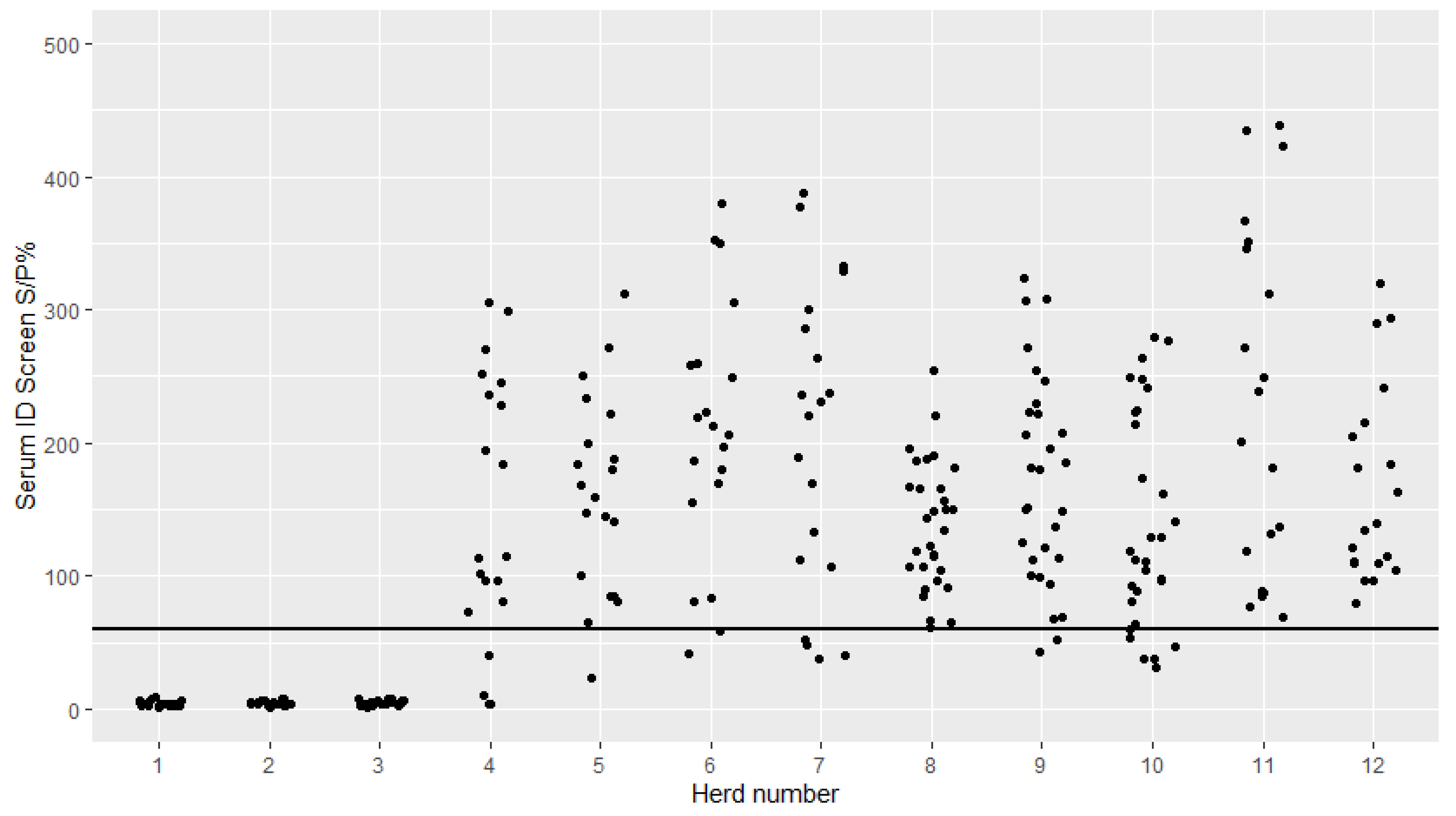
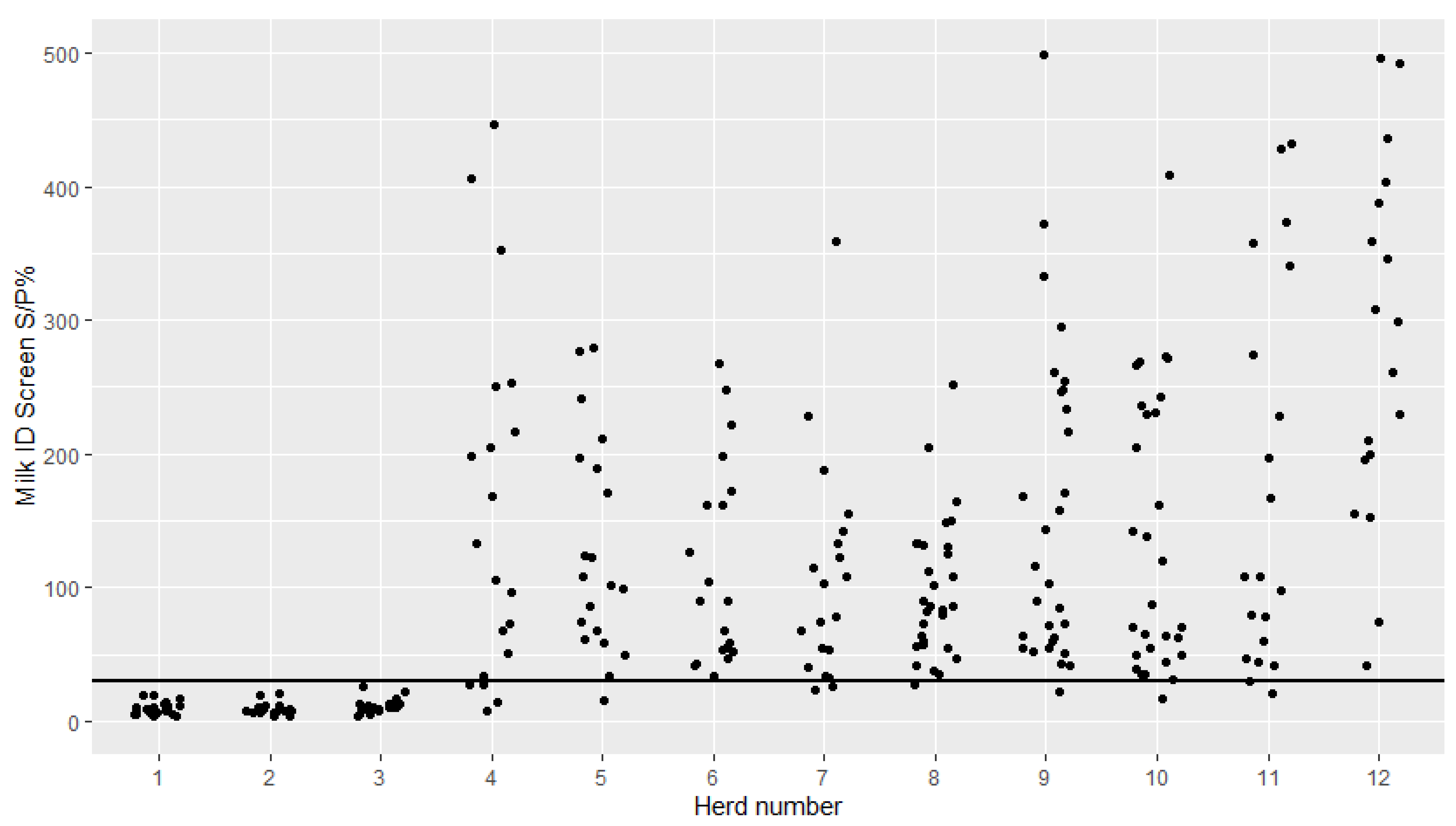
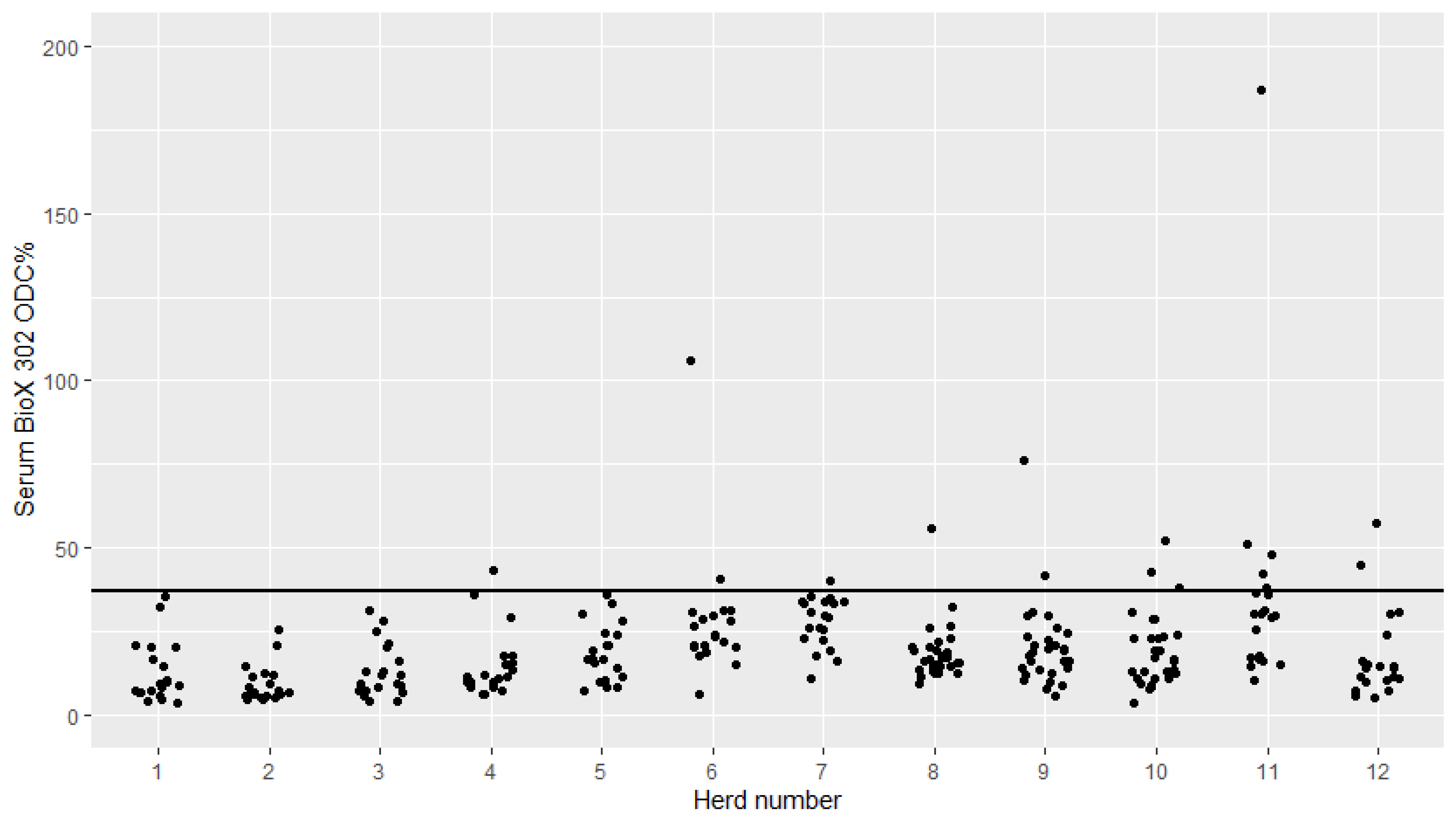
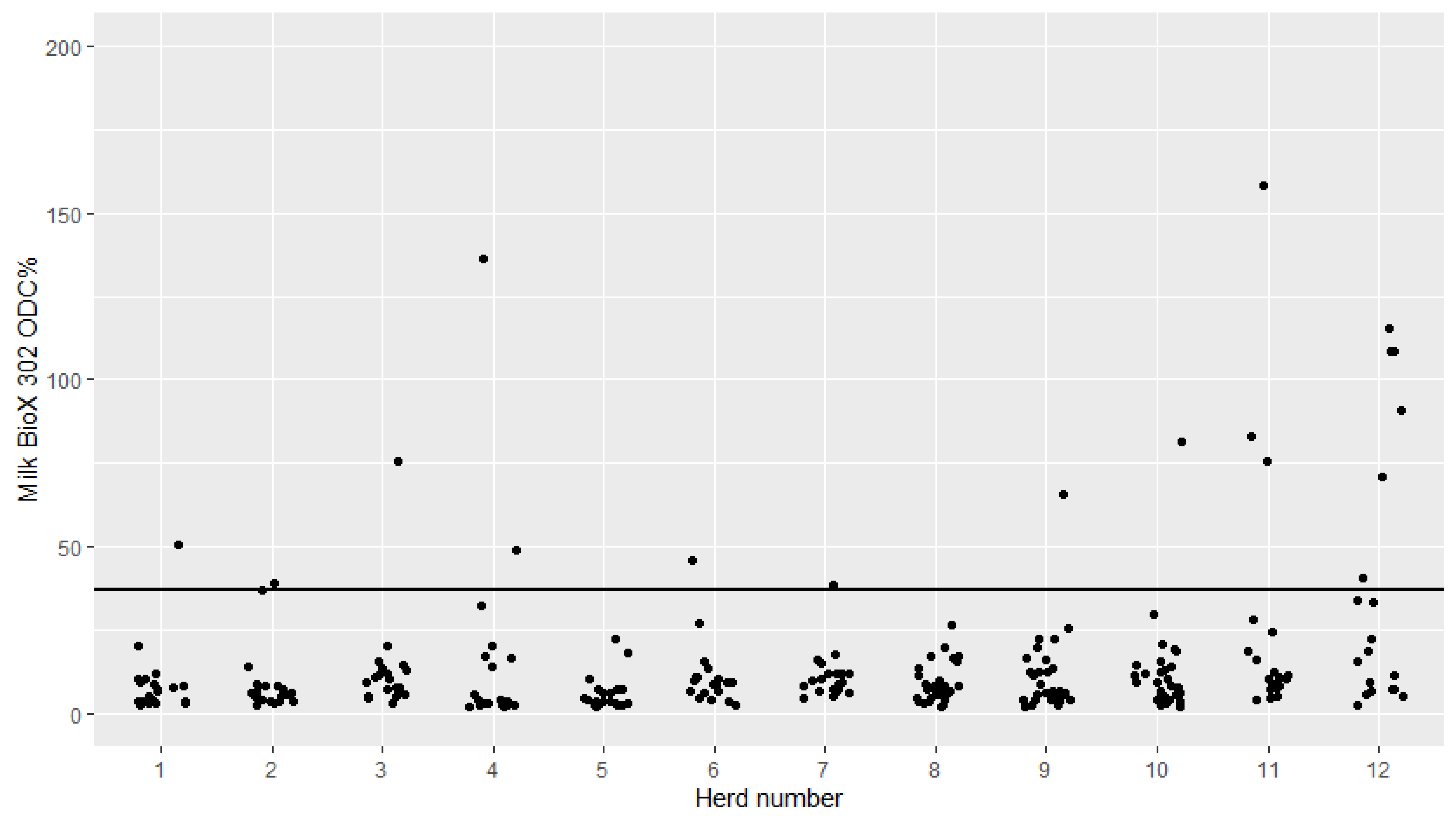
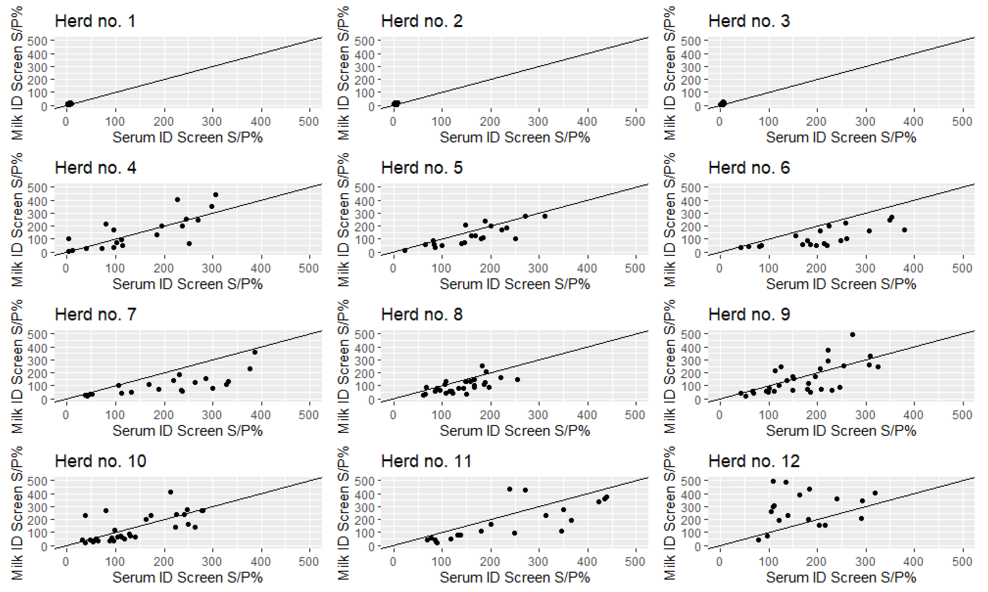
2. Results
Correlation between Serum and Milk S/P%
3. Discussion
3.1. Field Performance of the ID Screen and BioX 302
3.2. Correlation between Serum and Milk Samples
3.3. Uncertainty in Herd Classification
4. Materials and Methods
4.1. Study Herds
- Not infected—meaning that none (or very few, likely false positives) of the available test results were positive for M. bovis and the farmer stated that they had never had clinical signs of M. bovis-associated disease or that the clinical signs occurred more than 5 years prior to sampling;
- Infected within the last 5 years—meaning that there were multiple positive diagnostic test results in previously or recently collected samples and/or reporting of clinical signs of M. bovis within the last 5 years prior to sampling;
- Infected at sampling—meaning that diagnostic tests indicated an ongoing infection with M. bovis among one or more age groups at the time of sampling for the present study.
4.2. Sample Collection
4.3. Laboratory Analysis
4.4. Statistical Analysis
5. Conclusions
Author Contributions
Funding
Conflicts of Interest
References
- Nicholas, R.A.J.; Ayling, R.D. Mycoplasma bovis: Disease, diagnosis, and control. Res. Vet. Sci. 2003, 74, 105–112. [Google Scholar] [CrossRef]
- Maunsell, F.P.; Donovan, G.A. Mycoplasma bovis Infections in young calves. Vet. Clin. N. Am. Food Anim. Pract. 2009, 25, 139–177. [Google Scholar] [CrossRef] [PubMed]
- Maunsell, F.P.; Woolums, A.R.; Francoz, D.; Rosenbusch, R.F.; Step, D.L.; Wilson, D.J.; Janzen, E.D. Mycoplasma bovis Infections in Cattle. J. Vet. Intern. Med. 2011, 25, 772–783. [Google Scholar] [CrossRef] [PubMed]
- Petersen, M.B. Mycoplasma Bovis in Dairy Cattle—Clinical Epidemiology and Antibody Measurements for Decision Making. Ph.D. Thesis, University of Copenhagen, Frederiksberg, Denmark, 15 August 2018. [Google Scholar]
- Schibrowski, M.L.; Barnes, T.S.; Wawegama, N.K.; Vance, M.E.; Markham, P.F.; Mansell, P.D.; Marenda, M.S.; Kanci, A.; Perez-Casal, J.; Browning, G.F.; et al. The performance of three immune assays to assess the serological status of cattle experimentally exposed to Mycoplasma bovis. Vet. Sci. 2018, 5, 27. [Google Scholar] [CrossRef] [PubMed] [Green Version]
- Wawegama, N.K.; Markham, P.F.; Kanci, A.; Schibrowski, M.; Oswin, S.; Barnes, T.S.; Firestone, S.M.; Mahony, T.J.; Browning, G.F. Evaluation of an IgG enzyme-linked immunosorbent assay as a serological assay for detection of mycoplasma bovis infection in feedlot cattle. J. Clin. Microbiol. 2016, 54, 1269–1275. [Google Scholar] [CrossRef] [PubMed] [Green Version]
- Andersson, A.M.; Aspán, A.; Wisselink, H.J.; Smid, B.; Ridley, A.; Pelkonen, S.; Autio, T.; Lauritsen, K.T.; Kensø, J.; Gaurivaud, P.; et al. A European inter-laboratory trial to evaluate the performance of three serological methods for diagnosis of Mycoplasma bovis infection in cattle using latent class analysis. BMC Vet. Res. 2019, 15, 1–10. [Google Scholar] [CrossRef] [PubMed] [Green Version]
- Petersen, M.B.; Pedersen, J.; Holm, D.L.; Denwood, M.; Nielsen, L.R. A longitudinal observational study of the dynamics of Mycoplasma bovis antibodies in naturally exposed and diseased dairy cows. J. Dairy Sci. 2018, 101, 7386–7396. [Google Scholar] [CrossRef] [PubMed]
- Petersen, M.B.; Wawegama, N.K.; Denwood, M.; Markham, P.F.; Browning, G.F.; Nielsen, L.R. Mycoplasma bovis antibody dynamics in naturally exposed dairy calves according to two diagnostic tests. BMC Vet. Res. 2018, 14. [Google Scholar] [CrossRef] [Green Version]
- Bürki, S.; Frey, J.; Pilo, P. Virulence, persistence and dissemination of Mycoplasma bovis. Vet. Microbiol. 2015, 179, 15–22. [Google Scholar] [CrossRef] [Green Version]
- Vähänikkilä, N.; Pohjanvirta, T.; Haapala, V.; Simojoki, H.; Soveri, T.; Browning, G.F.; Pelkonen, S.; Wawegama, N.K.; Autio, T. Characterisation of the course of Mycoplasma bovis infection in naturally infected dairy herds. Vet. Microbiol. 2019, 231, 107–115. [Google Scholar] [CrossRef] [PubMed] [Green Version]
- Anonymous. Internal Validation Report ID Screen® Mycoplasma Bovis Indirect. Available online: https://www.id-vet.com/produit/id-screen-mycoplasma-bovis-indirect/ (accessed on 30 June 2020).
- Howard, C.J.; Gourlay, R.N. Imunne Response Of Calves Following The Inoculation Of Mycoplasma Dispar And Mycoplasma Bovis. Vet. Microbiol. 1983, 8, 45–56. [Google Scholar] [CrossRef]
- Jacobson, R.H. Validation of serological assays for diagnosis of infectious diseases. Rev. Sci. et Tech. de l’OIE 1998, 17, 469–486. [Google Scholar] [CrossRef] [PubMed]
- Nielsen, P.K.; Petersen, M.B.; Nielsen, L.R.; Halasa, T.; Toft, N. Latent class analysis of bulk tank milk PCR and ELISA testing for herd level diagnosis of Mycoplasma bovis. Prev. Vet. Med. 2015, 121, 338–342. [Google Scholar] [CrossRef] [PubMed]
- Parker, A.M.; House, J.K.; Hazelton, M.S.; Bosward, K.L.; Morton, J.M.; Sheehy, P.A. Bulk tank milk antibody ELISA as a biosecurity tool for detecting dairy herds with past exposure to Mycoplasma bovis. J. Dairy Sci. 2017, 100, 8296–8309. [Google Scholar] [CrossRef] [PubMed] [Green Version]
- Anonymous. Standard Operating Procedure for OIE Registration of Diagnostic Kits. Available online: https://www.oie.int/doc/ged/D12069.PDF (accessed on 29 July 2020).
- Petersen, M.B.; Krogh, K.; Nielsen, L.R. Factors associated with variation in bulk tank milk Mycoplasma bovis antibody-ELISA results in dairy herds. J. Dairy Sci. 2016, 99, 3815–3823. [Google Scholar] [CrossRef] [PubMed] [Green Version]
- Goecke, N.B.; Hjulsager, C.K.; Krog, J.S.; Skovgaard, K.; Larsen, L.E. Development of a high-throughput real-time PCR system for detection of enzootic pathogens in pigs. J. Vet. Diagn. Investig. 2020, 32, 51–64. [Google Scholar] [CrossRef] [PubMed]
- Wawegama, N.K.; Browning, G.F.; Kanci, A.; Marenda, M.S.; Markham, P.F. Development of a recombinant protein-based enzyme-linked immunosorbent assay for diagnosis of mycoplasma bovis infection in cattle. Clin. Vaccine Immunol. 2014, 21, 196–202. [Google Scholar] [CrossRef] [PubMed] [Green Version]
- Anonymous. IDVet Screen Mycoplasma Bovis Indirect Material Safety Data Sheet. Available online: https://www.id-vet.com/produit/id-screen-mycoplasma-bovis-indirect/ (accessed on 30 June 2020).
- Anonymous. BioX Diagnostics Monoscreen Ab ELISA. Available online: https://www.biox.com/en/bio-k-302-monoscreen-abelisa-mycoplasma-bovis-indirect-monowell-p-250/ (accessed on 30 June 2020).
- Lin, L.I.-K. A Concordance Correlation Coefficient to Evaluate Reproducibility. Biometrics 1989, 45, 255. [Google Scholar] [CrossRef] [PubMed]
- R Core Team. R: A Language and Environment for Statistical Computing. R Foundation for Statistical Computing: Vienna, Austria,2016. Available online: https://www.R-project.org/ (accessed on 30 June 2020).


| Herd No. | Herd Type | No. of Cows a | PCR—Individual Cows b (Positives/n) | ELISA—Individual Cows/ Calves c (Positives/n) | BTM PCR b (Positives/n) | BTM ELISA c (Positives/n) | RC-Calves—ID Screen d (Positives/n) | RC-Calves—BioX 302 c (Positives/n) | RC-Calves—PCR Test e (Positives/n) | Mycoplasma bovis Disease Outbreak | Mycoplasma bovis Classification |
|---|---|---|---|---|---|---|---|---|---|---|---|
| 1 | RC | 150 | N/A | N/A | 0/21 | 0/16 | 0/27 | 1/27 | 0/182 | No f | Not infected |
| 2 | RC | 190 | 0/348 | N/A | 0/14 | 0/8 | 0/27 | 2/27 | 1/209 | No f | Not infected |
| 3 | RC | 350 | 0/9 | 0/8 | 0/9 | 1/5 | 0/29 | 1/29 | 0/228 | Yes (2013) f | Not infected |
| 4 | RC | 220 | 0/1 | N/A | 0/10 | 0/6 | 0/29 | 0/29 | 0/172 | No f | Not infected |
| 5 | RC | 200 | 0/3 | N/A | 0/11 | 1/6 | 1/29 | 0/30 | 0/129 | Yes (2012) f | Not infected |
| 6 | RC | 700 | 1/398 | N/A | 0/9 | 2/5 | 16/30 | 2/30 | 3/431 | No f | Infected within the last 5 years |
| 7 | RC | 600 | 0/284 | N/A | 1/23 | 0/6 | 24/30 | 10/30 | 1/179 | Yes (2014–2015) f | Infected within the last 5 years |
| 8 | Outbreak | 190 | 7/140 | 85/372 | 0/34 | 0/23 | N/A | N/A | N/A | Yes (2015–2016) g | Infected within the last 5 years |
| 9 | Outbreak | 430 | 69/1188 | 70/282 | 18/327 | 1/9 | N/A | N/A | N/A | Yes (2015–2016) g | Infected within the last 5 years |
| 10 | Outbreak | 200 | 21/98 | 91/303 | 3/16 | 1/12 | N/A | N/A | N/A | Yes (2015–2016) g | Infected within the last 5 years |
| 11 | RC | 600 | 10/25 | 0/3 | 0/10 | 1/7 | N/A | N/A | 11/256 | Yes (2014) f | Infected at sampling |
| 12 | RC | 330 | 4/234 | N/A | 0/14 | 0/6 | N/A | N/A | 9/228 | No f | Infected at sampling |
© 2020 by the authors. Licensee MDPI, Basel, Switzerland. This article is an open access article distributed under the terms and conditions of the Creative Commons Attribution (CC BY) license (http://creativecommons.org/licenses/by/4.0/).
Share and Cite
Petersen, M.B.; Pedersen, L.; Pedersen, L.M.; Nielsen, L.R. Field Experience of Antibody Testing against Mycoplasma bovis in Adult Cows in Commercial Danish Dairy Cattle Herds. Pathogens 2020, 9, 637. https://0-doi-org.brum.beds.ac.uk/10.3390/pathogens9080637
Petersen MB, Pedersen L, Pedersen LM, Nielsen LR. Field Experience of Antibody Testing against Mycoplasma bovis in Adult Cows in Commercial Danish Dairy Cattle Herds. Pathogens. 2020; 9(8):637. https://0-doi-org.brum.beds.ac.uk/10.3390/pathogens9080637
Chicago/Turabian StylePetersen, Mette Bisgaard, Lars Pedersen, Lone Møller Pedersen, and Liza Rosenbaum Nielsen. 2020. "Field Experience of Antibody Testing against Mycoplasma bovis in Adult Cows in Commercial Danish Dairy Cattle Herds" Pathogens 9, no. 8: 637. https://0-doi-org.brum.beds.ac.uk/10.3390/pathogens9080637