Article

Monitoring the Starvation–Survival Response of Edwardsiella piscicida and E. tarda in Freshwater Microcosms, at Various Temperatures

Departamento de Microbiología y Ecología, Universitat de València, Campus de Ciencias Burjassot-Paterna, E-46100 Valencia, Spain
*
Author to whom correspondence should be addressed.
Submission received: 2 May 2022 / Revised: 13 May 2022 / Accepted: 16 May 2022 / Published: 18 May 2022
(This article belongs to the Section Environmental Microbiology)

Abstract

:
Edwardsiella piscicida is an important fish pathogen responsible for economic losses in global aquaculture, and E. tarda is also a human zoonotic pathogen. In this study, the survival of E. piscicida and E. tarda strains kept in filtered and sterilized lake water microcosms was investigated during a 20-week period at 7 °C, 15 °C and 25 °C, as well as its pathogenicity retention during a starvation period. E. tarda V43.2 stayed culturable for 6 weeks at 7 °C, 9 weeks at 25 °C and 12 weeks at 15 °C. Both E. piscicida strains (V12.1 and V57.2) stayed culturable even longer, for at least 12 weeks at 7 °C, 15 °C and 25 °C under the same starvation conditions. After Edwardsiella cells entered into the VBNC state, some became shorter and ”rounded up,” but others aggregated and retained a short rod shape. Aggregates of Edwardsiella cells were common throughout the VBNC period, and a well-formed biofilm was observed for all tested strains at the end of the experiment. The growth capacity of VBNC cells was restored by cultivating microcosm water samples in LB broth at 28 °C. Resuscitated E. piscicida cells were as virulent for the European eel as the controls. Natural waters can be a reservoir for Edwardsiella, and its underestimation in environmental samples poses a risk to public health and aquaculture.

1. Introduction

The genus Edwardsiella includes enteric gram-negative bacteria of the Hafniaceae family [1] that are causative agents of disease in homeothermic and poikilothermic animals [2,3]. The species E. tarda, E. ichtaluri, E. piscicida and E. anguillarum are related to the systemic disease Edwardsiellosis, which has been associated with mass mortality in more than 20 species of fish reared in freshwater and marine-supplied aquaculture systems worldwide [4,5].
The search for Edwardsiella reservoirs in wild aquatic vertebrates and natural surface waters has primarily been a public health issue [6,7] because E. tarda cause water- and foodborne infection in humans [2]. To date, the Edwardsiella have been effectively isolated from diseased aquatic vertebrates [8,9,10] and, to a lesser extent, from “healthy carriers” [6,10,11]. In addition, E. tarda have been isolated from natural surface freshwater on a few occasions [8,10]. Regarding E. piscicida, the only data on its presence in nature describe its isolation from wild eels (Anguilla anguilla), but scarcely from lagoon freshwater, with all samples being collected at the L’Albufera Lake (Mediterranean Spain) over a 12-year period [10,12]. Since Edwardsiellosis disease arises within populations in warm seasons [10,13,14], its continuous incidence in the fishery over a long time period is an intriguing problem [10,12].
The classic experiments to evaluate the role of water as a reservoir of many bacterial pathogens and, therefore, to study their survival response to lack of nutrients, have been carried out using microcosms of filtered and sterilized water [15,16,17,18]. Du et al. [15] studied the survival response of E. tarda strain CW7 in the microcosms of filtered sterilized seawater (FSSW) maintained at 4 °C for 42 days. They reported no more than 28 days of cultivability for strain E. tarda CW7 at 4 °C under nutrient deprivation, after which it entered the viable but non-cultivable (VBNC) state [15]. Many species of bacteria enter the VBNC state when exposed to stressful conditions such as starvation and low temperatures [15,16,17,18], and this bacterial adaptability could pose a risk to animal and human health. The risks emerge from the fact that pathogenic bacteria can be avirulent in the VBNC state but regain virulence after resuscitation into culturable cells under suitable conditions [16,17,18]. This was the case for resuscitate cells of E. tarda CW7 that were as virulent for fish as the original strain [15]. For now, the survival response to starvation of E. piscicida is unknown because no research has been conducted on this topic.
To better understand the environmental epidemiology of these pathogens, we studied the starvation response of two E. piscicida and one E. tarda strains in filtered and sterilized lake water (FSLW) microcosms maintained at cold (7 °C), temperate (15 °C) and warm (25 °C) temperatures. This study represents the first assessment of the response of E. piscicida and E. tarda to nutrient deprivation in water over a period of 20 weeks (i.e., 140 days), showing its ability to survive and remain pathogenic in freshwater at several temperatures (7 °C, 15 °C and 25 °C), as well as its ability to form biofilms under environmental stress.

2. Materials and Methods

2.1. Bacterial Strains and Culture Conditions

Three Edwardsiella strains were used in the study: E. piscicida V12.1 isolated from kidney of diseased European eel [19], E. piscicida V57.2 isolated from intestine of healthy European eel [10] and E. tarda V43.2 isolated from lagoon freshwater [10]. These strains were previously identified by biochemical traits as well as 16S rRNA sequencing [10]. Unless otherwise stated, cells were grown in Luria Bertani (LB, Pronadisa, Alcobendas, Spain) broth and agar (LBA, Pronadisa) at 28 °C for 24 h.

2.2. Preparation of the Filtered and Sterilized Lake Water (FSLW) and Inoculation of Microcosms

Natural fresh water collected at a depth of 20 cm from Lake L’Albufera (Mediterranean Spain) was used. In the laboratory, lagoon freshwater was filtered through membrane filters (Millipore, 0.45 µm pore size) to remove all particles, placed in 125 mL Erlenmeyer flasks with a stopper (each containing 75 mL of water) and finally autoclaved at 121 °C for 20 min. The physicochemical parameters of the FSLW were BOD5 = 8 mg O2/L regarding of dissolved organic matter; pH = 8.2; salinity = 0.6 g/L.
Bacteria were grown in LBA at 28 °C, and harvested and washed twice in quarter-strength Ringer’s solution (Oxoid, Hampshire, UK). Then, Edwardsiella cells pellet was re-suspended into FSLW, and added into the FSLW microcosms in a final concentration of 107–108 CFU/mL. The seeded microcosms were incubated without shaking (static microcosms) at 7 °C, 15 °C or 25 °C and maintained in the dark for 20 weeks. Experiments were performed in triplicate (three simultaneous microcosms) for each Edwardsiella strain and temperature condition. Temperature values were chosen according to water temperature records previously measured at the L’Albufera Lake [10].

2.3. Cell Counts, Viability Assessment and Staining Methods

Cell counts were determined immediately after microcosm inoculation and then at a weekly or biweekly interval over an 18-week period. A last counting was performed at the 20-week mark. Microcosms were gently shaken prior to take the sample for counting.
Direct counts of viable and total bacterial cells were determined in 7 °C and 25 °C microcosm samples by staining with Syto 9 and propidium iodide (PI) (Baclight LIVE/DEAD kit, Molecular Probes Inc., Eugene, OR, USA) for 20 min. The stained samples were added to 5 mL of sterile Scharlau water and filtered on 0.22 µm black Nucleopore filters (GTPB filter type, Millipore, Burlington, MA, USA). The prepared filters were observed using epifluorescence microscopy (Nikon ECLIPSE E800, Tokyo, Japan) in 20 to 30 randomly selected fields for a total count, and the number of total (reed and green cells) and viable (green cells) bacteria was calculated, respectively. Morphological changes were annotated and photographed.
The culturable cell counts were determined in 7 °C, 15 °C and 25 °C-microcosms by seeding 3 plates of Luria-Bertani Agar (LBA, Pronadisa) per each water microcosm dilution, and then incubated for 24–48 h at 28 °C. The dilution chose for counting was always between 20 and 200 colonies on LBA plates. The number of colony-forming units (CFU) per mL was calculated from the number of colonies, the volume of the inoculum used (0.1 mL) and the dilution factor. Finally, the CFU/mL for each Edwardsiella strain and temperature treatment was expressed as (average ± standard deviation) from the results obtained in the three simultaneous microcosms. Colonies counted in these LBA cultures were sub-cultured onto Salmonella Shigella agar (SSA, Oxoid) to check that they showed the typical Edwardsiella morphology (i.e., transparent colonies with a black center as it is a non-lactose-fermenting and H2S-producing organism). Once the plate counts were negative for Edwardsiella (below the detection limit of 1 CFU per 0.1 mL), samples of 1 mL were taken directly from the microcosm and plated on LBA plates (3 plates) that were incubated at 28 °C for 48 h. At this point, the cells were considered to be in the viable non-culturable state (VBNC) if no growth was achieved.

2.4. Resuscitation of VBNC Cells

Microcosm samples of 7 °C, 15 °C and 25 °C that did not grow in LBA plates were taken at 1, 2, 3, 4, 5 and 6 weeks after entering the VBNC state in order to restore the culture capacity of the strain, according to the method described by the authors of [20].
Briefly, aliquots of 1 mL samples of microcosm water were diluted (from 10−1 to 10−7) in 9 mL tubes of Luria Bertani broth (LBB, Pronadisa) to reduce the probability (P) of any culturable cell down to <0.0000001 CFU/mL. These broth cultures were incubated at 28 °C for 2–4 days with shaking. Those with positive growth were seeded on Salmonella Shigella agar (SSA, Oxoid), and the plates were incubated at 28 °C for 24–48 h. Typical Edwardsiella colonies in SSA were sub-cultured on LBA to obtain pure cultures that were identified [10].

2.5. Virulence of the Original Edwardsiella Strains and of Its Resuscitated Cultures for European Eel

Animal experiments were carried out in accordance with the EU Directive [21]. Twelve juvenile eels of 10–15 g were used for the challenge dose. All eels were supplied by the “Piscifactoria de Tuéjar (Azud de Tuéjar. Generalitat Valenciana)” and certified to be disease-free.
The virulence of each original Edwardsiella isolate and its resuscitated strains was assessed by an intraperitoneal (IP) injection challenge at a unique dose of 107.3 to 105.8 CFU/fish as previously described [10]. A control group was used for each trial in which the fish were injected IP with 0.1 mL of sterile saline. The mortalities were recorded daily for a 5-day period and were only considered if the challenged strain was recovered as pure cultures from the internal organs. In all experiments, the percentage of accumulative mortality was determined, the CFUs of the bacterial suspensions were obtained on LBA at 28 °C by the “drop-plate method” [22] and the survivors were sacrificed by 20 min inhalation of anesthesia with benzocaine at 200 mg/L (wt./vol.).

2.6. Data Analysis and Statistics

A one-way analysis of variance (ANOVA) with Tukey’s method was used to assess the statistical significance of the differences observed in VBC counts as a function of incubation temperature. For each strain, data were analyzed if VBC counts were obtained from all temperatures.

3. Results

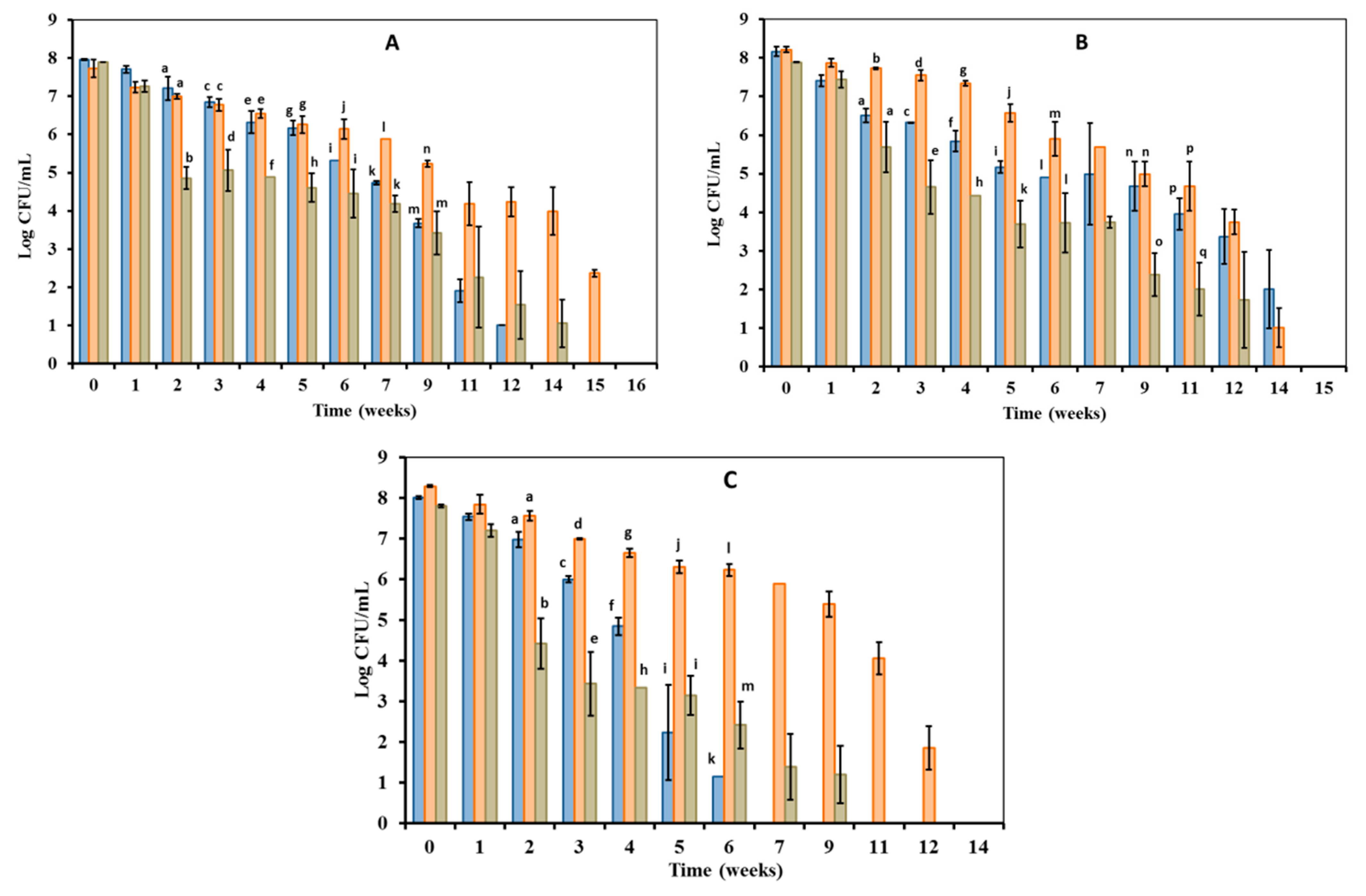
3.1. Culturability of Edwardsiella Maintained under Starvation Conditions at Various Temperatures

Culturable cell (VBC) numbers in Edwardsiella populations were around 108 CFU/mL at the beginning of the experiment (Figure 1), which lasted 20 weeks. In general, we found differences between E. piscicida (Figure 1A,B) and E. tarda (Figure 1C) regarding their culturability behavior when they were kept under starvation conditions.
The VBC numbers of E. piscicida kept at 7 °C decreased from 108 to 105 CFU/mL after 7 weeks of incubation (Figure 1A,B), while that of E. tarda were of 0 CFU/mL (Figure 1C). The two E. piscicida strains entered into viable non-culturable state (VBNC: 0 CFU/mL) significantly later than the E. tarda strain because they stayed culturable onto LBA plates until 12 weeks (Figure 1A) or 14 weeks (Figure 1B) of incubation.
All Edwardsiella kept at 15 °C under starvation conditions maintained VBC counts above 105 CFU/mL until at least 9 weeks of incubation (Figure 1). Then, the two E. piscicida strains stayed culturable for an additional 5 or 6 weeks, entering into the VBNC state (0 CFU/mL) after 15 weeks (Figure 1A) or week 14 (Figure 1B) of incubation. The E. tarda strain entered into VBNC state earlier than E. piscida ones because 0 CFU/mL counts were already recorded after the 12 weeks of incubation (Figure 1C).
All microcosms kept at 25 °C under starvation conditions showed an acute decreasing in VBC Edwardsiella numbers of at least 3-log units during the second week of incubation (Figure 1). After, the VBC numbers of the two E. piscicida strains slowly decreased along study period, and stayed culturable even at levels of around 103 CFU/mL at 9–11 weeks of incubation (Figure 1A,B). Finally, E. piscicida strains entered into the VBNC state after 14 weeks (Figure 1A) or 12 weeks (Figure 1B) of incubation. In contrast, VBC numbers of the E. tarda strain V43.2 decreased rapidly from the second week to the end of the experiment, and the strain entered into the VBNC (0 CFU/mL) after 9 weeks of incubation (Figure 1C).
Furthermore, the observed differences in VBC counts as a function of microcosm incubation temperature were statistically significant for Edwardsiella strains maintained under the same starvation conditions (Figure 1). Regadless of their taxonomic assignment, all Edwardsiella showed the highest numbers of VBC counts in microcosms at 15 °C (Figure 1). E. piscicida strains frequently showed significantly higher VBC counts in microcosms at 7 °C than in microcosms at 25 °C throughout the experimental period (Figure 1A,B). In contrast, E. tarda seemed to maintain more stable VBC numbers in microcosm at 25 °C than in microcosms at 7 °C (Figure 1C).

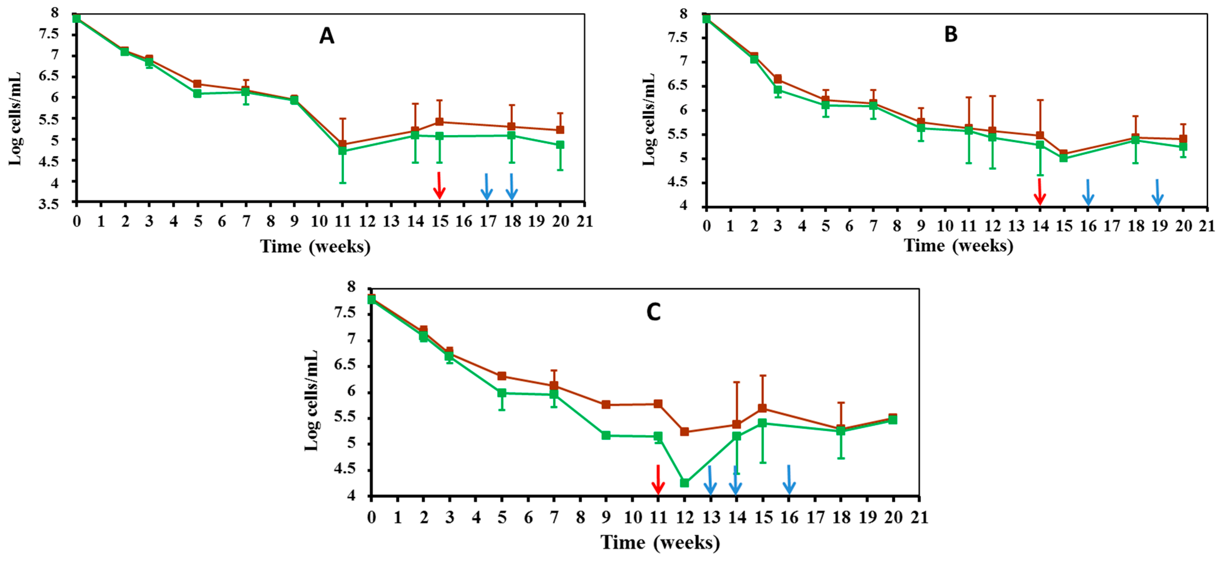
3.2. The VBNC State of Edwardsiella Maintained under Starvation Conditions at 7 °C and 25 °C

3.2.1. Direct Counts of Edwardsiella Cells during Culturable and VBNC Period

Throughout the period in which the tested strains decreased in cultivability (Figure 1), the initial size of direct Edwardsiella counts also showed a marked decrease, regardless of the strain taxonomic assignment and the incubation temperature (7 °C: Figure 2, 25 °C: Figure 3).
Such a numerical reduction could show a rapid initial collective death rate in the Edwardsiella population, which would then benefit viable cells, as they can recycle the biomass of dead ones [23]. In fact, direct Edwardsiella cell counts remained stable throughout the VBNC period, the beginning of which is marked on the graphs with a red arrow (Figure 2 and Figure 3). The direct total and viable Edwardsiella cell counts during the VBNC period and until the end of the experiment remained around 107 cells/mL in the 7 °C microcosms (Figure 2). These counts were at lower levels of 106–105 cells/mL in the 25 °C microcosms (Figure 3).

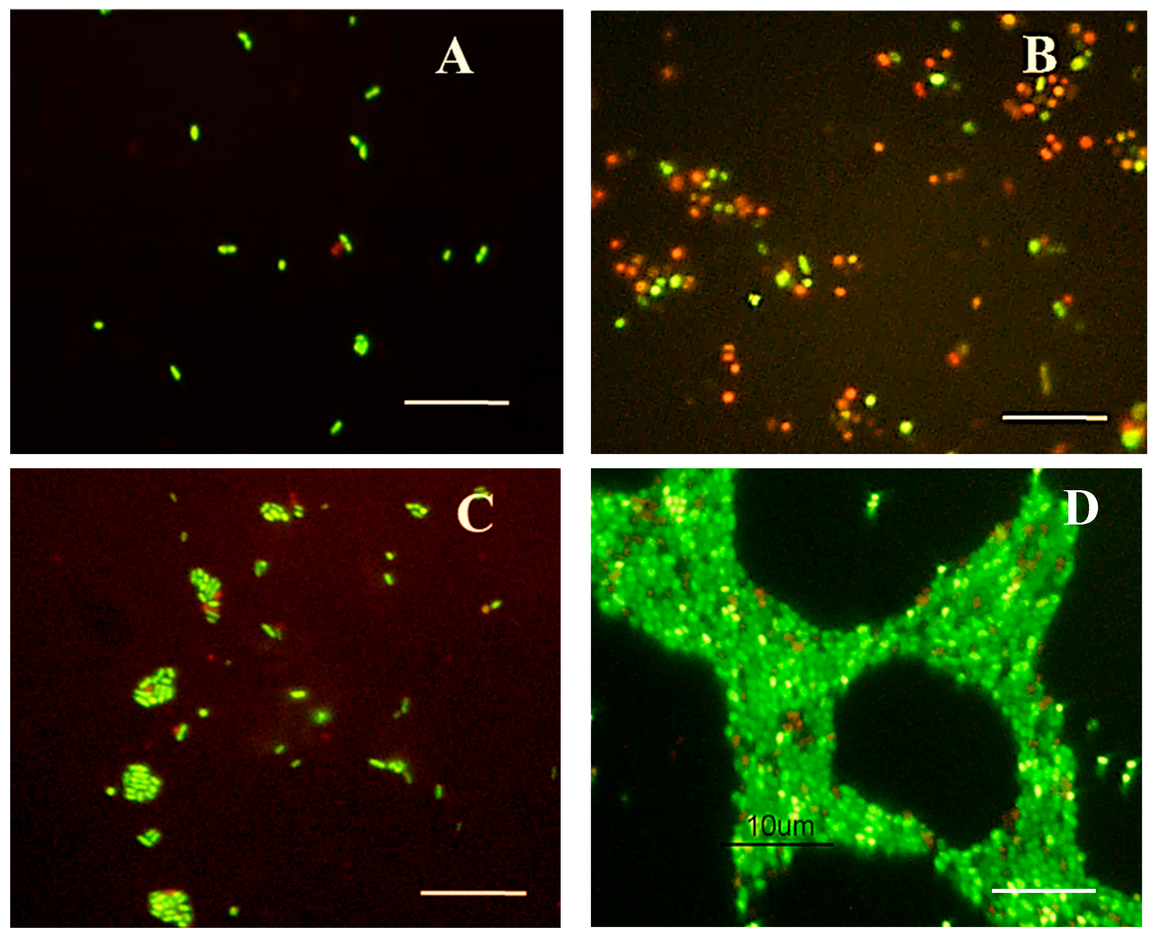
3.2.2. Morphological Changes Observed in Edwardsiella Cells during Starvation-Survival Response

Samples (100 µL) of microcosm water were stained with Syto 9 and propidium iodide (LIVE/DEAD staining) to reveal viable/live (green-stained) and dead (red-stained) Edwardsiella cells. Morphological changes of Edwardsiella cells maintained under starvation conditions for 20 weeks at 7 °C or 25 °C were observed under an epifluorescence microscopy (Figure 4).
The two strains of E. piscicida and the strain of E. tarda showed similar morphological changes upon entering into the VBNC state. Edwardsiella cells were single or paired rods (Figure 4A) that progressively changed over their cultivable period to rounded viable cells (Figure 4B), rounded non-viable cells (Figure 4B) or rod-shaped viable cells in aggregates (Figure 4C).
Figure 4. Morphological changes of Edwardsiella cells maintained under starvation conditions. (A) Viable bacillary cells in 2-day-old microcosms (10 µL were used for LIVE/DEAD staining); (B) Viable and non-viable rounded cells, and viable bacillary cells in 4-week-old microcosm; (C) Viable and non-viable rounded cells, and aggregates of viable bacillary cells observed in 10-week-old microcosm; (D) Biofilm composed primarily of short rod-shaped viable cells in 20-week-old microcosm. Bars = 10 µm.
Microorganisms 10 01043 g004
We observed that these three morphological types could coexist in the microcosms at the same time, at the beginning of the VBNC period. Finally, a biofilm, primarily including short rod-shaped viable cells and some non-viable rounded cells, was observed for all tested strains at the end of the experiment (Figure 4D). Those viable cells inside the biofilms presented a bright green color or a darker green color by Syto9. We did not find that LIVE/DEAD staining underestimated the viability of these adherent cells as others described [24].

3.2.3. Resuscitation of VBNC Edwardsiella Cells and Its Virulence for European Eel

Samples were taken from 7 °C, 15 °C and 25 °C microcosms that did not grow on LBA plates (VBNC: 0 CFU/mL, Figure 1) after 1, 2, 3, 4, and 5 weeks of entering the VBNC state (Figure 2 and Figure 3), with the purpose of restoring the culture capacity of the strain. The growth capacity of VBNC cells was restored in both E. piscicida and E. tarda strains using a rich general medium (i.e., LB broth) to culture the microcosm water samples. Furthermore, an increase in temperature could have positively affected Edwardsiella cultivability in the case of microcosms at 7 °C and 15 °C, because all samples were incubated at 28 °C. The “resuscitation window” [25] was different depending on the microcosm temperature. The longest resuscitation period was observed for the microcosms kept at 15 °C because growing Edwardsiella cells recovered 2–4 weeks after entering the VBNC state (Table 1). A narrower resuscitation window was observed for the VBNC cells from those microcosms maintained at 7 °C and 25 °C. The recovery of growing Edwardsiella after entering the VBNC state was limited to 1 week for microcosms maintained at 7 °C and 2 weeks for those maintained at 25 °C (Table 1).
The resuscitated E. piscicida strains were virulent for eels by intraperitoneal injection (IP) challenge at doses of 106 CFU/g of fish. They caused an accumulative mortality of 67–100% among challenged fish (Table 1). Dead and moribund fish showed pathological signs previously observed in sampled wild eels suffering from edwardsiellosis [10]. Although these strains were generally as pathogenic as the original strain, we observed that the longest lapse of time after the induction of the VBNC state could have contributed to reduction of the pathogenicity of one out of the five resuscitated E. piscicida tested (Table 1). The original E. tarda V43.2 strain, as well as its resuscitated strains, yielded 0% of accumulative mortality among challenged fish; therefore, they were avirulent for European eel (Table 1) in contrast to E. tarda CW7 [15]. The E. tarda CW7 was recovered from diseased fish [15]. Therefore, it could belong to another Edwardsiella species based on the current taxonomic viewpoint [5]. In fact, the genetic divergence observed for fish isolates of E. tarda [26,27] was the first evidence that later led to the proposals of E. piscicida and E. anguillarum [28,29]. The fish pathogenic Edwardsiella (i.e., E. ichtaluri, E. piscicida and E. anguillarum) constitute a clearly segregated branch of E. tarda and E. hoshinae in the Edwardsiella phylogenetic tree [29,30]. Thus, strains previously defined as “fish pathogenic E. tarda” belong to the species E. piscicida or E. anguillarum, whereas strains of E. tarda that are non-pathogenic for fish are consistent with the species E. tarda, as currently defined [5,30].

4. Discussion

A recent paper introduced that Edwardsiella exist in aquatic environments either as individual planktonic cells or in communal biofilms [31]. However, biofilm production by Edwardsiella has been only related to its life as a pathogen for vertebrate animals [32,33]. Moreover, E. piscicida and E. tarda have hardly been isolated from surface water in natural environments, and work on this subject is limited. White et al. [8] recovered Edwardsiella from 13 out of 108 water samples (12%), and Esteve and Alcaide [10] recovered Edwardsiella from 2 out of 27 water samples (7%). Some authors have highlighted the role of the intestine as a gateway for the Edwardsiella since they can cross intestinal mucosa, such as other enteric pathogens ingested through water or food [2,34]. Although the presence of Edwardsiella in the intestines of healthy wild fish has been reported [6,10,11], the role of water as a reservoir for these bacteria is still unknown. Specifically, it is unknown whether the Edwardsiella are capable of surviving in such an oligotrophic environment even after their exposure to different temperature conditions.
Du et al. [15] reported no more than 28 days of culturability for the strain E. tarda CW7 maintained in filtered and sterilized seawater (FSSW) at 4 °C under nutrient starvation. In contrast, our strain, E. tarda V43.2, remained cultivable for 42 days in filtered sterilized freshwater (FSLW) microcosms at 7 °C, and slightly longer at warm (25 °C) temperature, similar to that observed for other bacterial pathogens of humans and fish [16,17]. The two strains of E. piscicida tested (V12.1 and V57.2) remained cultivable even longer, for 84 days at 7 and 25 °C, and around 100 days at 15 °C, under the same conditions. In addition, the highest number of viable cultivable (VBC) cells was found in the microcosms kept at 15 °C, showing a statistically significant difference. Unlike other fish pathogens, E. piscicida did not prefer low temperatures [18] or warm temperatures [16,17]. After the tested E. piscicida and E. tarda strains lost the ability to grow on Luria-Bertani agar, however, all FSLW microcosms had live Edwardsiella cells (i.e., viable non-cultivable (VBNC) cells) until the end of the experiment (140 days). These results show for the first time that the species E. piscicida exists in the VBNC state in response to nutrient deprivation.
The entry into the VBNC state of E. piscicida V12.1 and V57.2 and E. tarda V43.2 caused a reduction in the size of some cells until they became coccoid cells, as described for other gram-negative pathogens [15,16,17,18]. This rounding and cell size reduction is well known to increase the cell surface area to volume ratio, which is an advantage in oligotrophic environments [35]. Furthermore, our starved Edwardsiella strains exhibited various cell types at the beginning of the VBNC period, as observed by epifluorescence microscopy, which showed cylindrical rods, shorter rod-shape cells and spherical-shaped cells similar to that described for Escherichia coli K-12 [36]. The coexistence of different morphological types within starving bacterial populations have been linked in Vibrio species to starvation-induced phenotypic diversification [37,38]. Rod-shaped Edwardsiella cells forming aggregates were also observed in the VBNC period. In fact, our results show for the first time that starved populations of E. piscicida and E. tarda produce a biofilm, primarily including short rod-shaped viable cells and some non-viable rounded cells, after prolonged incubation in the VBNC period. Such loss of motility, as well as the increase in adhesion properties, including biofilm production, has been reported for other intestinal pathogens maintained in lake water microcosms, suggesting that biofilm might has a role in their survival in natural waters [39].
It is important to note that bacteria that enter the VBNC state may become culturable again. Thus, this state may be reversible in the presence of a suitable stimulus (i.e., rich medium, temperature upshift, presence of host cells) [35]. Of the bacterial pathogens that can enter the VBNC state (52 reported species), resuscitation has been reported in only half [35]. The risk emerges from the fact that many of them are avirulent in the VBNC state but regain virulence after resuscitation into culturable cells [15,35]. Similarly, we found that E. piscicida cells resuscitated from the VBNC state were as virulent to European eel as the original strain. Such behavior was limited to the “resuscitation window” involving younger E. piscicida VBNC cells, because a reduction in resuscitable cells was observed over time, consistent with other reports [40]. Specifically, the length of the “resuscitation window” observed for our Edwardsiella strains ranged from 1 to 2 weeks after entering into the VBNC state. This corresponded to the 112–119 days after the E. piscicida strains began the nutrient depletion conditions in FSLW microcosms. This work represents the first description of the starvation response of E. piscicida, evidencing its survival and pathogenic potential in freshwater at a wide range of temperatures. Moreover, it also delves into the starvation response of E. tarda compared to what was previously reported [15]. These findings suggest that natural freshwater may be a reservoir for Edwardsiella because it can adapt to survive in such an oligotrophic environment. Therefore, its low recovery from these samples could be explained by the use of culture media that are not selective enough to reduce the growth of accompanying bacteria (i.e., Enterobacter spp., Citrobacter spp., Aeromonas spp., Plesiomonas shigelloides and Hafnia alvei) [7,10]. Even the effect of microbial interactions with other bacteria or phages should not be ruled out as relevant stress factors for the survival of Edwardsiella in natural waters. Future research will focus on the effect of accompanying bacteria on the survival of Edwardsiella in water.

5. Conclusions

Edwardsiella piscicida and E. tarda can survive in freshwater under nutrient deprivation for long time periods, evidencing its starvation response and pathogenic potential at a wide range of temperatures (7–25 °C). Specifically, a temperate temperature (15 °C) favors the persistence of E. piscicida in the aquatic environment in a culturable and pathogenic state. However, lower and higher temperatures in other periods of the year would induce a more rapid development of the VBNC response, with an enhanced resistance to environmental stress due to biofilm formation. These VBNC cells can recover their pathogenicity under appropriate conditions according to a resuscitation window that is also temperature-dependent. This knowledge provides new and valuable information on the biology of this pathogen; therefore, it leads us to a better understanding of the environmental epidemiology of diseases caused by Edwardsiella.

Author Contributions

Conceptualization, C.E.; methodology, C.E. and E.A.; software, C.E.; validation, C.E. and E.A.; formal analysis, C.E.; investigation, C.E. and E.A.; resources, C.E.; data curation, E.A.; writing—original draft preparation, C.E.; writing—review and editing, C.E. and E.A.; visualization, C.E.; supervision, C.E. and E.A.; project administration, C.E.; funding acquisition, C.E. All authors have read and agreed to the published version of the manuscript.

Funding

This research was founded by the “Ministerio de Educación y Ciencia” (grant CGL2007-60565).

Institutional Review Board Statement

All assays involving animals were approved by the Institutional Review Board (or Ethics Committee) of Universitat de València, following European Directive 2010/63/EU [21] and were performed in the SCSIE facilities by using the protocol 2016-USC-PEA-00033 type 2 (virulence in eels). We also have a permission from Generalitat Valenciana to use eel for scientific research purposes.

Data Availability Statement

Not applicable.

Acknowledgments

We thank M.A. Sánchez from the “Piscifactoria de Tuéjar (Azud de Tuéjar. Generalitat Valenciana)” for providing the juvenile eels used in the virulence assays. We thank N. Salas for her technical assistance.

Conflicts of Interest

The authors have no conflict of interest to declare.

References

  1. Adelou, M.; Alnajar, S.; Naushad, S.; Gupta, R.S. Genome-based phylogeny and taxonomy of the “Enterobacteriales”: Proposal for Enterobacterales ord. nov. divided into the families Enterobacteriaceae, Erwiniaceae fam. nov., Pectobacteriaceae fam. nov., Yersiniaceae fam. nov., Hafniaceae fam. nov., Morganellaceae fam. nov., and Budviciaceae fam. nov. Int. J. Syst. Evol. Microbiol. 2016, 66, 5575–5599. [Google Scholar] [CrossRef]
  2. Hirai, Y.; Asahata-Tago, S.; Ainoda, Y.; Fijita, T.; Kikuchi, K. Edwardsiella tarda bacteremia. A rare but fatal water- and foodborne infection: Review of the literatura and clinical cases from a single centre. Can. J. Infect. Dis. Med. Microbiol. 2015, 26, 313–318. [Google Scholar] [CrossRef] [PubMed] [Green Version]
  3. Mohanty, B.R.; Sahoo, P.K. Edwardsiellosis in fish: A bief review. J. Biosci. 2007, 32, 1331–1344. [Google Scholar] [CrossRef] [PubMed]
  4. Xu, T.; Zhang, X.-H. Edwardsiella tarda: An intringuing problem in aquaculture. Aquaculture 2014, 431, 129–135. [Google Scholar] [CrossRef]
  5. Reichley, S.R.; Ware, C.; Steadman, J.; Gaunt, P.S.; García, J.C.; LaFrentz, B.R.; Thachil, A.; Waldbieser, G.C.; Stine, C.B.; Arias, C.R.; et al. Phenotypic and genotypic analysis of Edwardsiella spp. isolates from different hosts and geographical origins, with an emphasis on isolates formerly classified as E. tarda and an evaluation of diagnostic methods. J. Clin. Microbiol. 2017, 55, 3466–3491. [Google Scholar] [CrossRef] [PubMed] [Green Version]
  6. Van Damme, L.R.; Vandepitte, J. Frequent isolation of Edwardsiella tarda and Plesiomonas shigelloides from healthy Zairese freshwater fish: A possible source of sporadic diarrhea in the Tropics. Appl. Environ. Microbiol. 1980, 39, 475–479. [Google Scholar] [CrossRef] [PubMed] [Green Version]
  7. Sechter, I.; Shmilovitz, M.; Altman, G.; Seligmann, R.; Kretzer, B.; Braunstein, I.; Gerichter, B. Edwardsiella tarda isolated in Israel between 1961 and 1980. J. Clin. Microbiol. 1983, 17, 669–671. [Google Scholar] [CrossRef] [Green Version]
  8. White, F.H.; Simpson, C.F.; Williams, I.E. Isolation of Edwardsiella tarda from aquatic animal species and surface waters in Florida. J. Wildl. Dis. 1973, 9, 204–208. [Google Scholar] [CrossRef]
  9. Baya, A.M.; Romalde, J.L.; Green, D.E.; Navarro, R.B.; Evans, J.; May, E.B.; Toranzo, A.E. Edwardsiellosis in wild striped bass from the Cheseapeake Bay. J. Wildl. Dis. 1997, 33, 517–525. [Google Scholar] [CrossRef] [Green Version]
  10. Esteve, C.; Alcaide, E. Seasonal recovery of Edwardsiella piscicida from wild European eels and natural waters: Isolation methods, virulence and reservoirs. J. Fish. Dis. 2018, 41, 1613–1623. [Google Scholar] [CrossRef]
  11. Kelly, E.; Martin, P.A.J.; Gibson-Kueh, S.; Morgan, D.L.; Ebner, B.C.; Donalson, J.; Buller, N.; Crook, D.A.; Davis, A.M.; Hammer, M.P.; et al. First detection of Edwardsiella ictaluri (Proteobacteria: Enterobacteriaceae) in wild Australian catfish. J. Fish. Dis. 2018, 41, 199–208. [Google Scholar] [CrossRef] [PubMed]
  12. Esteve, C.; Alcaide, E. Influence of diseases on the wild eel stock: The case of L’Albufera Lake. Aquaculture 2009, 289, 143–149. [Google Scholar] [CrossRef]
  13. Katharios, P.; Kokkari, C.; Dourala, N.; Smyrli, M. First report of Edwardsiellosis in cage-cultured sharpsnout sea bream, Diplodus puntazzo from the Mediterranean. BMC Vet. Res. 2015, 11, 155. [Google Scholar] [CrossRef] [PubMed] [Green Version]
  14. Camus, A.; Griffin, M.; Armwood, A.; Soto, E. A spontaneous outbreak of systemic Edwardsiella piscicida infection in largemounth bass Micropterus salmoides (Lacépède, 1802) in California, USA. J. Fish. Dis. 2019, 42, 759–763. [Google Scholar] [CrossRef] [PubMed]
  15. Du, M.; Chen, J.; Zhang, X.; Li, A.; Li, Y.; Wang, Y. Retention of virulence in a viable but nonculturable Edwardsiella tarda isolate. Appl. Environ. Microbiol. 2007, 73, 1349–1354. [Google Scholar] [CrossRef] [PubMed] [Green Version]
  16. Biosca, E.G.; Amaro, C.; Marco-Noales, E.; Oliver, J.D. Effect of low temperature on starvation-survival of the eel pathogen Vibrio vulnificus biotype 2. Appl. Environ. Microbiol. 1996, 62, 450–455. [Google Scholar] [CrossRef] [PubMed] [Green Version]
  17. Zhang, X.; Cai, W.; Tao, Z.; Arias, C.R. Survival of fish-pathogenic strains of Aeromonas hydrophila under starvation. J. Aquat. Anim. Health 2014, 26, 190–193. [Google Scholar] [CrossRef]
  18. Duodu, S.; Colquhoun, D. Monitoring the survival of fish-pathogenic Francisella in water microcosms. FEMS Microbiol. Ecol. 2010, 74, 534–541. [Google Scholar] [CrossRef] [Green Version]
  19. Alcaide, E.; Herraiz, S.; Esteve, C. Occurrence of Edwardsiella tarda in wild European eels Anguilla anguilla from Mediterranean Spain. Dis. Aquat. Org. 2006, 73, 77–81. [Google Scholar] [CrossRef] [Green Version]
  20. Whitesides, M.D.; Oliver, J.D. Resuscitation of Vibrio vulnificus from the viable but not culturable state. Appl. Environ. Microbiol. 1997, 63, 1002–1005. [Google Scholar] [CrossRef] [Green Version]
  21. Official Journal of European Union. Directive 2010/63/EU of the European Parliament and of the Council, of 22 September 2010 on the Protection of Animals Used for Scientific Purposes. 2010. Available online: https://ec.europa.eu/environment/chemicals/lab_animals/legislation_en.htm (accessed on 15 May 2022).
  22. Herigstad, B.; Hamilton, M.; Heersink, J. How to optimize the drop plate method for enumerating bacteria. J. Microbio. Methods 2001, 44, 121–129. [Google Scholar] [CrossRef]
  23. Schink, S.J.; Biselli, E.; Ammar, C.; Gerland, U. Death rate of E. coli during starvation is set by maintenance cost and biomass recycling. Cell Syst. 2019, 9, 64–73. [Google Scholar] [CrossRef] [PubMed]
  24. Rosenberg, M.; Azevedo, N.F.; Ivask, A. Propidium iodide staining underestimates viability of adherent bacterial cells. Sci. Rep. 2019, 9, 6483. [Google Scholar] [CrossRef] [PubMed] [Green Version]
  25. Pinto, D.; Santos, M.A.; Chambel, L. Thirty years of viable but nonculturable state research: Unsolved molecular mechanisms. Crit. Rev. Microbiol. 2015, 41, 61–76. [Google Scholar] [CrossRef]
  26. Abayneh, T.; Colquhoun, D.J.; Sørum, H. Multi-locus sequence snalysis (MLSA) of Edwardsiella tarda isolates from fish. Vet. Microbiol. 2012, 158, 367–375. [Google Scholar] [CrossRef]
  27. Griffin, M.J.; Quiniou, S.M.; Cody, T.; Tabuchi, M.; Ware, C.; Cipriano, R.C.; Mauel, M.J.; Soto, E. Comparative analysis of Edwardsiella isolates from fish in the eastern United States identifies two distinct genetic taxa amongst organisms phenotypically classified as E. tarda. Vet. Microbiol. 2013, 30, 358–372. [Google Scholar] [CrossRef] [Green Version]
  28. Abayneh, T.; Colquhoun, D.J.; Sørum, H. Edwardsiella piscicida sp. nov., a novel species pathogenic to fish. J. Appl. Microbiol. 2012, 114, 644–654. [Google Scholar] [CrossRef]
  29. Shao, S.; Lai, Q.; Liu, Q.; Wu, H.; Xiao, J.; Shao, Z.; Wang, Q.; Zhang, Y. Phylogenomics characterization of a highly virulent Edwardsiella strain ET080813T encoding two distinct T3SS and three gene clusters: Propose a novel species as Edwardsiella anguillarum sp. nov. Syst. Appl. Microbiol. 2015, 38, 36–47. [Google Scholar] [CrossRef]
  30. Buján, N.; Mohammed, H.; Balboa, S.; Romalde, J.L.; Toranzo, A.E.; Arias, C.R.; Magariños, B. Genetic studies to re-affiliate Edwardsiella tarda fish isolates to Edwardsiella piscicida and Edwardsiella anguillarum species. Syst. Appl. Microbiol. 2018, 41, 30–37. [Google Scholar] [CrossRef]
  31. Leung, K.Y.; Wang, Q.; Zheng, X.; Zhuang, M.; Yang, Z.; Shao, S.; Achmon, Y.; Siame, B.A. Versatile lifestyles of Edwardsiella: Free-living, pathogen, and core bacterium of the aquatic resistome. Virulence 2022, 13, 5–18. [Google Scholar] [CrossRef]
  32. Sun, Y.; Li, Y.; Luo, Q.; Huang, J.; Chen, J.; Zhang, R.; Wang, X. LuS/AI-2 Quorum sensing system in Edwardsiella piscicida promores biofilm formation and pathogenicity. Infect. Immun. 2020, 88, e00907–e00919. [Google Scholar] [CrossRef] [PubMed]
  33. Hassan, H.A.; Ding, X.; Zhang, X.; Zhu, G. Fish borne Edwardsiella tarda eha involved in the bacterial biofilm formation, hemolytic activity, adhesion capability and pathogenicity. Arch. Microbiol. 2020, 202, 835–842. [Google Scholar] [CrossRef] [PubMed]
  34. Leung, K.Y.; Siame, B.A.; Tenkink, B.J.; Noort, R.J.; Mok, Y.-K. Edwardsiella tarda—Virulence mechanisms of an emerging gastroenteritis pathogen. Microb. Infect. 2012, 14, 26–34. [Google Scholar] [CrossRef] [PubMed]
  35. Li, L.; Mendis, N.; Trigui, H.; Oliver, J.D.; Faucher, S.P. The importance of the viable but non-culturable state in human bacterial pathogens. Front. Microbiol. 2014, 5, 258. [Google Scholar] [CrossRef] [PubMed] [Green Version]
  36. Kim, J.-S.; Chowdhury, N.; Yamasaki, R.; Wood, T.K. Viable but non-culturable and persistence describe the same bacterial stress state. Environ. Microbiol. 2018, 20, 2038–2048. [Google Scholar] [CrossRef] [PubMed]
  37. Chen, H.; Chen, C.Y. Starvation induces phenotypic diversification and convergent evolution in Vibrio vulnificus. PLoS ONE 2014, 9, e88658. [Google Scholar] [CrossRef] [Green Version]
  38. Jubair, M.; Morris, G.J.J.; Ali, A. Survival of Vibrio cholera in nutrient-poor environments is associated with a novel “persister” phenotype. PLoS ONE 2012, 7, e45187. [Google Scholar] [CrossRef]
  39. Jubair, M.; Atanasova, K.R.; Rahman, M.; Klose, K.E.; Yasmin, M.; Yilmaz, Ö.; Morris, J.G., Jr.; Ali, A. Vibrio cholera persisted in microcosm for 700 days inhibits motility but promotes biofilm formation in nutrient-poor lake water microcosms. PLoS ONE 2014, 9, e92883. [Google Scholar] [CrossRef] [Green Version]
  40. Senoh, M.; Ghosh-Banerjee, J.; Ramamurthy, T.; Hamabata, T.; Kurakawa, T.; Takeda, M.; Colwell, R.R.; Balakrish Nair, G.; Takeda, Y. Conversion of viable but non culturable Vibrio cholera to the culturable state by co-culture with eukaryotic cells. Microbiol. Immunol. 2010, 54, 502–507. [Google Scholar] [CrossRef]
Figure 1. Culturable cell (VBC) numbers of Edwardsiella maintained under starvation conditions in FSLW microcosms at 7 °C (blue bars), 15 °C (orange bars) and 25 °C (green bars): (A) E. piscicida V12.1; (B) E. piscicida V57.2; (C) E. tarda V43.2. For each strain, values with different letters indicate significant differences at p < 0.05.
Microorganisms 10 01043 g001
Figure 2. Direct counts of total (brown line) and viable (green line) Edwardsiella cells maintained under starvation conditions in FSLW microcosms at 7 °C: (A) E. piscicida V12.1; (B) E. piscicida V57.2; (C) E. tarda V43.2. Red arrow indicates the beginning of the VBNC period. Blue arrows indicate the timing of resuscitation experiments of VBNC Edwardsiella cells.
Microorganisms 10 01043 g002
Figure 3. Direct counts of total (brown line) and viable (green line) Edwardsiella cells maintained under starvation conditions in FSLW microcosms at 25 °C: (A) E. piscicida V12.1; (B) E. piscicida V57.2; (C) E. tarda V43.2. Red arrow indicates the beginning of the VBNC period. Blue arrows indicate the timing of resuscitation experiments of VBNC Edwardsiella cells.
Microorganisms 10 01043 g003
Table 1. Mortality of juvenile eels (10–15 g) injected with Edwardsiella piscicida and E. tarda strains.
StrainsIP Challenges Dose
(CFU/g of Fish)
5 Days-Accumulative Mortality
Edwardsiella piscicida
V12.1 original1.20 × 10683%
V12.1_7 °C_R_1 w 10.64 × 106100%
V12.1_15 °C_R_2 w1.00 × 10680%
V12.1_25 °C_R_2 w1.00 × 10667%
Edwardsiella piscicida
V57.2 original2.30 × 10580%
V57.2_7 °C_R_1 w1.42 × 106100%
V57.2_15 °C_R_2 w0.70 × 10680%
Edwardsiella tarda
V43.2 original1.10 × 1070%
V43.2_7 °C_R_1 w2.16 × 1070%
V43.2_15 °C_R_1 w2.42 × 1070%
V43.2_25 °C_R_2 w1.86 × 1070%
1 Resuscitated (R) strains were recovered from microcosms kept at 7, 15 and 25 °C, after 1–4 weeks (1 w–4 w) of entry into the VBNC state of the Edwardsiella original strain.
Publisher’s Note: MDPI stays neutral with regard to jurisdictional claims in published maps and institutional affiliations.

