Recent Advances in Shiga Toxin-Producing Escherichia coli Research in Latin America

 ,
, 
Abstract
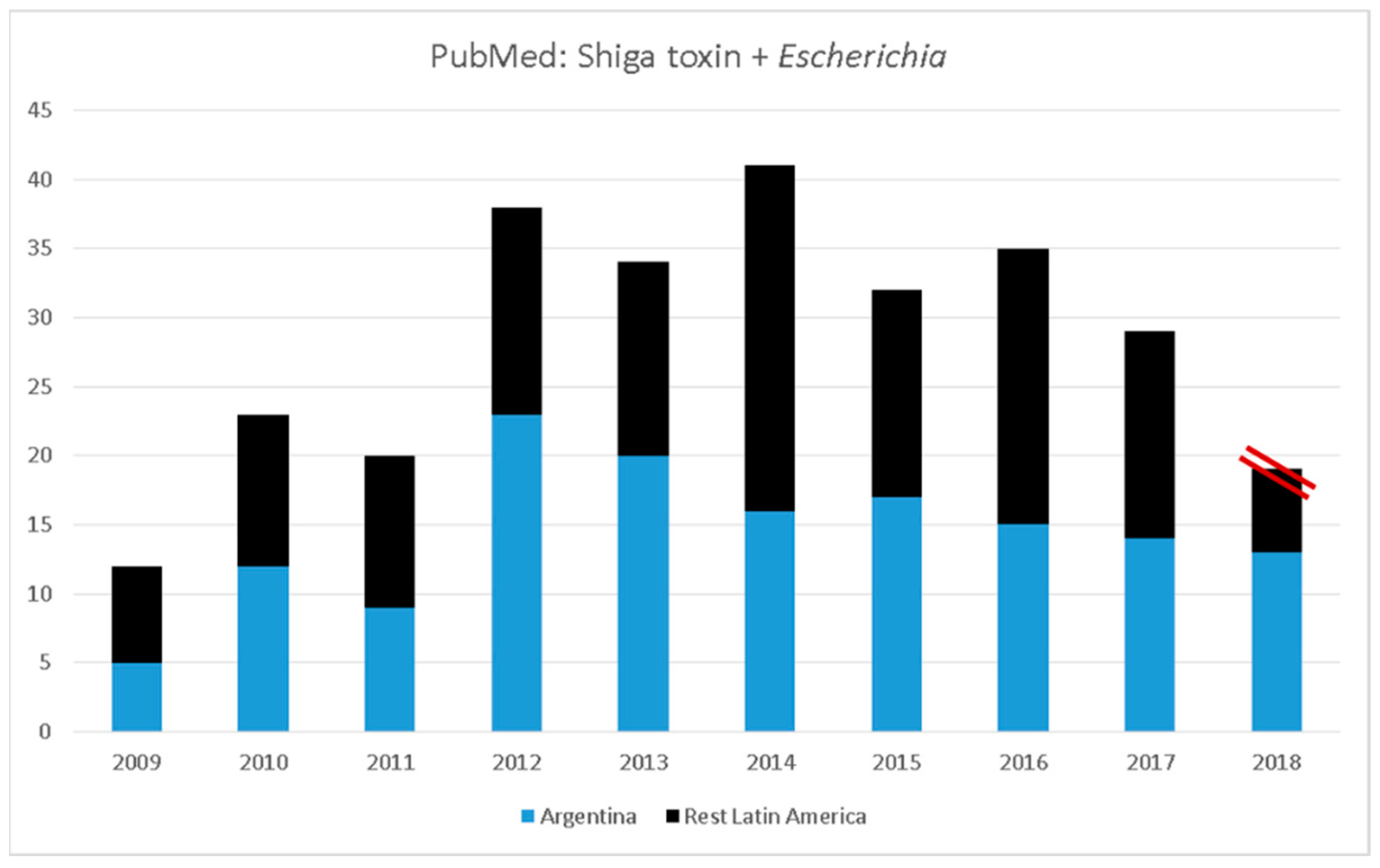
:1. Introduction
2. Discovery of STEC Serotypes and Novel Virulence Factors in Isolates from Humans and Animals
3. Novel Insights about Shiga Toxins and the Immune Responses
4. Progress in Phage Therapy and Stx Phage Biology
5. Current Advancements in Control of STEC in Food Products and Food Safety
6. Epidemiology of STEC in Cattle
7. New Treatments against STEC Infections (Antisera and Vaccines)
8. Discussion
9. Conclusions
Author Contributions
Funding
Acknowledgments
Conflicts of Interest
References
- Farfan, M.J.; Torres, A.G. Molecular mechanisms that mediate colonization of Shiga toxin-producing Escherichia coli strains. Infect. Immun. 2012, 80, 903–913. [Google Scholar] [CrossRef] [PubMed]
- Kaper, J.B.; O’Brien, A.D. Overview and historical perspectives. Microbiol. Spectr. 2014, 2. [Google Scholar] [CrossRef]
- Caprioli, A.; Scavia, G.; Morabito, S. Public health microbiology of Shiga toxin-producing Escherichia coli. Microbiol. Spectr. 2014, 2. [Google Scholar] [CrossRef] [PubMed]
- Rivas, M.; Chinen, I.; Guth, B.E.C. Enterohemorrhagic (Shiga toxin-producing) Escherichia coli. In Escherichia coli in the Americas; Torres, A.G., Ed.; Springer: Cham, Switzerland, 2016; pp. 97–123. [Google Scholar]
- Majowicz, S.E.; Scallan, E.; Jones-Bitton, A.; Sargeant, J.M.; Stapleton, J.; Angulo, F.J.; Yeung, D.H.; Kirk, M.D. Global incidence of human Shiga toxin-producing Escherichia coli infections and deaths: A systematic review and knowledge synthesis. Foodborne Pathog. Dis. 2014, 11, 447–455. [Google Scholar] [CrossRef] [PubMed]
- Torres, A.G. Pathogenic Escherichia coli in Latin America; Bentham Science: Bentham, UK, 2010. [Google Scholar]
- Torres, A.G. Escherichia coli in the Americas; Springer: Cham, Switzerland, 2016. [Google Scholar]
- Guth, B.E.C.; Prado, V.; Rivas, M. Shiga toxin-producing Escherichia coli. In Pathogenic Escherichia coli in Latin America; Torres, A.G., Ed.; Betham Science: Bentham, UK, 2010; pp. 65–83. [Google Scholar]
- Segura, A.; Auffret, P.; Bibbal, D.; Bertoni, M.; Durand, A.; Jubelin, G.; Kérourédan, M.; Brugère, H.; Bertin, Y.; Forano, E. Factors involved in the persistence of a Shiga toxin-producing Escherichia coli O157:H7 strain in bovine feces and gastro-intestinal content. Front. Microbiol. 2018, 9, 375. [Google Scholar] [CrossRef] [PubMed]
- Rivas, M.; Padola, N.L.; Lucchesi, P.M.A.; Masana, M. Diarrheagenic Escherichia coli in Argentina. In Pathogenic Escherichia coli in Latin America, Torres, A.G., Ed.; Bentham Science: Bentham, UK, 2010; pp. 141–161. [Google Scholar]
- Mohawk, K.L.; O’Brien, A.D. Mouse models of Escherichia coli O157:H7 infection and Shiga toxin injection. J. Biomed. Biotechnol. 2011, 2011, 258185. [Google Scholar] [CrossRef] [PubMed]
- Rojas-Lopez, M.; Monterio, R.; Pizza, M.; Desvaux, M.; Rosini, R. Intestinal pathogenic Escherichia coli: Insights for vaccine development. Front. Microbiol. 2018, 9, 440. [Google Scholar] [CrossRef] [PubMed]
- Krüger, A.; Lucchesi, P.M. Shiga toxins and stx phages: Highly diverse entities. Microbiology 2015, 161, 451–462. [Google Scholar] [CrossRef] [PubMed]
- Sperandio, V.; Hovde Bohach, C. Enterohemorrhagic Escherichia coli and other Shiga Toxin-Producing E. coli; ASM Press: Washington, DC, USA, 2015. [Google Scholar]
- Hall, G.; Kurosawa, S.; Stearns-Kurosawa, D.J. Shiga toxin therapeutics: Beyond neutralization. Toxins 2017, 9, 291. [Google Scholar] [CrossRef] [PubMed]
- Alconcher, L.F.; Coccia, P.A.; Suarez, A.D.C.; Monteverde, M.L.; Perez, Y.; Gutiérrez, M.G.; Carlopio, P.M.; Missoni, M.L.; Balestracci, A.; Principi, I.; Ramírez, F.B.; et al. Hyponatremia: A new predictor of mortality in patients with Shiga toxin-producing Escherichia coli hemolytic uremic syndrome. Pediatr. Nephrol. 2018, 33, 1791–1798. [Google Scholar] [CrossRef] [PubMed]
- Karmali, M.A. Emerging public health challenges of Shiga toxin-producing Escherichia coli related to changes in the pathogen, the population, and the environment. Clin. Infect. Dis. 2017, 64, 371–376. [Google Scholar] [PubMed]
- Pianciola, L.; D’Astek, B.A.; Mazzeo, M.; Chinen, I.; Masana, M.; Rivas, M. Genetic features of human and bovine Escherichia coli O157:H7 strains isolated in Argentina. Int. J. Med. Microbiol. 2016, 306, 123–130. [Google Scholar] [CrossRef] [PubMed]
- Carbonari, C.C.; Fittipaldi, N.; Teatero, S.; Athey, T.B.; Pianciola, L.; Masana, M.; Melano, R.G.; Rivas, M.; Chinen, I. Whole-genome sequencing applied to the molecular epidemiology of Shiga toxin-producing Escherichia coli O157:H7 in Argentina. Genome Announc. 2016, 4, E01341–E01316. [Google Scholar] [CrossRef] [PubMed]
- Oderiz, S.; Leotta, G.A.; Galli, L. Detection and characterization of Shiga toxin-producing Escherichia coli in children treated at an inter-zonal pediatric hospital in the city of La Plata. Rev. Argent. Microbiol. 2018, S0325–S7541. [Google Scholar] [CrossRef]
- Galli, L.; Brusa, V.; Rodríguez, R.; Signorini, M.; Oteiza, J.M.; Leotta, G.A. Escherichia coli in food products. In Escherichia coli in the Americas; Torres, A.G., Ed.; Springer: Cham, Switzerland, 2016; pp. 173–203. [Google Scholar]
- Cundon, C.; Carbonari, C.C.; Zolezzi, G.; Rivas, M.; Bentancor, A. Putative virulence factors and clonal relationship of O174 Shiga toxin-producing Escherichia coli isolated from human, food and animal sources. Vet. Microbiol. 2018, 215, 29–34. [Google Scholar] [CrossRef] [PubMed]
- Santos, R.F.C.; Nascimento, J.D.S.; Geimba, M.P.; Hessel, C.T.; Tondo, E.C. First report of human gastroenteritis caused by Escherichia coli O157:NM in Brazil. Foodborne Pathog. Dis. 2017, 14, 665–666. [Google Scholar] [CrossRef] [PubMed]
- Vieira, M.A.; dos Santos, L.F.; Dias, R.C.B.; Camargo, C.H.; Pinheiro, S.R.S.; Gomes, T.A.T.; Hernandes, R.T. Atypical enteropathogenic Escherichia coli as aetiologic agents of sporadic and outbreak-associated diarrhoea in Brazil. J. Med. Microbiol. 2016, 65, 998–1006. [Google Scholar] [CrossRef] [PubMed]
- Marchant, P.; Hidalgo-Hermoso, E.; Espinoza, K.; Retamal, P. Prevalence of Salmonella enterica and Shiga toxin-producing Escherichia coli in zoo animals from Chile. J. Vet. Sci. 2016, 17, 583–586. [Google Scholar] [CrossRef] [PubMed]
- Vasco, K.; Graham, J.P.; Trueba, G. Detection of zoonotic enteropathogens in children and domestic animals in a semirural community in Ecuador. Appl. Environ. Microbiol. 2016, 82, 4218–4224. [Google Scholar] [CrossRef] [PubMed]
- Sanches, L.A.; Gomes, M.D.S.; Teixeira, R.H.F.; Cunha, M.P.V.; Oliveira, M.G.X.; Vieira, M.A.M.; Gomes, T.A.T.; Knobl, T. Captive wild birds as reservoirs of enteropathogenic E. coli (EPEC) and Shiga-toxin producing E. coli (STEC). Braz. J. Microbiol. 2017, 48, 760–763. [Google Scholar] [CrossRef] [PubMed]
- Blanco Crivelli, X.; Bonino, M.P.; Von Wernich Castillo, P.; Navarro, A.; Degregorio, O.; Bentancor, A. Detection and characterization of enteropathogenic and Shiga toxin-producing Escherichia coli strains in Rattus spp. from Buenos Aires. Front. Microbiol. 2018, 9, 199. [Google Scholar] [CrossRef] [PubMed]
- Soledad-Cadona, J.; Bustamante, A.V.; González, J.; Mariel-Sanso, A. Pathogenicity islands distribution in Non-O157 Shiga toxin-producing Escherichia coli (STEC). Genes 2018, 9, 81. [Google Scholar] [CrossRef] [PubMed]
- Montero, D.A.; Velasco, J.; Del Canto, F.; Puente, J.L.; Padola, N.L.; Rasko, D.A.; Farfán, M.; Salazar, J.C.; Vidal, R. Locus of Adhesion and Autoaggregation (LAA), a pathogenicity island present in emerging Shiga toxin-producing Escherichia coli strains. Sci. Rep. 2017, 7, 7011. [Google Scholar] [CrossRef] [PubMed]
- Colello, R.; Vélez, M.V.; González, J.; Montero, D.A.; Bustamante, A.V.; Del Canto, F.; Etcheverría, A.I.; Vidal, R.; Padola, N.L. First report of the distribution of Locus of Adhesion and Autoaggregation (LAA) pathogenicity island in LEE-negative Shiga toxin-producing Escherichia coli isolates from Argentina. Microb. Pathog. 2018, 123, 259–263. [Google Scholar] [CrossRef] [PubMed]
- Gianantonio, C.A.; Vitacco, M.; Mendilaharzu, F.; Gallo, G.E.; Sojo, E.T. The hemolytic-uremic syndrome. Nephron 1973, 11, 174–192. [Google Scholar] [CrossRef] [PubMed]
- Henn, V.; Slupsky, J.R.; Grafe, M.; Anagnostopoulos, I.; Forster, R.; Muller-Berghaus, G.; Kroczek, R.A. CD40 ligand on activated platelets triggers an inflammatory reaction of endothelial cells. Nature 1998, 391, 591–594. [Google Scholar] [CrossRef] [PubMed]
- Abrey Recalde, M.J.; Alvarez, R.S.; Alberto, F.; Mejias, M.P.; Ramos, M.V.; Fernandez Brando, R.J.; Bruballa, A.C.; Exeni, R.A.; Alconcher, L.; Ibarra, C.A.; et al. Soluble CD40 ligand and oxidative response are reciprocally stimulated during Shiga toxin-associated hemolytic uremic syndrome. Toxins 2017, 9, 331. [Google Scholar] [CrossRef] [PubMed]
- Ruckerl, R.; Phipps, R.P.; Schneider, A.; Frampton, M.; Cyrys, J.; Oberdorster, G.; Wichmann, H.E.; Peters, A. Ultrafine particles and platelet activation in patients with coronary heart disease- Results from a prospective panel study. Part. Fibre. Toxicol. 2007, 4. [Google Scholar] [CrossRef] [PubMed]
- Yngen, M.; Ostenson, C.G.; Hu, H.; Li, N.; Hjemdahl, P.; Wallen, N.H. Enhanced P-selectin expression and increased soluble CD40 ligand in patients with type 1 diabetes mellitus and microangiopathy: Evidence for platelet hyperactivity and chronic inflammation. Diabetologia 2004, 47, 537–540. [Google Scholar] [CrossRef] [PubMed]
- Sipsas, N.V.; Sfikakis, P.P.; Kontos, A.; Kordossis, T. Levels of soluble CD40 ligand (CD154) in serum are increased in human immunodeficiency virus type 1-infected patients and correlate with CD4(+) T-cell counts. Clin. Diagn. Lab. Immunol. 2002, 9, 558–561. [Google Scholar] [CrossRef] [PubMed]
- Exeni, R.A.; Fernandez-Brando, R.J.; Santiago, A.P.; Fiorentino, G.A.; Exeni, A.M.; Ramos, M.V.; Palermo, M.S. Pathogenic role of inflammatory response during Shiga toxin-associated hemolytic uremic syndrome (HUS). Pediatr. Nephrol. 2018, 33, 2057–2071. [Google Scholar] [CrossRef] [PubMed]
- Márquez, L.B.; Araoz, A.; Repetto, H.A.; Ibarra, F.R.; Silberstein, C. Effects of Shiga toxin 2 on cellular regeneration mechanisms in primary and three-dimensional cultures of human renal tubular epithelial cells. Microb. Pathog. 2016, 99, 87–94. [Google Scholar] [CrossRef] [PubMed]
- Seyahian, E.A.; Oltra, G.; Ochoa, F.; Melendi, S.; Hermes, R.; Paton, J.C.; Paton, A.W.; Lago, N.; Castro Parodi, M.; Damiano, A.; et al. Systemic effects of Subtilase cytotoxin produced by Escherichia coli O113:H21. Toxicon 2017, 127, 49–55. [Google Scholar] [CrossRef] [PubMed]
- Amaral, M.M.; Sacerdoti, F.; Jancic, C.; Repetto, H.A.; Paton, A.W.; Paton, J.C.; Ibarra, C. Action of Shiga toxin type-2 and Subtilase cytotoxin on human microvascular endothelial cells. PLoS ONE 2013, 8, e70431. [Google Scholar] [CrossRef] [PubMed]
- Amaral, M.M.; Girard, M.C.; Álvarez, R.S.; Paton, A.W.; Paton, J.C.; Repetto, H.A.; Sacerdoti, F.; Ibarra, C.A. Ouabain protects human renal cells against the cytotoxic effects of Shiga toxin type 2 and Subtilase cytotoxin. Toxins 2017, 9, 226. [Google Scholar] [CrossRef] [PubMed]
- Álvarez, R.S.; Sacerdoti, F.; Jancic, C.; Paton, A.W.; Paton, J.C.; Ibarra, C.; Amaral, M.M. Comparative characterization of Shiga toxin type 2 and Subtilase cytotoxin effects on human renal epithelial and endothelial cells grown in monolayer and bilayer conditions. PLoS ONE 2016, 11, e0158180. [Google Scholar] [CrossRef] [PubMed]
- Goldstein, J.; Loidl, C.F.; Creydt, V.P.; Boccoli, J.; Ibarra, C. Intracerebroventricular administration of Shiga toxin type 2 induces striatal neuronal death and glial alterations: An ultrastructural study. Brain Res. 2007, 1161, 106–115. [Google Scholar] [CrossRef] [PubMed]
- Tironi-Farinati, C.; Loidl, C.F.; Boccoli, J.; Parma, Y.; Fernandez-Miyakawa, M.E.; Goldstein, J. Intracerebroventricular Shiga toxin 2 increases the expression of its receptor globotriaosylceramide and causes dendritic abnormalities. J. Neuroimmunol. 2010, 222, 48–61. [Google Scholar] [CrossRef] [PubMed]
- Tironi-Farinati, C.; Geoghegan, P.A.; Cangelosi, A.; Pinto, A.; Loidl, C.F.; Goldstein, J. A translational murine model of sub-lethal intoxication with Shiga toxin 2 reveals novel ultrastructural findings in the brain striatum. PLoS ONE 2013, 8, e55812. [Google Scholar] [CrossRef] [PubMed] [Green Version]
- Pinto, A.; Cangelosi, A.; Geoghegan, P.; Goldstein, J. Dexamethasone prevents motor deficits and neurovascular damage produced by Shiga-toxin 2 and lipopolysaccharide in the mouse striatum. Neuroscience 2017, 344, 25–38. [Google Scholar] [CrossRef] [PubMed]
- D’Alessio, L.; Pinto, A.; Cangelosi, A.; Geoghegan, P.A.; Tironi-Farinati, C.; Brener, G.J.; Goldstein, J. Sub-lethal dose of Shiga toxin 2 from enterohemorrhagic Escherichia coli affects balance and cerebellar cytoarchitecture. Front. Microbiol. 2016, 7, 133. [Google Scholar] [CrossRef] [PubMed]
- Goldstein, J.; Carden, T.; Perez, M.J.; Taira, C.A.; Hocht, C.; Gironacci, M.M. Angiotensin-(1-7) protects from brain damage induced by Shiga toxin 2-producing enterohemorrhagic Escherichia coli. Am. J. Physiol. Regul. Integr. Comp. Physiol. 2016, 311, R1173–R1185. [Google Scholar] [CrossRef] [PubMed]
- Ishida, S.; Yasukawa, K.; Koizumi, M.; Abe, K.; Hirai, N.; Honda, T.; Sakuma, H.; Tada, H.; Takanashi, J.I. Excitotoxicity in encephalopathy associated with STEC O-157 infection. Brain. Dev. 2018, 40, 357–360. [Google Scholar] [CrossRef] [PubMed]
- Exeni, R. Sindrome urémico hemolítico: Manifestaciones clínicas: Tratamiento. Medicina (Buenos Aires) 2006, 66, 6–10. [Google Scholar]
- Proulx, F.; Seidman, E.G.; Karpman, D. Pathogenesis of Shiga toxin-associated hemolytic uremic syndrome. Pediatr. Res. 2001, 50, 163–171. [Google Scholar] [CrossRef] [PubMed]
- Schmidt, H. Shiga-toxin-converting bacteriophages. Res. Microbiol. 2001, 152, 687–695. [Google Scholar] [CrossRef]
- Tyler, J.S.; Beeri, K.; Reynolds, J.L.; Alteri, C.J.; Skinner, K.G.; Friedman, J.H.; Eaton, K.A.; Friedman, D.I. Prophage induction is enhanced and required for renal disease and lethality in an EHEC mouse model. PLOS Pathog. 2013, 9, e1003236. [Google Scholar] [CrossRef] [PubMed]
- Bentancor, L.V.; Bilen, M.F.; Mejías María, P.; Fernández-Brando, R.J.; Panek, C.A.; Ramos, M.V.; Fernández, G.C.; Isturiz, M.; Ghiringhelli, P.D.; Palermo, M.S. Functional capacity of Shiga-toxin promoter sequences in eukaryotic cells. PLoS ONE 2013, 8, e57128. [Google Scholar] [CrossRef] [PubMed]
- Bentancor, L.V.; Mejías, M.P.; Pinto, A.; Bilen, M.F.; Meiss, R.; Rodriguez-Galán, M.C.; Baez, N.; Pedrotti, L.P.; Goldstein, J.; Ghiringhelli, P.D.; et al. Promoter sequence of Shiga toxin II (Stx2) is recognized in vivo leading to the production of biologically active Stx2. MBio 2013, 4, e00501–e00513. [Google Scholar] [CrossRef] [PubMed]
- Amorim, J.H.; Del Cogliano, M.E.; Fernandez-Brando, R.J.; Bilen, M.F.; Jesus, M.R.; Luiz, W.B.; Palermo, M.S.; Ferreira, R.C.C.; Servat, E.G.; Ghiringhelli, P.D.; et al. Role of bacteriophages in STEC infections: New implications for the design of prophylactic and treatment approaches. F1000 Res. 2014, 3, 74. [Google Scholar] [CrossRef]
- Del Cogliano, M.E.; Hollmann, A.; Martinez, M.; Semorile, L.; Ghiringhelli, P.D.; Maffía, P.C.; Bentancor, L.V. Cationic antimicrobial peptides inactivate Shiga toxin-encoding bacteriophages. Front. Chem. 2017, 5, 122. [Google Scholar] [CrossRef] [PubMed]
- Ly-Chatain, M.H.; Moussaoui, S.; Vera, A.; Rigobello, V.; Demarigny, Y. Antiviral effect of cationic compounds on bacteriophages. Front. Microbiol. 2013, 4, 46. [Google Scholar] [CrossRef] [PubMed]
- Leotta, G.A.; Brusa, V.; Galli, L.; Adriani, C.; Linares, L.; Etcheverría, A.; Sanz, M.; Sucari, A.; Peral García, P.; Signorini, M. Comprehensive evaluation and implementation of improvement actions in butcher shops. PLoS ONE 2016, 11, e0162635. [Google Scholar] [CrossRef] [PubMed]
- Kalchayanand, N.; Arthur, T.M.; Bosilevac, J.M.; Schmidt, J.W.; Wang, R.; Shackelford, S.; Wheeler, T.L. Efficacy of antimicrobial compounds on surface decontamination of seven Shiga toxin-producing Escherichia coli and Salmonella inoculated onto fresh beef. J. Food. Prot. 2015, 78, 503–510. [Google Scholar] [CrossRef] [PubMed]
- Kalchayanand, N.; Arthur, T.M.; Bosilevac, J.M.; Schmidt, J.W.; Wang, R.; Shackelford, S.D.; Wheeler, T.L. Evaluation of commonly used antimicrobial interventions for fresh beef inoculated with Shiga toxin–producing Escherichia coli serotypes O26, O45, O103, O111, O121, O145, and O157:H7. J. Food. Prot. 2012, 75, 1207–1212. [Google Scholar] [CrossRef] [PubMed]
- Kanankege, K.S.; Anklam, K.S.; Fick, C.M.; Kulow, M.J.; Kaspar, C.W.; Ingham, B.H.; Milkowski, A.; Dopfer, D. Evaluating the efficacy of beef slaughter line interventions by quantifying the six major non-O157 Shiga toxin producing Escherichia coli serogroups using real-time multiplex PCR. Food Microbiol. 2017, 63, 228–238. [Google Scholar] [CrossRef] [PubMed]
- Signorini, M.; Costa, M.; Teitelbaum, D.; Restovich, V.; Brasesco, H.; García, D.; Superno, V.; Petroli, S.; Bruzzone, M.; Arduini, V.; et al. Evaluation of decontamination efficacy of commonly used antimicrobial interventions for beef carcasses against Shiga toxin-producing Escherichia coli. Meat Sci. 2018, 142, 44–51. [Google Scholar] [CrossRef] [PubMed]
- Lenzi, L.J.; Lucchesi, P.M.; Medico, L.; Burgán, J.; Krüger, A. Effect of the food additives sodium citrate and disodium phosphate on Shiga toxin-producing Escherichia coli and production of stx-phages and Shiga toxin. Front. Microbiol. 2016, 7, 992. [Google Scholar] [CrossRef] [PubMed]
- García-Heredia, A.; García, S.; Merino-Mascorro, J.Á.; Feng, P.; Heredia, N. Natural plant products inhibits growth and alters the swarming motility, biofilm formation, and expression of virulence genes in enteroaggregative and enterohemorrhagic Escherichia coli. Food Microbiol. 2016, 59, 124–132. [Google Scholar] [CrossRef] [PubMed]
- Gómez-Aldapa, C.A.; Rangel-Vargas, E.; Torres-Vitela, M.R.; Villarruel-López, A.; Acevedo-Sandoval, O.A.; Gordillo-Martínez, A.J.; Godínez-Oviedo, A.; Castro-Rosas, J. Antibacterial activities of Hibiscus sabdariffa extracts and chemical sanitizers directly on green leaves contaminated with foodborne pathogens. J. Food. Prot. 2018, 81, 209–217. [Google Scholar] [CrossRef] [PubMed]
- Rangel-Vargas, E.; Gómez-Aldapa, C.A.; Falfan-Cortes, R.N.; Rodríguez-Marín, M.L.; Godínez-Oviedo, A.; Acevedo-Sandoval, O.A.; Castro-Rosas, J. Attachment of 13 types of foodborne bacteria to Jalapeño and Serrano peppers and antibacterial effect of roselle calyx extracts, sodium hypochlorite, colloidal silver, and acetic acid against these foodborne bacteria on peppers. J. Food. Prot. 2017, 80, 406–413. [Google Scholar] [CrossRef] [PubMed]
- Rangel-Vargas, E.; Luna-Rojo, A.M.; Cadena-Ramírez, A.; Torres-Vitela, R.; Gómez-Aldapa, C.A.; Villarruel-López, A.; Téllez-Jurado, A.; Villagómez-Ibarra, J.R.; Reynoso-Camacho, R.; Castro-Rosas, J. Behavior of 11 foodborne bacteria on whole and cut mangoes var. ataulfo and kent and antibacterial activities of Hibiscus sabdariffa extracts and chemical sanitizers directly onto mangoes contaminated with foodborne bacteria. J. Food. Prot. 2018, 81, 743–753. [Google Scholar] [CrossRef] [PubMed]
- Jaroni, D.; Ravishankar, S. Bactericidal effects of roselle (Hibiscus sabdariffa) against foodborne pathogens in vitro and on romaine lettuce and alfalfa sprouts. Qual. Assur. Saf. Crop. Foods 2012, 4, 33–40. [Google Scholar] [CrossRef]
- Gutiérrez-Alcántara, E.J.; Rangel-Vargas, E.; Gómez-Aldapa, C.A.; Falfan-Cortes, R.N.; Rodríguez-Marín, M.L.; Godínez-Oviedo, A.; Cortes-López, H.; Castro-Rosas, J. Antibacterial effect of roselle extracts (Hibiscus sabadariffa), sodium hypochlorite and acetic acid against multidrug-resistant Salmonella strains isolated from tomatoes. Lett. Appl. Microbiol. 2016, 62, 177–184. [Google Scholar] [CrossRef] [PubMed]
- Gómez-Aldapa, C.A.; Torres-Vitela, M.R.; Amaya-Acosta, M.A.; Rangel-Vargas, E.; Villaruel-López, A.; Castro-Rosas, J. Behavior of thirteen foodborne bacteria on whole Hass avocado and potential of roselle calyx extracts as alternative disinfectant agents of avocado. J. Food Saf. 2017, 37, e12351. [Google Scholar] [CrossRef]
- Smulders, F.J.M.; Greer, G.G. Integrating microbial decontamination with organic acids in HACCP programmes for muscle foods: Prospects and controversies. Int. J. Food Microbiol. 1998, 44, 149–169. [Google Scholar] [CrossRef]
- Islam, M.Z.; Musikiwa, A.; Islam, K.; Ahmed, S.; Chowdjury, S.; Ahad, A.; Biswas, P.K. Regional variation in the prevalence of E. coli O157 in cattle: A meta-analysis and meta-regression. PLoS ONE 2014, 9, e93299. [Google Scholar] [CrossRef] [PubMed]
- Cap, M.; Carbonari, C.C.; D’Astek, B.A.; Zolezzi, G.; Deza, N.; Palladino, M.P.; Masana, O.; Chinen, I.; Rivas, M. Frequency, characterization and genotypic analysis of Shiga toxin-producing Escherichia coli in beef slaughterhouses of Argentina. Rev. Argent. Microbiol. 2018. [Google Scholar] [CrossRef] [PubMed]
- Brusa, V.; Restovich, V.; Galli, L.; Teitelbaum, D.; Signorini, M.; Brasesco, H.; Londero, A.; García, D.; Padola, N.L.; Superno, V.; et al. Isolation and characterization of non-O157 Shiga toxin-producing Escherichia coli from beef carcasses, cuts and trimmings of abattoirs in Argentina. PLoS ONE 2017, 12, e0183248. [Google Scholar] [CrossRef] [PubMed]
- Etcheverría, A.I.; Lucchesi, P.M.A.; Krüger, A.; Bentancor, A.B.; Padola, N.L. Escherichia coli in animals. In Escherichia coli in the Americas; Torres, A.G., Ed.; Springer: Cham, Switzerland, 2016; pp. 149–172. [Google Scholar]
- Pianciola, L.; Rivas, M. Genotypic features of clinical and bovine Escherichia coli O157 strains isolated in countries with different associated-disease incidences. Microorganisms 2018, 6, 36. [Google Scholar] [CrossRef] [PubMed]
- Krüger, A.; Lucchesi, P.M.; Parma, A.E. Verotoxins in bovine and meat verotoxin-producing Escherichia coli isolates: Type, number of variants, and relationship to cytotoxicity. Appl. Environ. Microbiol. 2011, 77, 73–79. [Google Scholar] [CrossRef] [PubMed]
- Mellor, G.E.; Fegan, N.; Gobius, K.S.; Smith, H.V.; Jennison, A.V.; D’Astek, B.A.; Rivas, M.; Shringi, S.; Baker, K.N.; Besser, T.E. Geographically distinct Escherichia coli O157 isolates differ by lineage, Shiga toxin genotype, and total Shiga toxin production. J. Clin. Microbiol. 2015, 53, 579–586. [Google Scholar] [CrossRef] [PubMed]
- González, J.; Sanso, A.M.; Cadona, J.; Bustamante, A.V. Virulence traits and different nle profiles in cattle and human verotoxin-producing Escherichia coli O157:H7 strains from Argentina. Microb. Pathog. 2017, 102, 102–108. [Google Scholar] [CrossRef] [PubMed]
- Amigo, N.; Mercado, E.; Bentancor, A.; Singh, P.; Vilte, D.; Gerhardt, E.; Zotta, E.; Ibarra, C.; Manning, S.D.; Larzábal, M.; et al. Clade 8 and clade 6 strains of Escherichia coli O157:H7 from cattle in Argentina have hypervirulent-like phenotypes. PLoS ONE 2015, 10, e0127710. [Google Scholar] [CrossRef] [PubMed]
- Krüger, A.; Zhu, K.; Friedrich, A.W.; Lucchesi, P.M.; Rossen, J.W. Comparative analysis of Q-stx regions of stx-phages encoding stx1a, stx2a and stx2c subtypes. In Proceedings of the EMBO Conference: Viruses of Microbes: Structure and Function, from Molecules to Communities, Zurich, Switzerland, 14–18 July 2014. [Google Scholar]
- Burgán, J.; Lucchesi, P.M.; Krüger, A. Niveles de expresión de toxina Shiga 2a en cepas STEC O145:H- aisladas de humanos y de bovinos. In Proceedings of the XII Encuentro de Biólogos en Red., Mar del Plata, Argentina, 13–14 November 2017; pp. 1853–3426. [Google Scholar]
- Krüger, A.; Burgán, J.; Friedrich, A.W.; Rossen, J.W.; Lucchesi, P.M. ArgO145, a Stx2a prophage of a bovine O145:H-STEC strain, is closely related to phages of virulent human strains. Infect. Genet. Evol. 2018, 60, 126–132. [Google Scholar]
- Krüger, A.; Lucchesi, P.M.; Sanso, A.M.; Etcheverría, A.I.; Bustamante, A.V.; Burgán, J.; Fernández, L.; Fernández, D.; Leotta, G.; Friedrich, A.W.; et al. Genetic characterization of Shiga toxin-producing Escherichia coli O26:H11 strains isolated from animal, food, and clinical samples. Front. Cell. Infect. Microbiol. 2015, 5, 74. [Google Scholar] [CrossRef] [PubMed]
- Sanso, A.M.; Bustamante, A.V.; Krüger, A.; Cadona, J.S.; Alfaro, R.; Cáceres, M.E.; Fernández, D.; Lucchesi, P.M.; Padola, N.L. Molecular epidemiology of Shiga toxin-producing O113:H21 isolates from cattle and meat. Zoonoses Public Health 2018, 65, 569–577. [Google Scholar] [CrossRef] [PubMed]
- Oliveira, M.G.; Brito, J.R.; Gomes, T.A.; Guth, B.E.C.; Vieira, M.A.; Naves, Z.V.; Vaz, T.; Irino, K. Diversity of virulence profiles of Shiga toxin-producing Escherichia coli serotypes in food-producing animals in Brazil. Int. J. Food Microbiol 2008, 127, 139–146. [Google Scholar] [CrossRef] [PubMed]
- dos Santos, E.C.C.; Silva Castro, V.; Cunha-Neto, A.; dos Santos, L.F.; Vallim, D.C.; de Castro Lisbôa, R.; Tavares Carvalho, R.C.; Conte Junior, C.A.; de Souza Figueiredo, E.E. Escherichia coli O26 and O113:H21 on cand beef from a slaughterhouse located in Mato Grosso, Brazil. Foodborne Pathog. Dis. 2018. [Google Scholar] [CrossRef]
- Gonzalez, A.G.; Cerqueira, A.M.; Guth, B.E.; Coutinho, C.A.; Liberal, M.H.; Souza, R.M.; Andrade, J.R. Serotypes, virulence markers and cell invasion ability of Shiga toxin-producing Escherichia coli strains isolated from healthy dairy cattle. J. Appl. Microbiol. 2016, 121, 1130–1143. [Google Scholar] [CrossRef] [PubMed]
- Mejias, M.P.; Ghersi, G.; Craig, P.O.; Panek, C.A.; Bentancor, L.V.; Baschkier, A.; Goldbaum, F.A.; Zylberman, V.; Palermo, M.S. Immunization with a chimera consisting of the B subunit of Shiga toxin type 2 and brucella lumazine synthase confers total protection against Shiga toxins in mice. J. Immunol. 2013, 191, 2403–2411. [Google Scholar] [CrossRef] [PubMed]
- Mejias, M.P.; Cabrera, G.; Fernández-Brando, R.J.; Baschkier, A.; Ghersi, G.; Abrey-Recalde, M.J.; Miliwebsky, E.; Meiss, R.; Goldbaum, F.; Zylberman, V.; et al. Protection of mice against Shiga toxin 2 (Stx2)-associated damage by maternal immunization with a Brucella lumazine synthase-Stx2 B subunit chimera. Infect. Immun. 2014, 82, 1491–1499. [Google Scholar] [CrossRef] [PubMed]
- Sacerdoti, F.; Mejías, M.P.; Bruballa, A.C.; Alvarez, R.S.; Amaral, M.M.; Palermo, M.S.; Ibarra, C. Immunization with BLS-Stx2B chimera totally protects dams from early pregnancy loss induced by Shiga toxin type 2 (Stx2) and confers anti-Stx2 immunity to the offspring. Vaccine 2016, 34, 4732–4737. [Google Scholar] [CrossRef] [PubMed]
- Mejías, M.P.; Hiriart, Y.; Lauché, C.; Fernández-Brando, R.J.; Pardo, R.; Bruballa, A.; Ramos, M.V.; Goldbaum, F.A.; Palermo, M.S.; Zylberman, V. Development of camelid single chain antibodies against Shiga toxin type 2 (Stx2) with therapeutic potential against Hemolytic Uremic Syndrome (HUS). Sci. Rep. 2016, 6, 24913. [Google Scholar] [CrossRef] [PubMed]
- Hiriart, Y.; Pardo, R.; Bukata, L.; Lauché, C.; Muñoz, L.; Colonna, M.; Goldbaum, F.; Sanguineti, S.; Zylberman, V. Development of a product anti-Shiga toxin for prevention of the hemolytic uremic syndrome. Medicina (B Aires) 2018, 78, 107–112. [Google Scholar] [PubMed]
- Martorelli, L.; Garbaccio, S.; Vilte, D.A.; Albanese, A.A.; Mejías, M.P.; Palermo, M.S.; Mercado, E.C.; Ibarra, C.E.; Cataldi, A.A. Immune response in calves vaccinated with type three secretion system antigens and Shiga toxin 2B subunit of Escherichia coli O157:H7. PLoS ONE 2017, 12, e0169422. [Google Scholar] [CrossRef] [PubMed]
- Martorelli, L.; Garimano, N.; Fiorentino, G.A.; Vilte, D.A.; Garbaccio, S.G.; Barth, S.A.; Menge, C.; Ibarra, C.; Palermo, M.S.; Cataldi, A. Efficacy of a recombinant Intimin, EspB and Shiga toxin 2B vaccine in calves experimentally challenged with Escherichia coli O157:H7. Vaccine 2018, 36, 3949–3959. [Google Scholar] [CrossRef] [PubMed]
- Navarro, A.; Hernández-Chiñas, U.; Licona-Moreno, D.; Zenteno, E.; Cravioto, A.; Eslava-Campos, C.A. Immunogenic peptide mimotopes from an epitope of Escherichia coli O157 LPS. Biochem. J. 2016, 473, 3791–3804. [Google Scholar] [CrossRef] [PubMed]
- Fingermann, M.; Avila, L.; De Marco, M.B.; Vázquez, L.; Di Biase, D.N.; Müller, A.V.; Lescano, M.; Dokmetjian, J.C.; Fernández Castillo, S.; Pérez Quiñoy, J.L. OMV-based vaccine formulations against Shiga toxin producing Escherichia coli strains are both protective in mice and immunogenic in calves. Hum. Vaccines Immunother. 2018, 20, 1–6. [Google Scholar] [CrossRef] [PubMed]
- Torres, A.G. The Importance of international collaborations to advance research endeavors. PLoS Pathog. 2016, 13, e1006047. [Google Scholar] [CrossRef] [PubMed]
- Torres, A.G. Escherichia coli diseases in Latin America-a ‘One Health’ multidisciplinary approach. Pathog. Dis. 2017, 75. [Google Scholar] [CrossRef] [PubMed]


© 2018 by the authors. Licensee MDPI, Basel, Switzerland. This article is an open access article distributed under the terms and conditions of the Creative Commons Attribution (CC BY) license (http://creativecommons.org/licenses/by/4.0/).
Share and Cite
Torres, A.G.; Amaral, M.M.; Bentancor, L.; Galli, L.; Goldstein, J.; Krüger, A.; Rojas-Lopez, M. Recent Advances in Shiga Toxin-Producing Escherichia coli Research in Latin America. Microorganisms 2018, 6, 100. https://0-doi-org.brum.beds.ac.uk/10.3390/microorganisms6040100
Torres AG, Amaral MM, Bentancor L, Galli L, Goldstein J, Krüger A, Rojas-Lopez M. Recent Advances in Shiga Toxin-Producing Escherichia coli Research in Latin America. Microorganisms. 2018; 6(4):100. https://0-doi-org.brum.beds.ac.uk/10.3390/microorganisms6040100
Chicago/Turabian StyleTorres, Alfredo G., Maria M. Amaral, Leticia Bentancor, Lucia Galli, Jorge Goldstein, Alejandra Krüger, and Maricarmen Rojas-Lopez. 2018. "Recent Advances in Shiga Toxin-Producing Escherichia coli Research in Latin America" Microorganisms 6, no. 4: 100. https://0-doi-org.brum.beds.ac.uk/10.3390/microorganisms6040100