Article

Hydrocarbon-Degrading Bacteria Found Tightly Associated with the 50–70 μm Cell-Size Population of Eukaryotic Phytoplankton in Surface Waters of a Northeast Atlantic Region

1
Institute of Mechanical, Process and Energy Engineering (IMPEE), School of Engineering and Physical Sciences, Heriot-Watt University, Edinburgh EH14 4AS, UK
2
The Singapore Centre for Environmental Life Sciences Engineering, Nanyang Technological University, Singapore 637551, Singapore
3
Iain Fraser Cytometry Centre, Institute of Medical Sciences IMS, University of Aberdeen, Aberdeen AB25 2ZD, UK
4
Exeter Centre for Cytomics (EXCC), College of Life and Environmental Sciences, University of Exeter, Exeter, EX4 4QD, UK
*
Author to whom correspondence should be addressed.
Submission received: 19 November 2020 / Revised: 30 November 2020 / Accepted: 3 December 2020 / Published: 9 December 2020
(This article belongs to the Section Environmental Microbiology)

Abstract

:
The surface of marine eukaryotic phytoplankton can harbour communities of hydrocarbon-degrading bacteria; however, this algal–bacterial association has, hitherto, been only examined with non-axenic laboratory cultures of micro-algae. In this study, we isolated an operationally-defined community of phytoplankton, of cell size 50–70 μm, from a natural community in sea surface waters of a subarctic region in the northeast Atlantic. Using MiSeq 16S rRNA sequencing, we identified several recognized (Alcanivorax, Marinobacter, Oleispira, Porticoccus, Thalassospira) and putative hydrocarbon degraders (Colwelliaceae, Vibrionaceae) tightly associated with the phytoplankton population. We combined fluorescence in situ hybridisation with flow-cytometry (FISH-Flow) to examine the association of Marinobacter with this natural eukaryotic phytoplankton population. About 1.5% of the phytoplankton population contained tightly associated Marinobacter. The remaining Marinobacter population were loosely associated with either eukaryotic phytoplankton cells or non-chlorophyll particulate material. This work is the first to show the presence of obligate, generalist and putative hydrocarbonoclastic bacteria associated with natural populations of eukaryotic phytoplankton directly from sea surface water samples. It also highlights the suitability of FISH-Flow for future studies to examine the spatial and temporal structure and dynamics of these and other algal–bacterial associations in natural seawater samples.

Graphical Abstract

1. Introduction

As protagonist members at the base of the food chain, marine eukaryotic phytoplankton (micro-algae), together with cyanobacteria, contribute significantly to the ecology of the marine ecosystem, in addition to contributing almost half of the oxygen in our atmosphere [1] and to half of global carbon fixation [2]. Whilst we may refer to eukaryotic phytoplankton as independent entities (i.e., as micro-algal cells), their functioning and very existence is largely, and in some cases, almost entirely dependent on their “intimate” interaction with bacteria. This close algal–bacterial interaction mainly occurs on the algal cell surface (i.e., phycosphere), which is thought to be important to their ecological success [3,4,5]. This is proven by the fact that very few eukaryotic phytoplankton species can be maintained in the laboratory for long periods without their bacterial symbionts (i.e., in axenic state). For example, Amin et al. [6] reported a mutual sharing of iron and fixed carbon between several species of eukaryotic phytoplankton and their bacterial symbionts, mainly with members of the genus Marinobacter. Other bacterial symbionts have been reported to utilize eukaryotic phytoplankton exudates as carbon and energy sources [7,8], whereas the phytoplankton could benefit through bacterial-mediated trace metal/nutrient bioavailability [9]. A study by Kazamia and colleagues [10] reported the supply of bacterial-produced vitamin B12 to the eukaryote host in exchange for fixed carbon.
In recent years, members of three major eukaryotic phytoplankton lineages (dinoflagellates, diatoms, and coccolithophores) have been found to harbour obligate and generalist hydrocarbon-degrading (hydrocarbonoclastic) bacteria, including novel taxa of these organisms [11,12,13,14,15]. Whilst the underlying basis and mechanism(s) of this association remains unresolved, there is evidence suggesting that the enrichment of hydrocarbons on phytoplankton (micro-algal and cyanobacterial) cell surfaces [16,17,18] or their biogenic synthesis by the phytoplankton [16,17,19,20,21,22] plays a crucial role. Regardless of biogenic synthesis or adsorption of hydrocarbons from the surrounding seawater environment, the phycosphere of eukaryotic phytoplankton cells could be regarded as an important micro-biotope in the marine water column, and possibly also in shallow sediments, to which hydrocarbon-degrading bacteria are attracted to.
Quite often, studies aimed at exploring the co-existence and relationship of bacterial symbionts with their eukaryotic phytoplankton hosts use methods that tease them apart and/or study them as independent entities. Taking into account the intimate relationship that exists between these organisms, and the fact that the degradation of hydrocarbons can be enhanced when bacteria and phytoplankton coexist e.g., [23,24], studies assessing phytoplankton-bacterial communities would be more relevant when these organisms are investigated together as a microbiological unit, as they exist naturally. Many of the hydrocarbonoclastic bacteria reported in the literature, from isolation techniques or through sequencing surveys, were quite possibly directly associated with eukaryotic phytoplankton cells in the original water samples at the time of their collection. The development of methods for rapid species-specific quantification of these bacteria with eukaryotic phytoplankton, and that can be applied directly to field samples, would greatly expand this field and enhance our understanding of these algal–bacterial associations in the ocean.
Flow cytometry is a quantitative sub-microscopic particle analysis method commonly used for counting cells and detecting other characteristics of individual particles [25]. The power of this method is enhanced when combined with fluorescence in situ hybridization (FISH-Flow), allowing for the identification and quantification of a range of cell types of different sizes and fluorescent characteristics. Accurate quantification of targeted bacterial taxa within communities has previously been achieved by this method [26,27,28,29]. More recently, FISH-Flow has been combined with cell sorting [30] and protein detection [31] for high-throughput multi-parametric measurement of various cell types. Populations of marine picoeukaryotes have also been quantified using FISH-Flow [32]. The application of combining Catalyzed Reporter Deposition (CARD) FISH with flow cytometry has also been explored [33] and later further improved [34] with a focus on the identification and quantification of specific types of bacteria. However, so far no reports have explored combining FISH with flow cytometry to examine bacterial communities, including hydrocarbonoclastic bacteria, living associated on the phycosphere of eukaryotic phytoplankton, and neither directly to assess this in natural seawater samples.
In the present study, we used FISH-Flow to detect and quantify members of the genus Marinobacter in sea surface waters of the Faroe-Shetland Channel—a deep and highly hydrodynamic region of the subarctic northeast Atlantic (Figure 1). The genus Marinobacter comprises members with hydrocarbon-degrading activities [35,36] and which are commonly found associated with species of marine eukaryotic phytoplankton constituting the three major lineages—diatoms, dinoflagellates, coccolithophores [6]. Recent work by our group has shown Marinobacter to be present at relative abundances of around 0.5% of the total bacterial community in surface waters of the Faroe-Shetland Channel, and which become enriched on marine oil snow (MOS) particles when it forms in the presence of crude oil [37]. We hypothesized that a subset of the total Marinobacter community in sea surface waters exists closely associated with particulate organic matter, including the eukaryotic phytoplankton population. To identify the majority of Marinobacter spp. in surface waters of the Faroe-Shetland Channel, we used a FISH probe set that targets up to 75% of this genus [38]. Applying this probe set with FISH-Flow, we report on the abundance and tight coupling of Marinobacter cells associated with eukaryotic phytoplankton communities in the Faroe-Shetland Channel and which we suspect may reflect a similar association of these organisms in surface waters elsewhere in the global ocean.

2. Materials and Methods

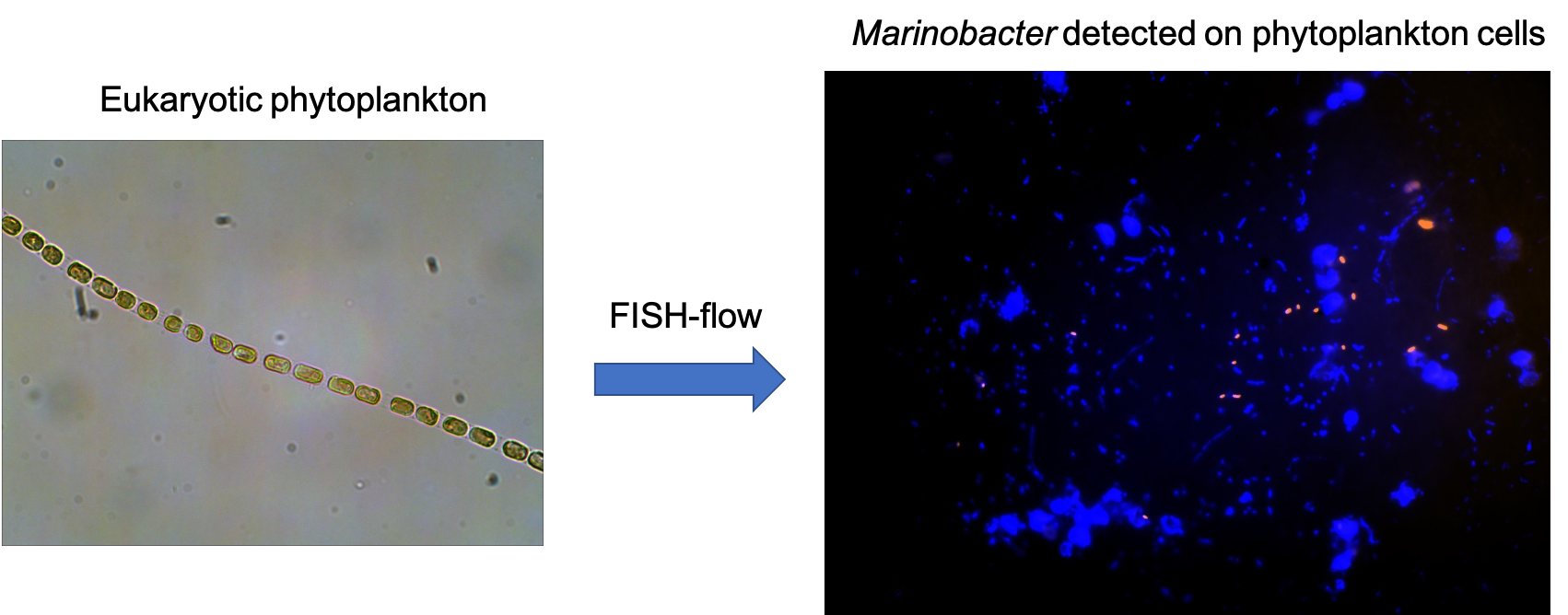
2.1. Study Site and Field Sampling

During a research cruise aboard MRV Scotia in April–May 2015, a sea surface phytoplankton sample was collected from a deep subarctic region called the Faroe-Shetland Channel (60°37.84′ N, 4°54.60′ W; water depth 1022 m). The Faroe-Shetland Channel is a dynamically complex region [39] that is the “spaghetti junction” of Icelandic, Norwegian and Atlantic currents, located approx. 200 miles north of Scotland’s most northerly point. The sampling site is located along the Fair Isle-Munken (FIM) line, which is a sampling transect that runs between the Faroe and Shetland Isles, north of Scotland. This site falls within a sea surface water mass defined as Modified North Atlantic Water (MNAW) which originates from the Faroe Islands and travels in a south-westerly direction through the Faroe-Shetland Channel before diverging south (Figure 1). MNAW is a warm and saltier Atlantic water mass compared to the underlying Arctic/Icelandic cold-water masses that are found at depths from 400 to 1500 m in the Faroe-Shetland Channel [39].
Sampling was performed using a phytoplankton net (50 μm mesh size) that was trawled 10 m below the sea surface (temp. 7.6 °C) for 20 min. Twelve equal volumes (1.5 mL) of the collected phytoplankton fraction were each gently centrifuged (300× g; 1 min.) to pellet the eukaryotic phytoplankton population. The supernatant fractions were collected and set aside at 4 °C. Each of the twelve phytoplankton pellets were washed once with filter-sterilized (0.2 μm) 0.05 M phosphate-buffered saline (PBS; pH 7.3) using the same gentle centrifugation step as before; the supernatant fractions from this wash step were combined with the first collected supernatant fractions stored at 4 °C. The resultant washed phytoplankton fractions were fixed with a filter-sterilized (0.2 μm) solution of 4% paraformaldehyde for 30 min at 4 °C. The fixed phytoplankton fractions were then each washed three times by centrifugation (8000× g; 10 min) with PBS to remove the fixative. The washed fractions were stored frozen onboard the ship at −20 °C in 1:1 (v/v) PBS and ethanol (1 mL total volume) for subsequent analysis by FISH-Flow upon return to the laboratory (described below).
Non-fixed samples of the trawled phytoplankton population, herein referred to as “Phyto”, were prepared in the same way as above, though without fixative, and stored frozen at −20 °C onboard. For the collected supernatant fractions above, these were combined (50 mL total volume) and filtered through a 0.2 μm Nuclepore filter (Whatman, Maidstone, UK) to collect the bacterial biomass representing the bacterial community loosely associated with the phytoplankton population, herein referred to as “Phyto_super”. All filters were stored at −20 °C onboard. Upon return to the laboratory, total genomic DNA was extracted from the frozen “Phyto” and “Phyto_super” filters for analysis by barcoded amplicon MiSeq sequencing (described below)—these samples, respectively, represented the bacterial community found tightly associated with the eukaryotic phytoplankton population, and the bacterial community that was loosely associated with the phytoplankton cells.

2.2. Barcoded-Amplicon Illumina MiSeq Sequencing and Bioinformatic Analysis

For DNA extractions, frozen filters were crushed into liquid nitrogen to a fine powder and DNA extracted according to the method of Tillet and Nielan [40] which utilizes chemical cell lysis with potassium xanthogenate buffer. Unfortunately, some of the replicates were lost due to a haphazard occurrence in the laboratory, but we were able to salvage and process two replicates of the “Phyto” and one of the “Phyto-super” samples. DNA extracts were resuspended in 20 μL of 1 mM TE buffer and stored at −20 °C for Illumina barcoded-amplicon sequencing. Extracted DNA was confirmed using a Nanodrop 3300 fluorescence spectrometer (Thermo, Waltham, MA, USA and further confirmed by gel electrophoresis. PCR amplifications of the V3-V4 16S rRNA gene fragment were performed on all genomic DNA using MyTaq polymerase (BioLine, Heidelberg, Germany), as described by the manufacturer’s protocol. Primer sequences of choice were the universal bacterial primers of forward or reverse direction 341F (CCTACGGGNGGCWGCAG) and 785R (GGACTACHVGGGTATCTAATCC) [41]. Target-specific primer pair coverage distribution was tested with TestPrime 1.0 tool against SILVA RefNR database [42]. Negative controls (molecular grade water) were employed to check for DNA contamination during the PCR procedure. PCR products were quantified by nanodrop (as above) and checked by gel electrophoresis. PCR products (of ~440 bp size) were enzymatically cleaned of polymerase and residual primers using 10 U Exonuclease I and 1 U FastAP (ThermoFisher, Waltham, MA, USA) according to the manufacturer’s protocol (ThermoFisher, Waltham, MA, USA).
Illumina MiSeq sequencing was performed at the University of Liverpool, Centre for Genomic Research. At the sequencing facility, the PCR products underwent a second round of PCR (8 cycles), adding barcodes and the Illumina Nextera XT adapters of the amplified sequences as per the adapted protocol of Berry et al. [43]. This two-round PCR approach has been shown to reduce PCR bias and sequencing artifacts, especially common for complex environmental multi-bacterial samples [43]. Prepared sequencing libraries were qualified and quantified using Qubit Fluorometer and Bioanalyzer systems (ThermoFisher, Waltham, MA, USA) and run on a 2 × 250 paired-end run of the Illumina MiSeq platform. De-multiplexed and primer trimmed data files were returned for downstream processing, as described below.
All reads were processed using Mothur and following the MiSeq SOP [44]. In brief, all sequences were trimmed to 420 bases or less and low-quality sequences were discarded. All reads were aligned to the 16S rRNA gene of E. coli (J01859.1) and trimmed to equal lengths. Each sequence was clustered to 97% similarity to form OTUs and a representative sequence from each OTU was compared to the Silva nr database (v138) to provide taxonomic identification. These data were standardized for read count, resulting in 16S rRNA relative abundance counts for each treatment. All nucleotide sequence data have been deposited in NCBI Sequence Read Archive (SRA) and are available under BioSample accession numbers SAMN16737052, SAMN16737053 and SAMN16737054.

2.3. FISH-Flow Procedures

2.3.1. Preparation of Pure Cell Culture Controls for FISH-Flow

A pure culture of Marinobacter algicola (DSM 16394) was used as a positive control to test the FISH protocol and to calibrate the flow cytometer (BD LSR Fortessa, USA; see below) for the detection of Marinobacter in the collected field samples. For this, M. algicola was grown on marine broth medium (ZM/10) composed of ¾-strength naturally-aged seawater, peptone (0.05%), yeast extract (0.01%), and supplemented after autoclaving with filter-sterilized (0.2 μm) trace elements and vitamins to final concentrations as previously described [45]. Samples of the cells in the exponential phase were fixed with a filter-sterilized (0.2 μm) solution of 3% paraformaldehyde for 30 min at 4 °C. The fixed cells were then washed three times (8000× g; 10 min) using filter-sterilized (0.2 μm) 0.05 M PBS. The washed fractions were stored at −20 °C in 1:1 (v/v) PBS and ethanol (1 mL total volume) for FISH and subsequent calibration of the flow cytometer for the detection and quantification of Marinobacter (see below).

2.3.2. FISH Protocol

Fixed samples of M. algicola cell suspensions and the twelve fixed phytoplankton field samples were thawed and washed three times with 0.05M PBS (8000× g; 10 min) at 21 °C. For hybridization, this was performed in solution using a modified version of the FISH-Flow protocol described by Nettmann et al. [28]. The samples were gently re-suspended in 221 μL of 46 °C preheated hybridization buffer, which contained 900 mM NaCl, 20 mM Tris/HCl (pH 7.2), 0.1% SDS, 25% formamide [38] and 21 μL of the Marinobacter-specific probes (50 ng μL−1 final concentration) Mrb-0625-a and competitor Hal-0625-a [38], or the non-sense probe NON338 [29]. Probe Mrb-0625-a (labeled at the 5′-end with the sulfoindocyanine dye CY3), the labeled NON338 and unlabeled Hal-0625-a probes were all obtained from IDT (Integrated DNA Technologies, Coralville, IA, USA). The Marinobacter-specific probe (Mrb-0625-a) currently targets > 62% of the Marinobacter genus and is used together with the competitor probe to block hybridization to six Halomonas species which share a one base pair mismatch [38]. Hybridisations were also performed in the absence of these probes in order to subtract any autofluorescence as background during the subsequent analysis of these samples by FISH-Flow. Samples were incubated at 46 °C for 2 h whilst inverting periodically. Immediately following hybridization, samples were centrifuged (as before), the supernatant fractions discarded, and then 500 μL of preheated (48 °C) washing buffer (0.149 M NaCl, 20 mM Tris/HCl (pH 7.2), 5 mM EDTA) was added to the samples. This washing buffer maintained slightly more stringent conditions than hybridization buffer. Samples were incubated for 15 min at 48 °C prior to washing twice with 0.05 M PBS (pH 7) [28] and maintained at 4 °C for immediate microscopic observation and analysis by FISH-Flow. To confirm successful hybridization, hybridized samples of M. algicola (as positive control) were visualized using a Zeiss epifluorescence microscope (Axio Scope.A1) equipped with a Zeiss digital fluorescence imaging camera (AxioCam MRm).

2.3.3. Flow Cytometry

Hybridized samples for flow cytometry were counterstained with 4′,6-diamidino-2-phenylindole (DAPI) to a final concentration of 1 μg mL−1, following standard methods in order to identify DNA containing species [46]. The samples were then passed through a 70 μm nylon mesh filter to remove cell aggregates that may cause blockage of the flow cytometer during analysis. For determining the relative size of particles in the samples, the flow cytometer was set to the most useful scatter profile (Forward Scatter FSC vs Side Scatter FSC) by using size-standardized beads covering a range of 1.3 μm to 15 μm (Thermo Fisher Scientific, Waltham, MA, USA).
Samples were analysed using a BD LSR Fortessa Flow Cytometer with five lasers (λex: 355 nm, 405 nm, 488 nm, 561 nm and 640 nm) and 18 fluorescence detection units, in addition to the parameters Forward Scatter (FSC) and Side Scatter (SSC). FSC is defined as the scattered light in the forward direction, with the signal proportional to cell size. SSC is defined as the signal intensity (measured at 90° to the FSC signal) and is proportional to cell complexity/granularity. Scatter profiles (FSC and SSC) were optimized by measuring the background using filtered solutions (0.22 μm for Sheath fluid; 0.1 μm for dH2O and PBS) and size calibration beads in order to accurately gauge the size and complexity of each cell in the samples. The phycoerythrin (PE) channel was used for Cy3 fluorescence (λex = 561 nm (yellow/green laser) and a 585/15 nm emission band-pass filter), whilst the peridinin-chlorophyll protein (PerCP) channel was used to measure chlorophyll (Chl) as auto-fluorescence emitted by eukaryotic phytoplankton (micro-algal) cells (λex = 488 nm [blue laser] and a 670/14 nm emission band pass filter). Spectral overlay of the Cy3 into the Chl channel, and vice versa, was compensated by using a sample of Cy3-labelled M. algicola and non-stained marine eukaryotic phytoplankton sample for chlorophyll auto-fluorescence. DAPI was excited by a 355 nm UV laser and measured using an emission filter 450/50 nm.

2.3.4. Data Processing and Statistical Analysis

For each particle/cell that passed through the flow cytometer, information from all five signals (FSC, SSC, Cy3, Chl and DAPI) of the above channels/lasers was collected and saved as a FCS (Flow Cytometry Standard) file. Using De Novo Software FlowJo V10 (FlowJo, LLC, USA), the following gating strategy was implemented to quantify the target population of total eukaryotic phytoplankton cells, and subsequently those with associated signals for Marinobacter cells (Cy3). Using plot 1 (see below) created above, fluorescence intensity of chlorophyll-a (PerCP Channel, 670 nm emission) was plotted against FSC. Eukaryotic phytoplankton cell signals (Big Chl+) were discriminated using chlorophyll fluorescence (Chl) and “big” size (FSC), as determined using M. algicola alone as Chl-negative control (treatment E; Table S1). The gated population “Big Chl+” was further analysed to identify and quantify Cy3 fluorescence signals when plotted against DAPI signals. Analysing the negative control treatments C and D together with the positive control treatment B for M. algicola cells (Table S1), positive Cy3 signals were observed. Particles of interest were labelled Cy3+ DAPI+ and gated, representing the presence of Marinobacter together with eukaryotic phytoplankton. Using this gating strategy, the frequency (%) of Marinobacter that were within the Big Chl+ gate was determined.
One-way ANOVA was used with Tukey’s post hoc test to compare groups using Statistical Package for the Social Sciences (SPSS). Student’s t-tests were used for comparing between two groups (Treatments A, B, C and D; Table S1) using SPSS. When necessary, data were log10-transformed in order to conform to parametric assumptions of normality and homogeneity of variance.

3. Results and Discussion

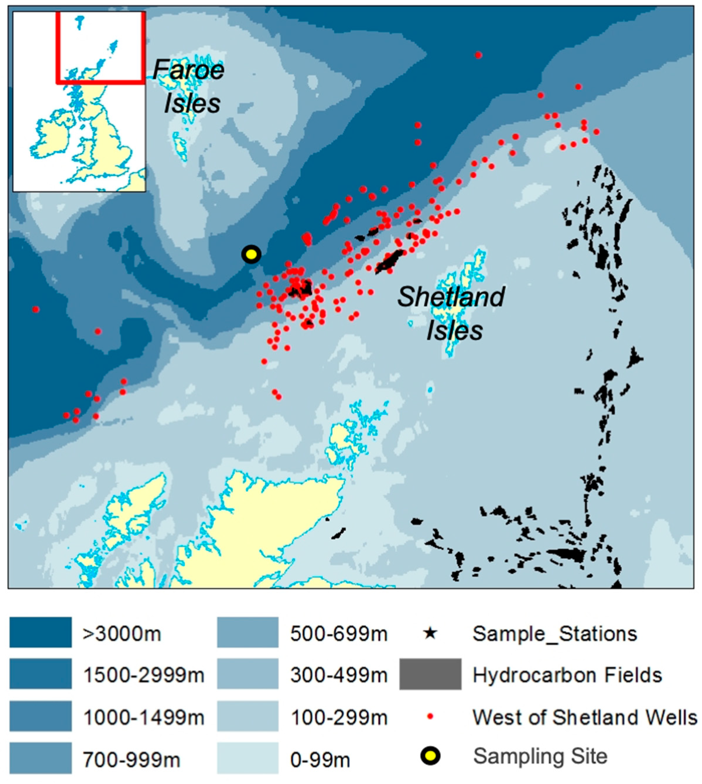
3.1. MiSeq 16S rRNA Sequencing Analysis of the Bacterial Community Associated with the Eukaryotic Phytoplankton Population in the FSC

The “Phyto” bacterial community structure—i.e., that found tightly associated with the eukaryotic phytoplankton cell population in surface waters of the FSC—was dominated by Alcanivorax and members of unclassified Colwelliaceae—40–63% combined contribution to the total 16S rRNA gene sequence reads in both “Phyto” libraries (Figure 2; Table S1). Minor contributions, between 1–5% of relative abundance, were observed for Thioglobaceae, SAR11, Oleispira, Vibrionaceae, Psychrobacter, Marinobacter, Massilia, Erythrobacter, Stenotrophomonas, Thalassospira, and members of the order Rhodospirillales (Marine Group AEGEAN-169) and unclassified Flavobacteriaceae, Rhodobacteraceae, Gammaproteobacteria, Pirellulaceae and Alteromonadaceae. This bacterial community profile reflects that typically found associated with eukaryotic phytoplankton from laboratory cultures and in the field [5,47,48,49], including both obligate and putative oil-degrading taxa, and substantiates our methodological approach for isolating the phytoplankton population collected from the field by net trawl. Alcanivorax are often strongly selected for in oil-impacted environments [35,36] and have been found associated with marine phytoplankton [47]. The family Colwelliaceae is represented by four genera, which includes Colwellia—a genus of psychrophilic marine heterotrophic generalists [50]. However, non-psychrophilic members of Colwellia, able to degrade PAHs, were identified in sea surface oil slicks in the Gulf of Mexico during the active phase of the Deepwater Horizon oil spill [51]. During this spill, Colwellia had rapidly become enriched in sea surface oil slicks and the subsurface oil plume [51]; some members of the genus were shown to having a predilection for degrading and growing on the dispersant Corexit and crude oil [52]. In surface waters of the FSC, we previously reported strains of the genus as susceptible to hydrocarbons and/or to a synthetic chemical dispersant used to treat oil spills [37]. In a more recent report, we showed Colwellia from the FSC as a member of the bacterial community associated with marine oil snow (MOS) [53], similarly to a hydrocarbon-degrading Colwellia strain, RC25, that was isolated from deep waters in the Gulf of Mexico following the Deepwater Horizon oil spill and shown to dominate the bacterial community associated with MOS [54]. Our results and those of past studies suggest that some members of the genus have an affinity for planktonic organic matter, such as MOS (i.e., marine snow containing oil) or with eukaryotic phytoplankton cells (this study).
A number of organisms associated with the eukaryotic phytoplankton population of the FSC were also identified in a previous study to be associated with a phytoplankton population in surface waters of the west coast of Scotland and which had become enriched by crude oil [55]. These organisms included members of Rhodobacteraceae, Thalassospira, Cycloclasticus, Oleispira, Flavobacteriaceae, and Verrucomicrobium. Of these taxa, Thalassospira contain members with reported hydrocarbon-degrading activity [56], and Oleispira is a genus comprising members of obligate hydrocarbon-degraders with a preference for utilizing straight-chain aliphatics [35,36]. The family Rodobacteraceae includes the Roseobacter clade, which is commonly found in high abundance during algal blooms [57] and is recognized for encoding multiple ring-cleaving pathways that participate in the degradation of monocyclic and PAHs [58]. The Flavobacteriaceae contain members with hydrocarbon-degrading activity, such as Arenibacter (not detected in this study) which is a genus comprising members with the ability to degrade PAHs [15]. Our results show that the autochthonous population of eukaryotic phytoplankton, of cell size 50–70 μm, in surface waters of the sampled region in the northeast Atlantic, is a natural source for a rich diversity of oil-degrading bacteria. It could be assumed that this population of phytoplankton may act as an important seed source of oil-degrading bacteria, of which some would be expected to respond strongly in the event of an oil spill in this region where there exists a high presence of oil and gas extraction activity.
Of the less represented phyla found tightly associated with the phytoplankton population of the FSC, a few are worth mentioning. Marinobacter was found almost entirely associated with the “Phyto” sampled fraction (up to 1.75%), as its presence in the “Phyto_super” library was insignificant with only two Marinobacter sequences. As discussed below, this 1.75% relative cell abundance of Marinobacter found tightly bound to the phytoplankton population corroborates the results we subsequently found using FISH-Flow. Though its relative abundance in the “Phyto” libraries was <1%, the detection of members of the family Porticoccaceae is also notable as only two genera are included in this family, one of which is represented by Porticcoccus hydrocarbonoclasticus—an obligate hydrocarbon degrader recognized for having an almost exclusive preference for polycyclic aromatic hydrocarbons (PAHs) as the sole source of carbon and energy [12,59]. The detection of Methylophaga sequences in the phytoplankton community, albeit <0.1% relative abundance, is also notable as these organisms were also found associated with a natural phytoplankton community in a previous study where they were shown to become significantly enriched by crude oil [55]. Members of this genus are recognized for their almost exclusive requirement for C1 sources (e.g., methanol, methylamine, dimethylsulfide) as sole growth substrates, with the exception of some strains that are also capable of metabolizing fructose [60]. However, employing DNA-based stable-isotope probing, Mishamandani et al. [61] provided substantiated evidence implicating members of this genus in also being able to utilize hydrocarbons as a sole source of carbon and energy.
The “Phyto_super” community structure, representing the bacterial population loosely associated with the eukaryotic phytoplankton cell population in surface waters of the FSC, was largely dominated by Psychrobacter and, to a lesser extent, Synechococcus—respectively contributing 75% and 15.9% to the total 16S rRNA gene sequence reads in both “Phyto_super” libraries (Figure 2; Table S1). Minor contributors were identified to belong to Pirellulaceae of the Planctomycetes and unclassified Flavobacteriaceae (collectively 3.8%). Members of all these phyla have been found associated with laboratory cultures of eukaryotic phytoplankton or with phytoplankton blooms in the field [5,47,49]. Therefore, they can be assumed to have been loosely associated with the phytoplankton cell population collected by the net trawl and had become dissociated during its manipulation.
Our identification of Synechococcus associated with the eukaryotic phytoplankton population (15% relative abundance of total bacterial community) is the first report documenting the association of these organisms in a region of the northeast Atlantic. The significance of this follows a recent report by Lea-Smith et al. [17], where the authors discovered the production of alkanes, predominantly pentadecane and heptadecane, by Synechococcus and Prochlorococcus [17]—the two most abundant cyanobacteria in the global ocean [62]. Like these cyanobacteria, some micro-algae from the sunlit, near-surface ocean are also known to produce hydrocarbons, suggesting that these organisms collectively produce a highly significant hydrocarbon input to the global ocean. This is believed to support communities of hydrocarbon-degrading bacteria, even in the absence of any obvious hydrocarbon contamination [63]. In the study by Lea-Smith et al. [17], the cyanobacterial hydrocarbons alone were calculated to provide an input of 308–771 million tons (280–699 million metric tonnes) of pentadecane and heptadecane to the sea per year—a quantity of hydrocarbons that is ~1% of bioavailable dissolved organic carbon in the ocean. Indeed, the abundance and ubiquity of cyanobacteria in the marine environment suggest hydrocarbon production in the oceans could be considerable and broadly distributed geographically. In the laboratory, the authors showed that these hydrocarbons supported the rapid growth of the n-alkane-degrading species Alcanivorax borkumensis.
As mentioned, members of Alcanivorax have been reported associated with laboratory cultures of marine micro-algae [47], and here in the present study we show for the first time that they are tightly associated with a natural community of eukaryotic phytoplankton in the field. Notably, their abundance was much higher (up to 31%) than expected and reported in seawater environments where there is no obvious petrochemical contamination. However, the FSC is a body of water that for many years has been an oil-producing region. Hence, small discharges from industry activity, as well as frequent shipping and oil transportation activities in the region, could be a continued source of petroleum into this region that feeds a higher-than-normal population of hydrocarbon-degrading bacteria. Though no confirmed oil seeps are known along the seabed of the FSC, nor in adjacent water bodies such as the North Sea, satellite surveys suggest the presence of subsurface oil seeps on the east and west of Scotland and offshore in the North Sea (Peter Browning-Stamp, pers. comm.). Such persistent surface oil slicks suggest naturally occurring oil seepage, which may contribute to elevated (above background) levels of an oil-degrading bacterioplankton population, including Alcanivorax, in the FSC; a similar situation is well documented in the Gulf of Mexico [64]. However, we do not exclude the likelihood that such a community is in part also supported by the close coupling of these organisms with hydrocarbon-producing phytoplankton, such as Synechococcus.

3.2. FISH-Flow Optimisation and Methodological Considerations

In the present study, FISH-Flow was employed with probe MRB625a to identify Marinobacter cells associated with the eukaryotic phytoplankton population (of cell size >50 μm) in sea surface waters of a region in the northeast Atlantic. With FISH, there is an inherent possibility of nonspecific binding. However, the significant differences between negative controls and positive controls demonstrate the hybridization worked effectively and that nonspecific binding levels were within acceptable standards. This provided confidence that the Cy3 channel fluorescence was attributed to the probe (MRB625a), and that detected Marinobacter signals were very closely associated with larger eukaryotic phytoplankton population.
Whilst we present here quantitative data for Marinobacter cells found tightly or loosely associated with a natural community of eukaryotic phytoplankton, we are aware that this is not an accurate representation of the complete phytoplankton community in the sea surface sample collected at the time from the northeast Atlantic. However, this was unavoidable as it was necessary to use a 70 μm size nylon mesh filtration step to remove large (>70 μm) particulates from the 50 μm net trawl sample in order to avoid clogging of the flow cytometer’s flow cell—hence, the seawater sample that was used for FISH-Flow contained cells (and other particulates) of size range 50–70 μm. Whilst our analysis also did not account for the possibility of finding Marinobacter associated with the eukaryotic phytoplankton population of cell sizes <50 and >70 μm, as the pore size of our net trawl was 50 μm and we used a >70 μm pre-FISH-Flow filtration step, our aim was principally to determine whether these bacteria are found associated with natural communities of eukaryotic phytoplankton in sea surface waters.
Some authors have in the past reported issues with clogging of their flow cell due to the presence of large aggregates, such as when using flow cytometry coupled with CARD-FISH to quantify nanoplankton and picoplankton communities [32,33,65,66]. Such studies also reported cell loss due to multiple centrifugation steps and re-suspension during hybridization. Our methodology attempted to minimize these shortcomings by reducing the number of centrifugation steps and the processing time using a modified version of the Nettmann et al. [28] protocol (without catalyzed reporter deposition). Gentle centrifugation was intended to maintain the natural microbial assemblage as much as possible in order to obtain a near accurate representation of the Marinobacter population associated with the eukaryotic phytoplankton community, albeit represented by the population of size range 50–70 μm.

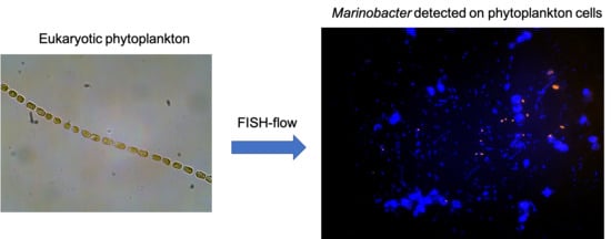
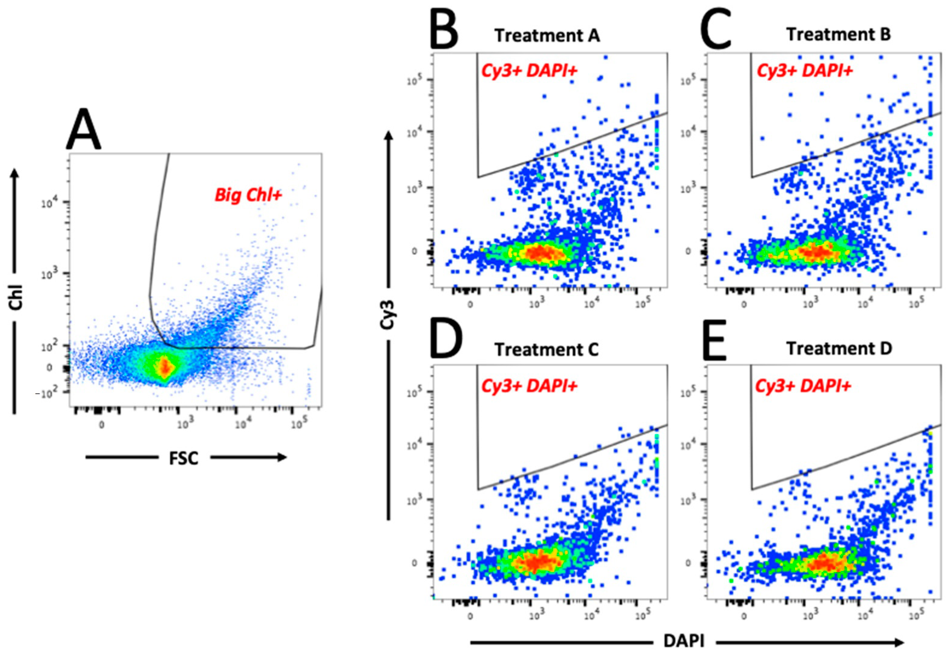
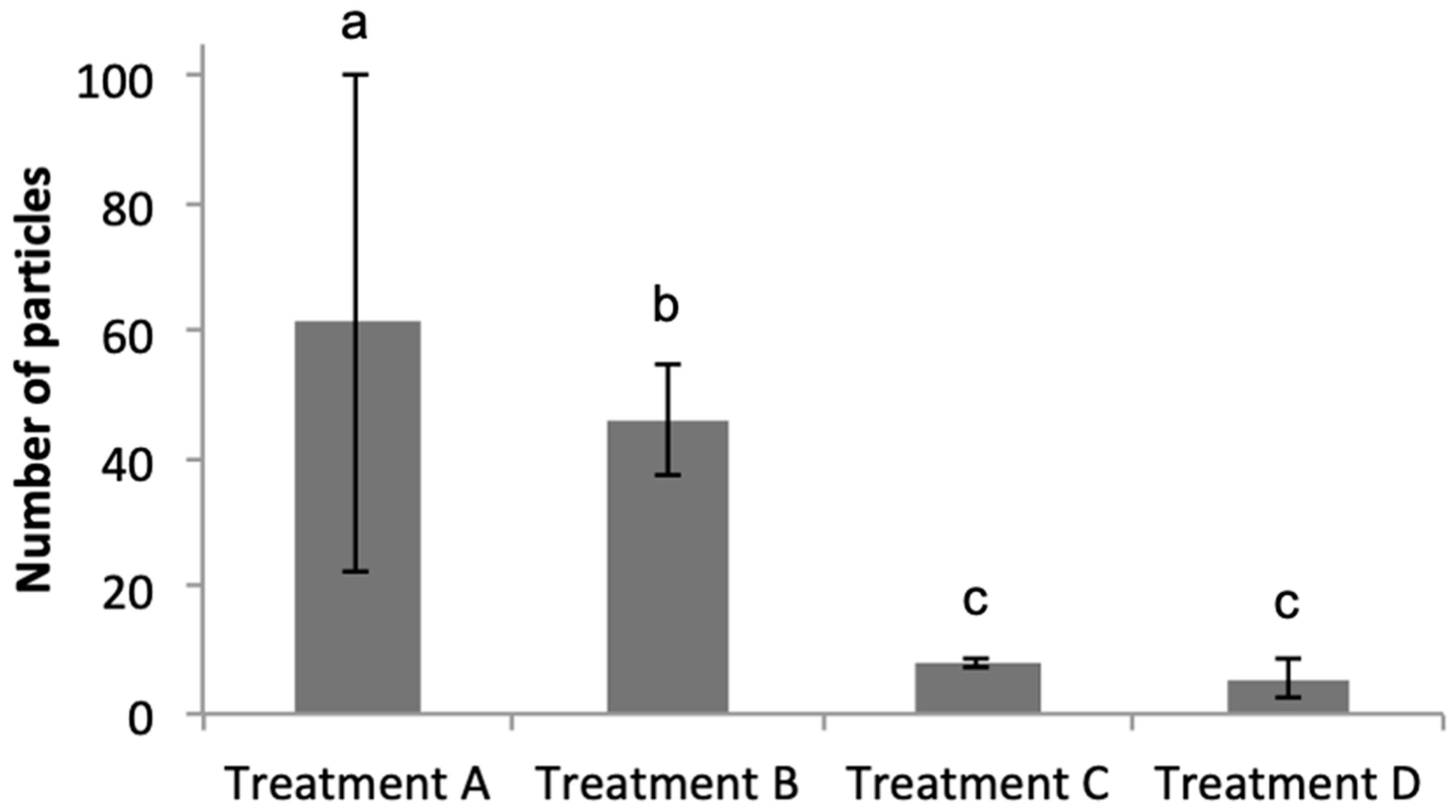
3.3. FISH-Flow Identification of Marinobacter Associated with the Eukaryotic Phytoplankton Population

As shown in Figure 3A, the eukaryotic phytoplankton population (“Big Chl+”, left hand plot) were first gated, and subsequently data from this gate were analysed to identify the Marinobacter population by plotting Cy3 (y-axis) versus DAPI (x-axis)—shown in the “Cy3+ DAPI+” gated regions of Figure 3B–E; total particles in “Cy3+ DAPI+” gates are also shown. As expected, the negative control treatments (C and D) showed a clear lack of particles in these gates, and log10-transformed data confirm that the number of particles were significantly lower (p < 0.01) compared to that in treatments A and B. Results of a Tukey’s post hoc test (Figure 4) show that in Treatment A, which represents the field sample data, there were 60.4 ± 38.7 particles in the Cy3+ DAPI+ gate (Figure 3B); significantly more compared to the 7.59 ± 0.49 particles in the non-sense treatment C. In Treatment A, a total of 1.53 ± 1.33% eukaryotic phytoplankton cells (Big Chl+) tested positive for the Marinobacter Cy3 signal. These results confirm the close physical attachment of Marinobacter on the phycosphere of about 1.5% of the eukaryotic phytoplankton community (of cell size >50 μm) in sea surface waters of the northeast Atlantic during the time of sampling in the spring of 2015. This Marinobacter-phytoplankton association is consistent with studies that have explored this with laboratory cultures of marine micro-algae [6,67], and supports the view of Marinobacter as ubiquitous microorganisms in the marine environment [35,36].
Whilst Marinobacter were found tightly associated with ~1.5% of the eukaryotic phytoplankton community in surface waters of the Faroe-Shetland Channel that may be assuming symbiotic relationships between these organisms [6], other Marinobacter cells were found associated with 2.19 ± 0.64% of the total particulate fraction (of <50 μm size) exhibiting no chlorophyll-a signals. This population may be involved in nutrient cycling, as has been reported for this genus [67,68]. We also identified a large population of Marinobacter cells (54.5 ± 1.6%) that were not associated with eukaryotic phytoplankton or other particulate matter. It should be noted that during flow cytometry, individual particles identified of size <1 μm can be assumed to be cells that were loosely attached to larger particles (i.e., eukaryotic phytoplankton cells or other non-chlorophyll-containing particulate matter) originating from the field samples and that had become detached considering the use of a 50 μm mesh net to collect the phytoplankton field sample. We assume that this population of Marinobacter cells that were not detected as epibiotic on eukaryotic phytoplankton cells may have originated from the phycosphere, and likely were loosely associated and consequently became detached during the FISH-Flow processing steps.
Interestingly, some of the highest Cy3 fluorescence intensities were found on large eukaryotic phytoplankton cells. Possibly individual phytoplankton cells contained high numbers of epibiotic Marinobacter cells attached to them. Alternatively, since with FISH the fluorescence intensity is an indicator of ribosomal abundance inside of cells, and therefore also metabolic activity [26,28], we can assume that this population of Marinobacter cells producing high Cy3 intensities were highly active at the time of our sampling—i.e., 1.55 ± 1.33% of the larger-sized (>50 μm) eukaryotic phytoplankton cells. This is supported by the fact that the mutual symbiosis of Marinobacter and eukaryotic phytoplankton is, at least in part, mediated by the sharing of iron and fixed carbon [6].

4. Conclusions

Members of the genus Marinobacter are known as ubiquitous, opportunistic heterotrophs [67] in marine environments that can use a wide range of substrates [69,70]. These organisms are most widely known for their ability to utilize hydrocarbons and in responding positively to marine oil spills [35,36,69]. Their distribution in the marine environment is largely confined to oligotrophic (i.e., nutrient-/substrate-limited) waters which make up at least 18% (estimated oligotrophic gyre area [71,72]) of the global ocean. Species of Marinobacter may thus be well adapted to survive long periods of low nutrient/substrate availability [67,73,74], especially for species that exist in a free-living state. However, using FISH-Flow and MiSeq sequencing, we have found that the total Marinobacter population in surface waters of the FSC is apparently tightly bound to the eukaryotic phytoplankton population of cell size between 50–70 μm. Isolated strains of this genus have been found living associated on the phycosphere of laboratory cultures of phytoplankton as part of a mutual symbiosis [6]. This adaptation of Marinobacter to physically coupling together with phytoplankton cells may play importantly to its advantage in oligotrophic waters compared to bacteria that exist in a free-living state where such organisms will be limited by the availability of nutrients. The phycosphere offers what can be described as a protective habitat and a source of nutrients provisioned by the algal cell partner, such as algal exudates (vitamin B12, fixed carbon) as carbon and energy sources [6,7,8,10]. This is supported by the fact that very few phytoplankton species can be maintained for long periods in an axenic state in the laboratory.
In conclusion, this work highlights FISH-Flow as an effective method for detecting and quantifying hydrocarbonoclastic bacteria on eukaryotic phytoplankton in seawater samples, and for potentially other epibiotic bacteria where a FISH probe set is available. Using multiple probes for differential targeting of bacterial groups would enable their rapid quantification in water samples, including their association with different size populations of eukaryotic phytoplankton and other planktonic particulate matter such as marine snow and marine oil snow (MOS) where Marinobacter (and other hydrocarbonoclastic taxa) have been found associated [37,75]. Whilst our sampling was limited to the spring 2015 and from one location in the global ocean targeting a specific size range (50–70 μm) of the eukaryotic phytoplankton population, our findings show for the first time the diversity and close association between hydrocarbon-degrading bacteria with a natural population of eukaryotic phytoplankton. Future work is planned to explore this in other ocean sites and across seasons of the year.

Supplementary Materials

The following are available online at https://0-www-mdpi-com.brum.beds.ac.uk/2076-2607/8/12/1955/s1, Table S1: Probes used and their purpose for FISH.

Author Contributions

T.G. conceived the study, and together with H.F.T. designed the experiments and wrote the paper. H.F.T. performed all the experiments, and together with R.Y. analyzed and interpreted the FISH-Flow results of the study. S.S. performed the bioinformatic analysis and curated all the MiSeq sequencing data. All authors have read and agreed to the published version of the manuscript.

Funding

This work was support by a Royal Society Research Grant (project RG2014R2) and a MASTS PECRE grant (project NEADMICRO) awarded to T.G. Partial support was also provided by a Natural Environment Research Council grant (NE/N006526/1).

Acknowledgments

We express our gratitude to chief scientist Alejandro Gallego of Marine Scotland Science, and the ship and scientific crew of MRV Scotia for their hard work, facilities and support to accommodate our research needs during the research cruise to the Faroe-Shetland Channel. We also thank Kate Gormley for help with the production of the map presented in Figure 1. This manuscript contains work conducted during a PhD study sponsored by Heriot-Watt University via their James-Watt Scholarship Scheme to H.F.T. and whose support is gratefully acknowledged. We would also like to thank two anonymous reviewers who provided valuable comments that helped to improve this manuscript.

Conflicts of Interest

The authors declare no conflict of interest.

References

  1. Field, C.B.; Behrenfield, M.J.; Randerson, J.T.; Falkowski, P. Primary production of the biosphere: Integrating terrestrial oceanic components. Science 1998, 281, 237–240. [Google Scholar] [CrossRef] [PubMed] [Green Version]
  2. Ducklow, H.W.; Steinberg, D.K.; Buesseler, K.O. Upper ocean carbon export and the biological pump. Oceanography 2001, 14, 50–58. [Google Scholar] [CrossRef]
  3. Amin, S.A.; Hmelo, L.R.; van Tol, H.M.; Durham, B.P.; Carlson, L.T.; Heal, K.R.; Morales, R.L.; Berthiaume, C.T.; Parker, M.S.; Djunaedi, B.; et al. Interactions and signalling between a cosmopolitan phytoplankton and associated bacteria. Nature 2015, 522, 98–101. [Google Scholar] [CrossRef] [PubMed]
  4. Amin, S.A.; Parker, M.S.; Ambrust, E.V. Interactions between diatoms and bacteria. Microbiol. Mol. Biol. Rev. 2012, 76, 667–684. [Google Scholar] [CrossRef] [PubMed] [Green Version]
  5. Buchan, A.; LeCleir, G.R.; Gulvik, C.A.; González, J.M. Master recyclers: Features and functions of bacteria associated with phytoplankton blooms. Nat. Rev. Microbiol. 2014, 12, 686–698. [Google Scholar] [CrossRef]
  6. Amin, S.A.; Green, D.H.; Hart, M.C.; Küpper, F.C.; Sunda, W.G.; Carrano, C.J. Photolysis of iron-siderophore chelates promotes bacterial-algal mutualism. Proc. Natl. Acad. Sci. USA 2009, 106, 17071–17076. [Google Scholar] [CrossRef] [Green Version]
  7. Bell, W.; Mitchell, R. Chemotactic and growth responses of marine bacteria to algal extracellular products. Biol. Bull. 1972, 143, 265–277. [Google Scholar] [CrossRef]
  8. Myklestad, S.M. Release of extracellular products by phytoplankton with special emphasis on polysaccharides. Sci. Total Environ. 1995, 165, 155–164. [Google Scholar] [CrossRef]
  9. McGenity, T.J.; Folwell, B.D.; McKew, B.A.; Sanni, G.O. Marine crude-oil biodegradation: A central role for interspecies interactions. Aquat. Biosyst. 2012, 8, 10. [Google Scholar] [CrossRef] [PubMed] [Green Version]
  10. Kazamia, E.; Czesnick, H.; Van Nguyen, T.T.; Croft, M.T.; Sherwood, E.; Sasso, S.; Hodson, S.J.; Warren, M.J.; Smith, A.G. Mutualistic interactions between vitamin B12-dependent algae and heterotrophic bacteria exhibit regulation. Environ. Microbiol. 2012, 14, 1466–1476. [Google Scholar] [CrossRef]
  11. Green, D.H.; Bowman, J.P.; Smith, E.A.; Gutierrez, T.; Bolch, C.J.S. Marinobacter algicola sp. nov., isolated from laboratory cultures of paralytic shellfish toxin-producing dinoflagellates. Int. J. Syst. Evol. Microbiol. 2006, 56, 523–527. [Google Scholar] [CrossRef] [PubMed] [Green Version]
  12. Gutierrez, T.; Nichols, P.D.; Whitman, W.B.; Aitken, M.D. Porticoccus hydrocarbonoclasticus sp. nov., an aromatic hydrocarbon-degrading bacterium identified in laboratory cultures of marine phytoplankton. Appl. Environ. Microbiol. 2012, 78, 628–637. [Google Scholar] [CrossRef] [PubMed] [Green Version]
  13. Gutierrez, T.; Green, D.H.; Whitman, W.B.; Nichols, P.D.; Semple, K.T.; Aitken, M.D. Algiphilus aromaticivorans gen. nov., sp. nov., an aromatic hydrocarbon-degrading bacterium isolated from a culture of the marine dinoflagellate Lingulodinium polyedrum, and proposal of Algiphilaceae fam. nov. Int. J. Syst. Evol. Microbiol. 2012, 62, 2743–2749. [Google Scholar] [CrossRef] [PubMed]
  14. Gutierrez, T.; Green, D.H.; Nichols, P.D.; Whitman, W.B.; Semple, K.T.; Aitken, M.D. Polycyclovorans algicola gen. nov., sp. nov., an aromatic-hydrocarbon-degrading marine bacterium found associated with laboratory cultures of marine phytoplankton. Appl. Environ. Microbiol. 2013, 79, 205–214. [Google Scholar] [CrossRef] [PubMed] [Green Version]
  15. Gutierrez, T.; Rhodes, G.; Mishamandani, S.; Berry, D.; Whitman, W.B.; Nichols, P.D.; Semple, K.T.; Aitken, M.D. Polycyclic aromatic hydrocarbon degradation of phytoplankton-associated Arenibacter spp. and description of Arenibacter algicola sp. nov., an aromatic hydrocarbon-degrading bacterium. Appl. Environ. Microbiol. 2014, 80, 618–628. [Google Scholar] [CrossRef] [Green Version]
  16. Andelman, J.B.; Suess, M.J. Polynuclear aromatic hydrocarbons in the water environment. Bull. World Health Organ. 1970, 43, 479–508. [Google Scholar]
  17. Lea-Smith, D.J.; Biller, S.J.; Davey, M.P.; Cotton, C.A.R.; Perez Sepulveda, B.M.; Turchyn, A.V.; Scanlan, D.J.; Smith, A.G.; Chisholm, S.W.; Howe, C.J. Contribution of cyanobacterial alkane production to the ocean hydrocarbon cycle. Proc. Natl. Acad. Sci. USA 2015, 112, 13591–13596. [Google Scholar] [CrossRef] [Green Version]
  18. Mallet, L.; Sardou, J. Examination of the presence of the polybenzic hydrocarbon benzo-3,4-pyrene in the plank-tonic environment of the Bay of Villefranche. Symposium, committee on international scientific exploration of the mediterranean sea, Monaco. CR Acad. Sci. 1964, 258, 5264–5267. [Google Scholar]
  19. Exton, D.A.; Steinke, M.; Suggett, D.J.; McGenity, T.J. Spatial and temporal variability of biogenic isoprene emissions from a temperate estuary. Glob. Biogeochem. Cycl. 2012, 26, GB2012. [Google Scholar] [CrossRef] [Green Version]
  20. Gunnison, D.; Alexander, M. Basis for the resistance of several algae to microbial decomposition. Appl. Microbiol. 1975, 29, 729–738. [Google Scholar] [CrossRef]
  21. Marlowe, I.T.; Green, J.C.; Neal, A.C.; Brassell, S.C.; Eglinton, G.; Course, P.A. Long chain (n-C37-C39) alkenones in the Prymnesiophyceae. Distribution of alkenones and other lipids and their taxonomic significance. Br. Phycol. J. 1984, 19, 203–216. [Google Scholar] [CrossRef] [Green Version]
  22. Shaw, S.L.; Gantt, B.; Meskhidze, N. Production and emissions of marine isoprene and monoterpenes: A review. Adv. Meteorol. 2010, 2010, 408696. [Google Scholar] [CrossRef]
  23. Abed, R.M.M.; Köster, J. The direct role of aerobic heterotrophic bacteria associated with cyanobacteria in the degradation of oil compounds. Int. Biodeterior. Biodegrad. 2005, 55, 29–37. [Google Scholar] [CrossRef]
  24. Warshawsky, D.; LaDow, K.; Schneider, J. Enhanced degradation of benzo[a]pyrene by Mycobacterium sp. in conjunction with green alga. Chemosphere 2007, 69, 500–506. [Google Scholar] [CrossRef] [PubMed]
  25. Steen, H.B. Flow cytometer for measurement of the light scattering of viral and other submicroscopic particles. Cytom. A 2004, 57, 94–99. [Google Scholar] [CrossRef] [PubMed]
  26. Amann, R.I.; Binder, B.J.; Olson, R.J.; Chisholm, S.W.; Devereux, R.; Stahl, D.A. Combination of 16S rRNA-targeted oligonucleotide probes with flow cytometry for analyzing mixed microbial populations. Appl. Environ. Microbiol. 1990, 56, 1919–1925. [Google Scholar] [CrossRef] [Green Version]
  27. Friedrich, U.; Lenke, J. Improved enumeration of lactic acid bacteria in mesophilic dairy starter cultures by using multiplex quantitative real-time pcr and flow cytometry-fluorescence in situ hybridization. Appl. Environ. Microbiol. 2006, 72, 4163–4171. [Google Scholar] [CrossRef] [Green Version]
  28. Nettmann, E.; Fröhling, A.; Heeg, K.; Klocke, M.; Schlüter, O.; Mumme, J. Development of a flow-fluorescence in situ hybridization protocol for the analysis of microbial communities in anaerobic fermentation liquor. BMC Microbiol. 2013, 13, 278. [Google Scholar] [CrossRef] [Green Version]
  29. Wallner, G.; Amann, R.; Beisker, W. Optimizing fluorescent in situ hybridization with rRNA-targeted oligonucleotide probes for flow cytometric identification of microorganisms. Cytometry 1993, 14, 136–143. [Google Scholar] [CrossRef]
  30. Li, M.; Yang, Y.; He, Y.; Mathieu, J.; Yu, C.; Li, Q.; Alvarez, P.J.J. Detection and cell sorting of Pseudonocardia species by fluorescence in situ hybridization and flow cytometry using 16S rRNA-targeted oligonucleotide probes. Appl. Microbiol. Biotechnol. 2018, 102, 3375–3386. [Google Scholar] [CrossRef]
  31. Arrigucci, R.; Bushkin, Y.; Radford, F.; Lakehal, K.; Vir, P.; Pine, R.; Martin, D.; Sugarman, J.; Zhao, Y.; Yap, G.S.; et al. FISH-Flow, a protocol for the concurrent detection of mRNA and protein in single cells using fluorescence in situ hybridization and flow cytometry. Nat. Prot. 2017, 12, 1245–1260. [Google Scholar] [CrossRef] [PubMed]
  32. Biegala, I.C.; Not, F.; Vaulot, D.; Simon, N. Quantitative assessment of picoeukaryotes in the natural environment by using taxon-specific oligonucleotide probes in association with tyramide signal amplification-fluorescence in situ hybridization and flow cytometry. Appl. Environ. Microbiol. 2003, 69, 5519–5529. [Google Scholar] [CrossRef] [PubMed] [Green Version]
  33. Sekar, R.; Fuchs, B.M.; Amann, R.; Pernthaler, J. Flow sorting of marine bacterioplankton after fluorescence in situ hybridization. Appl. Environ. Microbiol. 2004, 70, 6210–6219. [Google Scholar] [CrossRef] [PubMed] [Green Version]
  34. Manti, A.; Boi, P.; Amalfitano, S.; Puddu, A.; Papa, S. Experimental improvements in combining CARD-FISH and flow cytometry for bacterial cell quantification. J. Microbiol. Methods 2011, 87, 309–315. [Google Scholar] [CrossRef] [PubMed]
  35. Head, I.M.; Jones, D.M.; Röling, W.F.M. Marine microorganisms make a meal of oil. Nat. Rev. Microbiol. 2006, 4, 173–182. [Google Scholar] [CrossRef] [PubMed]
  36. Yakimov, M.M.; Timmis, K.N.; Golyshin, P.N. Obligate oil-degrading marine bacteria. Curr. Opin. Biotechnol. 2007, 18, 257–266. [Google Scholar] [CrossRef]
  37. Suja, L.D.; Summers, S.; Gutierrez, T. Role of EPS, dispersant and nutrients on the microbial response and MOS formation in the subarctic northeast Atlantic. Front. Microbiol. 2017, 8, 676. [Google Scholar] [CrossRef] [Green Version]
  38. McKay, L.J.; Gutierrez, T.; Teske, A.P. Development of a group-specific 16S rRNA-targeted probe set for the identification of Marinobacter by fluorescence in situ hybridization. Deep Sea Res. Part II Top. Stud. Oceanogr. 2016, 129, 360–367. [Google Scholar] [CrossRef] [Green Version]
  39. Berx, B.; Hansen, B.; Østerhus, S.; Larsen, K.M.; Sherwin, T.; Jochumsen, K. Combining in situ measurements and altimetry to estimate volume, heat and salt transport variability through the Faroe–Shetland Channel. Ocean Sci. 2013, 9, 639–654. [Google Scholar] [CrossRef] [Green Version]
  40. Tillett, D.; Neilan, B. Xanthogenate nucleic acid isolation from cultured and environmental cyanobacteria. J. Phycol. 2000, 36, 251–258. [Google Scholar] [CrossRef]
  41. Klindworth, A.; Pruesse, E.; Schweer, T.; Peplies, J.; Quast, C.; Horn, M.; Glöckner, F.O. Evaluation of general 16S ribosomal RNA gene PCR primers for classical and next-generation sequencing-based diversity studies. Nucleic Acids Res. 2013, 41, e1. [Google Scholar] [CrossRef] [PubMed]
  42. Quast, C.; Pruesse, E.; Yilmaz, P.; Gerken, J.; Schweer, T.; Glo, F.O.; Yarza, P. The SILVA ribosomal RNA gene database project: Improved data processing and web-based tools. Nucleic Acids Res. 2013, 41, D590–D596. [Google Scholar] [CrossRef] [PubMed]
  43. Berry, D.; Ben Mahfoudh, K.; Wagner, M.; Loy, A. Barcoded primers used in multiplex amplicon pyrosequencing bias amplification. Appl. Environ. Microbiol. 2011, 78, 612. [Google Scholar] [CrossRef] [Green Version]
  44. Kozich, J.J.; Westcott, S.L.; Baxter, N.T.; Highlander, S.K.; Schloss, P.D. Development of a dual-index sequencing strategy and curation pipeline for analyzing amplicon sequence data on the MiSeq Illumina sequencing platform. Appl. Environ. Microbiol. 2013, 79, 5112–5120. [Google Scholar] [CrossRef] [PubMed] [Green Version]
  45. Blackburn, S.I.; Hallegraeff, G.M.; Bolch, C.J. Vegetative reproduction and sexual life cycle of the toxic dinoflagellate Gymnodinium catenatum from Tasmania. Aust. J. Phycol. 1989, 25, 577–590. [Google Scholar] [CrossRef]
  46. Porter, K.G.; Feig, Y.S. The use of DAPI for identifyinh and counting aquatic microflora. Limnol. Oceanogr. 1980, 25, 943–948. [Google Scholar] [CrossRef]
  47. Chernikova, T.N.; Bargiela, R.; Toshchakov, S.V.; Shivaraman, V.; Lunev, E.A.; Yakimov, M.M.; Thomas, D.N.; Golyshin, P.N. Hydrocarbon-degrading bacteria Alcanivorax and Marinobacter associated with microalgae Pavlova lutheri and Nannochloropsis oculata. Front. Microbiol. 2020, 11, 572931. [Google Scholar] [CrossRef]
  48. Fei, C.; Ochsenkühn, M.A.; Shibi, A.A.; Isaac, A.; Wang, C.; Amin, S.A. Quorum sensing regulates ‘swim-or-stick’ lifestyle in the phycosphere. Environ. Microbiol. 2020, 22, 4761–4778. [Google Scholar] [CrossRef]
  49. Needham, D.M.; Fuhrman, J.A. Pronounced daily succession of phytoplankton, archaea and bacteria following a spring bloom. Nat. Microbiol. 2016, 1, 16005. [Google Scholar] [CrossRef]
  50. Methé, B.A.; Nelson, K.E.; Deming, J.W.; Momen, B.; Melamud, E.; Zhang, X.; Moult, J.; Madupu, R.; Nelson, W.C.; Dodson, R.J.; et al. The psychrophilic lifestyle as revealed by the genome sequence of Colwellia psychrerythraea 34H through genomic and proteomic analyses. Proc. Natl. Acad. Sci. USA 2005, 102, 10913–10918. [Google Scholar] [CrossRef] [Green Version]
  51. Yang, T.; Nigro, L.M.; Gutierrez, T.; D’Ambrosio, L.; Joye, S.B.; Highsmith, R.; Teske, A.P. Pulsed blooms and persistent oil-degrading bacterial populations in the water column during and after the Deepwater Horizon blowout. Deep Sea Res. Part II Top. Stud. Oceanogr. 2016, 129, 282–291. [Google Scholar] [CrossRef] [Green Version]
  52. Kleindienst, S.; Seidel, M.; Ziervogel, K.; Grim, S.; Loftis, K.; Harrison, S.; Malkin, S.Y.; Perkins, M.J.; Field, J.; Sogin, M.L. Chemical dispersants can suppress the activity of natural oil-degrading microorganisms. Proc. Natl. Acad. Sci. USA 2015, 112, 14900–14905. [Google Scholar] [CrossRef] [PubMed] [Green Version]
  53. Suja, L.D.; Chen, X.; Summers, S.; Paterson, D.; Gutierrez, T. Chemical dispersant enhances microbial exopolymer (EPS) production and formation of marine oil/dispersant snow in surface waters of the subarctic northeast Atlantic. Front. Microbiol. 2019, 10, 553. [Google Scholar] [CrossRef] [PubMed]
  54. Bælum, J.; Borglin, S.; Chakraborty, R.; Fortney, J.L.; Lamendella, R.; Mason, O.U.; Auer, M.; Zemla, M.; Bill, M.; Conrad, M.E.; et al. Deep-sea bacteria enriched by oil and dispersant from the Deepwater Horizon spill. Environ. Microbiol. 2012, 14, 2405–2416. [Google Scholar] [CrossRef] [PubMed]
  55. Thompson, H.; Angelova, A.; Bowler, B.; Jones, M.; Gutierrez, T. Enhanced crude oil biodegradative potential of natural phytoplankton-associated hydrocarbonoclastic bacteria. Environ. Microbiol. 2017, 19, 2843–2861. [Google Scholar] [CrossRef] [PubMed]
  56. Zhao, B.; Wang, H.; Li, R.; Mao, X. Thalassospira xianhensis sp. nov., a polycyclic aromatic hydrocarbon-degrading marine bacterium. Int. J. Syst. Evol. Microbiol. 2010, 60, 1125–1129. [Google Scholar] [CrossRef]
  57. González, J.M.; Simó, R.; Massana, R.; Covert, J.S.; Casamayor, E.O.; Pedrós-Alió, C.; Moran, M.A. Bacterial community structure associated with a dimethylsulfoniopropionate-producing North Atlantic algal bloom. Appl. Environ. Microbiol. 2000, 66, 4237–4246. [Google Scholar] [CrossRef] [Green Version]
  58. Moran, M.A.; Belas, R.; Schell, M.A.; Gonzalez, J.M.; Sun, F.; Sun, S.; Binder, B.J.; Edmonds, J.; Ye, W.; Orcutt, B.; et al. Ecological genomics of marine roseobacters. Appl. Environ. Microbiol. 2007, 73, 4559–4569. [Google Scholar] [CrossRef] [Green Version]
  59. Gutierrez, T. Aerobic hydrocarbon-degrading Gammaproteobacteria–Porticoccus. In Taxonomy, Genomics and Ecophysiology of Hydrocarbon-Degrading Microbes; Section 6.10; Handbook of Hydrocarbon and Lipid Microbiology; McGenity, T.J., Prince, R., Eds.; Springer: Berlin, Germany, 2018; Volume 6. [Google Scholar]
  60. Janvier, M.; Grimont, P.A.D. The genus Methylophaga, a new line of descent within phylogenetic branch γ of Proteobacteria. Res. Microbiol. 1995, 146, 543–550. [Google Scholar] [CrossRef]
  61. Mishamandani, S.; Gutierrez, T.; Aitken, M.D. DNA-based stable isotope probing coupled with cultivation methods implicates Methylophaga in hydrocarbon degradation. Front. Microbiol. 2014, 8, 10. [Google Scholar] [CrossRef]
  62. Scanlan, D.J.; Ostrowski, M.; Mazard, S.; Dufresne, A.; Garczarek, L.; Hess, W.R.; Post, A.F.; Hagemann, M.; Paulsen, I.; Partensky, F. Ecological genomics of marine picocyanobacteria. Microbiol. Mol. Biol. Rev. 2009, 73, 249–299. [Google Scholar] [CrossRef] [PubMed] [Green Version]
  63. Gutierrez, T. Occurrence and roles of the obligate hydrocarbonoclastic bacteria in the ocean when there is no obvious hydrocarbon contamination. In Taxonomy, Genomics and Ecophysiology of Hydrocarbon-Degrading Microbes; Section 6.34; Handbook of Hydrocarbon and Lipid Microbiology; McGenity, T.J., Prince, R., Eds.; Springer: Berlin, Germany, 2018; Volume 6. [Google Scholar]
  64. Joye, S.; Kostka, J. Microbial Genomics of the Global Ocean System. Earth and Space Science Open Archive. 2020. Available online: https://www.asmscience.org/content/colloquia.58 (accessed on 1 October 2020).
  65. Lebaron, P.; Catala, P.; Fajon, C.; Joux, F.; Baudart, J.; Bernard, L. A new sensitive, whole-cell hybridization technique for detection of bacteria involving a biotinylated oligonucleotide probe targeting rRNA and tyramide signal amplification. Appl. Environ. Microbiol. 1997, 63, 3274–3278. [Google Scholar] [CrossRef] [PubMed] [Green Version]
  66. Schönhuber, W.; Fuchs, B.; Juretschko, S.; Amann, R. Improved sensitivity of whole-cell hybridization by the combination of horseradish peroxidase-labeled oligonucleotides and tyramide signal amplification. Appl. Environ. Microbiol. 1997, 63, 3268–3273. [Google Scholar] [CrossRef] [PubMed] [Green Version]
  67. Singer, E.; Webb, E.A.; Nelson, W.C.; Heidelberg, J.F.; Ivanova, N.; Pati, A.; Edwards, K.J. Genomic potential of Marinobacter aquaeolei, a biogeochemical “opportunitroph”. Appl. Environ. Microbiol. 2011, 77, 2763–2771. [Google Scholar] [CrossRef] [PubMed] [Green Version]
  68. Handley, K.M.; Lloyd, J.R. Biogeochemical implications of the ubiquitous colonization of marine habitats and redox gradients by Marinobacter species. Front. Microbiol. 2013, 4, 136. [Google Scholar] [CrossRef] [Green Version]
  69. Duran, R. Marinobacter. In Handbook of Hydrocarbon and Lipid Microbiology; Timmis, K.N., Ed.; Springer: Heidelberg, Germany, 2010; pp. 1725–1736. [Google Scholar]
  70. Mounier, J.; Camus, A.; Mitteau, I.; Vaysse, P.J.; Goulas, P.; Grimaud, R.; Sivadon, P. The marine bacterium Marinobacter hydrocarbonoclasticus SP17 degrades a wide range of lipids and hydrocarbons through the formation of oleolytic biofilms with distinct gene expression profiles. FEMS Microbiol. Ecol. 2014, 90, 816–831. [Google Scholar] [CrossRef] [Green Version]
  71. Polovina, J.J.; Howell, E.A.; Abecassis, M. Ocean’s least productive waters are expanding. Geophys. Res. Lett. 2008, 35, L03618. [Google Scholar] [CrossRef] [Green Version]
  72. Signorini, S.R.; Franz, B.A.; McClain, C.R. Chlorophyll variability in the oligotrophic gyres: Mechanisms, seasonality and trends. Front. Mar. Sci. 2015, 2. [Google Scholar] [CrossRef]
  73. Sebastian, M.; Estrany, M.; Ruiz-Gonzalez, C.; Forn, I.; Sala, M.M.; Gasol, J.M.; Marrase, C. High growth potential of long-term starved deep ocean opportunistic heterotrophic bacteria. Front. Microbiol. 2019, 10, 760. [Google Scholar] [CrossRef]
  74. Vaysse, P.J.; Sivadon, P.; Goulas, P.; Grimaud, R. Cells dispersed from Marinobacter hydrocarbonoclasticus SP17 biofilm exhibit a specific protein profile associated with a higher ability to reinitiate biofilm development at the hexadecane–water interface. Environ. Microbiol. 2011, 13, 737–746. [Google Scholar] [CrossRef]
  75. Arnosti, C.; Ziervogel, K.; Yang, T.; Teske, A. Oil-derived marine aggregates—Hot spots of polysaccharide degradation by specialized bacterial communities. Deep Sea Res. Part II Top. Stud. Oceanogr. 2016, 129, 179–186. [Google Scholar] [CrossRef]
Figure 1. Map representing the Faroe-Shetland Channel showing bathymetry contours, hydrocarbon gas/oil wells and production fields, and the sampling location.
Microorganisms 08 01955 g001
Figure 2. Abundant bacterial phyla in the “Phyto” and “Phyto-super” fractions.
Microorganisms 08 01955 g002
Figure 3. Bivariate plot and gating sequence using scatter (FSC) and chlorophyll (Chl) parameters to gate big eukaryotic phytoplankton ((A) Big Chl+), followed by fluorescence (DAPI and Cy3) parameters to gate viable (DAPI+) Marinobacter cells (Cy3+) in gate (BE) Cy3+ DAPI+ (examples from treatments A–D). Gate names are shown in red text on each plot. Treatment A represents the field sample data, whereas treatments B represents the positive (M. algicola enrichment) control. Non-sense probe and negative (probe absent) control treatments (C and D, respectively) clearly contain less Cy3 fluorescence in gate “Big Chl+”. The red, green and blue colouration in each plot represents the signal intensity, from strong to weak respectively.
Microorganisms 08 01955 g003
Figure 4. Total number of particles (mean values and standard deviation for three replicates) in gate “Cy3+ DAPI+” of Figure 2 comparing all treatments A–D. Lower case letters show the results of Tukey’s post hoc test: a treatment sharing the same letter with another treatment denotes the treatments were not significantly different from each another. See Figure 3 caption for key to treatments A–D.
Microorganisms 08 01955 g004
Publisher’s Note: MDPI stays neutral with regard to jurisdictional claims in published maps and institutional affiliations.

