Article

Occurrence of Hybrid Diarrhoeagenic Escherichia coli Associated with Multidrug Resistance in Environmental Water, Johannesburg, South Africa

Water and Health Research Centre, University of Johannesburg, Doornfontein 2092, South Africa
*
Author to whom correspondence should be addressed.
Submission received: 27 August 2021 / Revised: 25 September 2021 / Accepted: 27 September 2021 / Published: 17 October 2021

Abstract

:
This study was undertaken to determine the virulence and antibiotic resistance profiles of diarrhoeagenic Escherichia coli (DEC) in environmental waters of Johannesburg, South Africa. Samples were collected and cultured on selective media. An 11-plex PCR assay was used to differentiate five DEC, namely: enteroaggregative (EAEC), enterohaemorrhagic (EHEC), enteroinvasive (EIEC), enteropathogenic (EPEC) and enterotoxigenic (ETEC). The antibiotic resistance profile of isolates was determined using the VITEK®-2 automated system. The virulence profiles of 170 E. coli tested showed that 40% (68/170) were commensals and 60% (102/170) were pathogenic. EPEC had a prevalence of 19.2% (32/170), followed by ETEC 11.4% (19/170), EAEC 6% (10/170) and EHEC 3% (5/170). Hybrid DEC carrying a combination of simultaneously two and three pathogenic types was detected in twenty-eight and nine isolates, respectively. The antibiotic susceptibility testing showed isolates with multidrug resistance, including cefuroxime (100%), ceftazidime (86%), cefotaxime (81%) and cefepime (79%). This study highlighted the widespread occurrence of DEC and antibiotic resistance strains in the aquatic ecosystem of Johannesburg. The presence of hybrid pathotypes detected in this study is alarming and might lead to more severe diseases. There is a necessity to enhance surveillance in reducing the propagation of pathogenic and antibiotic-resistant strains in this area.

1. Introduction

Diarrhoea is one of the common causes of morbidity and mortality among infants and children in most developing countries [1]. It is estimated that diarrhoea causes 526,000 deaths of children younger than five per year [2,3]. Escherichia coli (E. coli) is an anaerobic Gram-negative, typically rod-shaped bacterium considered as part of the normal flora of the gut of humans and warm-blooded animals, and can also be found in the environment, e.g., in water and soil [4,5]. According to its biological significance, E. coli is classified into harmless commensals, pathogenic and extra-intestinal pathogenic strains [6]. Pathogenic or diarrhoeagenic Escherichia coli (DEC) strains are among the causative agents of diarrhoea outbreaks and other serious waterborne and foodborne infections in humans [7,8]. Seven major DEC have been described: (i) adherent-invasive E. coli (AIEC), (ii) diffusely adherent E. coli (DAEC), (iii) enteroaggregative E. coli (EAEC), (iv) enterohaemorrhagic E. coli (EHEC), (v) enteroinvasive E. coli (EIEC), (vi) enteropathogenic E. coli (EPEC) and (vii) enterotoxigenic E. coli (ETEC) [9,10,11]. These pathotypes are classified based on clinical features, epidemiological evidence, phenotypic traits and specific virulence factors [12,13].
EAEC is a pathotype associated with persistent diarrhoea in humans and has been identified as possessing a plasmid of aggregative adhesion (pAA), for fimbriae production, which contains the aggregative adherence fimbriae type R (aggR) [12]. EHEC is an important enteric pathogen that produces Shiga toxin and causes a variety of clinical syndromes in humans, including bloody and non-bloody diarrhoea, haemorrhagic colitis and haemolytic uremic syndrome (HUS) [14,15]. EIEC is a pathotype that possesses invasion plasmid antigen H (ipaH) and invasive antigen locus (ial) genes, which encode the virulence regulon, the key for severe/invasive intestinal infections and dysentery [16,17]. EPEC and ETEC cause significant diarrhoeal illness and mortality in children, mostly in the developing world. EPEC strains are classified as typical or atypical, according to the presence or absence of the E. coli adherence factor plasmid (EAF) that carries the bfpA gene, which encodes for the bundle-forming pili [18]. Typical EPEC (tEPEC) contain both the locus of enterocyte effacement (LEE) region for attaching and effacing lesion (eaeA gene) and the bfpA gene, while atypical EPEC (aEPEC) are strains that do not contain the bfpA gene [19]. ETEC is a pathotype that colonises the small intestine and causes watery diarrhoea, known as traveller’s diarrhoea, in humans by producing plasmid-encoded heat-stable (ST) and/or heat-labile enterotoxin (LT) [20].
Hybrid DEC harbouring a combination of virulence genes have emerged worldwide and have been reported as a public health concern [21,22]. This is due to horizontal gene transfer (HGT) among diarrhoeagenic groups of E. coli [22,23] or to the fact that most E. coli virulence genes are generally found on plasmids, transmissible by means of conjugation [24]. The first hybrid pathotype (EAEC/EHEC or EAHEC) with genetic recombination was reported during a German diarrhoea outbreak [25]. Since then, several other studies have reported molecular evidence and genomics of hybrid pathotypes worldwide, including EPEC/ETEC in India [21] and in the USA [19], EPEC/EHEC in Brazil [14] and STEC/ETEC in Sweden [13,22].
The widespread occurrence of waterborne infections and the increase in antibiotic-resistant strains have become global health concerns. According to the Centres for Disease Control and Prevention, at least 2.8 million people acquire an antibiotic-resistant infection, and more than 35,000 people die each year in the United States alone [26]. In addition to its role as an indicator of microbiological purity of water, E. coli is also widely recognised due to its role in spreading antibiotic resistance in the water environment [27]. Several studies have reported antimicrobial-resistant strains’ transmission between animals, humans and water environments, including streams, rivers and lakes as well as from discharge that flows from hospitals, farms or sewage systems [28,29,30]. Understanding the different ways that antimicrobial resistance-determining genes spread and their transmission between various components of the ecosystem will contribute to the development of new concepts to prevent this process [31]. Studies have described several antibiotic resistance mechanisms in E. coli bacteria, including conjugation and horizontal gene transfer among the isolates [5,27]. However, the level of risks caused by antimicrobial-resistant strains to human health is still not fully documented.
In South Africa, only a few studies have reported the virulence and antibiotic resistance profile of DEC isolated in the environment. Therefore, this study aimed to determine the virulence profile of five DEC with special attention to the hybrid strains with the potential genetic combinations and their antibiotic resistance profile in the environmental water of Johannesburg, South Africa.

2. Materials and Methods

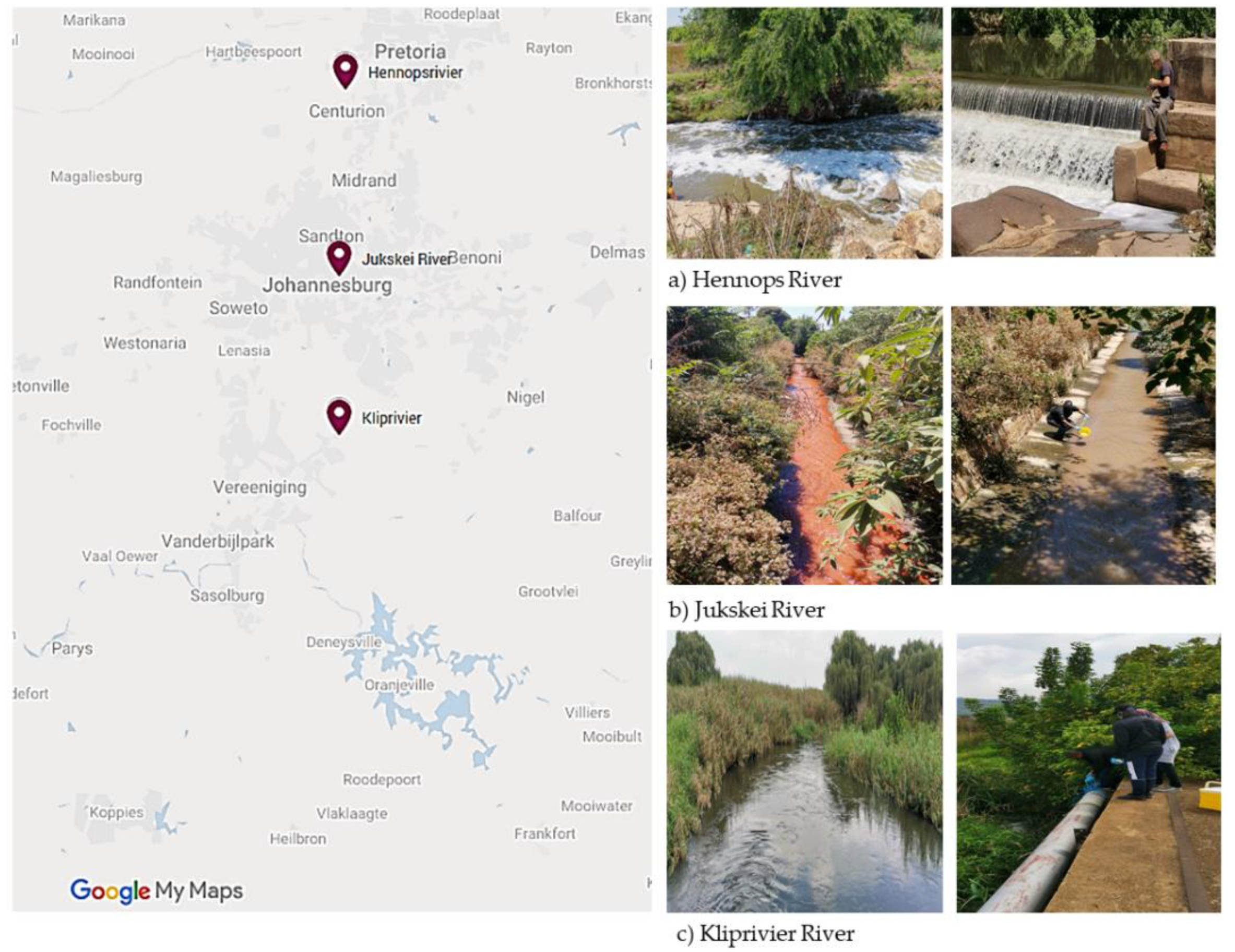
2.1. Ethical Consideration and Sample Collection

Ethical clearance to conduct this study was obtained from the Research Ethics Committees, Faculty of Health Sciences, University of Johannesburg (REC-168-2019), before sample collection. Environmental water samples (n = 101) were collected from nine water sources between August 2020 and February 2021. These included: (i) surface water from Jukskei River (n = 46) and Kliprivier River (Eikenhof) (n = 33), (ii) run-off water, upstream and downstream from the six Hennops river sites (n = 19), and (iii) sewage water from a stream and a well (n = 3), all in the Johannesburg, Gauteng Province, South Africa (as shown in Figure 1). Water samples were collected aseptically in sterile 1 litre glass bottles and properly labelled, and the temperature (°C) and potential of hydrogen ion (pH) were tested in situ by using a waterproof tester (Hanna, Sigma-Aldrich, Saint Louis, MI, USA). Samples were transported in an icebox to the laboratory of Water and Health Research Centre, the University of Johannesburg, for processing within 4 h.

2.2. Bacterial Isolation

Escherichia coli were recovered from the water samples by a standard membrane filtration procedure. Briefly, 100 mL of water samples were serially diluted and filtered through 0.45 μm S-PAK® membrane filters (Millipore, Sigma-Aldrich, Darmstadt, Germany). The membrane filters were aseptically placed directly onto HiChrome® Coliform chromogenic media (Sigma-Aldrich, St. Louis, MO, USA) and incubated (Scientific Series 2000, USA) at 37 °C for 18 to 24 h. Bluish colonies were selected as presumptive E. coli (as indicated on the manufacturer’s manual) and were sub-cultured onto Müller-Hinton agar plates (Oxoid, UK) and re-incubated at 37 °C for 24 h. Pure colonies were Gram-stained and identified using the VITEK-2® automated system (bioMérieux, Marcy-l’Étoile, France). Escherichia coli isolates were inoculated into Luria Bertani (LB) broth (HiMedia® Laboratories Pvt. Ltd., India), grown overnight (at 37 °C for 24 h) and stored long-term at −80 °C in a biofreezer (ThermoScientific, Waltham, MA, USA) in a 50% (v/v) sterile glycerol solution (Associated Chemical Enterprises (Pty) Ltd., Gauteng, South Africa) until further analysis.

2.3. DNA Extraction from Escherichia coli Isolates

Two mL of presumptive isolates were grown in LB broth (HiMedia® Laboratories Pvt. Ltd., Maharashtra, India) at 37 °C overnight and the total genomic DNA was extracted using the silica/guanidium thiocyanate (Sigma-Aldrich, Saint Louis, MI, USA) method adapted from the article previously published [32]. The extracted DNA was quantified using the Nanodrop instrument (Jenway Genova Nano, USA).

2.4. Multiplex PCR for the Detection of Virulence Profile of Escherichia coli

A single-step 11-gene multiplex PCR assay was performed on E. coli isolates for the detection of the virulence genes based on the methods and conditions previously described [28], using primer sequences targeting the E. coli genes presented in Table 1. All the primers used in this study were synthesised by WhiteHead Scientific (Pty) Ltd., South Africa.
The 20 μL Hotstart multiplex PCR reaction consisted of 10 μL of Qiagen master mix (Qiagen, Germany), 1 μL of the primer mixture (forward and reverse) (0.1 μM of lt primers, 0.2 μM of asta, bfp, eagg, ial, and gapdh primers, 0.3 μM of eaeA and stx2 primers, 0.5 μM of stx1 and sta primers)), 2 μL of MgCl2, 1 μL of Q-solution, 4.0 μL of PCR-grade water and 2 μL of sample DNA. The following conditions were used during PCR amplification performed in a Bio-Rad MyCyclerTM Thermal cycler (Bio-Rad, Hercules, CA, USA): an initial activation at 95 °C for 15 min, followed by 35 cycles consisting of denaturation at 94 °C for 45 s, annealing at 55 °C for 45 s, extension at 68 °C for 2 min and final extension at 72 °C for 5 min.

2.5. Singleplex PCR for the Confirmation of Escherichia coli

A singleplex PCR assay was performed for the confirmation of genes present in each positive E. coli isolate using the primer sequences presented in Table 1. The 20 μL PCR reaction consisted of 2 μL of 10× buffer (Qiagen, Germany), 2 μL of the primer mixture (forward and reverse), 2 μL of MgCl2, 4 μL of Q-solution, 0.4 μL of dNTPs, 0.1 μL of Taq polymerase, 7.5 μL of PCR-grade water and 2 μL of genomic DNA. The PCR conditions were the same as described in Section 2.4.

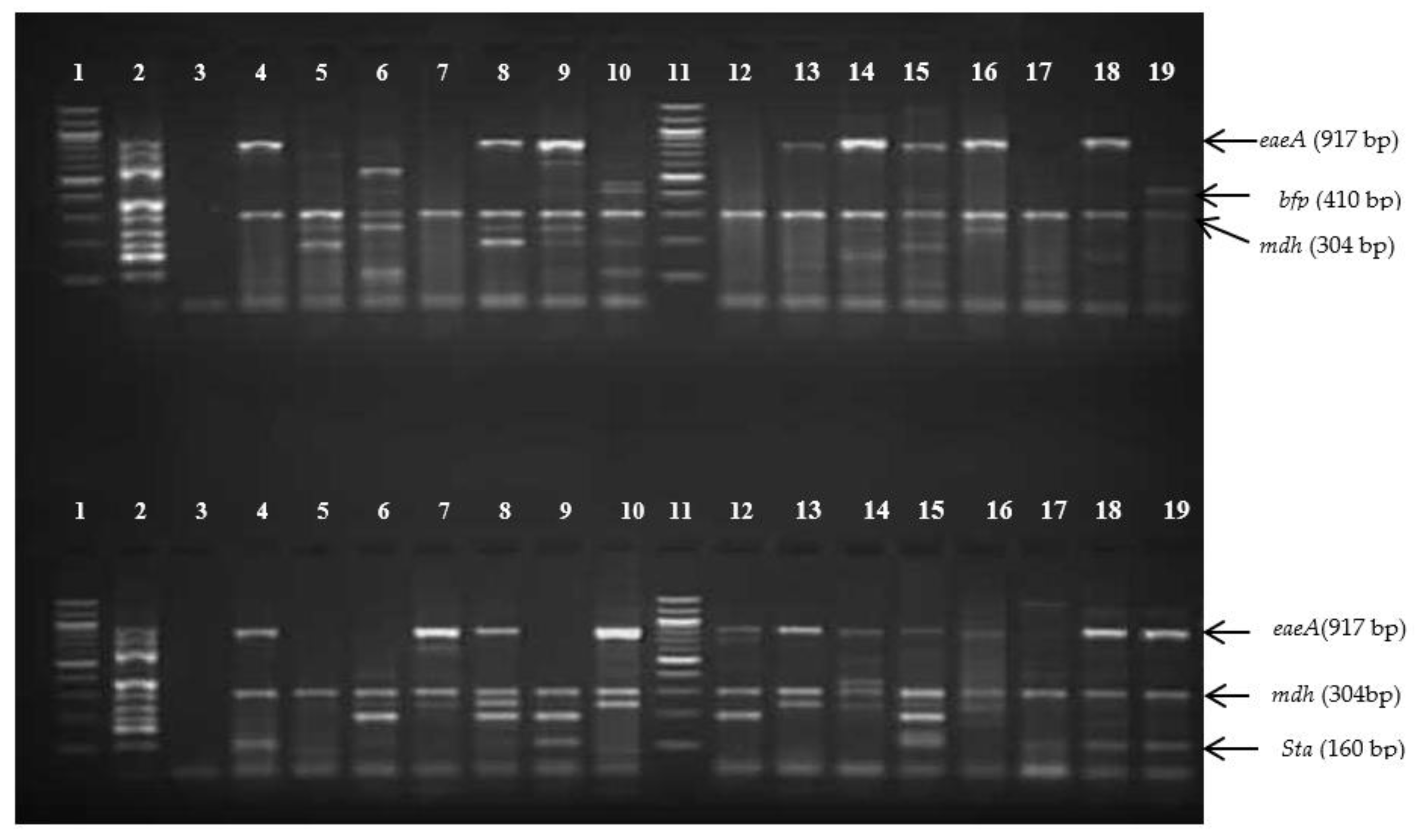
2.6. Visualisation of PCR Products

The PCR-amplified products were separated on a 2.5% (m/v) agarose gel (Bioline, Taunton, MA, USA), and stained with 5 µL of a 10 mg/mL stock solution of ethidium bromide (Merck, New York, NY, USA) using TAE buffer (40 mM Tris-acetate, 2 mM EDTA, pH 8.3). Gel electrophoresis was performed at 90 volts for 120 min. A 100 bp molecular weight marker (Fermentas, Thermo Scientific, Waltham, MA, USA) was included as a reference in all gels. The gel images were visualised and captured using a Gel DocTM EZ system (Bio-Rad, Hercules, CA, USA).

2.7. Control Strains and Data Analysis

The control strains used in this study included E. coli: (Commensal) ATCC 25922, enteroinvasive (EIEC) ESCCOS ATCC 43893, enteroaggregative (EAEC) ESCCO 14, enterohaemorrhagic (EHEC) ESCCO 21, enterotoxigenic (ETEC) ESCCO 22 and enteropathogenic (EPEC) S-ESCCO 16 Pl. All these strains were purchased from the National Health Laboratory Service (NHLS), South Africa, and were confirmed in a previous study [28]. The positive m-PCR was made up (in-house) by combining the extracted plasmids that contained the targeted virulence genes (eaeA, eagg, Asta, bfp, gapdh, ial, lt, mdh, sta, stx1, stx2). The malate dehydrogenase (mdh) housekeeping gene was used as an internal control and the glyceraldehyde 3-phosphate dehydrogenase (gapdh) gene was used as an external control. The gel images were analysed by reading the presence of bands detected. All data obtained were recorded and exported into a Microsoft Excel sheet for analysis.

2.8. Antibiotic Susceptibility Testing of Escherichia coli Isolates

All pathogenic Escherichia coli and some commensal isolates were selected and tested for antibiotic susceptibility using the VITEK®2 automated system (bioMérieux, Marcy-l’Étoile, France). Briefly, a bacterial suspension with an optical density (turbidity) of 0.5 McFarland was prepared in saline (0.85%) (bioMérieux, France) from an overnight bacterial culture incubated (Vacutec, Roodepoort, South Africa) for 18 h. The antibiotics included in the VITEK®2 automated system panel were: ampicillin, amikacin, amoxicillin/clavulanic acid, cefepime, cefotaxime, cefoxitin, ceftazidime, cefuroxime, cefuroxime-Axetil, ciprofloxacin, colistin, ertapenem, gentamicin, imipenem, meropenem, piperacillin/tazobactam, tigecycline, tobramycin and trimethoprim/sulfamethoxazole. The minimum inhibitory concentration (MIC) for each isolate tested was interpreted according to the Clinical and Laboratory Standards Institute (CLSI) 2021 guidelines [35].

3. Results

3.1. Isolation and Detection of E. coli Isolates from Environmental Water Samples

In total, 101 environmental water samples were collected and analysed in this study. The HiChrome® Coliform chromogenic media (Sigma-Aldrich, Saint Louis, MI, USA) identified 288 presumptive E. coli, which appeared bluish, as from the manufacturer. The PCR assay confirmed 170 E. coli isolates with the detection of the mdh housekeeping gene. The presence of the gapdh gene used as an external control excluded any possible PCR inhibition in all samples. The 11-gene multiplex PCR assays indicated that 40% (68/170) were commensals E. coli (ComEC) and 60% (102/170) were positive for at least one pathogenic E. coli type. Figure 2 shows the gel image of m-PCR with different genes detected in this study.

3.2. Detection of Pathogenic Types of E. coli Isolates by Multiplex PCR

3.2.1. Escherichia coli (Single Pathogenic Type) Detected in This Study

Enteropathogenic E. coli (EPEC) was the most detected in this study, with 19.2% (32/170) of isolates, of which atypical EPEC (aEPEC) harbouring only the eaeA gene accounted for 18% (30/170) while typical EPEC (tEPEC) harbouring both the eaeA and bfp genes accounted for 0.6% (1/170), and the bfp gene alone was present in 0.6% (1/170). Enterotoxigenic E. coli was detected in 11.4% (19/170) of isolates, with only a limited number of isolates harbouring both lt and sta genes (1.2%, 2/170), while sta alone was present in 9% (15/170) and lt alone in 1.2% (2/170). EAEC harbouring the eagg gene was present in 6% (10/170), and EHEC was detected in 3% (5/170), of which 1.2% (2/170) of isolates harboured stx1 and stx2 each, whereas both stx1 and stx2 were detected in 0.6% (1/170). EIEC harbouring the ial gene was not detected in this study. Table 2 shows the details of single E. coli pathotypes and their virulence genes detected.

3.2.2. Hybrid Pathogenic Types (Two Pathotypes) Detected in This Study

Hybrid pathogenic types forming two pathotypes harbouring a combination of virulent genes were detected in 28 isolates, including EAEC/EPEC (7.6%, 13/170), EAEC/ETEC (3%, 5/170), EPEC/ETEC (2.4%, 4/170), EPEC/EHEC (1.8%, 3/170) and EHEC/ETEC (1.8%, 3/170). The complete distribution of virulence genes forming a hybrid of two pathotypes is illustrated in Table 3.

3.2.3. Hybrid Pathogenic Types (Three Pathotypes) Detected in This Study

A hybrid of three different pathotypes simultaneously harbouring a combination of virulent genes was detected in nine isolates, including EAEC/aEPEC/ETEC (2.4%, 4/170), EHEC/ETEC/aEPEC (1.8%, 3/170) and EAEC/EHEC/ETEC (1.2%, 2/170). Table 4 shows the full distribution of virulence genes identified in E. coli isolates forming three pathotypes. Singleplex PCRs were run on all hybrid isolates as confirmatory tests and the results showed the presence of the single genes detected by different pathogenic types (Supplementary Materials).

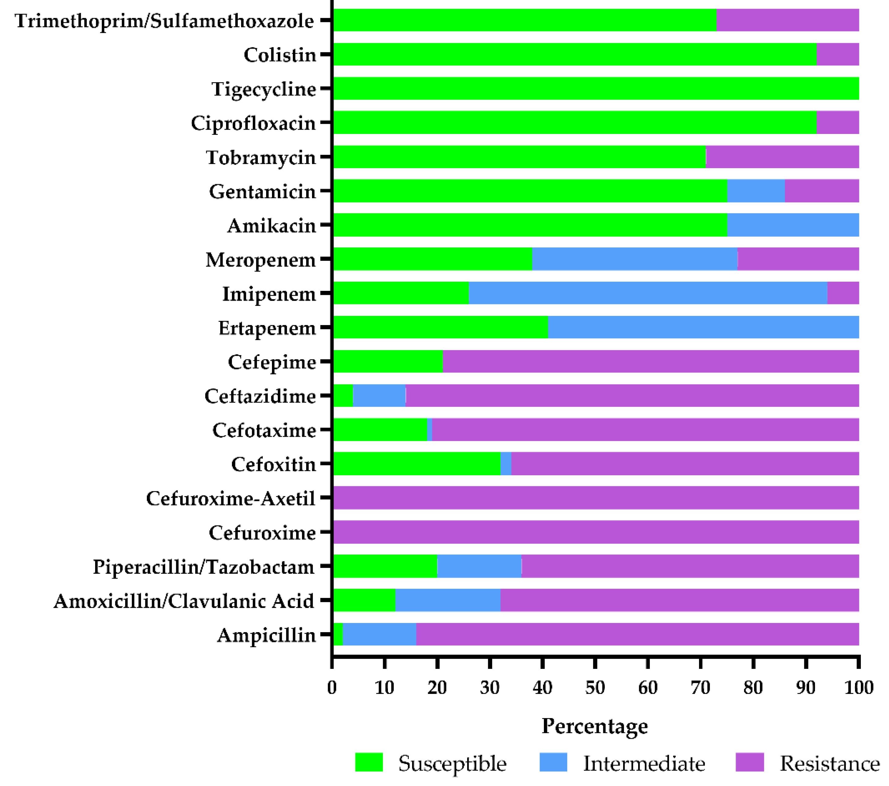
3.2.4. Antimicrobial Susceptibility Testing Results of Escherichia coli Isolates

Antibiotic resistance patterns of selected E. coli isolates (n = 100) using the VITEK®2 automated system (bioMérieux, France) showed isolates with multidrug resistance (Figure 2). All isolates were 100% (100/100) resistant to cefuroxime, 98% (98/100) to ampicillin and 96% (96/100) to ceftazidime. In total, 88%, 82%, 80%, 79% and 68% of isolates were resistant to amoxicillin/clavulanic acid, cefotaxime, piperacillin/tazobactam, cefepime and cefoxitin, respectively. Imipenem, meropenem and ertapenem resistance were 74%, 62% and 59%, respectively. Resistance to amikacin and gentamicin was observed in 25% (15/50) each. No tigecycline resistance was reported in this study. All intermediately resistant isolates were reported as resistant (Figure 3). At least 8% (8/100) of E. coli isolates showed an increased colistin MIC using the VITEK®2 automated system (bioMérieux, France).

4. Discussion

Diarrhoea outbreaks are a persistent problem with significant economic and potential public health impacts worldwide. In most developing countries, people continue to use environmental water (river water and stream) for domestic activities such as bathing, washing clothes, cooking and drinking [36]. It has also been reported that the spread of bacteria in the environment can be affected by the discharge of municipal sewage into surface water and soil [27]. Consequently, waterborne pathogens including diarrhoeagenic E. coli (DEC) can pass from the environment to humans, causing severe diseases. Studies have reported waterborne diarrhoeal disease claiming two million deaths worldwide each year, mostly in children below 5 years of age [37].
This study was carried out to determine the virulence profile of DEC and their antibiotic resistance profile in the environmental water of the Johannesburg region, South Africa. In total, 101 samples were included in this study, from which 288 presumptive E. coli were identified using HiChrome® Coliform selective chromogenic media (Sigma-Aldrich, USA) and the VITEK®-2 automated system (bioMérieux, France). Studies have reported these techniques to generate many errors and they can accommodate the growth of other species, leading to the misidentification of colonies, especially in environmental samples [38]. As such, molecular testing was performed on presumptive isolates as a confirmatory test. The single-step 11-gene multiplex PCR was used and identified DEC strains in environmental water. Overall, 170 E. coli were isolated, and the prevalence of commensal and DEC identified was 40% (68/170) and 60% (102/170), respectively. Among the DEC identified, EPEC was the most common single enteropathogen, with 19.2% (32/170) of cases. Studies have shown that infections due to EPEC are usually endemic in developing countries [37,39]. The proportion of EPEC detected in this study is consistent with other studies in South Africa [36,40] and elsewhere in the world [8,37,41]. In total, 18% (30/170) of isolates were atypical EPEC (aEPEC) harbouring the single gene eaeA, and 1.2% (2/170) were typical EPEC (tEPEC), of which 0.6% harboured both eaeA and bfp genes and 0.6% harboured the single bfp gene. The importance of distinguishing typical and atypical EPEC is that tEPEC causes infections mostly in infants, while aEPEC has been reported to cause infections in both children and adults [18]. The proportion of aEPEC in this study was lower compared to the 86% reported by Traoré et al. [42] in Vhembe district, Limpopo, South Africa. This difference might be due to the difference in geographic region and to the increase in human activity observed in environmental water of Vhembe district by villagers compared to the city of Johannesburg [42].
This study detected ETEC in 11.4% (19/170) of isolates. The genes that encode both heat-liable (LT) and heat-stable (ST) enterotoxins in ETEC are generally found on plasmids, transmissible and causing severe diarrhoea [43]. Our finding on ETEC was lower than the 47% and 83% reported by Nontongana et al. [44] and Traoré et al. [42] in the rural areas of the Eastern Cape and Limpopo provinces of South Africa, respectively. However, the proportion of ETEC detected in this study correlated with the previous findings in the surface water of northwest Mexico [45].
This study has identified 6% (10/170) of EAEC pathotypes in environmental water. Infection due to the EAEC pathotype is dangerous in immuno-compromised individuals and children [36] and has also been reported as one of the leading causes of DEC-associated food- and water-borne enteric infection [46]. The prevalence of EAEC identified in this study correlated with the study by Tanih et al. [47]. However, this proportion was lower when compared to the 87%, 34.4% and 58.3% reported by Traoré et al. [36], Canizalez-Roman et al. [45] and Mbanga et al. [48], respectively.
The current study detected the EHEC pathotype in 3% (5/170). Two isolates harboured each single stx1 and stx2 gene, while one isolate harboured both stx1 and stx2. The detection of the stx gene in river water is of concern because the colonisation of the human large intestine with EHEC stx even in low proportions can result in potentially fatal complications, such as haemolytic uremic syndrome (HUS) [20]. Previous studies conducted in South Africa have reported EHEC rates of 15.0% [40], 15.08% [49] and 8.3% [47].
A similar study conducted in the USA has reported 14% of EHEC in water samples [38]. In Georgia, Cho and co-workers reported a low rate (0.2%) of EHEC in watershed [50]. In this study, EIEC harbouring ial virulence genes was not detected in environmental water. This is not surprising because this pathotype is mostly reported as causing dysentery in humans and sometimes in animals [16,51].
Our study revealed hybrid pathotypes with virulence combinations among the isolates. This might be due to the mechanism of conjugation in virulence-associated genes which define a pathotype and are carried on mobile genetic elements (plasmids) [20]. Hybrids of Shiga toxin-producing and enterotoxigenic E. coli (EHEC/ETEC) have previously been reported associated with diarrhoeal disease in humans and animals [24,52]. In the present study, hybrid EHEC/ETEC was detected in 1.8% of isolates. This is serious and should be considered for epidemiological surveillance. Similarly, studies have reported 2.05% and 34% of hybrid EHEC/ETEC in Sweden [13] and in Bangladesh [43], respectively. This current study reported hybrids EAEC/aEPEC in 7.6% (13/170). A similar situation has been reported in Mexico [53]. The present study detected hybrids EPEC/ETEC and EPEC/EHEC in 2.4% and 1.8%, respectively. In India, Dutta and co-workers [21] identified hybrid EPEC/ETEC in a child with acute diarrhoea, while EPEC/EHEC strains were detected in Iran [54] and Mexico [14]. All these strains detected are virulent and might contribute to severe diarrhoea outbreaks and could pose a potential public health threat to consumers of untreated environmental water. Studies have reported molecular evidence of such hybrid pathotypes in humans, animals and environmental origins.
Interestingly, in this study, three different DEC strains were simultaneously detected in isolates. Hybrids of aEPEC + EHEC + ETEC harbouring eaeA + stx1 + stx2 + lt + sta were detected in two isolates. This indicates that hypervirulent strains are circulating in our area. Further confirmatory tests such as sequencing and comparative genomics would need to be performed on these strains to characterise and determine their phylogenetic position. Similarly, in Finland, three hybrid pathotypes were detected among E. coli strains harbouring up to six gene products [55]; while in Mexico, Patzi-Vargas et al. [53] detected three different DEC strains (EAEC + ETEC + DAEC) in one isolate.
In recent times and in modern medicine, the increasing microbial drug resistance has been identified as the biggest public health challenge and listed as one of the public health priorities among CDC fights [26,27,56]. According to the Global Antimicrobial Resistance Surveillance System (GLASS) report, E. coli is one of the pathogens that cause common hospital-acquired and community-acquired infections worldwide. Consequently, treatment is becoming increasingly difficult due to high rates of antimicrobial resistance [57]. In this study, the antibiotic resistance profile of E. coli isolates was determined and resistance to multiple antibiotics was identified in the environmental water using the VITEK®2 automated system (bioMérieux, France). Resistance to the most common antibiotics used for the treatment of infections due to DEC, including last-resort antibiotics, was observed among the isolates. The most abundant resistances were against cefuroxime (100%) and both ampicillin and ceftazidime (94%), followed by cefotaxime (88%), cefepime (84%), amoxicillin/clavulanic acid (82%) and imipenem (70%). These findings are higher as compared to previous study findings in the KwaZulu-Natal (Durban), Western Cape (Stellenbosch) and Eastern Cape provinces of South Africa [28,30,48]. This is alarming and shows that antibiotic resistance strains are circulating in South Africa. A similar situation was observed in Poland, where numerous multidrug-resistant strains were detected in water samples, which did not exhibit the ESBL phenotype [27]. The presence of antibiotic-resistant bacteria in source water is considered to be an emerging health concern in humans [52]. To prevent the spread of drug resistance in the region, several actions should be taken, including the monitoring of antimicrobial consumption, measuring antibiotic use and antimicrobial resistance genes’ surveillance in the environment [57,58].
One of the limitations of this study was the use of the VITEK®-2 automated system to assess drug susceptibility. This phenotypic method is mostly used in routine diagnosis laboratories to determine the bacterial susceptibility/resistance to antimicrobials (which sometimes generates errors). It has also been reported that some of the VITEK®-2 cards’ susceptible MIC breakpoints differ from one another as recommended for therapy of some infections per CLSI [59]. Furthermore, the VITEK®-2 automated system does not detect the presence of antibiotic-resistant genes as performed in scientific research laboratories. Further tests including sequencing would need to be performed on these isolates for the detection of extended-spectrum beta-lactamases, carbapenemases and plasmid-mediated colistin antibiotic resistance genes.

5. Conclusions

This present study highlighted the widespread occurrence of potentially DEC and antibiotic resistance genes in the aquatic ecosystem of Johannesburg, South Africa. Among the enteropathogens tested, EPEC was the most dominant, followed by ETEC, EAEC and EHEC. The presence of hybrid pathotypes detected in this study can pose a potential public health risk to consumers of untreated water in the region. The single-step 11-gene multiplex PCR system used in this study is potentially a quick, powerful and useful method for routine monitoring, virulence gene screening and risk assessment of water quality in developing countries. This study reported the presence of numerous multidrug-resistant strains among the isolates, of which resistance to the most common antibiotics used for the treatment of infections due to DEC was observed. There is a necessity to enhance surveillance in reducing the propagation of pathogenic and antibiotic-resistant E. coli bacteria, which are environmental and public health concerns.

Supplementary Materials

Author Contributions

Conceptualization, T.G.B. and A.S.; Formal analysis, J.Y.B., A.S., N.A. and T.G.B.; Funding acquisition, T.G.B. and A.S.; Methodology, J.Y.B. and N.A.; Project administration, T.G.B.; Resources, T.G.B.; Software, T.G.B. and A.S.; Supervision, T.G.B. and A.S.; Validation, T.G.B.; Writing—original draft, J.Y.B.; Writing—review and editing, J.Y.B., A.S., N.A. and T.G.B. All authors have read and agreed to the published version of the manuscript.

Funding

The authors thank the University of Johannesburg (UJ) Global Excellence and Stature (GES) for the post-doctoral fellowship and the UJ Research Committee (Water and Health Grant) for all financial support.

Institutional Review Board Statement

Not applicable.

Data Availability Statement

All data generated or analyzed during this study are included in this article and its Supplementary Materials files.

Acknowledgments

The authors acknowledge the technical support accorded to this study by the staff of the Water and Health Research Centre (WHRC), Faculty of Health Sciences, University of Johannesburg; more specifically, Kousar Hoorzook and Riahaanah Paulse for assisting with sample collection and antibiotic resistance testing, respectively.

Conflicts of Interest

The authors declare no conflict of interest. The funders had no role in the design of the study; in the collection, analyses, or interpretation of data; in the writing of the manuscript, or in the decision to publish the results.

References

  1. Khalil, I.A.; Troeger, C.; Blacker, B.F.; Rao, P.C.; Brown, A.; Atherly, D.E.; Brewer, T.G.; Engmann, C.M.; Houpt, E.R.; Kang, G.; et al. Morbidity and mortality due to shigella and enterotoxigenic Escherichia coli diarrhoea: The Global Burden of Disease Study 1990–2016. Lancet Infect. Dis. 2018, 18, 1229–1240. [Google Scholar] [CrossRef] [Green Version]
  2. The United Nations International Children’s Emergency Fund. One Is too Many: Ending Child Deaths from Pneumonia and Diarrhoea; UNICEF: New York, NY, USA, 2016; pp. 1–74. [Google Scholar]
  3. Bailey, E.S.; Beetsch, N.; Wait, D.A.; Oza, H.H.; Ronnie, N.; Sobsey, M.D. Methods, protocols, guidance and standards for performance evaluation for point-of-use water treatment technologies: History, current status, future needs and directions. Water 2021, 13, 1094. [Google Scholar] [CrossRef]
  4. Tenaillon, O.; Skurnik, D.; Picard, B.; Denamur, E. The population genetics of commensal Escherichia coli. Nat. Rev. Microbiol. 2010, 8, 207–217. [Google Scholar] [CrossRef]
  5. Wolny-Koładka, K.A.; Zdaniewicz, M. Antibiotic resistance of Escherichia coli isolated from processing of brewery waste with the addition of bulking agents. Sustainability 2021, 13, 10174. [Google Scholar] [CrossRef]
  6. Molina, F.; López-Acedo, E.; Tabla, R.; Roa, I.; Rebolo, J.E. Improved detection of Escherichia coli and coliform bacteria by multiplex PCR. BMC Biotechnol. 2015, 15, 1–9. [Google Scholar] [CrossRef] [PubMed] [Green Version]
  7. Mokdad, A. Estimates of global, regional and national morbidity, mortality and aetiologies of diarrhoeal diseases: A systematic analysis for the global burden of disease Study 2015. Lancet Infect. Dis. 2017, 17, 909–948. [Google Scholar]
  8. Park, J.; Kim, J.S.; Kim, S.; Shin, E.; Oh, K.-Y.; Kim, Y.; Kim, C.H.; Hwang, M.A.; Jin, C.M.; Na, K.; et al. A waterborne outbreak of multiple diarrhoeagenic Escherichia coli infections associated with drinking water at a school camp. Int. J. Infect. Dis. 2018, 66, 45–50. [Google Scholar] [CrossRef] [PubMed] [Green Version]
  9. Toma, C.; Lu, Y.; Higa, N.; Nakasone, N.; Chinen, I.; Baschkier, A.; Rivas, M.; Iwanaga, M. Multiplex PCR assay for identification of human diarrhoeagenic Escherichia coli. J. Clin. Microbiol. 2003, 41, 2669–2671. [Google Scholar] [CrossRef] [Green Version]
  10. Croxen, M.A.; Law, R.J.; Scholz, R.; Keeney, K.M.; Wlodarska, M.; Finlay, B.B. Recent advances in understanding enteric pathogenic Escherichia coli. Clin. Microbiol. Rev. 2013, 26, 822–880. [Google Scholar]
  11. Chique, C.; Hynds, P.; Burke, L.P.; Morris, D.; Ryan, M.P.; O’Dwyer, J. Contamination of domestic groundwater systems by verotoxigenic Escherichia coli (VTEC), 2003–2019: A global scoping review. Water Res. 2021, 188, 116496. [Google Scholar] [CrossRef]
  12. Cabal, A.; García-Castillo, M.; Cantón, R.; Gortázar, C.; Domínguez, L.; Álvarez, J. Prevalence of Escherichia coli virulence genes in patients with diarrhoea and a subpopulation of healthy volunteers in Madrid, Spain. Front. Microbiol. 2016, 7, 1–6. [Google Scholar] [CrossRef] [Green Version]
  13. Bai, X.; Zhang, J.; Ambikan, A.; Jernberg, C.; Ehricht, R.; Scheutz, F.; Xiong, Y.; Matussek, A. Molecular characterisation and comparative genomics of clinical hybrid Shiga toxin-producing and enterotoxigenic Escherichia coli (STEC/ETEC) strains in Sweden. Nat. Sci. Rep. 2019, 9, 5619. [Google Scholar] [CrossRef] [Green Version]
  14. Gioia-Di Chiacchio, R.M.; Cunha, M.; de Sá, L.; Davies, Y.M.; Pereira, C.; Martins, F.H.; Munhoz, D.D.; Abe, C.M.; Franzolin, M.R.; Dos Santos, L.F.; et al. Novel Hybrid of Typical Enteropathogenic Escherichia coli and Shiga-Toxin-Producing E. coli (tEPEC/STEC) Emerging from Pet Birds. Front. Microbiol. 2018, 9, 2975. [Google Scholar] [CrossRef]
  15. García, A.; Fox, J.G.; Besser, T.E. Zoonotic enterohemorrhagic Escherichia coli: A One Health perspective. ILAR J. 2010, 51, 221–232. [Google Scholar] [CrossRef]
  16. Lan, R.; Alles, M.C.; Donohoe, K.; Martinez, M.B.; Reeves, P.R. Molecular evolutionary relationships of enteroinvasive Escherichia coli and Shigellaspp. Infect. Immun. 2004, 72, 5080–5088. [Google Scholar] [CrossRef] [Green Version]
  17. Newitt, S.; MacGregor, V.; Robbins, V.; Bayliss, L.; Chattaway, M.A.; Dallman, T.; Ready, D.; Aird, H.; Puleston, R.; Hawker, J. Two Linked Enteroinvasive Escherichia coli Outbreaks, Nottingham, UK, June 2014. Emerg. Infect. Dis. 2016, 22, 1178–1184. [Google Scholar] [CrossRef] [Green Version]
  18. Nakhjavani, F.A.; Emaneini, M.; Hosseini, H.; Iman-Eini, H.; Aligholi, M.; Jabalameli, F.; Haghi-Ashtiani, M.T.; Taherikalani, M.; Mirsalehian, A. Molecular analysis of typical and atypical enteropathogenic Escherichia coli (EPEC) isolated from children with diarrhoea. J. Med. Microbiol. 2013, 62, 191–195. [Google Scholar] [CrossRef] [Green Version]
  19. Hazen, T.H.; Michalski, J.; Luo, Q.; Shetty, A.C.; Daugherty, S.C.; Fleckenstein, J.M.; Rasko, D.A. Comparative genomics and transcriptomics of Escherichia coli isolates carrying virulence factors of both enteropathogenic and enterotoxigenic Escherichia coli. Sci. Rep. 2017, 7, 3513. [Google Scholar] [CrossRef] [PubMed]
  20. Leonard, S.R.; Mammel, M.K.; Rasko, D.A.; Lacher, D.W. Hybrid Shiga Toxin-Producing and Enterotoxigenic Escherichia sp. Cryptic Lineage 1 Strain 7v Harbors a Hybrid Plasmid. Appl. Environ. Microbiol. 2016, 82, 4309–4319. [Google Scholar] [CrossRef] [PubMed] [Green Version]
  21. Dutta, S.; Pazhani, G.P.; Nataro, J.P.; Ramamurthy, T. Heterogenic virulence in a diarrheagenic Escherichia coli: Evidence for an EPEC expressing heat-labile toxin of ETEC. Int. J. Med. Microbiol. 2015, 305, 47–54. [Google Scholar] [CrossRef] [PubMed]
  22. Santos, A.; Santos, F.F.; Silva, R.M.; Gomes, T. Diversity of Hybrid- and Hetero-Pathogenic Escherichia coli and their potential implication in more severe diseases. Front. Cell. Infect. Microbiol. 2020, 10, 339. [Google Scholar] [CrossRef]
  23. Aref, N.M.; Abdel-Raheem, A.A.; Kamaly, H.F.; Hussien, S.Z. Clinical and sero-molecular characterization of Escherichia coli with an emphasis on hybrid strain in healthy and diarrheic neonatal calves in Egypt. Open Vet. J. 2018, 8, 351–359. [Google Scholar] [CrossRef] [Green Version]
  24. Yang, X.; Bai, X.; Zhang, J.; Sun, H.; Fu, S.; Fan, R.; He, X.; Scheutz, F.; Matussek, A.; Xiong, Y. Escherichia coli strains producing a novel Shiga toxin 2 subtype circulate in China. Int. J. Med. Microbiol. 2020, 310, 151377. [Google Scholar] [CrossRef]
  25. Bielaszewska, M.; Mellmann, A.; Zhang, W.; Köck, R.; Fruth, A.; Bauwens, A.; Peters, G.; Karch, H. Characterisation of the Escherichia coli strain associated with an outbreak of haemolytic uraemic syndrome in Germany: A microbiological study. Lancet Infect. Dis. 2011, 11, 671–676. [Google Scholar] [CrossRef] [Green Version]
  26. The Centers for Disease Control and Prevention. The Antibiotic Resistance Threats in the United States. 2019. Available online: www.cdc.gov/DrugResistance/Biggest-Threats.html (accessed on 22 June 2021).
  27. Wolny-Koładka, K.A.; Lenart-Boroń, A. Phenotypic and molecular assessment of drug resistance profile and genetic diversity of waterborne Escherichia coli. Water Air Soil Pollut. 2016, 227, 1–11. [Google Scholar] [CrossRef] [Green Version]
  28. Lamprecht, C.; Romanis, M.; Huisamen, N.; Carinus, A.; Schoeman, N.; Sigge, G.O.; Britz, T.J. Escherichia coli with virulence factors and multidrug resistance in the Plankenburg River. S. Afr. J. Sci. 2014, 110, 1–6. [Google Scholar] [CrossRef] [Green Version]
  29. Morel, C. Transmission of antimicrobial resistance from livestock agriculture to humans and from humans to animals. In Food, Agriculture and Fisheries Papers, No. 133; OECD Publishing: Paris, France, 2019. [Google Scholar] [CrossRef]
  30. Msolo, L.; Iweriebor, B.C.; Okoh, A.I. Antimicrobial resistance profiles of diarrhoeagenic E. coli (DEC) and Salmonella species recovered from diarrhoeal patients in selected rural communities of the Amathole District Municipality, Eastern Cape province, South Africa. Infect. Drug Resist. 2020, 13, 4615–4626. [Google Scholar] [CrossRef] [PubMed]
  31. Wolny-Koładka, K.A.; Lenart-Boroń, A. Antimicrobial resistance and the presence of extended-spectrum beta-lactamase genes in Escherichia coli isolated from the environment of horse riding centers. Environ. Sci. Pollut. Res. 2018, 25, 21789–21800. [Google Scholar] [CrossRef] [PubMed]
  32. Omar, K.B.; Barnard, T.G. Detection of diarrhoeagenic Escherichia coli in clinical and environmental water sources in South Africa using single-step 11-gene m-PCR. World J. Microbiol. Biotechnol. 2014, 30, 2663–2671. [Google Scholar] [CrossRef] [PubMed] [Green Version]
  33. Mbene, A.B.; Houreld, N.N.; Abrahamse, H. DNA damage after phototherapy in wounded fibroblast cells irradiated with 16 J/cm2. J. Photochem. Photobiol. 2009, 94, 131–137. [Google Scholar] [CrossRef] [PubMed]
  34. Pass, M.A.; Odedra, R.; Batt, R.M. Multiplex PCRs for identification of Escherichia coli virulence genes. J. Clin. Microbiol. 2000, 38, 2001–2004. [Google Scholar] [CrossRef] [Green Version]
  35. Clinical Laboratory Standards Institute. Performance Standards for Antimicrobial Susceptibility Testing. M100-S22, M2–7 and M–7; Clinical Laboratory Standards Institute: Wayne, PA, USA, 2021. [Google Scholar]
  36. Potgieter, N.; Karambwe, S.; Mudau, L.S.; Barnard, T.G.; Traore, A. Human enteric pathogens in eight rivers used as rural household drinking water sources in the northern region of South Africa. Int. J. Environ. Res. Public Health 2020, 17, 2079. [Google Scholar] [CrossRef] [Green Version]
  37. Cooley, M.B.; Jay-Russell, M.; Atwill, E.R.; Carychao, D.; Nguyen, K.; Quinones, B.; Patel, R.; Walker, S.; Swimley, M.; Pierre-Jerome, E.; et al. Development of a robust method for isolation of Shiga toxin-positive Escherichia coli from faecal, plant, soil and water samples from a leafy greens production region in California. PLoS ONE 2013, 8, e65716. [Google Scholar] [CrossRef] [Green Version]
  38. Lim, M.A.; Kim, J.Y.; Acharya, D.; Bajgain, B.B.; Park, J.H.; Yoo, S.J.; Lee, K. A diarrhoeagenic enteropathogenic Escherichia coli (EPEC) infection outbreak that occurred among elementary school children in Gyeongsangbuk-Do province of South Korea was associated with consumption of water-contaminated food items. Int. J. Environ. Res. Public Health 2020, 17, 3149. [Google Scholar] [CrossRef]
  39. Pienaar, J.A.; Singh, A.; Barnard, T.G. Acid-happy: Survival and recovery of enteropathogenic Escherichia coli (EPEC) in simulated gastric fluid. Microb. Pathog. 2019, 128, 396–404. [Google Scholar] [CrossRef]
  40. Ndlovu, T.; Le Roux, M.; Khan, W.; Khan, S. Co-detection of virulent Escherichia coli genes in surface water sources. PLoS ONE 2015, 10, e0116808. [Google Scholar] [CrossRef]
  41. Sidhu, J.P.S.; Ahmed, W.; Hodgers, L.; Toze, S. Occurrence of virulence genes associated with diarrhoeagenic pathotypes in Escherichia coli isolates from surface water. Appl. Environ. Microbiol. 2013, 79, 328–335. [Google Scholar] [CrossRef] [Green Version]
  42. Traoré, A.N.; Mulaudzi, K.; Chari, G.J.E.; Foord, S.H.; Mudau, L.S.; Barnard, T.G.; Potgieter, N. The impact of human activities on microbial quality of rivers in the Vhembe District, South Africa; Int. J. Environ. Res. Public Health 2016, 13, 817. [Google Scholar] [CrossRef] [PubMed] [Green Version]
  43. Johura, F.T.; Parveen, R.; Islam, A.; Sadique, A.; Rahim, M.N.; Monira, S.; Khan, A.R.; Ahsan, S.; Ohnishi, M.; Watanabe, H.; et al. Occurrence of hybrid Escherichia coli strains carrying Shiga toxin and heat-stable toxin in livestock of Bangladesh. Front. Public Health 2017, 4, 287. [Google Scholar] [CrossRef] [PubMed] [Green Version]
  44. Nontongana, N.; Sibanda, T.; Ngwenya, E.; Okoh, A.I. Prevalence and antibiogram profiling of Escherichia coli pathotypes isolated from the Kat River and the Fort Beaufort Abstraction Water. Int. J. Environ. Res. Public Health 2014, 11, 8213–8227. [Google Scholar] [CrossRef] [PubMed] [Green Version]
  45. Canizalez-Roman, A.; Velazquez-Roman, J.; Valdez-Flores, M.A.; Flores-Villaseñor, H.; Vidal, J.E.; Muro-Amador, S.; Guadrón-Llanos, A.M.; Gonzalez-Nuñez, E.; Medina-Serrano, J.; Tapia-Pastrana, G.; et al. Detection of antimicrobial-resistance diarrheagenic Escherichia coli strains in surface water used to irrigate food products in the northwest of Mexico. Int. J. Food Microbiol. 2019, 304, 1–10. [Google Scholar] [CrossRef]
  46. Aijuka, M.; Santiago, A.E.; Giron, J.A.; Nataro, J.P.; Buys, E.M. Enteroaggregative Escherichia coli is the predominant diarrhoeagenic E. coli pathotype among irrigation water and food sources in South Africa. Int. J. Food Microbiol. 2018, 278, 44–51. [Google Scholar] [CrossRef] [Green Version]
  47. Tanih, N.F.; Bolick, D.T.; Samie, A.; Nyathi, E.; Dillingham, R.; Pinkerton, R.C.; Guerrant, R.L.; Bessong, P.O. Prevalence of virulence genes in Enteroaggregative E. coli isolates from young children from rural South Africa. Am. J. Trop. Med. Hyg. 2019, 101, 1027–1033. [Google Scholar] [CrossRef]
  48. Mbanga, J.; Amoako, D.G.; Abia, A.L.K.; Allam, M.; Ismail, A.; Essack, S.Y. Genomic insights of multidrug-resistant Escherichia coli from wastewater sources and their association with clinical pathogens in South Africa. Front. Vet. Sci. 2021, 8, 636715. [Google Scholar] [CrossRef]
  49. Ramlal, P.S.; Kistnasamy, E.J. An ecological study of diarrhoeagenic Escherichia coli associated with indiscriminate waste dumps and under five diarrhoea in six informal settlements in Durban, South Africa. Int. J. Environ. Waste Manag. 2017, 20, 300–323. [Google Scholar] [CrossRef]
  50. Cho, S.; Hiott, L.M.; Barrett, J.B.; McMillan, E.A.; House, S.L.; Humayoun, S.B.; Adams, E.S.; Jackson, C.R.; Frye, J.G. Prevalence and characterization of Escherichia coli isolated from the upper Oconee watershed in Northeast Georgia. PLoS ONE 2018, 13, e0197005. [Google Scholar] [CrossRef] [Green Version]
  51. Bonkoungou, I.J.O.; Somda, N.S.; Traoré, O.; Zoma, B.S.; Garba, Z.; Drabo, K.M.; Barro, N. Detection of diarrhoeagenic Escherichia coli in human diarrhoeic stool and drinking water samples in Ouagadougou, Burkina Faso. Afr. J. Infect. Dis. 2021, 15, 53–58. [Google Scholar] [PubMed]
  52. Nyholm, O.; Heinikainen, S.; Pelkonen, S.; Hallanvuo, S.; Haukka, K.; Siitonen, A. Hybrids of Shigatoxigenic and Enterotoxigenic Escherichia coli (STEC/ETEC) among human and animal isolates in Finland. Zoonoses Public Health 2015, 62, 518–524. [Google Scholar] [CrossRef] [PubMed]
  53. Patzi-Vargas, S.; Zaidi, M.B.; Perez-Martinez, I.; León-Cen, M.; Michel-Ayala, A.; Chaussabel, D.; Estrada-Garcia, T. Diarrheagenic Escherichia coli carrying supplementary virulence genes are an important cause of moderate to severe diarrhoeal disease in Mexico. PLoS Negl. Trop. Dis. 2015, 9, e0003510. [Google Scholar] [CrossRef] [PubMed]
  54. Badouei, M.A.; Morabito, S.; Najafifar, A.; Mazandarani, E. Molecular characterisation of enterohemorrhagic Escherichia coli haemolysin gene (EHEC-hlyA)-harboring isolates from cattle reveals a diverse origin and hybrid diarrheagenic strains. Infect. Genet. Evol. 2016, 39, 342–348. [Google Scholar] [CrossRef] [PubMed]
  55. Antikainen, J.; Tarkka, E.; Haukka, K.; Siitonen, A.; Vaara, M.; Kirveskari, J. The new 16-plex PCR method for rapid detection of diarrhoeagenic Escherichia coli directly from stool samples. Eur. J. Clin. Microbiol. Infect. Dis. 2009, 28, 899–908. [Google Scholar] [CrossRef] [PubMed]
  56. The World Health Organization. Antibiotic Resistance. 2020. Available online: www.who.int/news-room/fact-sheets/detail/antimicrobial-resistance (accessed on 26 June 2021).
  57. The World Health Organization. Global Antimicrobial Resistance Surveillance System (GLASS) Report. Early Implementation. 2021. Available online: www.who.int/publications/i/item/9789240027336 (accessed on 14 September 2021).
  58. Bezuidenhout, C.C.; O’Reilly, G.; Sigudu, M.V.; Ncube, E.J. A Scoping Study on the Levels of Antimicrobials and Presence of Antibiotic Resistant Bacteria in Drinking Water; WRC Report No. KV 360/16; Water Research Commission: Lynnwood, South Africa, 2016; ISBN 978-1-4312-0823-4. [Google Scholar]
  59. Hogan, C.A.; Watz, N.; Budvytiene, I.; Banaei, N. Rapid antimicrobial susceptibility testing by VITEK®2 directly from blood cultures in patients with gram-negative rod bacteremia. Diagnost. Microbiol. Infect. Dis. 2019, 94, 116–121. [Google Scholar] [CrossRef] [PubMed]
Figure 1. Map of the water sampling sites for study areas within the Johannesburg region. Shown are the locations’ approximate sampling points of the (a) Hennops, (b) Jukskei and (c) Kliprivier Rivers investigated in this study.
Microorganisms 09 02163 g001
Figure 2. Gel image of m-PCR products of E. coli isolates: Top and Bottom wells: Lanes 1 and 11: 100 bp molecular marker (ladder), Lane 2: positive control, Lane 3: negative control, Lanes 4–10 and Lanes 12–19: E. coli isolates showing different genes.
Microorganisms 09 02163 g002
Figure 3. Antibiotic resistance patterns of E. coli isolates (n = 100) using the VITEK®2 automated system (bioMérieux, Marcy-l’Étoile, France) in Johannesburg, South Africa.
Microorganisms 09 02163 g003
Table 1. Primers used for multiplex PCR in this study.
PathogenGene PrimersPrimer Sequence (5′– 3′)Size (bp) Reference
Internal controlmdh (F)GGT ATG GAT CGT TCC GAC CT304[32]
mdh (R)GGC AGA ATG GTA ACA CCA GAG T
External controlgapdh (F)GAG TCA ACG GAT TTG GTC GT238[33]
gapdh (R)TTG ATT TTG GAG GGA TCT CG
EIECial (F)GGT ATG ATG ATG ATG AGT CCA650[34]
ial (R)GGA GGC CAA CAA TTA TTT CC
EHECstx1 (F)ACA CTG GAT GAT CTC AGT GG614[32]
stx1 (R)CTG AAT CCC CCT CCA TTA TG
stx2 (F)CCA TGA CAA CGG ACA GCA GTT779
stx2 (R)CCT GTC AAC TGA GCA CTT TG
EHEC/aEPECeaeA (F)CTG AAC GGC GAT TAC GCG AA917[34]
eaeA (R)CCA GAC GAT ACG ATC CAG
tEPECbfpA (F)AAT GGT GCT TGC GCT TGC TGC410
bfpM (R)TAT TAA CAC CGT AGC CTT TCG CTG AAG TAC CT
EAECeagg (F)AGA CTC TGG CGA AAG ACT GTA TC194[34]
eagg (R)ATG GCT GTC TGT AAT AGA TGA GAA C
ETEClt-1 (F)TGG ATT CAT CAT GCA CCA CAA GG360[34]
lt-1 (R)CCA TTT CTC TTT TGC CTG CCA TC
sta (F)TTT CCC CTC TTT TAG TCA GTC AAC TG160
sta(R)GGC AGG ATT ACA ACA AAG TTC ACA
E. coli toxinastA (F)GCC ATC AAC ACA GTA TAT CC106[32]
astA (R)GAG TGA CGG CTT TGT AGT C
Table 2. Escherichia coli (single pathotype) and their virulence genes detected in this study.
Pathogenic TypesGenes Detectedn (170)%
ComECmdh6840
EPEC 3219.2
aEPECeaeA(30)(18.0)
tEPECbfp(1)(0.6)
bfp + eaeA(1)(0.6)
ETEC 1911.4
lt(2)(1.2)
sta(15)(9.0)
lt + sta(2)(1.2)
EHEC 53.0
stx1(2)(1.2)
stx2(2)(1.2)
stx1 + stx2(1)(0.6)
EAECeagg106.0
EIECial00.0
n: number of isolates; ComEC: commensal E. coli; aEPEC: atypical EPEC; tEPEC: typical EPEC.
Table 3. Escherichia coli hybrid pathogenic types (two pathotypes) and their virulence genes detected in this study.
Hybrid PathotypesGene Combinations Detected n (170)%
EAEC/aEPECeaeA + eagg137.6
EAEC/ETEC 53.0
eagg + sta(3)(1.8)
eagg + lt + sta(2)(1.2)
EPEC/ETEC 42.4
eaeA + lt(1)(0.6)
eaeA + lt + sta(3)(1.8)
EHEC/ETEC 31.8
Stx1 + sta(1)(0.6)
Stx2 + sta(1)(0.6)
Stx1 + lt + sta(1)(0.6)
EPEC/EHEC 31.8
aEPEC/EHECeaeA + stx2(2)(1.2)
tEPEC/EHECeaeA + bfp + stx1 + stx2(1)(0.6)
n: number of isolates.
Table 4. Hybrid pathogenic type (three pathotypes) of E. coli and their virulence genes detected in this study.
Hybrid PathotypesGene Combinations Detected n
(170)
%
EAEC/aEPEC/ETECeagg + eaeA + sta42.4
EAEC/EHEC/ETECeagg + stx1 + sta21.2
EHEC/ETEC/aEPEC 31.8
eaeA + stx1 + sta(1)(0.6)
eaeA + stx1 + stx2 +lt + sta(2)(1.2)
Publisher’s Note: MDPI stays neutral with regard to jurisdictional claims in published maps and institutional affiliations.

