Article

The Presence of Treponema spp. in Equine Hoof Canker Biopsies and Skin Samples from Bovine Digital Dermatitis Lesions

1
Clinic of Ruminants, University Veterinary Hospital, University of Veterinary Medicine and Pharmacy in Košice, Komenského 73, 041 81 Košice, Slovakia
2
Department of Microbiology and Immunology, University of Veterinary Medicine and Pharmacy in Košice, Komenského 73, 041 81 Košice, Slovakia
3
Clinic of Horses, University Veterinary Hospital, University of Veterinary Medicine and Pharmacy in Košice, Komenského 73, 041 81 Košice, Slovakia
*
Author to whom correspondence should be addressed.
Submission received: 1 September 2021 / Revised: 5 October 2021 / Accepted: 18 October 2021 / Published: 20 October 2021
(This article belongs to the Section Medical Microbiology)

Abstract

:
Equine hoof canker and bovine digital dermatitis are infectious inflammatory diseases of the hooves with an unknown etiology. However, anaerobic spirochetes of the genus Treponema are considered to be potential etiological agents. The aim of this study was to find a suitable way to isolate DNA and to detect the presence of treponemal DNA in samples of equine hoof canker and bovine digital dermatitis. DNAzol®® Direct and column kits were used to isolate DNA from samples of equine hoof canker and bovine digital dermatitis. The presence of Treponema spp. was detected using PCR and Sanger sequencing. DNAzol®® Direct is suitable for isolating DNA from these types of samples. Treponemal DNA was detected in equine hoof samples as well as in bovine digital dermatitis skin samples. In equine hoof biopsies, the most frequently detected was Treponema pedis (8/13). Treponema brennaborense (2/13) and Treponema denticola (2/13) were also found. In the case of bovine digital dermatitis, Treponema medium ssp. bovis was confirmed in 14 of 36 skin samples. Treponema pedis (9/36), Treponema vincentii (1/36), Treponema phagedenis (1/36), and Treponema brennaborense (1/36) were detected as well. DNAzol®® Direct was more appropriate for isolation of treponemal DNA because the columns isolation method was more equipment and time-consuming. The presence of several Treponema spp. was determined in the samples. In horses, the most commonly detected species was a T. pedis, while in cattle it was T. medium ssp. bovis.

1. Introduction

Bovine digital dermatitis (BDD) manifests as painful, ulcerative or proliferative lesions. Typical lesions have been classified based on clinical appearance into five stages [1] and are located mainly on the plantar aspect of the hind foot. This inflammatory disease of interdigital skin is one of the most common causes of lameness [2,3]. Pain and lameness disrupt animal welfare and have a negative impact on the economy of breeding such as a reduction in milk production [4,5].
The pathognomonic condition of equine hoof canker (pododermatitis chronic verrucosa) has a very similar clinical course, clinical signs and pathological appearance to BDD [6]. It endangers the use and welfare of horses due to the instability of the affected hoof capsule and the subsequent lameness [7]. This disease is described as an infectious process that is characterized by chronic, hypertrophic, moist subdermatitis which affects the horn-producing tissues. Hoof canker generally originates from the frog and associated sulcus region. It may remain in focus, but if left untreated, it can diffuse and invade the bars, adjacent sole, and even the hoof wall [8,9]. Hoof canker can occur in one or more hooves, but more often on the hindlimbs, although this is not common in horses. This disease can affect all breeds but working draft horses are affected more often [10,11].
Treponema spp., as a potential etiological agent, may play a role in the development and pathogenesis of both diseases [12,13]. This anaerobic or microaerophilic spirochetal bacterium can infect a wide range of hosts and tissues and cause a range of diseases from periodontal diseases in companion animals and humans [14,15] to digital dermatitis of cattle [16]. The shape and composition of the outer membrane, which has transmembrane proteins on its surface, probably contributes to the ability of bacteria to escape from the host’s immune system [17].
Several studies detected the presence of treponemal DNA in tissues from affected hooves, suggesting that treponemes may play an important role in the etiology and/or pathogenesis of diseases. [18,19,20]. The most common Treponema phylogroups associated with polytreponemal etiology of BDD are Treponema medium/vincentii-like, Treponema phagedenis, Treponema denticola-like/putidum and Treponema pedis [21,22,23], whereas Treponema brennaborense is rarely found in lesions [24]. The fragile and fastidious nature of genus Treponema makes their isolation and cultivation very difficult. Therefore, molecular approaches such as PCR are more commonly used to detect difficult-to-culture bacteria [25].
The aim of the present study was to find a suitable method for the isolation of treponemal DNA from equine hoof canker biopsies and BDD lesions, further standardization of PCR reactions to detect Treponema spp. and determine their presence in the samples.

2. Materials and Methods

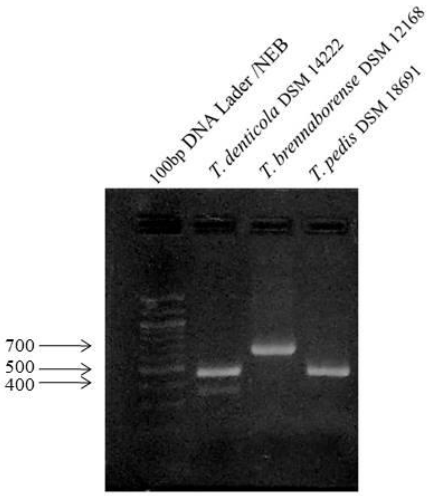
2.1. Standardization of PCRs

PCR to detect T. pedis, T. brennaborense and group of Treponema, which include T. denticola, T. vincentii, T. medium ssp. bovis, and T. phagedenis ssp. vaccae were standardized. DNA samples, namely T. denticola DSM 14222, T. brennaborense DSM 12168, and T. pedis DSM 18691, obtained from the Deutsche Sammlung von Mikroorganismen und Zelikultren GmbH (DSMZ, Braunschweig, Germany) were used as positive controls. The primers designed in the study of Brandt et al. [20] and synthesized by Merck were used. The primers with modified PRC protocols are shown in Table 1. The products of the PCR reactions were purified and sequenced on both sides by the Sanger method (Microsynth, Vienna, Austria).

2.2. Collection of Equine Hoof Biopsies

Horses from two stud farms located in Slovakia and the Czech Republic were included in this study (Table 2). From five Czech warmblood mares, one Muran type norik mare and one Austrian norik stallion were collected hoof biopsies during debridement for histopathological examination, determination of the DNA presence of Treponema spp. and biopsy swabs for mycological examination. Hoof samples and swabs from one healthy Muran type norik stallion, one Austrian norik stallion and one Czech warmblood mare were taken as controls during regular trimming. Histopathological examination of hoof biopsies was provided by IDEXX laboratories (Leipzig, Germany) and mycological examination by the State Veterinary and Food Institute (Košice, Slovakia). Swabs for mycological examination were taken from the hoof biopsies and placed into the Amies agar gel without carbon (Copan, Mantua, Italy). To examine the presence of bovine papillomavirus 1 and 2 (BPV 1, 2), hoof tissue and hair with roots were taken from each horse from both the affected and healthy areas. The samples were sent to Laboklin s.r.o. (Bratislava, Slovakia).

2.3. Collection of Skin Samples from BDD Lesions

Samples were obtained from four farms in Eastern Slovakia (1—beef, 2, 3, 4—dairy). On the first farm breeding Charolais beef cattle, a sporadic occurrence of digital dermatitis was recorded at the time of sampling. Beef cattle spent the majority of the year on the pasture. Digital dermatitis on the second farm breeding dairy cattle had a high prevalence and was a long-term problem. The third farm was characterized by indoor breeding of dairy cattle and also the presence of digital dermatitis. The fourth farm had a loose housing system without pasture access and BDD is a permanent problem there. A more detailed description of the skin samples obtained from cattle patients with digital dermatitis is shown in Table 3.

2.4. Processing of Clinical Samples for Isolation and Detection of Treponemal DNA

Two different types of sample processing were used to isolate DNA from bioptic hoof samples. The first was dependent on isolation by DNAzol®® Direct (Molecular Research Center Inc., Cincinnati, SA, USA) and the second one was based on Nucleo Spin Tissue columns (Macherey-Nagel GmbH & Co. KG, Düren, Germany). In the first case, shave biopsies of the hooves were taken from predilection sites using a scalpel. After collection, biopsies were placed in a 2 mL Eppendorf tube (Eppendorf, Hamburg, Germany) containing 100 μL DNAzol®® Direct. The samples were then incubated for 15 min at 95 °C. The 1 μL DNA sample was used directly in the PCR reaction. In the second case of DNA isolation, the samples were frozen with liquid nitrogen immediately after collection and subsequently homogenized in stomacher bags using a mortar. DNA was isolated according to the manufacturer’s instructions Nucleo Spin Tissue (Macharey-Nagel GmbH & Co. KG). The same sample volume as DNAzol®® Direct method was used in the PCR reaction. The One Taq®® 2X Master Mix with Standard Buffer (New England Biolabs, Foster City, USA) in a final volume of 50 µL was used for the PCR reaction. Primers diluted to a concentration of 33 µM were used in a volume of 1 µL each. The DNA isolation process for bovine digital dermatitis was based on DNAzol®® Direct and the PCRs were the same as used for the hoof canker biopsies. PCR products were visualized in 2% agarose gel electrophoresis under UV light and subsequently purified and sequenced on both sides by the Sanger method (Microsynth, Austria).

3. Results

To accelerate the detection of treponemes in the samples, the annealing temperature was adjusted based on the melting temperature of primers. The same annealing temperature and the parameters of the PCR protocol suitable for the detection of T. pedis were also suitable for T. brennaborense. The presence of the products after amplification (Figure 1) and their subsequent sequencing verified the suitability of the PCR reaction even at the temperatures adjusted by us. Comparison of the obtained sequences with GenBank®® DNA database using BLASTn analysis confirmed the sensitivity and specificity of these PCR reactions (Figures S1–S3).

3.1. Results of PCRs for Detection of Treponema spp., BPV (1, 2) Presence and Mycological Examination Results in Equine Hoof Biopsies

The clinically suspected diagnosis of pododermatitis verrucosa chronica (hoof canker), was confirmed by histopathological examination. The presence of treponemal DNA was confirmed in both stud farms (Table 4). T. pedis were detected in 8 out of 13 samples. T. brennaborense (2/13) and T. denticola (2/13) were also identified. T. pedis was found in horses with but also without hoof canker. All samples were negative for the presence of bovine papillomavirus (BPV 1, 2). Cultivation for dermatophytes on Sabouraud dextrose agar was negative in all samples. The mycological analysis revealed Candida glabrata (7/13), Trichosporon spp. (4/13), Aspergillus terreus (1/13), and Penicillium spp. (1/13).

3.2. Results of PCRs for Treponema spp. in Skin Samples from BDD Lesions

Representatives of Treponema spp. were detected in samples collected from skin lesions of cattle using specific PCRs. In cattle, T. medium ssp. bovis was detected in 14 samples from 36 samples (Table 5). T. vincentii (1/36) and T. phagedenis (1/36) were also identified. T. pedis was detected in nine samples and T. brennaborense in only one sample.

4. Discussion

Our results showed that DNA isolation for subsequent PCR using DNAzol®® Direct was better for bioptic or skin samples, in contrast to the more time-consuming and necessary equipment (liquid nitrogen, centrifuge etc.) for DNA isolation with the Nucleospin tissue kit (Macharey-Nagel GmbH & Co. KG). During the collection and testing of clinical samples, it was found that the DNA isolation method using DNAzol®® Direct is not suitable for long-term storage of DNA isolated from samples at 4 °C or −20 °C. Since the DNA is not purified and the samples remain in the DNAzol®® Direct, which acts as a lysis solution, eventually the nature of the sample aids in their DNA degradation. Prior to processing with DNAzol®® Direct, bioptic samples could be stored at −20 °C. Another limitation of the use of DNAzol is that it is not possible to measure the concentration of DNA obtained by spectrometry, as in the case of DNA extraction kits. However, this method was most suitable for rapid sample processing. According to Brandt et al. [20], we amplified a specific fragment of the 16S rRNA gene for the detection of T. brennaborense and the gene encoding flagellin flaB2 for the detection of T. pedis and group Treponema, namely T. denticola, T. vincentii, T. medium ssp. bovis, and T. phagedenis spp. vaccae. The primers used to detect the group Treponema encode a variable region, and sequencing is necessary to identify treponemal species. While PCR for detection T. brennaborense and T. pedis is species-specific.
Equine hoof canker is usually associated with unhygienic conditions. However, it has also been reported in horses with well-maintained hygienic conditions [6]. Microorganisms such as fungi, anaerobic bacteria, viruses, especially bovine papillomavirus, as well as genetic predisposition and autoimmune reactions are involved in the development of this disease [7]. The presence of Treponema spp. has been determined not only in equine hoof canker but also in horses without hoof canker. Sequences of DNA identical or similar to bovine digital dermatitis associated with T. medium ssp. bovis and T. denticola were exclusively detected in equine hoof canker samples′, while Treponema refringens, T. pedis, and T. phagedenis were detected in horses with healthy hooves [12]. In this study, the presence of the treponemal DNA was detected in samples of hooves with and without equine hoof canker, which is consistent with previous findings. Moe et al. [19] reported that treponemal DNA is not only present in hooves affected with equine hoof canker. Bacteria of genus Treponema are a physiological part of the intestinal microbiota of horses, with most commonly present Treponema bryantii and Treponema succinifaciens [26,27]. The most detected species of Treponema in our tested samples from horses was T. pedis, while T. brennaborense DNA and T. denticola DNA were detected, but in a few cases. The presence of T. brennaborense and T. denticola was also reported in a previous study using clonal sequence analysis of 16S rRNA [19]. Brandt et al. [11], revealed the presence of BPV 1 and 2 in all samples with equine hoof canker, whereas there were no BPV 1 and 2 detected in our samples. Although BDD is considered to be a multifactorial polymicrobial disease, Treponema spp. are the most common bacteria associated with this disease [28]. Detection of these difficult-to-cultivate bacteria by PCR is considered a rapid and sensitive diagnostic method. In the study by Brandt et al. [20], Treponema spp. were detected in 38 samples from 45 samples by quantitative PCR. Qualitative PCR revealed Treponema spp. in 42 samples from 45 samples. T. pedis was the most frequently detected treponemal species found in 51% of the samples, while T. brennaborense was not found in any of the samples in their study. In the present study, the most common species was T. medium ssp. bovis detected in 39% of the samples. We detected only one positive sample for T. brennaborense, which is reported as less frequent also by Wilson and Welder [29]. In the study by Mamuad et al. [16], samples were positively tested for T. phagedenis-like (86.2%), T. medium/T. vincentii-like (75%), and T. pedis (68.8%). In 25% of the lesions all the phylogroups were detected and phylogroup T. putidum/T. denticola-like was the most frequent in the lesions. Nascimento et al. [25] detected all phylogroups in 81.8% of lesions. Bomjardin et al. [23] explain that lower frequency with low diversity of Treponema found in the lesions may be related to the lower environmental pressure to these animals raised on pasture compared to intensively reared dairy cows from the study of Nacimiento et al. [25]. Moreira et al. [30] reported a lower prevalence (72.9%) of treponemal species in BDD lesions from dairy cows in different regions of Brazil. This difference indicates the influence of the breeding environment on the frequency of treponemes. In the Alsaaod et al. [31] study, the most prevalent was T. phagedenis (65.1%). Beninger et al. [32] reported significantly higher amounts of Treponema spp. in the active ulcerative lesions than in healing and chronic ones. Bomjadrim et al. [23] and Moreira et al. [30] suggest the influence of the stages of BDD lesions on the possibility to detect etiological agents. Their results suggest that BDD lesions in the early stages of the disease and in the healing stage may have a low concentration of bacteria in the affected tissues or may even lack any presence of Treponema. Treponemes can also be localized in a non-homogenous manner in which biopsies are not able to obtain proper genetic material for diagnosis. Above mentioned reasons and long-term storage of samples in −20 °C could be a possible reason for the low number of positive results in our study compared to other referred.

5. Conclusions

DNA isolation with DNAzol®® Direct, PCR, Sanger sequencing, and genotyping are suitable for finding the etiological agents of equine hoof canker and bovine digital dermatitis. In the present study, several species of Treponema have been detected in samples of equine hoof canker and bovine digital dermatitis. T. pedis was the most commonly detected species in horses. T. medium ssp. bovis and T. pedis were frequent in cattle. Future analysis of either BDD or equine hoof canker could focus on other types of samples that could be a potential source of Treponema spp. such as fecal samples from the rectum, fecal samples over the lesion, various skin layers, and others.

Supplementary Materials

The following are available online at www.mdpi.com/article/10.3390/microorganisms9112190/s1, Figure S1: The consensus sequence of flaB2 gene obtained after sequencing and assembling Treponema pedis DSM 18691 as a positive control, Figure S2: The consensus sequence of flaB2 gene obtained after sequencing and assembling Treponema denticola DSM 14222 as a positive control, Figure S3: The consensus sequence of 16S rRNA gene obtained after sequencing and assembling Treponema brennaborense DSM 12168 as a positive control.

Author Contributions

P.M. (Paulína Marčeková): investigation, writing—original draft preparation; M.M.: methodology, investigation, formal analysis, writing—original draft preparation; E.S.: writing—original draft preparation, conceptualization, project administration, funding acquisition, supervision; J.K.: investigation, formal analysis, writing—review and editing; M.S.: investigation, data curation, writing—review and editing; P.M. (Pavol Mudroň): conceptualization, project administration, funding acquisition, supervision, resources, methodology; Z.Ž.: resources, writing—review and editing. All authors have read and agreed to the published version of the manuscript.

Funding

This research was funded by the Slovak Research and Development Agency, grant number APVV-16-0203, and the Slovak Research and Development Agency, grant number APVV-19-0462.

Institutional Review Board Statement

The study was conducted according to the guidelines of the Declaration of Helsinki, and all procedures involving animals followed the guidelines stated in the Guide for the Care and Use of Animals (Protocol No. 3323/16-221/3), approved by the State Veterinary and Food Administration of the Slovak Republic and by the Ethics Commission of the University of Veterinary Medicine and Pharmacy in Košice, Slovakia. All applicable international, national and institutional guidelines for the care and use of animals were observed.

Informed Consent Statement

Not applicable.

Data Availability Statement

The data presented in this study are available on request from the corresponding author.

Conflicts of Interest

The authors declare no conflict of interest.

References

  1. Gillespie, A.V.; Carter, S.D.; Blowey, R.W.; Staton, G.J.; Evans, N.J. Removal of bovine digital dermatitis-associated treponemes from hoof knives after foot-trimming: A disinfection field study. BMC Vet. Res. 2020, 16, 330. [Google Scholar] [CrossRef]
  2. Staton, G.J.; Sullivan, L.E.; Blowey, R.W.; Carter, S.D.; Evans, N.J. Surveying bovine digital dermatitis and non-healing bovine foot lesions for the presence of Fusobacterium necrophorum, Porphyromonas endodontalis and Treponema pallidum. Vet Rec. 2020, 186, 450. [Google Scholar] [CrossRef] [Green Version]
  3. Newbrook, K.; Carter, S.D.; Crosby-Durrani, H.; Evans, N.J. Challenge of Bovine Foot Skin Fibroblasts with Digital Dermatitis Treponemes Identifies Distinct Pathogenic Mechanisms. Front. Cell. Infect. Microbiol. 2021, 10, 538591. [Google Scholar] [CrossRef]
  4. Hartshorn, R.E.; Thomas, E.C.; Anklam, K.; Lopez-Benavides, M.G.; Buchalova, M.; Hemling, T.C.; Döpfer, D. Short communication: Minimum bactericidal concentration of disinfectants evaluated for bovine digital dermatitis-associated Treponema phagedenis-like spirochetes. J. Dairy Sci. 2013, 96, 3034–3038. [Google Scholar] [CrossRef] [Green Version]
  5. Anklam, K.; Kulow, M.; Yamazaki, W.; Döpfer, D. Development of real-time PCR and loop-mediated isothermal amplification (LAMP) assays for the differential detection of digital dermatitis associated treponemes. PLoS ONE 2017, 12, e0178349. [Google Scholar] [CrossRef] [Green Version]
  6. Kuwano, A.; Niwa, H.; Higuchi, T.; Mitsui, H.; Agne, R.A. Treponemes-infected canker in a Japanese racehorse: Efficacy of maggot debridement therapy. J. Equine Sci. 2012, 23, 41–46. [Google Scholar] [CrossRef] [Green Version]
  7. Apprich, V.; Licka, T.; Zipfl, N.; Tichy, A.; Gabriel, C. Equine Hoof Canker: Cell Proliferation and Morphology. Vet. Pathol. 2017, 54, 661–668. [Google Scholar] [CrossRef] [PubMed] [Green Version]
  8. Oosterlinck, M.; Deneut, K.; Dumoulin, M.; Gasthuys, F.; Pille, F. Retrospective study on 30 horses with chronic proliferative pododermatitis (canker). Equine Vet. Educ. 2011, 23, 466–471. [Google Scholar] [CrossRef]
  9. Azzolini, E.F.O.T.; Bastos, S.F.; Barros, R.M. Chronic equine proliferative pododermatitis: Case report Braz. J. Vet. Res. Anim. Sci. 2019, 56, e160249. [Google Scholar] [CrossRef] [Green Version]
  10. Fürst, A.E.; Lische, C.J. Chapter 90-Foot. In Equine Surgery, 4th ed.; Auer, J.A., Stick, J.A., Eds.; Elsevier Saunders: Philadelphia, PA, USA, 2012; pp. 1264–1299. [Google Scholar] [CrossRef]
  11. Brandt, S.; Schoster, A.; Tober, R.; Kainzbauer, C.; Burgstaller, J.P.; Haralambus, R.; Steinborn, R.; Hinterhofer, C.; Stanek, C. Consistent detection of bovine papillomavirus in lesions, intact skin and peripheral blood mononuclear cells of horses affected by hoof canker. Equine Vet. J. 2011, 43, 202–209. [Google Scholar] [CrossRef]
  12. Sykora, S.; Brandt, S. Occurrence of Treponema DNA in equine hoof canker and normal hoof tissue. Equine Vet. J. 2015, 47, 627–630. [Google Scholar] [CrossRef]
  13. Espiritu, H.M.; Mamuad, L.L.; Jin, S.J.; Kim, S.H.; Kwon, S.W.; Lee, S.S.; Lee, S.M.; Cho, Y.I. Genotypic and Phenotypic Characterization of Treponema phagedenis from Bovine Digital Dermatitis. Microorganisms 2020, 8, 1520. [Google Scholar] [CrossRef] [PubMed]
  14. Nises, J.; Rosander, A.; Pettersson, A.; Backhans, A. The occurrence of Treponema spp. in gingival plaque from dogs with varying degree of periodontal disease. PLoS ONE. 2018, 13, e0201888. [Google Scholar] [CrossRef]
  15. Tanno-Nakanishi, M.; Kikuchi, Y.; Kokubu, E.; Yamada, S.; Ishihara, K. Treponema denticola transcriptional profiles in serum-restricted conditions. FEMS Microbiol. Lett. 2018, 365, fny171. [Google Scholar] [CrossRef] [PubMed] [Green Version]
  16. Mamuad, L.L.; Seo, B.J.; Faruk, M.S.A.; Espiritu, H.M.; Jin, S.J.; Kim, W.I.; Lee, S.S.; Cho, Y.I. Treponema spp., the dominant pathogen in the lesion of bovine digital dermatitis and its characterization in dairy cattle. Vet. Microbiol. 2020, 245, 108696. [Google Scholar] [CrossRef] [PubMed]
  17. Buyuktimkin, B.; Zafar, H.; Saier, M.H., Jr. Comparative genomics of the transportome of Ten Treponema species. Microb. Pathog. 2019, 132, 87–99. [Google Scholar] [CrossRef]
  18. Nagamine, C.M.; Castro, F.; Buchanan, B.; Schumacher, J.; Craig, J.L.E. Proliferative pododermatitis (canker) with intralesional spirochetes in three horses. Vet. Diagn. Investig. 2005, 17, 269–271. [Google Scholar] [CrossRef] [PubMed] [Green Version]
  19. Moe, K.K.; Yano, T.; Kuwano, A.; Sasaki, S.; Misawa, N. Detection of treponemes in canker lesions of horses by 16S rRNA clonal sequencing analysis. J. Vet. Med. Sci. 2010, 72, 235–239. [Google Scholar] [CrossRef] [Green Version]
  20. Brandt, S.; Apprich, V.; Hackl, V.; Tober, R.; Danzer, M.; Kainzbauer, C.; Gabriel, C.; Stanek, C.; Kofler, J. Prevalence of bovine papillomavirus and Treponema DNA in bovine digital dermatitis lesions. Vet. Microbiol. 2011, 148, 161–167. [Google Scholar] [CrossRef]
  21. Klitgaard, K.; Nielsen, M.W.; Ingerslev, H.C.; Boye, M.; Jensen, T.K. Discovery of bovine digital dermatitis-associated Treponema spp. in the dairy herd environment by a targeted deep-sequencing approach. Appl. Environ. Microbiol. 2014, 80, 4427–4432. [Google Scholar] [CrossRef] [Green Version]
  22. Kuhnert, P.; Brodard, I.; Alsaaod, M.; Steiner, A.; Stoffel, M.H.; Jores, J. Treponema phagedenis (ex Noguchi 1912) Brumpt 1922 sp. nov., nom. rev., isolated from bovine digital dermatitis. Int. J. Syst. Evol. Microbiol. 2020, 70, 2115–2123. [Google Scholar] [CrossRef] [PubMed]
  23. Bomjardim, H.A.; Oliveira, M.C.; Cordeiro, M.D.; Brito, M.F.; Fonseca, A.H.; Oliveira, C.; Silva, N.S.; Barbosa, J.D. Detection of Treponema spp. in bovine digital dermatitis in the Amazon biome, Brazil. Pesquisa Veterinária Brasileira 2020, 40, 430–437. [Google Scholar] [CrossRef]
  24. Nordhoff, M.; Moter, A.; Schrank, K.; Wieler, L.H. High prevalence of treponemes in bovine digital dermatitis-a molecular epidemiology. Vet. Microbiol. 2008, 131, 293–300. [Google Scholar] [CrossRef] [PubMed] [Green Version]
  25. Nascimento, L.V.; Mauerwerk, M.T.; Dos Santos, C.L.; Barros Filho, I.R.; BirgelJúnior, E.H.; Sotomaior, C.S.; Madeira, H.M.; Ollhoff, R.D. Treponemes detected in digital dermatitis lesions in Brazilian dairy cattle and possible host reservoirs of infection. J. Clin. Microbiol. 2015, 53, 1935–1937. [Google Scholar] [CrossRef] [PubMed] [Green Version]
  26. Daly, K.; Stewart, C.S.; Flint, H.J.; Shirazi-Beechey, S.P. Bacterial diversity within the equine large intestine as revealed by molecular analysis of cloned 16S rRNA genes. FEMS Microbiol. Ecol. 2001, 38, 141–151. [Google Scholar] [CrossRef]
  27. Steelman, S.M.; Chowdhary, B.P.; Dowd, S.; Suchodolski, J.; Janečka, J.E. Pyrosequencing of 16S rRNA genes in fecal samples reveals high diversity of hindgut microflora in horses and potential links to chronic laminitis. BMC Vet. Res. 2012, 8, 231. [Google Scholar] [CrossRef] [Green Version]
  28. Demirkan, I.; Erdoğan, M.; Demirkan, A.Ç.; Bozkurt, F.; Altındiş, M.; Navruz, F.Z.; Köse, Z. Isolation and identification of Treponema pedis and Treponema phagedenis-like organisms from bovine digital dermatitis lesions found in dairy cattle in Turkey. J. Dairy Sci. 2018, 101, 10317–10326. [Google Scholar] [CrossRef] [Green Version]
  29. Wilson-Welder, J.H.; Alt, D.P.; Nally, J.E. Digital Dermatitis in Cattle: Current Bacterial and Immunological Findings. Animals 2015, 5, 1114–1135. [Google Scholar] [CrossRef]
  30. Moreira, T.F.; Facury Filho, E.J.; Carvalho, A.U.; Strube, M.L.; Nielsen, M.W.; Klitgaard, K.; Jensen, T.K. Pathology and bacteria related to digital dermatitis in dairy cattle in all year round grazing system in Brazil. PLoS ONE 2018, 13, e0193870. [Google Scholar] [CrossRef] [Green Version]
  31. Alsaaod, M.; Locher, I.; Jores, J.; Grimm, P.; Brodard, I.; Steiner, A.; Kuhnert, P. Detection of specific Treponema species and Dichelobacter nodosusfrom digital dermatitis (Mortellaro’s disease) lesions in Swiss cattle. Schweiz. Arch. Tierheilkd. 2019, 161, 207–215. [Google Scholar] [CrossRef]
  32. Beninger, C.; Naqvi, S.A.; Naushad, S.; Orsel, K.; Luby, C.; Derakhshani, H.; Khafipour, E.; De Buck, J. Associations between digital dermatitis lesion grades in dairy cattle and the quantities of four Treponema species. Vet. Res. 2018, 49, 111. [Google Scholar] [CrossRef] [PubMed] [Green Version]
Figure 1. Detection of positive controls on agarose gel after amplification.
Microorganisms 09 02190 g001
Table 1. PCR conditions for detection of Treponema spp.
Detected Species (Gene)PrimersPCR ProtocolProduct
Treponema pedis
(flaB2)
TPed32f:
5′-CTTACTTACAGGAAACTACGGAC-3′;
Tped-500r:
5′-GCAATGTTAATTCCTACAACCGTAAG-3′
94 °C 5min
35x (94 °C 30 sec, 61 °C 30 s,72 °C 40 sec)
72 °C 5 min
424 bp
Treponema brennaborense (16SrRNA)TBrenn-418f:
5′-GACAGCGTGGTGACAGTAGG-3′;
TBrenn-1080r:
5′-CTTGCTGGTAACTGGCAGTAGG-3′
94 °C 5 min
35x (94 °C 30 s, 61 °C 30 s,72 °C 40 s)
72 °C 5 min
663 bp
Treponema denticola, Treponema vincentii, Treponema medium ssp. bovis, Treponema phagedenis spp. vaccae
(flaB2)
TMult-2f:
5′-ACGGYATTTCYTTTATTCAAGTTGC-3′;
TMult-472r:
5′-CGAGTCTGTTYTGGTATGCACC-3′
94 °C 5 min,
45x (94 °C 30 s, 63 °C 30 s, 72 °C 40 s)
72 °C 5 min
471 bp
Table 2. Specification of equine hoof biopsies samples.
HorseAge (years)SexBreedDiagnosisAffected LegLocation
1.114CWcankerRH frogPardubice region
1.214CWcankerLH frogPardubice region
212CWcankerLH frogPardubice region
3.120CWcankerRH frogPardubice region
3.220CWcankerLH frogPardubice region
4.113CWcankerRH heelPardubice region
4.213CWcankerLH heelPardubice region
519CWcankerLH frogPardubice region
65CWhealthyLF frogPardubice region
716MTNcankerRF frogKošice region
89ANcankerRF frogKošice region
912ANhealthyLH frogKošice region
108MTNhealthyRF frogKošice region
CW: Czech warmblood; AN: Austrian norik; MTN: Muran type norik; RF: right foreleg; LF: left foreleg; LH: left hindleg.
Table 3. Specification of skin samples in cattle with digital dermatitis.
CowAge (years)SexBreedFarm
13Ch1
22Ch1
34Ch1
44Ch1
53Ch1
62HF2
73HF2
83HF2
97HF2
102HF2
112S2
123S2
136HF2
147HF2
153HF2
164HF2
173HF2
185HF2
192HF2
203HF2
215HF2
227S3
234S3
247S3
253HF2
265HF2
273HF2
283HF2
295HF2
304HF2
314S4
325S4
333S4
346S4
353S4
364S4
Ch: Charolais; HF: Holstein Friesian; S: Siemental.
Table 4. Treponema spp. detection and mycological examination results of equine hoof biopsies.
HorseDiagnosisAffected LegClosest Related Sequence (BLASTn Homology)Identity (%)Mycological Examination
1.1cankerRH frogTreponema pedis
Treponema brennaborense
100
99
Trichosporon spp.
Candida glabrata
1.2cankerLH frogTreponema pedis100Candida glabrata
2.cankerLH frogTreponema pedis
Treponema denticola
100
98
Candida glabrata
3.1cankerRH frogTreponema pedis
Treponema brennaborense
Treponema denticola
100
98
99
Trichosporon spp.
Aspergillus terreus
3.2cankerLH frogTreponema pedis100Candida glabrata
Trichosporon spp.
4.1cankerRH heelnegative Candida glabrata
4.2cankerLH heelTreponema pedis100Candida glabrata
5cankerLH frognegative Trichosporon spp.
Penicillium spp.
6healthyLF frogTreponema pedis100Candida glabrata
7cankerRF frognegative ND
8.cankerRF frogTreponema pedis100ND
9healthyLH frognegative ND
10healthyRF frognegative ND
RF: right foreleg; LF: left foreleg; LH: left hindleg; ND: not detected.
Table 5. Treponema spp. detection of samples from BDD lesions.
CowClosest Related Sequence (BLASTn Homology)Identity (%)
1Treponema medium ssp. bovis98
2Treponema pedis100
3Treponema medium ssp. bovis98
4Treponema pedis
Treponema brennaborense
100
100
5Treponema pedisTreponema medium ssp. bovis100
98
6negative
7Treponema medium ssp. bovis98
8negative
9negative
10negative
11Treponema medium ssp. bovis100
12Treponema medium ssp. bovis100
13Treponema medium ssp. bovis99
14negative
15Treponema medium ssp. bovis100
16Treponema medium ssp. bovis97
17negative
18negative
19negative
20negative
21Treponema medium ssp. bovis99
22negative
23Treponema pedis100
24negative
25Treponema pedisTreponema medium ssp. bovis100
100
26Treponema medium ssp. bovis99
27Treponema vincentii89
28Treponema pedisTreponema phagedenis100
95
29Treponema pedisTreponema medium ssp. bovis100
99
30Treponema pedisTreponema medium ssp. bovis100
100
31negative
32negative
33negative
34negative
35negative
36Treponema pedis100
Publisher’s Note: MDPI stays neutral with regard to jurisdictional claims in published maps and institutional affiliations.

