Article

CsrA Regulates Swarming Motility and Carbohydrate and Amino Acid Metabolism in Vibrio alginolyticus

1
CAS Key Laboratory of Tropical Marine Bio-Resources and Ecology (LMB), Guangdong Provincial Key Laboratory of Applied Marine Biology (LAMB), South China Sea Institute of Oceanology, Chinese Academy of Sciences, Guangzhou 510301, China
2
College of Earth and Planetary Sciences, University of Chinese Academy of Sciences, Beijing 101408, China
3
School of Environment, South China Normal University, Guangzhou 510006, China
4
College of Marine Sciences, South China Agricultural University, Guangzhou 510600, China
5
Xisha and Nansha Ocean Observation and Research Station, South China Sea Institute of Oceanology, Chinese Academy of Sciences, Guangzhou 510301, China
*
Author to whom correspondence should be addressed.
Submission received: 14 October 2021 / Revised: 11 November 2021 / Accepted: 15 November 2021 / Published: 18 November 2021

Abstract

:
Vibrio alginolyticus, like other vibrio species, is a widely distributed marine bacterium that is able to outcompete other species in variable niches where diverse organic matters are supplied. However, it remains unclear how these cells sense and adjust metabolic flux in response to the changing environment. CsrA is a conserved RNA-binding protein that modulates critical cellular processes such as growth ability, central metabolism, virulence, and the stress response in gamma-proteobacteria. Here, we first characterize the csrA homolog in V. alginolyticus. The results show that CsrA activates swarming but not swimming motility, possibly by enhancing the expression of lateral flagellar associated genes. It is also revealed that CsrA modulates the carbon and nitrogen metabolism of V. alginolyticus, as evidenced by a change in the growth kinetics of various carbon and nitrogen sources when CsrA is altered. Quantitative RT-PCR shows that the transcripts of the genes encoding key enzymes involved in the TCA cycle and amino acid metabolism change significantly, which is probably due to the variation in mRNA stability given by CsrA binding. This may suggest that CsrA plays an important role in sensing and responding to environmental changes.

1. Introduction

Vibrio alginolyticus is a common gram-negative opportunistic pathogen present in a large variety of marine animals and human beings. It is widely distributed in marine waters, sediments, and on the surfaces of marine organisms. It is one of the predominant cultivable bacteria in the tropical or subtropical coastal waters, and outbreaks of this species often cause great losses in the marine aquaculture industry [1,2,3,4,5,6,7,8,9]. A distinguishing feature of V. alginolyticus is its ability to grow rapidly in the environment where abundant organic matters are supplied, which provides it with advantages over other bacteria. However, it remains unclear how it incorporates metabolic pathways in response to diverse carbon and nitrogen sources.
The carbon storage regulator (Csr) or repressor of stationary phase metabolites (Rsm) system of gamma-proteobacteria is among the most complex and best-studied post-transcriptional regulatory systems [10]. CsrA is an RNA binding protein that acts by binding to a consensus sequence containing the GGA motif of single-stranded RNA, thus affecting the translation and/or stability of a wide range of target mRNAs. Therefore, it plays an important role in regulating metabolism, virulence, motility, and other cellular processes [11,12,13,14,15,16]. CsrA activity is controlled by multiple small noncoding RNAs (sRNAs), namely CsrB/CsrC in Escherichia coli and CsrB/CsrC/CsrD in V. cholerae, which contain dozens of high affinity GGA motifs that compete with those of mRNA targets [17,18,19,20].
Only a few studies concerning CsrA have been carried out in Vibrio, among which V. cholerae has been most extensively explored. In V. cholerae, CsrA is indispensable for cell survival and is critical for quorum sensing, virulence, and amino acid metabolism [21,22]. RNA-seq analysis showed that CsrA affects the transcripts of as many as 22% of the genes in the V. cholera genome, whose functions include amino acid transport and metabolism, central metabolism, lipid metabolism, iron uptake, and flagellum-dependent motility. [23].
In the present study, we constructed a point-mutation strain and an overexpression strain of csrA and subsequently compared their growth ability, motility, and metabolism of various carbon and nitrogen sources. We showed that CsrA activates swarming rather than swimming motility, possibly by enhancing the expression of lateral flagellar associated genes. Additionally, CsrA is extensively involved in carbohydrate and amino acid metabolism, as evidenced by changes in the growth kinetics of different carbon and nitrogen sources when csrA is altered or overexpressed. In addition, the transcript abundance and mRNA stability of related genes were quantified. The transcripts of the genes encoding key enzymes involved in the pyruvate and TCA cycles and amino acid metabolism were found to be significantly changed, probably due to the variation in mRNA stability given by CsrA binding. These results suggest that CsrA is a global regulator that modulates motility and central metabolism in V. alginolyticus, and this study may shed light on the regulatory role of CsrA in V. alginolyticus.

2. Materials and Methods

2.1. Bacterial Strains, Plasmids, and Media

All bacterial strains and plasmids used in this study are listed in Table 1. All strains were maintained at −80 °C in Tryptic Soy Broth (TSB) (BD, USA) plus 25% glycerol. V. alginolyticus and derivatives were routinely cultured in TSB or Luria-Bertani (LB) broth (1% tryptone, 0.5% yeast extract, 1% NaCl [wt/vol]) (VWR International, LLC, Poland) plus 2.5% NaCl at 30 °C. Escherichia coli strains were cultured in LB medium supplemented with appropriate antibiotics at 37 °C. For the selection of transconjugants, TCBS medium (HuanKai, Guangzhou, China) was used with 5 μg/mL chloramphenicol (Cm) and 0.2% D-glucose. The amplified fragments were cloned into a suicide vector, pSW7848 [24] encoding the ccdB toxin gene under the control of an arabinose-inducible and glucose-repressible promoter. To select transconjugants that had undergone plasmid excision and allelic exchange, TCBS medium plus 0.2% arabinose plus 5 μg·mL−1 chloramphenicol (Cm) or TCBS medium plus 0.2% arabinose alone was used to induce the ccdB gene and to select bacteria that had lost the inserted plasmid. Unless otherwise indicated, M63 minimum medium [25] was made according to the following recipe: 3 g·L−1 KH2PO4, 7 g·L−1 K2HPO4, 2 g·L−1 (NH4)2SO4, 0.5 × 10⁻³ g·L−1 FeSO4, 30 g·L−1 NaCl, 2 × 10⁻³ M MgSO4, 5 × 10⁻³ g·L−1 thiamine, and 4 g·L−1 D-glucose. Antibiotics were used at the following concentrations: chloramphenicol (Cm) at 5 μg/mL for V. alginolyticus and 20 μg/mL for E. coli. When necessary, diaminopimelate (DAP) was added to the growth media at a final concentration of 0.3 mM.

2.2. Mutant and Plasmid Construction

To create the V. alginolyticus csrA point mutation, two 1000-bp PCR products flanking the target of interest were generated by splice overlap PCR using two pairs of the primers csrAR6H-up_fwd and csrAR6H-up_rev, csrAR6H-down_fwd and csrAR6H-down_rev respectively (Table S1). These two flanking products were engineered to have overlapping sequences, enabling the products to anneal and generate a ligated product. The amplified linearized plasmid pSW7848 (which can only replicate in a Pir-positive (Pir+) strain [24]) and purified PCR products corresponding to 1000 bp upstream and downstream of the target gene were mixed using the MultiS One Step Cloning Kit (Vazyme Biotech Co., Ltd., Nanjing, China), generating plasmid pSW7848-csrA.R6H (Table 1), which was then confirmed using the primers annotated Del-check-pSW7848-F and Del-check-pSW7848-R.
To generate deletions, pSW7848-csrA.R6H with E. coli GEB883 as an intermediate host contained the upstream and downstream flanking sequences of the gene of interest; the gene itself was not included. The recombinant plasmid was transferred by conjugation from strain GEB883 (Table 1) to V. alginolyticus wild-type strain ZJ-T before allelic exchange. Conjugations and selection of mutants were carried out as follows. The donor strain (GEB883 with the mobilizable plasmid) and the recipient strain V. alginolyticus ZJ_T were grown to the stationary phase in LB plus DAP plus Cm (E. coli) and in LBS (ZJ_T) at 37 and 30 °C, respectively. Overnight cultures of the donor and recipient strains were diluted 100-fold in broth and grown to an optical density of 0.3 at 600 nm. A 1.5 mL volume of donor cells was centrifuged and washed twice with LB and then resuspended in 100 μL LBS, whereas recipient cells were centrifuged and resuspended in 250 μL of LBS. For conjugation, 10 μL of donor cells and 50 μL of concentrated recipient cells were spotted on an LBS agar plate plus 0.3 mM DAP overnight at 30 °C.
Cells were then resuspended from the agar plate in 1 mL of LBS, centrifuged, resuspended in 100 μL of LBS medium, and spread for selection of the exconjugants on a TCBS-plus-Cm agar plate in the presence of 0.2% glucose before incubation at 30 °C until colonies appeared. Potential exconjugants were purified twice on the same selective medium at 30 °C and then restreaked on TCBS plus 0.2% arabinose to induce the ccdB gene and to select for bacteria that had lost the inserted plasmid. Colonies were repurified twice on the same medium, and PCR and sequencing were used on colonies to check for the presence or absence of the target genes. To create the strain ZJ_T/overcsrA-pSCT32 (the wild-type strain ZJ_T, carrying the CsrA expression plasmid pSCT32-over csrA), a PCR fragment containing the csrA gene and its flanking regions (including the target gene transcript and terminator sequence, excluding its native promoter) was amplified using the primers over-csrA_fwd and over-csrA_rev. The relaxed plasmid pSCT32 was amplified with linearized primer pairs over_pSCT32_fwd and over_pSCT32_rev, and the fragment was then inserted into plasmid pSCT32 with a one-step cloning kit (Sangon Biotech, Shanghai, China) to obtain a recombinant plasmid, which was transformed into GEB883-competent cells. The recombinant plasmid pSCT32-over csrA was transferred into V. alginolyticus ZJ-T by conjugation. The resulting strain ZJ_T/over-csrA-pSCT32 was confirmed by PCR analysis and sequencing (Table S1).

2.3. Motility

Overnight cell cultures of ZJ_T, ZJ_T- csrA.R6H, and ZJ_T/over csrA-pSCT32 were adjusted to OD600 = 5.0 in LBS medium. Cultures of 5 μL were spotted on the swimming motility agar plates (0.3% agar) and swarming motility agar plates (1.5% agar). The plates were placed at 30 °C for 16 h. Relative rates of motility were then determined by measuring the diameter of growth. Statistical significance was assayed by the one-way ANOVA LDS method (* p < 0.05, ** p < 0.01).
A 0.1 to 10-ml pipette tip was used to stab a single colony into the motility agar. The motility medium was T medium supplemented with NRES, and contained 0.3% agar. The inoculated plates were placed upside-down at 37°C for 24 h. Relative rates of motility were then determined by measuring the diameter of growth

2.4. Growth Measurement

Bacterial strains were grown overnight in LBS medium at 30 °C with shaking at 200 rpm. To investigate the effect of CsrA on growth in rich medium, each culture was brought to OD600 = 1.0 using fresh LBS and then diluted into fresh LBS (1:1000). To investigate the effects of various carbon sources on growth, M63 was modified by replacing D-glucose with 0.4% (w/v) of different carbohydrates (D-maltose, D-trehalose, D-fructose, D-sucrose, and glycerol). To probe the effect of amino acid(s) on growth, D-glucose and (NH4)2SO4 in M63 were left out and replaced by the amino acids L-alanine (150 mM), L-threonine (50 mM), L-proline (50 mM), and L-serine (50 mM) as carbon and nitrogen sources. Minimal medium assays were carried out as follows: Overnight cultures were collected by centrifugation and washed once with M63 without D-glucose and (NH4)2SO4. Then, they were individually resuspended in the modified M63 to the same cell density. Cultures (3 replicates in each case) were then incubated at 30 °C with continuous shaking at 200 rpm in 96 well plates. OD600 was measured at regular time intervals using the Multiskan Ascent plate reader (Thermo Fisher Scientific, Waltham, MA, USA).

2.5. RNA Isolation and Quantitative Reverse Transcription PCR (qRT-PCR) Analysis

LBS cultures from single colonies were grown overnight and then diluted 1:1000 in LBS medium and grown to the mid-log phase (optical density at 600 nm [OD600] of approximately 0.6), and 1 × 10⁹ bacterial cells were collected. Total RNA was isolated using the TransZol Up Plus RNA Kit (TransGen Biotech, Beijing, China). A total of 1 μg of RNA was reverse-transcribed using a PrimeScriptTM RT reagent Kit with the gDNA Eraser, and quantitative PCR (qPCR) was performed with TB Green® Premix Ex Taq™ II (Takara Bio Inc., Kusatsu City, Japan). The relative expression of genes was detected by qPCR using gene-specific primers (Table S2), and 16S rRNA was used as an internal reference, unless otherwise specified. An Thermal Cycler Dice Real Time System III (Takara Bio Inc.) instrument was used for qPCR with the following parameters: for the holding stage, 95 °C for 30 s; for the two step PCR stage, 95 °C for 5 s and 60 °C for 30 s (repeated 40 times) with the fluorescence recorded at 60 °C; and for the melting curve stage, 90 °C for 15 s, 60 °C for 30 s, and then 95 °C for 15 s with the fluorescence recorded every 0.05 s. Relative levels were calculated using the threshold cycle (ΔΔCT) method [29] and normalized to the wild type ZJ-T value. Each reaction only produced one melting curve, indicating that only one target had been amplified during the qPCR reaction. Measurements were done in triplicate. Statistical significance was determined by the one-way ANOVA LDS method (* p < 0.05, ** p < 0.01).

2.6. RNA Stability Measurement

Overnight cultures from a single colony were diluted 1:1000 into LB medium plus 2.5% NaCl (LBS). Cultures were grown to the early-log phase (OD600 = 0.5~0.6), and 200 μg/mL of rifampin was added to the culture to stop transcription. Cells were harvested immediately (t = 0) and at 4, 8, 16, and 64 min following the addition of rifampin and were put directly into liquid nitrogen. RNA was then purified from the samples as described above and used to generate cDNA. Those genes, along with control 16S rRNA, were detected by qRT-PCR. The percentage of each of the RNAs remaining at each time point was calculated relative to t = 0 (100%). Given that the percentage remaining of RNA at 4 min after rifampin addition was generally lower than 50%, this point was chosen to represent RNA stability. For the statistical analyses, the one-way ANOVA LDS method was used to compare the rates of decay.

3. Results

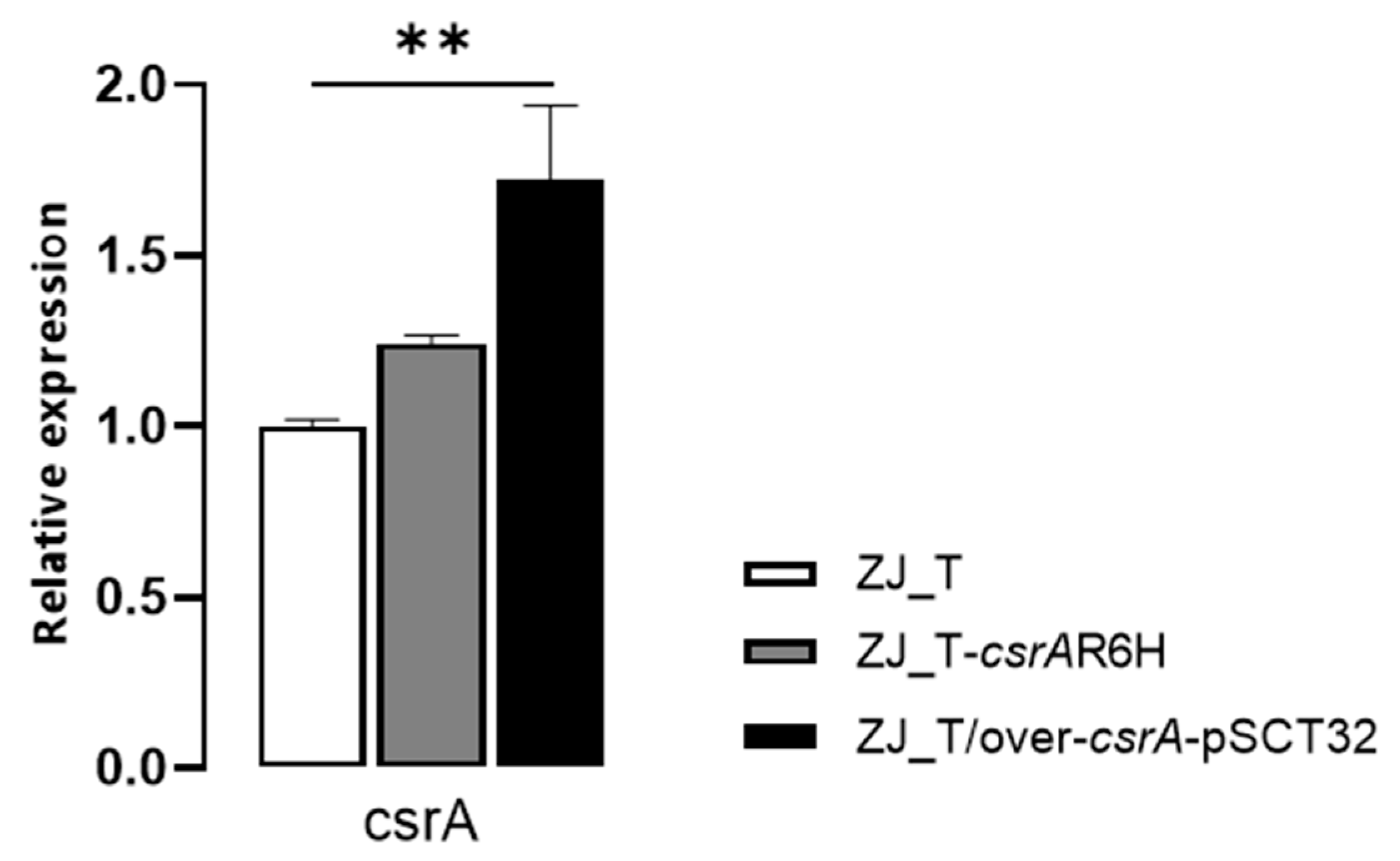
3.1. Construction of csrA Pointmutant and Overexpression Strain

In order to examine the role of CsrA in V. alginolyticus, we first made numerous attempts to construct an in-frame deletion of csrA in the ZJ-T strain. However, none of these attempts succeeded. It has been reported that severe growth defects make it impossible to make an in-frame deletion of csrA in several other Gram-negative bacterial species, including V. cholerae [21]. Therefore, we constructed a csrA point mutation where the sixth codon of CGC (encoding arginine) was replaced by CAC (encoding histidine), which has been shown to dramatically decrease CsrA activity in V. cholerae [21]. The mutant was named ZJ-T-csrA.R6H. In addition, an overexpression strain ZJ_T/over csrA -pSCT32 was constructed by introducing a multiple copy plasmid pSCT32 that harbors a csrA gene with the tac promoter. Quantitative RT-PCR was performed to examine the relative expression of csrA in WT, ZJ-T-csrA.R6H, and ZJ_T/over csrA -pSCT32. The results showed that csrA is upregulated in ZJ_T/over csrA -pSCT32, but not in ZJ-T-csrA.R6H (Figure 1), which is not surprising since CsrA has been shown to regulate its own translation rather than transcription.

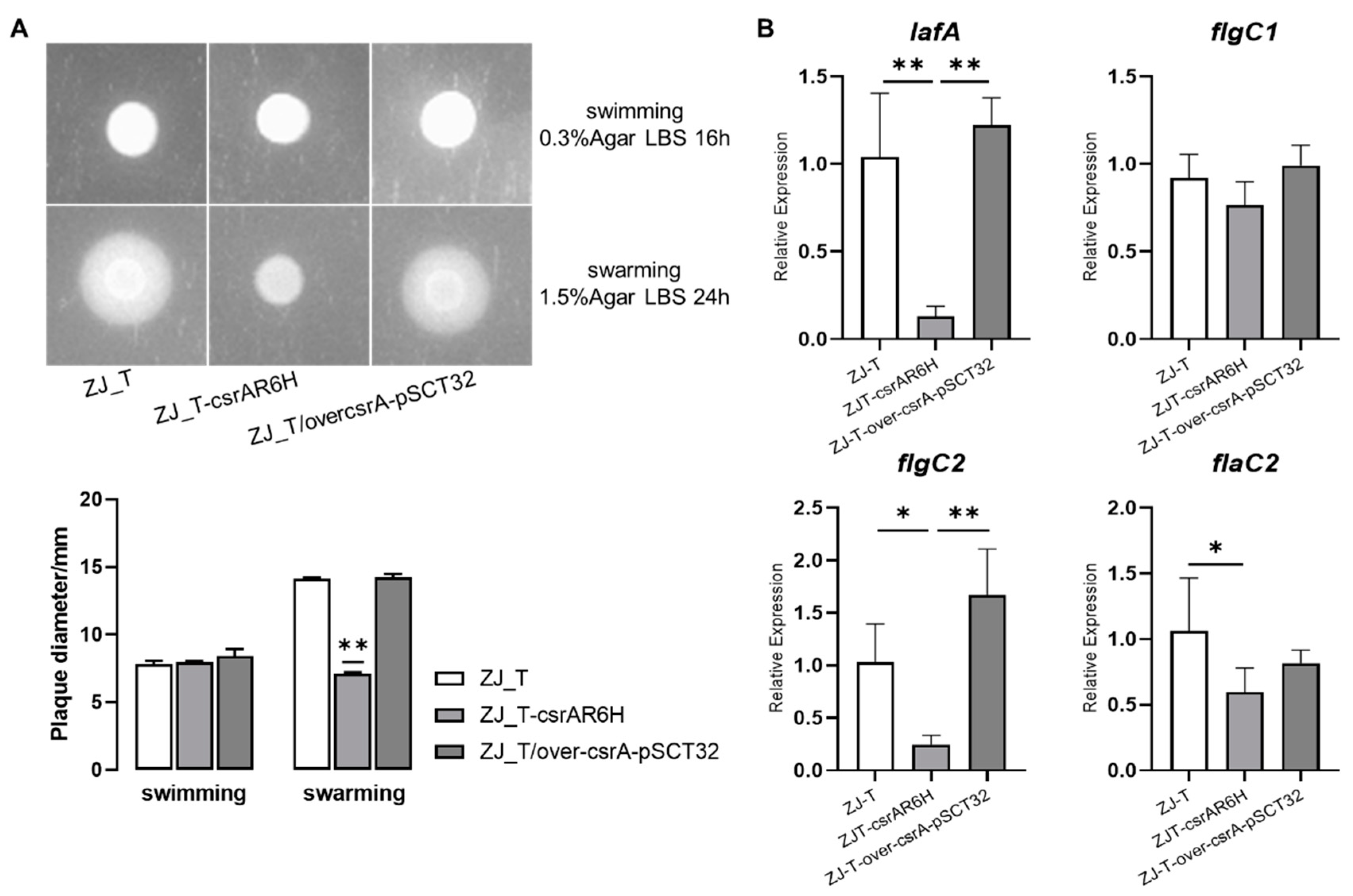
3.2. CsrA Positively Regulates Swarming but Not Swimming Motility in V. alginolyticus

Most Vibrio species possess two distinct sets of flagella: the polar flagellum, an organ that drives swimming motility in liquid or semi-solid surfaces, and the lateral flagellum, which drives swarming motility on solid surfaces [30,31,32]. To investigate the role of CsrA in the regulation of motility, 0.3% and 1.5% agar plates were used to evaluate the swimming and swarming motility, respectively. As shown in Figure 2A, the csrA mutant was not motile on the 1.5% agar plate. This was in contrast with the wild type and overexpression strains which formed large and similar cell motile zones outwards of the spots. However, on 0.3% agar plates, the strains showed no significant differences from each other (Figure 2A). This indicated that csrA is required for the swarming motility of V. alginolyticus, which may be mediated by the modulation of lateral-flagella-associated genes. The expression levels of genes encoding lateral flagellin (lafA), flagellar basal-body rod protein (flgC1), lateral flagellar basal-body rod protein C (flgC2), and polar flagellin (flaC2) were determined when the bacteria were cultured on 1.5% agar plates. As shown in Figure 2B, lafA and flgC2, which are required for lateral flagellar biosynthesis, were downregulated by more than 2-fold (p < 0.05) in the csrA mutant, compared with in the wild-type and csrA overexpression strains, while flgC1 and flaC2, which are involved in polar flagella biosynthesis, showed no difference between strains. Taken together, these results may suggest that CsrA regulates the swarming motility by positively affecting the transcript abundance of the genes related to lateral flagella biosynthesis in V. alginolyticus.

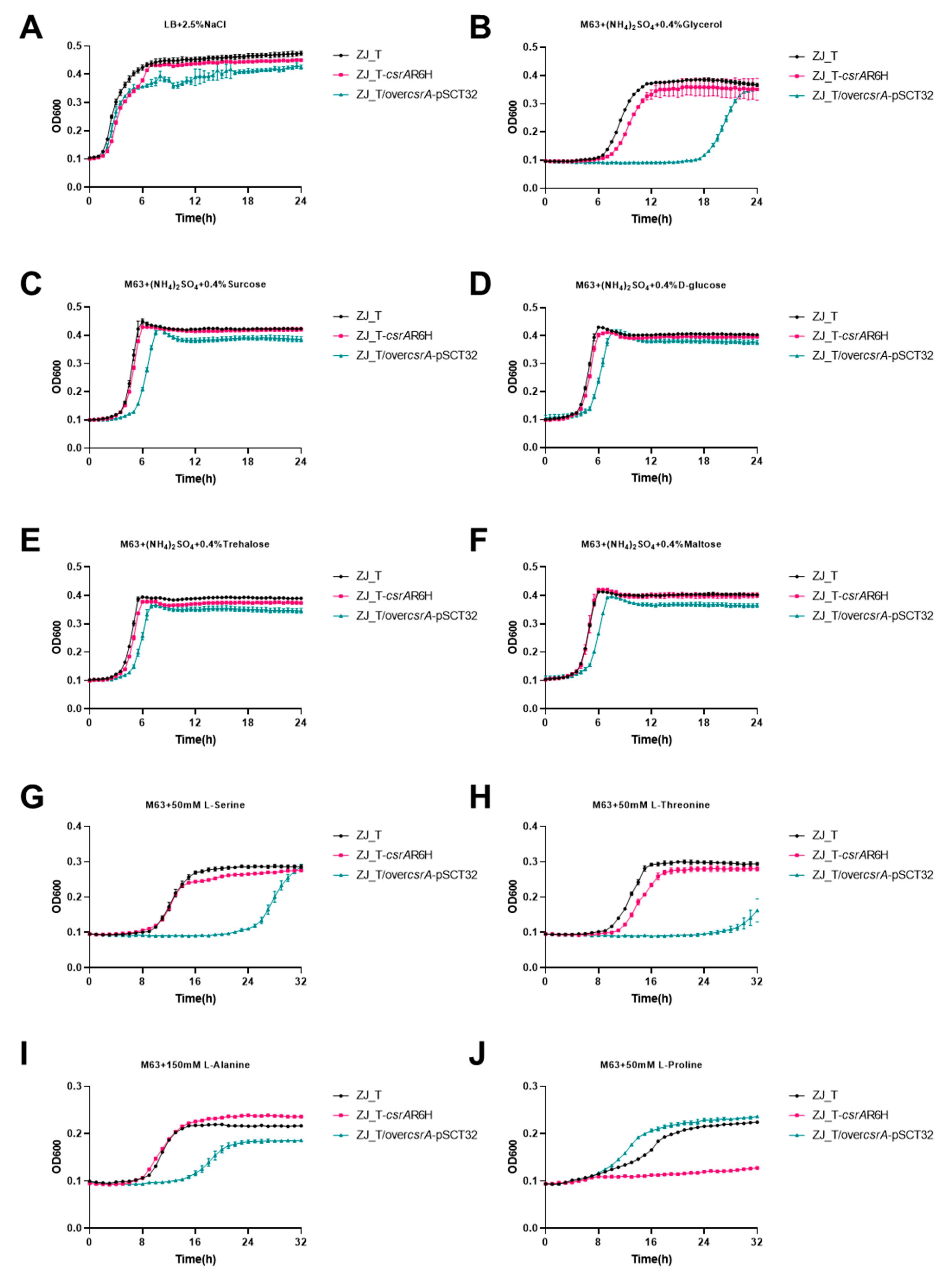
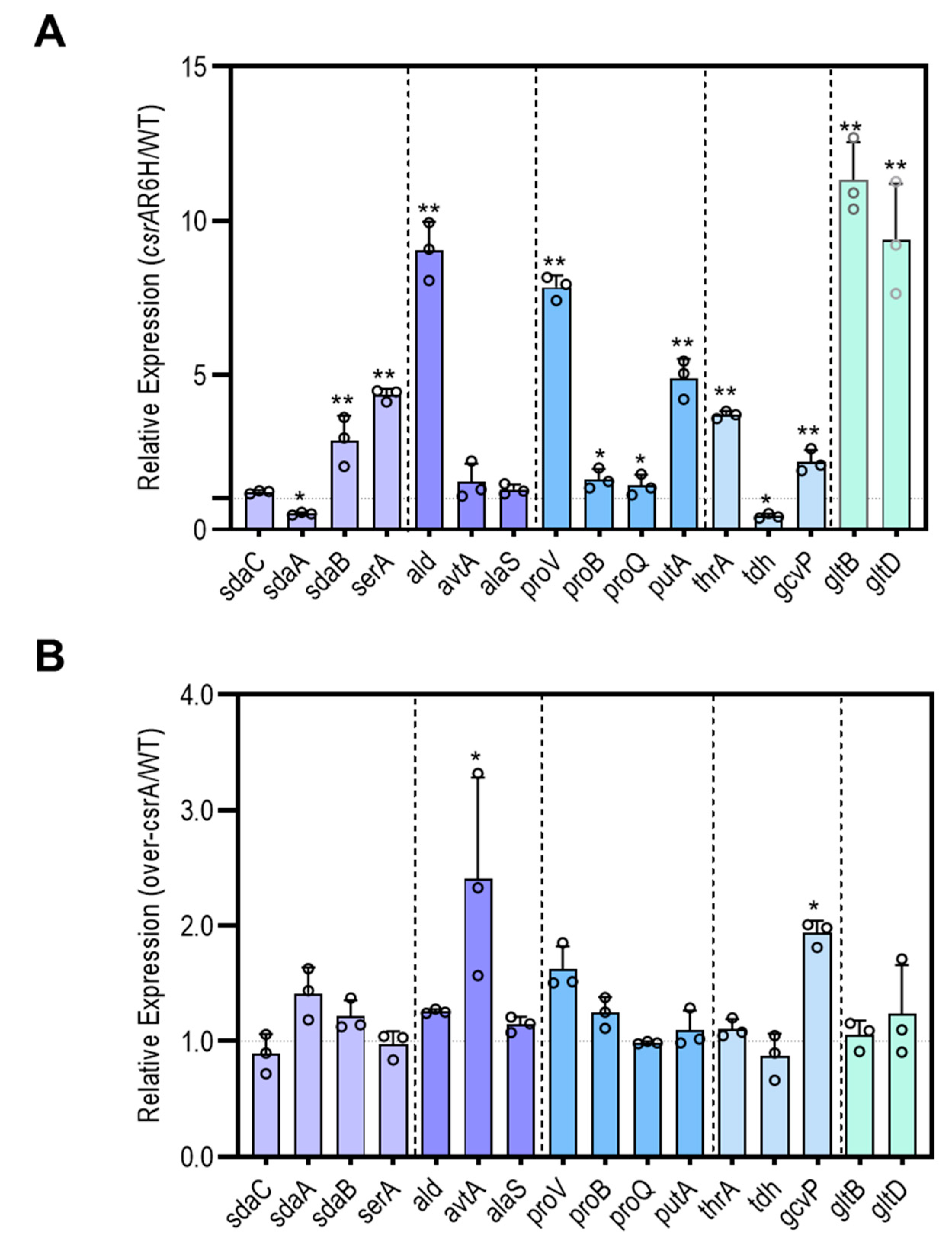
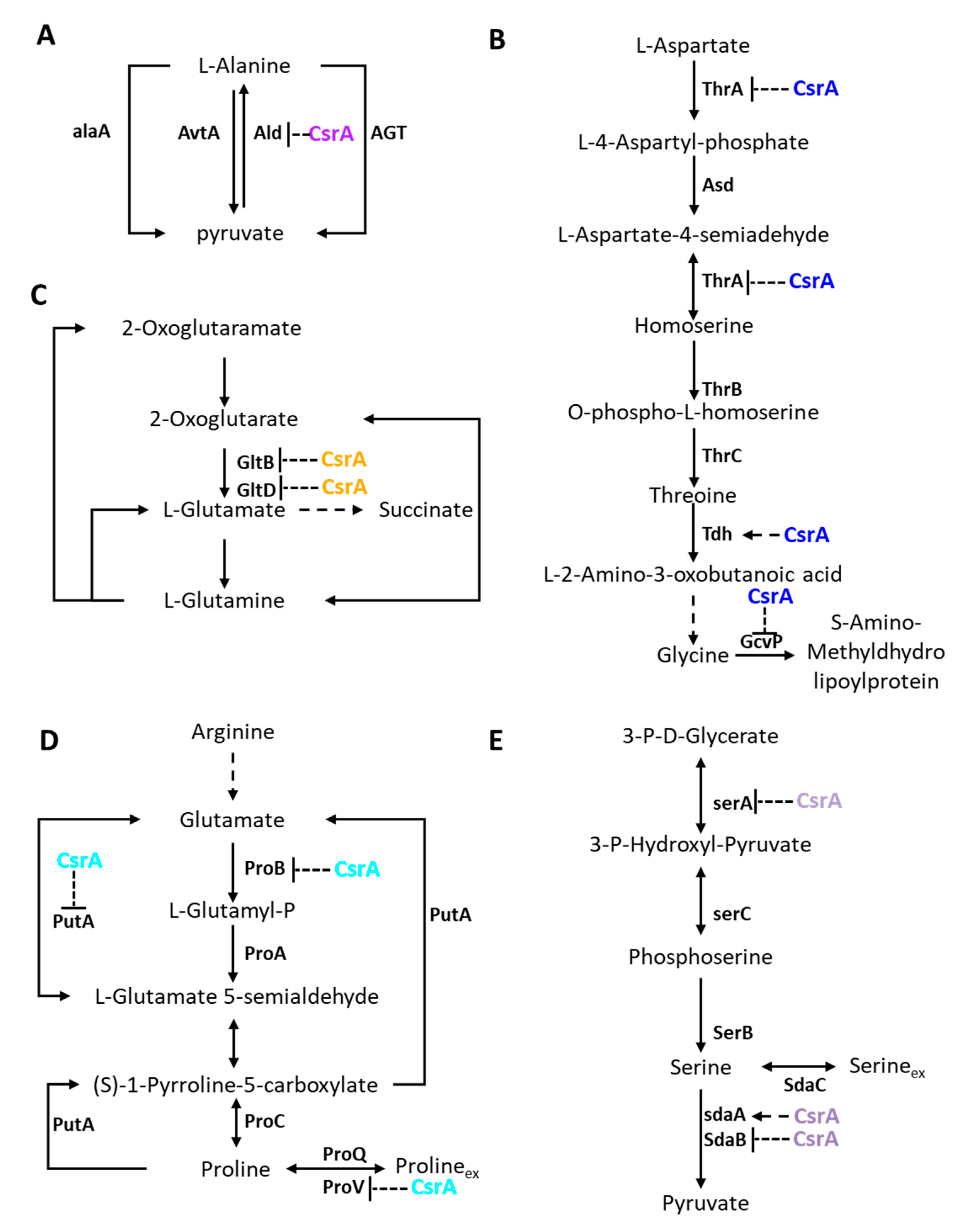
3.3. CsrA Is Extensively Involved in Carbon and Nitrogen Metabolism

To determine the effect of csrA on the carbon and nitrogen metabolism of V. alginolyticus, the growth characteristics of ZJ_T, ZJ_T-csrAR6H and ZJ_T/overcsrA-pSCT32 were compared in media containing different carbon and nitrogen sources. As shown in Figure 3, there was no significant difference between ZJ_T and ZJ_T-csrAR6H when the cells were grown in rich medium LBS, while ZJ_T/overcsrA-pSCT32 showed a decrease in optical density during the stationary phase. This suggests that excessive CsrA mitigates cellular resistance to the unfavorable environment of the stationary phase, which is characterized by a low pH, low oxygen, and a shortage of carbon/nitrogen sources.
When the cells were grown in minimum media M63 supplemented with NH4+ ((NH4)2SO4) as the sole nitrogen source as well as glycerol, glucose, sucrose, trehalose, or maltose as sole carbon source, ZJ_T/overcsrA-pSCT32 exhibited a prolonged lag phase compared with the other two strains. However, the growth kinetics at the log phase were similar. This suggests that the overexpression of csrA may slow down the uptake and accumulation of nutrients, which are the major cellular processes that occur during the lag phase, but not cell division. Interestingly, both the csrA mutant and overexpression strains showed decreased maximum cell densities during the stationary phase when glycerol was the sole carbon source. Under these conditions, the cells must reorganize two major biophysical processes, the catabolism of carbohydrates and the anabolism of amino acids de novo, to provide sufficient nucleotides and amino acids, which are the building blocks of macro-molecules required for fast growth. Given that the carbohydrates tested were taken up through different pathways, it can be hypothesized that CsrA may primarily regulate amino acid anabolism.
To investigate the impact of CsrA on the metabolism of amino acids, bacterial growth was measured in M63 supplemented with serine, threonine, alanine, or proline. As shown in Figure 3, in the medium containing serine, ZJ_T-csrAR6H had the longest lag phase, followed by ZJ_T/overcsrA-pSCT32, while ZJ_T had the shortest lag phase, but all strains exhibited similar growth rates during the log phase and had maximum cell density during the stationary phase. When cultured in medium containing threonine, ZJ_T-csrAR6H and ZJ_T/overcsrA-pSCT32 showed similar prolonged lag phases compared with ZJ_T, but ZJ_T-csrAR6H underwent a second growth phase when the other two reached the steady stationary phase, leading to a higher maximum cell density. In M63 with alanine, ZJ_T and ZJ_T-csrAR6H exhibited the same growth curve, but ZJ_T/overcsrA-pSCT32 showed a prolonged lag phase, slow growth rate, and low cell density during the stationary phase. When proline was used as the sole carbon and nitrogen source, ZJ_T-csrAR6H did not grow at all, but ZJ_T/overcsrA-pSCT32 showed a shorter lag phase, a faster growth rate, and a similar cell density during the stationary phase. These results indicate that CsrA may regulate the catabolism of amino acids in an amino-acid-specific manner in V. alginolyticus. In the situation tested above, amino acids were presumably taken up into the cell at first, before undergoing oxidative deamination to form their corresponding α-keto acids and ammonia, a process catalyzed by L-amino acid deaminases. The α-keto acids were then utilized for central metabolism, producing ATP, NAD(P)H, and carbon intermediates for the biosynthesis of other amino acids together with ammonia. Therefore, it is possible that csrA functions in the importation and/or the degradation of amino acids.

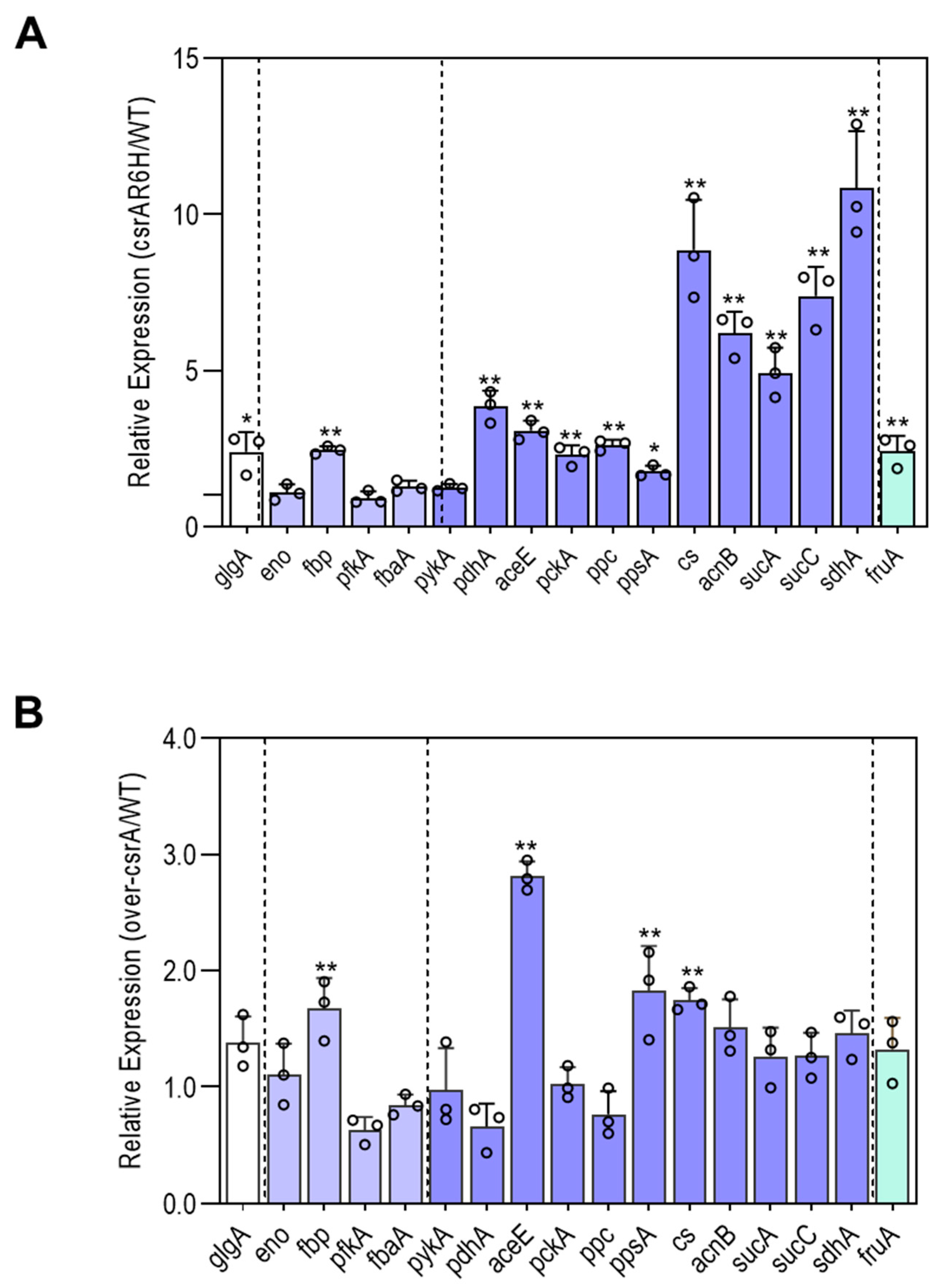
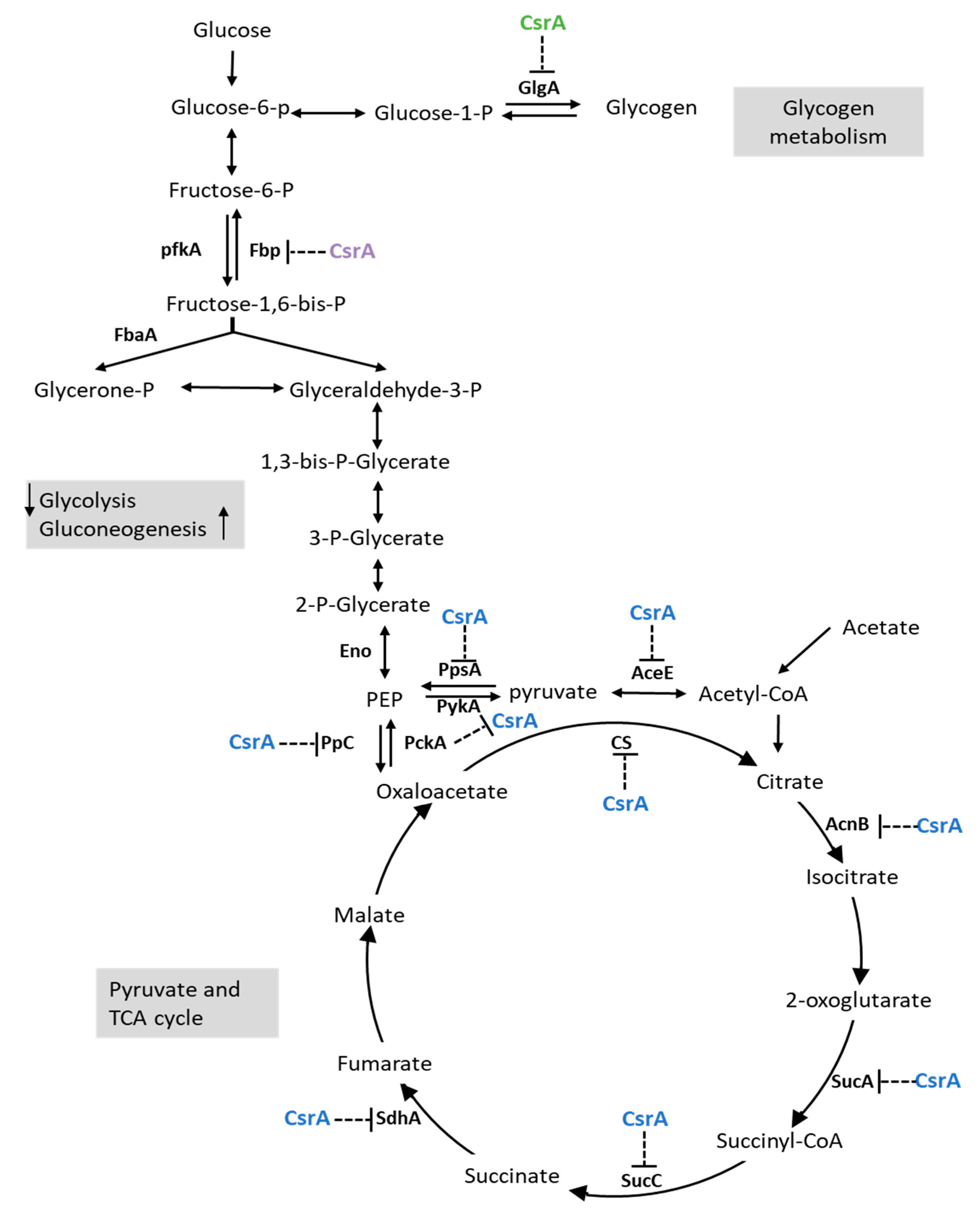
3.4. CsrA Has a Significant Impact on the Transcript Abundance of Genes Involved in Central Metabolism

To better understand the mechanism responsible for the phenotypical changes observed above, the expression profiles of genes associated with carbon and nitrogen metabolism were selected (Table S3) and determined by qRT-PCR. The results are presented and discussed in categories based on their functions.
The expression profiles of fbp, pfkA, fbaA, eno, and glgA were selected and measured (Figure 4). Except for fbp and glgA, the genes showed similar expression profiles in all strains, indicating that CsrA does not affect the transcription and/or mRNA stability of the genes involved in glycolysis. However, fbp, which encodes fructose-1,6-bisphosphatase, and glgA, which encodes glycogen synthase, were upregulated by more than 2-fold in ZJ_T-csrAR6H compared with ZJ-T. This suggests that CsrA may inhibit gluconeogenesis and glycogen biosynthesis.
Compared with ZJ_T, ZJ_T-csrAR6H displayed 3–4 fold increases in pdhA and aceE. These genes have been reported to encode two different sets of pyruvate dehydrogenase (acetyl-transferring) E1 component subunit alpha. However, aceE was also upregulated 3-fold in ZJ_T/overcsrA-pSCT32. This implies that csrA may negatively regulate the reaction from PEP to pyruvate, leading to the restriction of carbon flux into the TCA cycle. The genes cs, acnB, sucA, sucC, and sdhA which are responsible for the TCA cycle were all upregulated in ZJ_T-csrAR6H by over 5-fold. In addition, ppc, pckA, and ppsA, which are involved in the conversion of oxaloacetate, PEP, and pyruvate increased by more than 2-fold in ZJ_T-csrAR6H. These results indicate that CsrA negatively regulates the pyruvate and TCA cycles at multiple points.
As shown above, csrA is apparently involved in the regulation of serine, threonine, alanine, and proline metabolism. To determine its effects on serine metabolism, the expression profiles of sdaA, sdaB, and sdaC, which encode the serine transporter (sdaC) and two serine deaminases (sdaA and sdaB) were measured. As shown in Figure 5, the expression of sdaC was at the same level regardless of whether csrA was tuned up or down, but sdaA was downregulated by 2-fold and sdaB was upregulated by 3-fold in the csrA mutant. However, no significant difference was found between ZJ-T and ZJ_T/overcsrA-pSCT32. For alanine metabolism, the expression profiles of ald and avtA, which are involved in the utilization and conversion of alanine, were quantified. ald, which encodes alanine dehydrogenase, was increased by more than 8-fold in the csrA mutant compared with the wild-type strain, while avtA showed no difference between strains. proV and proB are involved in proline transport and utilization. When csrA was altered, ProV was upregulated by 7-fold, but proB remained at a similar expression level. In addition, gltB and gltD, encoding for glutamate synthase, which is central to the biosynthesis of amino acids, were upregulated by over 8-fold in csrA mutant ZJ_T-csrAR6H.
In summary, CsrA extensively represses the transcript abundance of genes involved in central metabolism, which may have profound effects on pathways such as gluconeogenesis, glycogen biosynthesis, the pyruvate cycle, and the TCA cycle (Figure 6), as well as amino acid uptake and utilization (Figure 7).

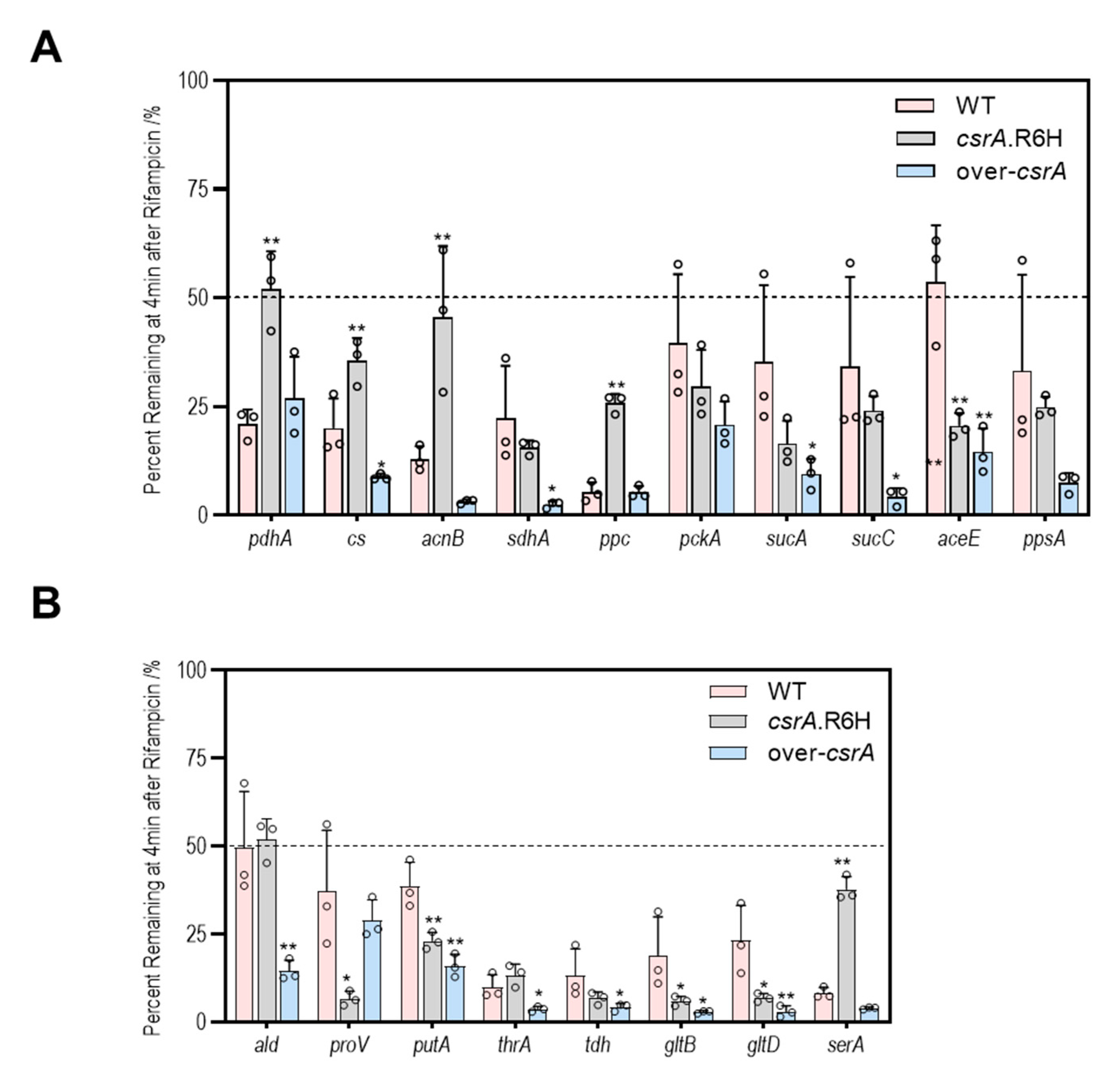
3.5. CsrA Changes the Transcipt Abundance of Genes Involved in Central Metabolism Likely by Alternation of Their mRNA Stability

CsrA is an RNA-binding protein that acts on single-stranded RNA molecules, so it is unlikely that it alters the abundance of transcripts by working on their transcription. A possible reason is that binding to CsrA may change the half-life of the target mRNA. To test this hypothesis, we measured the RNA stability of serA, pdhA, acnB, sdhA, ppc, sucC, ald, gltD, and cs. As shown in Figure 8, less than half of the transcripts of all genes remained after 4 min of rifampicin addition, but the quantity of residuals showed great variation among genes, ranging from 50% to less than 10%, indicating that the half-life of most mRNAs in V.alginolyticus may be less than 4 min. In addition, changes in the csrA abundance significantly altered the stability of the mRNA. For example, after 4 min, only 10.1% of serA transcripts remained in the wild type, while 37.8% remained in the csrA mutant. Similar results were obtained for pdhA (20.9% vs. 51.9%), acnB (12.8% vs. 45.5%), ppC (5.3% vs. 25.7%), and cs (20% vs. 35.5%). Meanwhile, the numbers of residual transcripts of ald (49.5% vs. 14.6%), cs (20.0% vs. 8.7%), gltD (23.2% vs. 3.1%), sdhA (22.2% vs. 2.4%), and sucC (34.2% vs. 4.2%) significantly reduced in the csrA overexpressed strain. These results suggest that binding to CsrA may accelerate target mRNA decay, leading to a decrease in mRNA abundance.
The consensus sequence of CsrA binding is ARGGAN, in which GGA is mandatory. To identify the possible CsrA binding site(s) in the genes measured above, we retrieved sequences ranging from −50 to +50 of the initiation codon and searched for the GGA motifs. As shown in Table S3 and Figure 9, out of 33 genes, only five genes (avtA, proB, sdaC, pfkA and sdhA) were not found to contain a GGA motif. Except for acnB, tdh, pfkA, flgC2 and ppsA, the genes (22) enclosed one or two GGA motifs located within the range of −20 to the initiation codon, which covers the ribosome binding sites. Nine genes showed no difference in the number of transcripts when csrA was mutated, among which four had no GGA motif(s) and four had only one in their sequences. Taken together, these results imply a strong correlation between the potential csrA binding sites and repression of CsrA.

4. Discussion

It has been widely reported that CsrA is a global regulator of gamma-proteobacteria that affects essential biochemical and biophysical processes. In vibrio species, it has been reported to modulate biofilm formation, quorum sensing, virulence, and amino acid metabolism [21,33,34,35]. In this study, the manipulation of CsrA resulted in variations in swarming motility and carbon and nitrogen metabolism but did not alter biofilm formation. Correspondingly, extensive changes in the expression of genes associated with these phenotypes were also observed. These results suggest that CsrA is a global regulator that modulates motility and central metabolism in V. alginolyticus.
The genome of V. alginolyticus has two sets of genes, which encode a polar flagellum and numerous lateral flagella, respectively. Expression of the polar flagellum is induced in liquid, which enables cells to swim in a liquid environment, while lateral flagella are synthesized only when cells are grown on solid surfaces or in a high-viscosity environment [30,31,32]. In this study, the mutation of csrA resulted in a lower swarming motility but no difference in swimming motility, which corresponded to the reduced expression of lateral flagellar genes, i.e., lafA, flgC2 in the csrA mutant, indicating that CsrA may positively regulate swarming motility by upregulating the expression of lateral flagellar genes in V. alginolyticus. In E. coli, CsrA was reported to enhance bacterial motility by binding to the 5′ UTR of flhDC transcript, which encodes an activator of flagella biosynthesis and chemotaxis, thus protecting it from RNase E-mediated cleavage [16,36]. Although no experiment has been carried out to study the regulatory effect of CsrA on motility in Vibrio species, RNA-Seq revealed that over half of all annotated flagellum-dependent motility genes, including rpoN and flrC, are significantly downregulated in the csrA mutant in V. cholera. CsrA was shown to bind directly to both rpoN and flrC mRNA, which encode master activators of flagella synthesis and chemotaxis in vivo [23]. However, unlike V. alginolyticus, V. cholerae possesses only one polar flagellum, so the mechanisms underlying their motility regulation may be very different, making it necessary to further explore the role of CsrA in motility regulation in V. alginolyticus.
Vibrio is widely distributed in the marine environment and has a very fast growth rate [37], suggesting it has the ability to effectively uptake, utilize, and convert nutrients. In this study, the overexpression of csrA led to a decrease in the maximum cell density during the stationary phase in rich medium, which may suggest its involvement in the stress response at the stationary phase. It has been reported that rpoS, which encodes the major sigma factor during the stationary phase, is negatively regulated by CsrA in V. cholerae [23]. Interestingly, a prolonged lag phase was observed when cells with overexpressed csrA grew in minimum media supplemented with various carbohydrates as the sole carbon source and ammonium as the sole nitrogen source. However, no difference in growth rate was shown during the exponential phase. This indicated that csrA may repress major biochemical processes during the lag phase, a distinctive growth phase in bacterial culture. The duration of lag phase depends on a lot of factors, but it is mainly influenced by the inoculum history [38]. When the inoculum has more viable cells during the last stationary phase, the lag period is shorter, which may be one of the reasons why the overproduction of CsrA resulted in prolongation of the lag phase.
In addition, the cells do not replicate during the lag phase, but active cellular processes are undertaken to produce cellular components that are prepared for exponential growth [39,40]. Therefore, optimizing primary metabolism to establish maxim carbon flow in response to available nutrients is the significant characteristic of the lag phase. Yamamotoya et al. demonstrated that intracellular glycogen, instead of glucose, imported from the environment is the primary source of central metabolism during the lag phase [41]. This implies that the speed of carbohydrate uptake may not be the cause of the prolonged lag phase in the csrA overexpression strain of V. alginiolyticus. Under the conditions measured above, active metabolic pathways of glycolysis, the pyruvate cycle, and amino acid biosynthesis de novo must be required for the growth of bacteria. It is reasonable to speculate that the repression of one or more of these genes would result in a prolonged lag phase. Indeed, the expression profiles of genes associated with these pathways provide preliminary evidence in support of this notion. Based on the abundance of transcripts, the mutation of csrA may significantly promote gluconeogenesis, glycogen synthesis, and the pyruvate cycle, but not glycolysis (Figure 6).
CsrA is essential for carbohydrate metabolism in E. coli and other gram-negative bacteria. It has long been reported that CsrA promotes glycolysis but inhibits gluconeogenesis and glycogen synthesis [42]. The interconversion of fructose 6-phosphate and fructose 1,6-diphosphate, catalyzed by 6-phosphofructokinase (PfkAB) and Fructose-1,6-bisphosphatase (Fbp), respectively, is the limiting step of glycolysis and gluconeogenesis. Thus, these processes are the critical regulatory checkpoints of central metabolism regulation. In E. coli, CsrA positively regulates pfkA at the levels of both mRNA stability and translational efficiency, leading to increased carbon flux into glycolysis [43]. However, in V. cholerae, CsrA has been shown to be a repressor rather than an activator of pfkA that only works during the stationary phase [23]. In this study, alternation of csrA had no effect on the expression of pfkA at the early-log phase, and it was not found to contain a potential CsrA binding site in the sequence between −50 and +50 nt around the initiation codon (Figure 9). In addition, other enzymes involved in glycolysis/gluconeogenesis displayed no difference in the abundance of transcripts, indicating that CsrA may not participate in the regulation of glycolysis in V. alginolyticus at the early-log phase. However, CsrA was shown to repress gluconeogenesis and glycogen synthesis through the inhibition of fbp and glgA during the early exponential growing period, which coincides with the results of other studies on E. coli and V. cholerae etc. [42].
Carbon flux from the PEP-pyruvate-AcCoA to TCA cycle is crucial for the integration of carbohydrates with energy production and amino acid metabolism. Recent studies have suggested a new pathway of central metabolism called the pyruvate cycle, or P cycle, which merges the PEP-pyruvate-AcCoA pathway with the TCA cycle [44]. In this study, CsrA inhibited the expression of genes involved in the P cycle extensively, a feature that has also observed in other studies [45]. It appears that CsrA-mediated multipoint inhibition of the P-cycle is a conserved feature of central metabolic regulation, so it can be expected that the overexpression of csrA will lead to a deceleration of energy production, reducing the concentration of carbon intermediates undergo amino acid biosynthesis de novo. This may be one of the reasons why the csrA overexpressed strain showed a prolonged lag phase when grown in the media with ammonium as the sole nitrogen source.
Amino acids are critical components that not only act as building blocks for protein synthesis but also serve as both carbon and nitrogen sources for growth. Here, we found that the alternation of CsrA significantly changed growth rates, the duration of the lag phase, or the maximum cell density of V. alginolyticus when serine, alanine, threonine, or proline served as the sole carbon and nitrogen source. Q-RTPCR has also shown that genes encoding for specific transporters and enzymes for their degradation had altered expression levels in the csrA mutant. It is not well known how CsrA regulates amino acid catabolism, but Ina et al. [46] suggested that CsrA regulates a switch from amino acid to glycerolipid metabolism in Legionella pneumophila and in V. cholerae, extensive changes in transcripts involved in amino acid uptake and degradation were found when csrA was impaired [23]. This may suggest that CsrA is extensively involved in the regulation of amino acid transport and catabolism by changing the expression levels of relevant genes.
In V. alginolyticus, the levels of a wide range of gene transcripts were significantly altered in the csrA mutant, which is in agreement with the results of other studies [42,47,48,49]. However, as an RNA binding protein, CsrA is not expected to function as a transcriptional regulator that affects the transcription of target genes. Heidi et al. 2021 suggested that this is because CsrA is able to regulate other global regulators, such as rpoS/rpoE [23]. Another plausible explanation is that binding to CsrA may affect mRNA stability. In this study, we found that impairment or overexpression of csrA resulted in longer or shorter half-lives for dozens of mRNAs, suggesting that binding to CsrA may accelerate mRNA decay, leading to the destabilization of target mRNAs in V. alginolyticus, which is in agreement with the overall repression of RNA abundance. However, in E. coli, CsrA was suggested to be a global positive regulator of mRNA stability, although possible mechanism(s) behind were not addressed [50].
In summary, this study provides the first insights into the regulatory role of CsrA in V. alginolyticus. Our results highlight the importance of post-transcriptional controls mediated by CsrA that likely contribute to the fast growth and adaptation of this bacteria in response to a changing environment. However, since it is a post-transcriptional global regulator, transcriptomic and proteomic analyses are needed to further reveal its role in V. alginolyticus.

Supplementary Materials

The following are available online at https://0-www-mdpi-com.brum.beds.ac.uk/article/10.3390/microorganisms9112383/s1, Table S1: Primers used for construction in this study, Table S2: Primers used for qPCR, Table S3: Genes used for the q-PCR and mRNA stability experiments.

Author Contributions

C.C. contributed reagents, materials; conceived and designed the experiments. B.L., Q.G., H.C., Y.Z., Y.S. and S.Y. performed the experiments; B.L. analyzed the data and X.Z. performed the data visualization. B.L. prepared the original draft and C.C. reviewed and edited the manuscript. All authors have read and agreed to the published version of the manuscript.

Funding

This study was funded by the National Natural Science Foundation of China (31872605) and China Agriculture Research System of MOF and MARA.

Data Availability Statement

All datasets generated for this study are included in the article/Supplementary Materials.

Acknowledgments

We thank the data archive support from South China Sea Ocean Data Center (http://data.scsio.ac.cn, accessed on 16 November 2021), National science & Technology infrastructure of China (http://www.geodata.cn, accessed on 16 November 2021).

Conflicts of Interest

The authors declare no conflict of interest.

References

  1. Xie, J.; Bu, L.; Jin, S.; Wang, X.; Zhao, Q.; Zhou, S.; Xu, Y. Outbreak of vibriosis caused by Vibrio harveyi and Vibrio alginolyticus in farmed seahorse Hippocampus kuda in China. Aquaculture 2020, 523, 735168. [Google Scholar] [CrossRef]
  2. Xie, Z.; Ke, S.; Hu, C.; Zhu, Z.; Wang, S.; Zhou, Y. First Characterization of Bacterial Pathogen, Vibrio alginolyticus, for Porites andrewsi White Syndrome in the South China Sea. PLoS ONE 2013, 8, e75425. [Google Scholar] [CrossRef] [Green Version]
  3. Taylor, R.; Mcdonald, M.; Russ, G.; Carson, M.; Lukaczynski, E. Vibrio alginolyticus peritonitis associated with ambulatory peritoneal dialysis. Br. Med. J. 1981, 283, 275. [Google Scholar] [CrossRef] [Green Version]
  4. Opal, S.M.; Saxon, J.R. Intracranial infection by Vibrio alginolyticus following injury in salt water. J. Clin. Microbiol. 1986, 23, 373–374. [Google Scholar] [CrossRef] [Green Version]
  5. Barbarossa, V.; Kucisec-Tepes, N.; Aldova, E.; Matek, D.; Stipoljev, F. Ilizarov technique in the treatment of chronic osteomyelitis caused by Vibrio alginolyticus. Croat. Med. J. 2002, 43, 346–349. [Google Scholar]
  6. Chien, J.; Shih, J.; Hsueh, P.; Yang, P.; Luh, K. Vibrio alginolyticus as the cause of pleural empyema and bacteremia in an immunocompromised patient. Eur. J. Clin. Microbiol. Infect. Dis. 2002, 21, 401–403. [Google Scholar] [CrossRef]
  7. Feingold, M.H.; Kumar, M.L. Otitis media associated with Vibrio alginolyticus in a child with pressure-equalizing tubes. Pediatric Infect. Dis. J. 2004, 23, 475–476. [Google Scholar] [CrossRef]
  8. Li, X.C.; Xiang, Z.Y.; Xu, X.M.; Yan, W.H.; Ma, J.M. Endophthalmitis Caused by Vibrio alginolyticus. J. Clin. Microbiol. 2009, 47, 3379–3381. [Google Scholar] [CrossRef] [Green Version]
  9. Hong, G.L.; Dai, X.Q.; Lu, C.J.; Liu, J.M.; Zhao, G.J.; Wu, B.; Li, M.F.; Lu, Z.Q. Temporizing surgical management improves outcome in patients with Vibrio necrotizing fasciitis complicated with septic shock on admission. Burns 2014, 40, 446–454. [Google Scholar] [CrossRef]
  10. Pourciau, C.; Lai, Y.-J.; Gorelik, M.; Babitzke, P.; Romeo, T. Diverse Mechanisms and Circuitry for Global Regulation by the RNA-Binding Protein CsrA. Front. Microbiol. 2020, 11, 601352. [Google Scholar] [CrossRef]
  11. Sabnis, N.A.; Yang, H.; Romeo, T. Pleiotropic regulation of central carbohydrate metabolism in Escherichia coli via the gene csrA. J. Biol. Chem. 1995, 270, 29096–29104. [Google Scholar] [CrossRef] [PubMed] [Green Version]
  12. Altier, C.; Suyemoto, M.; Lawhon, S.D. Regulation of Salmonella enterica serovar typhimurium invasion genes by csrA. Infect. Immun. 2000, 68, 6790–6797. [Google Scholar] [CrossRef] [PubMed] [Green Version]
  13. Jackson, D.W.; Suzuki, K.; Oakford, L.; Simecka, J.W.; Hart, M.E.; Romeo, T. Biofilm Formation and Dispersal under the Influence of the Global Regulator CsrA of Escherichia coli. J. Bacteriol. 2002, 184, 290–301. [Google Scholar] [CrossRef] [Green Version]
  14. Mukherjee, A.; Cui, Y.Y.; Liu, Y.; Dumenyo, C.K.; Chatterjee, A.K. Global regulation in Erwinia species by Erwinia carotovora rsmA, a homologue of Escherichia coli csrA: Repression of secondary metabolites, pathogenicity and hypersensitive reaction. Microbiology 1996, 142, 427–434. [Google Scholar] [CrossRef] [Green Version]
  15. Romeo, T.; Gong, M.; Liu, M.Y.; Brunzinkernagel, A.M. Identification and molecular characterization of csrA a pleiotropic gene from Escherichia coli that affects glycogen biosynthesis, gluconeogenesis, cell-size, and surface-properties. J. Bacteriol. 1993, 175, 4744–4755. [Google Scholar] [CrossRef] [Green Version]
  16. Wei, B.; Brun-Zinkernagel, A.M.; Simecka, J.W.; Prüss, B.; Romeo, T. Positive regulation of motility and flhDC expression by the RNA-binding protein CsrA of Escherichia coli. Mol. Microbiol. 2010, 40, 245–256. [Google Scholar] [CrossRef]
  17. Gudapaty, S.; Suzuki, K.; Wang, X.; Babitzke, P.; Romeo, T. Regulatory Interactions of Csr Components: The RNA Binding Protein CsrA Activates csrB transcription in Escherichia coli. J. Bacteriol. 2001, 183, 6017–6027. [Google Scholar] [CrossRef] [Green Version]
  18. Ikeda, M. Towards bacterial strains overproducing L-tryptophan and other aromatics by metabolic engineering. Appl. Microbiol. Biotechnol. 2006, 69, 615. [Google Scholar] [CrossRef] [Green Version]
  19. Romeo, T.; Vakulskas, C.A.; Babitzke, P. Post-transcriptional regulation on a global scale: Form and function of Csr/Rsm systems. Environ. Microbiol. 2013, 15, 313–324. [Google Scholar] [CrossRef]
  20. Weilbacher, T.; Suzuki, K.; Dubey, A.K.; Wang, X.; Romeo, T. A novel sRNA component of the carbon storage regulatory system of Escherichia coli. Mol. Microbiol. 2003, 48, 657–670. [Google Scholar] [CrossRef]
  21. Mey, A.R.; Butz, H.A.; Payne, S.M. Vibrio cholerae CsrA Regulates ToxR Levels in Response to Amino Acids and Is Essential for Virulence. mBio 2015, 6, e01064-15. [Google Scholar] [CrossRef] [PubMed] [Green Version]
  22. Butz, H.A.; Mey, A.R.; Ciosek, A.L.; Payne, S.M. Vibrio cholerae CsrA Directly Regulates varA to Increase Expression of the Three Nonredundant Csr Small RNAs. mBio 2019, 10, e01042-19. [Google Scholar] [CrossRef] [Green Version]
  23. Butz, H.A.; Mey, A.R.; Ciosek, A.L.; Crofts, A.A.; Davies, B.W.; Payne, S.M. Regulatory Effects of CsrA in Vibrio cholerae. mBio 2021, 12, e03380-20. [Google Scholar] [CrossRef] [PubMed]
  24. Val, M.E.; Skovgaard, O.; Ducos-Galand, M.; Bland, M.J.; Mazel, D. Genome engineering in Vibrio cholerae: A feasible approach to address biological issues. PLoS Genet. 2012, 8, e1002472. [Google Scholar] [CrossRef] [PubMed] [Green Version]
  25. Neidhardt, F.C.; Bloch, P.L.; Smith, D.F. Culture Medium for Enterobacteria. J. Bacteriol. 1974, 119, 736–747. [Google Scholar] [CrossRef] [PubMed] [Green Version]
  26. Chang, C.; Jin, X.; Hu, C. Phenotypic and genetic differences between opaque and translucent colonies of Vibrio alginolyticus. Biofouling 2009, 25, 525–531. [Google Scholar] [CrossRef]
  27. Nguyen, A.N.; Disconzi, E.; Charriere, G.M.; Destoumieux-Garzon, D.; Bouloc, P.; Le Roux, F.; Jacq, A. csrB Gene Duplication Drives the Evolution of Redundant Regulatory Pathways Controlling Expression of the Major Toxic Secreted Metalloproteases in Vibrio tasmaniensis LGP32. mSphere 2018, 3, e00582-18. [Google Scholar] [CrossRef] [Green Version]
  28. Zhang, Y.-J.; Chen, G.; Lin, H.; Wang, P.; Kuang, B.; Liu, J.; Chen, S. Development of a regulatable expression system for the functional study of Vibrio vulnificus essential genes. Antonie Leeuwenhoek 2017, 110, 607–614. [Google Scholar] [CrossRef]
  29. Livak, K.J.; Schmittgen, T.D. Analysis of relative gene expression data using real-time quantitative PCR and the 2−ΔΔCT method. Methods 2001, 25, 402–408. [Google Scholar] [CrossRef]
  30. Ulitzur, S. The mechanism of swarming of Vibrio alginolyticus. Arch. Microbiol. 1975, 104, 67–71. [Google Scholar] [CrossRef]
  31. Atsumi, T.; Maekawa, Y.; Yamada, T.; Kawagishi, I.; Imae, Y.; Homma, M. Effect of viscosity on swimming by the lateral and polar flagella of Vibrio alginolyticus. J. Bacteriol. 1996, 178, 5024–5026. [Google Scholar] [CrossRef] [PubMed] [Green Version]
  32. Kogure, K.; Ikemoto, E.; Morisaki, H. Attachment of Vibrio alginolyticus to glass surfaces is dependent on swimming speed. J. Bacteriol. 1998, 180, 932–937. [Google Scholar] [CrossRef] [Green Version]
  33. Lenz, D.H.; Miller, M.B.; Zhu, J.; Kulkarni, R.V.; Bassler, B.L. CsrA and three redundant small RNAs regulate quorum sensing in Vibrio cholerae. Mol. Microbiol. 2005, 58, 1186–1202. [Google Scholar] [CrossRef] [PubMed]
  34. Mey, A.R.; Craig, S.A.; Payne, S.M. Effects of Amino Acid Supplementation on Porin Expression and ToxR Levels in Vibrio cholerae. Infect. Immun. 2012, 80, 518–528. [Google Scholar] [CrossRef] [Green Version]
  35. Jones, M.K.; Warner, E.B.; Oliver, J.D. csrA Inhibits the Formation of Biofilms by Vibrio vulnificus. Appl. Environ. Microbiol. 2008, 74, 7064–7066. [Google Scholar] [CrossRef] [PubMed] [Green Version]
  36. Yakhnin, A.V.; Baker, C.S.; Vakulskas, C.A.; Yakhnin, H.; Babitzke, P. CsrA activates flhDC expression by protecting flhDC mRNA from RNase E-mediated cleavage. Mol. Microbiol. 2013, 87, 851–866. [Google Scholar] [CrossRef] [Green Version]
  37. Weinstock, M.T.; Hesek, E.D.; Wilson, C.M.; Gibson, D.G. Vibrio natriegens as a fast-growing host for molecular biology. Nat. Methods 2016, 13, 849–851. [Google Scholar] [CrossRef]
  38. Jacobsen, T.; Koch, A.G. Influence of different histories of the inoculum on lag phase and growth of Listeria monocytogenes in meat models. J. Food Prot. 2006, 69, 532–541. [Google Scholar] [CrossRef]
  39. Yates, G.T.; Smotzer, T. On the lag phase and initial decline of microbial growth curves. J. Theor. Biol. 2007, 244, 511–517. [Google Scholar] [CrossRef]
  40. Bertrand, R.L. Lag Phase Is a Dynamic, Organized, Adaptive, and Evolvable Period That Prepares Bacteria for Cell Division. J. Bacteriol. 2019, 201, e00697-18. [Google Scholar] [CrossRef]
  41. Yamamotoya, T.; Dose, H.; Tian, Z.Y.; Faure, A.; Toya, Y.; Honma, M.; Igarashi, K.; Nakahigashi, K.; Soga, T.; Mori, H.; et al. Glycogen is the primary source of glucose during the lag phase of E. coli proliferation. Biochim. Biophys. Acta-Proteins Proteom. 2012, 1824, 1442–1448. [Google Scholar] [CrossRef] [PubMed] [Green Version]
  42. Potts, A.H.; Vakulskas, C.A.; Pannuri, A.; Yakhnin, H.; Babitzke, P.; Romeo, T. Global role of the bacterial post-transcriptional regulator CsrA revealed by integrated transcriptomics. Nat. Commun. 2017, 8, 1596. [Google Scholar] [CrossRef] [Green Version]
  43. Morin, M.; Ropers, D.; Letisse, F.; Laguerre, S.; Portais, J.-C.; Cocaign-Bousquet, M.; Enjalbert, B. The post-transcriptional regulatory system CSR controls the balance of metabolic pools in upper glycolysis of Escherichia coli. Mol. Microbiol. 2016, 100, 686–700. [Google Scholar] [CrossRef] [Green Version]
  44. Su, Y.B.; Peng, B.; Li, H.; Cheng, Z.X.; Zhang, T.T.; Zhu, J.X.; Li, D.; Li, M.Y.; Ye, J.Z.; Du, C.C.; et al. Pyruvate cycle increases aminoglycoside efficacy and provides respiratory energy in bacteria. Proc. Natl. Acad. Sci. USA 2018, 115, E1578–E1587. [Google Scholar] [CrossRef] [PubMed] [Green Version]
  45. Durica-Mitic, S.; Goepel, Y.; Goerke, B. Carbohydrate Utilization in Bacteria: Making the Most Out of Sugars with the Help of Small Regulatory RNAs. Microbiol. Spectr. 2018, 6. [Google Scholar] [CrossRef]
  46. Hauslein, I.; Sahr, T.; Escoll, P.; Klausner, N.; Eisenreich, W.; Buchrieser, C. Legionella pneumophila CsrA regulates a metabolic switch from amino acid to glycerolipid metabolism. Open Biol. 2017, 7, 170149. [Google Scholar] [CrossRef] [Green Version]
  47. Lawhon, S.D.; Frye, J.G.; Suyemoto, M.; Porwollik, S.; McClelland, M.; Altier, C. Global regulation by CsrA in Salmonella typhimurium. Mol. Microbiol. 2003, 48, 1633–1645. [Google Scholar] [CrossRef] [PubMed] [Green Version]
  48. Brencic, A.; Lory, S. Determination of the regulon and identification of novel mRNA targets of Pseudomonas aeruginosa RsmA. Mol. Microbiol. 2009, 72, 612–632. [Google Scholar] [CrossRef] [Green Version]
  49. Potts, A.H.; Guo, Y.; Ahmer, B.M.M.; Romeo, T. Role of CsrA in stress responses and metabolism important for Salmonella virulence revealed by integrated transcriptomics. PLoS ONE 2019, 14, e0211430. [Google Scholar] [CrossRef] [Green Version]
  50. Esquerre, T.; Bouvier, M.; Turlan, C.; Carpousis, A.J.; Girbal, L.; Cocaign-Bousquet, M. The Csr system regulates genome-wide mRNA stability and transcription and thus gene expression in Escherichia coli. Sci. Rep. 2016, 6, 25057. [Google Scholar] [CrossRef]
Figure 1. The csrA mutant did not affect transcription. The relative expression of csrA behaved at the wildtype level in terms of expression in the csrA mutant, while it was upregulated in the overexpression strain (One-way ANOVA, FDR ** p < 0.01). The levels of csrA were normalized to the internal control recA level. Error bars indicate standard deviations.
Microorganisms 09 02383 g001
Figure 2. CsrA positively regulates swarming but not swimming motility in V. alginolyticus. (A) The ability of ZJ_T, ZJ_T-csrAR6H, ZJ_T/overcsrA-pSCT32 to swim or swarm through 0.3% and 1.5% LBS agar plates, which was observed with a BIO-RAD Gel DocTM XR+ imager. The presence of the csrA mutant did not change the swimming ability, but it decreased the swarming ability. The bars represent the mean of three biological replicates, the error bars are the standard deviations, and the p values were calculated using one-way ANOVA (* p < 0.05, ** p < 0.01) comparing ZJ_T-csrAR6H to both the wild type and the overexpression strain. (B) Relative expression levels of lateral flagellin (lafA), flagellar basal-body rod protein (flgC1), flagellar basal-body rod protein C (flgC2), flagellin (flaC2). lafA, flgC2, flaC2 were significantly downregulated and displayed changes of greater than 2-fold (Baggerley’s test, FDR p value of <0.05) in the expression in the csrA mutant compared with the wild-type strain, while flgC1 was downregulated but not significantly. Error bars indicate standard deviations.
Microorganisms 09 02383 g002
Figure 3. CsrA is extensively involved in carbon and nitrogen metabolism. Growth curves of the wild type, csrA point mutation, and overexpression strains grown in LB + 2.5% NaCl rich medium (A), minimal medium M63 containing (NH4)2SO4 and 0.4% glycerol (B), M63 plus (NH4)2SO4 plus sucrose (C), M63 plus (NH4)2SO4 plus glucose (D), M63 plus (NH4)2SO4 plus trehalose (E), and M63 plus (NH4)2SO4 plus maltose (F) and in M63 which D-glucose and (NH4)2SO4 were left out and replaced by the amino acids L-Serine (50 mM) (G), L-Threonine (50 mM) (H), L-Alanine (150 mM) (I), and L-Proline (50 mM) (J) as the sole carbon and nitrogen sources. For growth curves, three biological replicates are shown as points with their average values connected by lines. Error bars indicate the standard error of mean (SEM). ZJ_T-csrAR6H exhibited wild-type growth in rich medium and M63 modified by 0.4% (w/v) of different carbohydrates, but overexpression strains showed longer lag phases for all carbon sources tested. When M63 was supplemented with serine, the lag phase of the other strains was prolonged compared with that of the wild type, but the total growth was the same in general. When M63 was supplemented with Threonine, the lag phase of the csrA mutant and overexpression strains was prolonged like in serine, but the csrA mutant displayed an increased growth yield. When M63 was supplemented with Alanine, csrA.R6H exhibited wild-type growth, but the duration of the lag phase and final biomass of the overexpression strain decreased obviously. In addition, When M63 was supplemented with Proline, the mutation of csrA affected the ability of V. alginolyticus to grow on L-Proline.
Microorganisms 09 02383 g003
Figure 4. CsrA has a significant impact on the transcript abundance of genes involved in central metabolism. The color code matches the glucogenesis pathway (glgA), glycolysis pathway (eno, fbp, pfkA, fbaA), pyruvate cycle, TCA cycle (pykA, aceE, pckA, pdhA, ppc, ppsA, cs, acnB, sucA, sucC, sdhA), and nutrient transporter (fruA), respectively. (A) The relative expression of the csrA.R6H strain divided by that of the WT. (B) The relative expression of the over-csrA strain divided by that of the WT. The relative expression level of a set of genes was obtained for each strain from the integration of three biological replicates (see Materials and Methods for details). Data are presented as the mean ± SD; * p < 0.05, ** p < 0.01 by one-way ANOVA with the FDR post hoc test comparing ZJ_T-csrAR6H and ZJ_T/over-csrA-pSCT32 to the wild type.
Microorganisms 09 02383 g004
Figure 5. CsrA affects the expression of genes related to amino acid metabolism. The color code matches the genes involved in amino acid metabolism, serine metabolism (serA, sdaC, sdaB, sdaA), alanine metabolism (ald, avtA, alaS), proline metabolism (proV, proQ, proB, putA), threonine (thrA, tdh, gcvP), and glutamine metabolism (gltD, gltB). (A) The relative expression of the csrA.R6H strain divided by that of the WT. (B) The relative expression of the over-csrA strain divided by that of the WT. Data are presented as the mean ± SD. * p < 0.05, ** p < 0.01 by one-way ANOVA with the FDR post hoc test, comparing the ZJ_T-csrAR6H and overexpression strain to the wild type.
Microorganisms 09 02383 g005
Figure 6. Post-transcriptional regulation of the central carbon metabolic pathways in V. alginolyticus. Effects of the RNA-binding protein CsrA on the synthesis of enzymes involved in glycogen metabolism, glycolysis, gluconeogenesis, and the pyruvate and TCA cycles. Green, blue, and purple represent glycogen metabolism, glycolysis and gluconeogenesis, and the pyruvate and TCA cycles respectively. GlgA, glycogen synthase; PfkA, 6-phosphofructokinase; FbaA, fructose-bisphosphate aldolase; FBP, Fructose-1,6-bisphosphatase; Eno, phosphopyruvate hydratase; PykA, pyruvate kinase II; AceE, pyruvate dehydrogenase (acetyl-transferring); AcnB, bifunctional aconitate hydratase 2/2-methylisocitrate dehydratase; CS, citrate (Si)-synthase; SucA, 2-oxoglutarate dehydrogenase; SucC, succinyl-CoA synthetase, SdhA, succinate dehydrogenase catalytic; PckA, phosphoenolpyruvate carboxykinase; PpC, phosphoenolpyruvate carboxylase; PpsA, phosphoenolpyruvate synthase; FruA, PTS fructose transporter subunit IIBC.
Microorganisms 09 02383 g006
Figure 7. Schematic of metabolic reactions and relevant enzymes involved in amino acid metabolism. Effects of the RNA-binding protein CsrA on the synthesis of enzymes involved in alanine (A), glutamine (B), threonine (C), proline (D), and serine (E) biosynthesis and utilization. Purple, blue, orange, cyan, and dark goldenrod represent alanine, glutamine, threonine, proline, and serine biosynthesis and utilization respectively. Ald, alanine dehydrogenase; AlaA, alanine-synthesizing transaminase; AvtA, valine--pyruvate transaminase; AGT, pyruvate transaminase; GltD, glutamate synthase, GltB, glutamate synthase. ThrA, bifunctional aspartate kinase/homoserine dehydrogenase I; Asd, aspartate-semialdehyde dehydrogenase; ThrB, homoserine kinase; ThrC, threonine synthase; Tdh, L-threonine 3-dehydrogenase; GcvP, glycine dehydrogenase; ProV, ABC superfamily (glycine/betaine/proline transport protein); ProQ, RNA chaperone; ProB, gamma-glutamyl kinase; PutA, bifunctional proline dehydrogenase/L-glutamate gamma-semialdehyde dehydrogenase; ProA, glutamate-5-semialdehyde dehydrogenase; ProC, pyrroline-5-carboxylate reductase; SdaC, serine transporter; SdaA, L-serine dehydratase 1; SdaB, L-serine ammonia-lyase; SerA, D-3-phosphoglycerate dehydrogenase; SerB, phosphoserine phosphatase; SerC, phosphoserine aminotransferase.
Microorganisms 09 02383 g007
Figure 8. CsrA changes the transcript abundance of genes involved in central metabolism, likely by alternation of their mRNA stability. (A) The percent remaining at 4 min after rifampin addition. The rates of decay for pdhA, cs, acnB, sdhA, ppc, pckA, sucA, sucC, aceE, and ppsA (which were up-regulated or downregulated significantly and displayed greater than 3-fold changes in expression in the csrA mutant or over csrA strain compared with the wild-type strain) was measured by qRT-PCR from wild-type, csrA.R6H, or over-csrA cells grown to the mid-log phase (OD600 ~0.5) in LBS medium. (B) Percent remaining at 4 min after rifampin addition. The rates of decay for ald, proV, putA, thrA, tdh, gltB, gltD, and serA (which were up-regulated or downregulated significantly and displayed greater than 2-fold changes in expression in the csrA mutant or over csrA strain compared with the wild-type strain) were measured by qRT-PCR from wild-type, csrA.R6H, and over-csrA cells grown to the mid-log phase (OD600 ~0.5) in LBS medium. Rifampin was added to stop transcription, and cells were harvested immediately (t = 0) and at 4 min after rifampin addition. The displayed values correspond to the percentage of RNA remaining at 4 min after rifampin addition in data showing the degree of RNA decay. The error bars are the standard deviations, and the p values were calculated using one-way ANOVA (p values: *, <0.05, **, <0.01), comparing the ZJ_T-csrAR6H and overexpression strains to the wild type.
Microorganisms 09 02383 g008
Figure 9. GGA motifs of sequences ranging from −50 to +50 bp of the initiation codon. Bold letters (depicted in black) indicate the start codon and bold letters (depicted in red) indicate GGA motifs. A short horizontal line represents the base of a gene.
Microorganisms 09 02383 g009
Table 1. Strains and plasmids used in this study.
Strains or PlasmidsRelevant CharacteristicsSource
Vibrio alginolyticus
ZJ_TApr (ampicillin-resistant), translucent/smooth variant of wild strain ZJ51; isolated from diseased Epinephelus coioides off the Southern China coast[26]
ZJ_T-csrA R6HApr; ZJ-T carrying a point mutation that replaces the arginine residue at amino acid position 6 with a histidine (R6H).This study
ZJ_T/over-csrA-pSCT32Cmr; ZJ-T carrying the CsrA expression plasmid pSCT32- over-csrAThis study
E. coli
GEB883 WT; E.coli K12 ΔdapA::ermpir RP4-2 ΔrecA gyrA462, zei298::Tn10; donor strain for conjugation[27]
Plasmids
pSW7848Cmr; suicide vector with an R6K origin, requiring the Pir protein for its replication, and the ccdB toxin gene [24]
pSW7848-CsrAR6HCmr; pSW7848 containing the mutant allele of CsrAThis study
pSCT32Cmr; expression plasmid with a pBR322 and a f1 origin at the same time and a tac promoter[28]
pSCT32-over csrACmr; pSCT32 containing the WT allele of CsrAThis study
Note: Cmr and Apr indicate chloramphenicol and ampicillin resistance, respectively.
Publisher’s Note: MDPI stays neutral with regard to jurisdictional claims in published maps and institutional affiliations.

