
Sugarcane Mosaic Disease: Characteristics, Identification and Control


1
Key Laboratory of Sugarcane Biology and Genetic Breeding, Ministry of Agriculture and Rural Affairs, Fujian Agriculture and Forestry University, Fuzhou 350002, China
2
USDA-ARS, Sugarcane Research Unit, Houma, LA 70360, USA
*
Author to whom correspondence should be addressed.
Microorganisms 2021, 9(9), 1984; https://0-doi-org.brum.beds.ac.uk/10.3390/microorganisms9091984
Submission received: 11 August 2021
/
Revised: 13 September 2021
/
Accepted: 14 September 2021
/
Published: 17 September 2021
(This article belongs to the Section Plant Microbe Interactions)
Abstract
:Mosaic is one of the most important sugarcane diseases, caused by single or compound infection of Sugarcane mosaic virus (SCMV), Sorghum mosaic virus (SrMV), and/or Sugarcane streak mosaic virus (SCSMV). The compound infection of mosaic has become increasingly serious in the last few years. The disease directly affects the photosynthesis and growth of sugarcane, leading to a significant decrease in cane yield and sucrose content, and thus serious economic losses. This review covers four aspects of sugarcane mosaic disease management: first, the current situation of sugarcane mosaic disease and its epidemic characteristics; second, the pathogenicity and genetic diversity of the three viruses; third, the identification methods of mosaic and its pathogen species; and fourth, the prevention and control measures for sugarcane mosaic disease and potential future research focus. The review is expected to provide scientific literature and guidance for the effective prevention and control of mosaic through resistance breeding in sugarcane.
1. Introduction
Sugarcane (Saccharum spp. hybrids), the most important sugar and energy crop originating in the tropics, is a perennial, high biomass herb ratoon C4 crop. It is widely cultivated in more than 100 countries or regions in the tropics and subtropics, with a total area of about 27 million hectares. The world annual output is about 1.95 billion tons of fresh cane, which provides nearly 80% of sugar, 60% of bioethanol, and a total economic value of 75 billion U.S. dollars (FAO, 2019, http://www.fao.org/faostat/zh/#data/, accessed on 25 May 2021). Furthermore, the pressed cane juice can be used to produce diesel, jet fuel, and other high-value products [1,2]. Sugarcane by-products can also be used for direct-fired power generation, field fertilizers, and culture substrate for fruit tree seedlings [3,4].
Mosaic is one of the main viral sugarcane diseases. Systemic infection is caused by the virus after it invades sugarcane. The incubation period is generally about 10 d, but can be up to 20–30 d. The disease may even manifest in the second year of infection [5]. The disease was first described in 1892 by Musschenbroek [6] in Java as “yellow stripe disease”. Subsequently, it was found in Australia [7], Puerto Rico, the United States [8], and India [9]. In 1920, Brandes identified the disease as a transmissible viral disease that could be transmitted by aphis (Rhopalosiphum maidis Fitch) [10]. Summers et al. [11] speculated that the disease started in New Guinea and was introduced into Java from infected sugarcane, and then further spread to the Americas and other countries [12]. So far, mosaic has been widely discovered in most sugarcane planting regions around the world [13,14].
Before the 1990s, scientists generally agreed that mosaic was caused by the Sugarcane mosaic virus (SCMV). Since then, the Sorghum mosaic virus (SrMV) [15] and the Sugarcane streak mosaic virus (SCSMV) [16,17] have been independently classified as the new mosaic-causing viral species by the International Committee on Taxonomy of Viruses (ICTV) based on their molecular characteristics. SCMV and SrMV are distributed worldwide [13,18], and SCSMV mainly exists in Asia, including Bangladesh, China [19], India, Indonesia [20], Pakistan, Sri Lanka, Thailand, and Vietnam [21,22]. More recently, the virus has also been reported in Cote d’lvoire in West Africa [23]. In addition, compound infection incidences with different combinations of SCMV, SrMV and SCSMV were frequently reported [24,25,26].
Sugarcane is an asexually propagated crop. If infected stalks are ratooned or used as propagating material, the virus can accumulate in large quantities. Although viruses transfer slowly between plant cells, they move quickly in vascular bundles, along with the flow of plant nutrients [20,27,28]. As a result, the virus can spread to almost every tissue, even the whole stool [29]. In infected sugarcane plants, chlorophyll is destroyed, photosynthesis is weakened, and growth is significantly inhibited [30,31], resulting in shorter internodes, fewer mill-able stems, shorter roots, and a significantly lower sprouting rate and lower yield of cane stems [32,33,34]. Moreover, the disease also reduces juice content, sucrose content, and the crystallization rate [35], which can ultimately reduce sugarcane yield by 10–50% [36], or even 60–80% [12]. The disease has become a pandemic in many countries or regions, including the United States, China, Cuba, Puerto Rico, Argentina, Brazil, and Australia, causing huge economic losses and even bankruptcies to the sugarcane industries [13,14,37].
In recent years, the prevailing sugarcane cultivars, such as ROC22 and Liucheng 05-136, are highly susceptible to the mosaic disease in China [38,39]. In addition, growers’ long-term use of self-produced propagating material, successive cropping, frequent introduction, and improper production management have contributed to the increasing seriousness of mosaic disease in almost all sugarcane growing areas, with the worst infection rate being as high as 100% in some areas [5,14]. The sugarcane region of India faces the same dilemma [40]. In this paper, the pathogenic characteristics, identification methods, and control strategies of sugarcane mosaic disease were reviewed to facilitate the understanding and precise management by providing a reference for green control and resistance breeding in the future.
2. Characteristics of Mosaic Disease
2.1. Disease Symptoms
The symptoms of mosaic disease caused by SCMV, SrMV and SCSMV are similar, especially in the middle and lower sections of new leaves. In comparison to healthy leaves (Figure 1a), there are many irregular yellow and green inlays, stripes, or mottles alternate with parallel veins on symptomatic leaves, more clearly visible against the sunlight (Figure 1b). Some are mostly normal green with only a few narrow pale-yellow streaks, some show very obvious whole leaf chlorosis, and the seriously infected leaves turn yellow or yellow white, leaving only a few green islets or a small amount of red punctate necrosis (Figure 1c) [12,13], or the tips of new leaves are abnormally twisted (Figure 1d). Some varieties show cryptic or indistinct phenomena at a high temperature, but the symptoms recur as the temperature drops [41].
2.2. Hosts
All three viruses infect sugarcane, sorghum (Sorghum bicolor L.), and corn (Zea mays L.) [12,13,42]. The natural hosts of SCMV include panicum (Panicum miliaceum L.), millet (Setaria italica L.), green bristlegrass (Setaria viridis L.), Johnson grass (Sorghum halepense L.), Sudan grass [Sorghum sudanense (Piper) Stapf.], and more than 100 species in 40 genera of the Gramineae family [15,43,44,45]. Recent reports show that in nature, SCMV can infect St. Augustine grass [Stenotaphrum secundatum (Walt.) Kuntze] [46], Columbus grass (Sorghum almum Parodi.) [47], pumpkin [Cucurbita moschata (Duch. ex Lam.) Duch.] [48], red-veined prayer plant (Maranta leuconeura erythroneura) [49], and canna (Canna indica L.) [50]. SrMV can infect Miscanthus (Miscanthus sinensis cv.) [51] and cause the typical mosaic symptoms. The hosts of SCSMV include panicum (Panicum miliaceum L.), buttercup (Ranunculus japonicus Thunb.), Sudan grass, Johnson grass, and some other grasses of the Gramineae family [52,53,54].
2.3. Transmissions
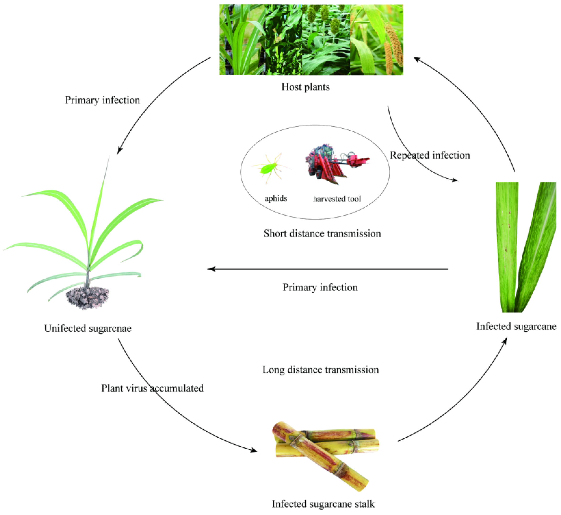
The primary infectious sources of mosaic mainly include infected plants of sugarcane and other Gramineae hosts. In nature, transmission of SCMV and SrMV is primarily by several aphid vectors including Dactynotus ambrosiae [55], Hysteroneura setariae [56], Longiunguis sacchari [57,58], Rhopalosiphum maidis [10,59], and Toxoptera graminum [60,61,62] in a non-persistent manner. Ants also have indirect transmission effect if they interact actively with aphids in diseased sugarcane fields [5,63]. However, insect borne SCSMV has not yet been detected [53,64,65], although Triticum mosaic virus (TriMV) and Wheat streak mosaic virus (WSMV), which belong to the same genus and share a high sequence similarity with SCSMV, can be transmitted by wheat curl mites (Aceria tosichella Keifer) [16,66]. The three viruses are easily transmitted over a short distance by machines, cutting tools, and juice fluid friction, but transmission over long-distance is mainly through infected materials [12,20]. A diagram of the specific transmission pathway is shown in Figure 2.
2.4. Epidemiology
The severity of mosaic disease in sugarcane fields is closely associated with sugarcane variety, infected setts, climatic conditions, and intermediate hosts. Among the six Saccharum species, S. officinarum are highly susceptible, S. sinense, S. spontaneum [67] and S. barber [68] are highly resistant or immune, S. robustum [67,68] are susceptible to mosaic. This disease was also found on S. edule [69], but the resistance to viruses is still uncertain. However, a recent study showed that most of Saccharum species have poor resistance to SCSMV; only three out of eight accessions of S. robustum among all of the 210 tested clones of Saccharum are identified to be resistant [70]. Generally, sugarcane cultivars with more resistant consanguinity also tend to show stronger resistance [65]. Young sugarcane plants are more susceptible than old, mature plants [71]. Drought and less rainfall environments are beneficial to the reproduction and activities of aphids, which promote the spread of mosaic. However, an extremely hot climate is not conducive to disease transmission, leading to slow virus proliferation, less disease symptoms, and less severe disease incidence [5,13]. In general, mosaic often occurs seriously in weedy or intercropping sugarcane fields [35]. High susceptibility of main varieties, relatively high temperatures and less rain, different planting periods in the same region, long-term rotation, and single variety and long-term successive cropping can all lead to a serious occurrence or epidemic of mosaic [72].
3. Pathogenicity Characteristics
3.1. Taxonomic Status
SCMV, SrMV, and SCSMV belong to the Potyviridae family, of which SCMV and SrMV belong to Potyvirus. Maize dwarf mosaic virus (MDMV), Johnsongrass mosaic virus (JGWV), Zea mosaic virus (ZnMV), Cocksfoot streak virus (CSV), and Pennisetum mosaic virus (PenMV) are grouped together under SCMV subgroup [15,73]. SCSMV belongs to Poacevirus, as do TriMV and Caladenia virus A (CalVA) [17].
3.2. Morphology, Size, and Viability
3.3. Genome Structure
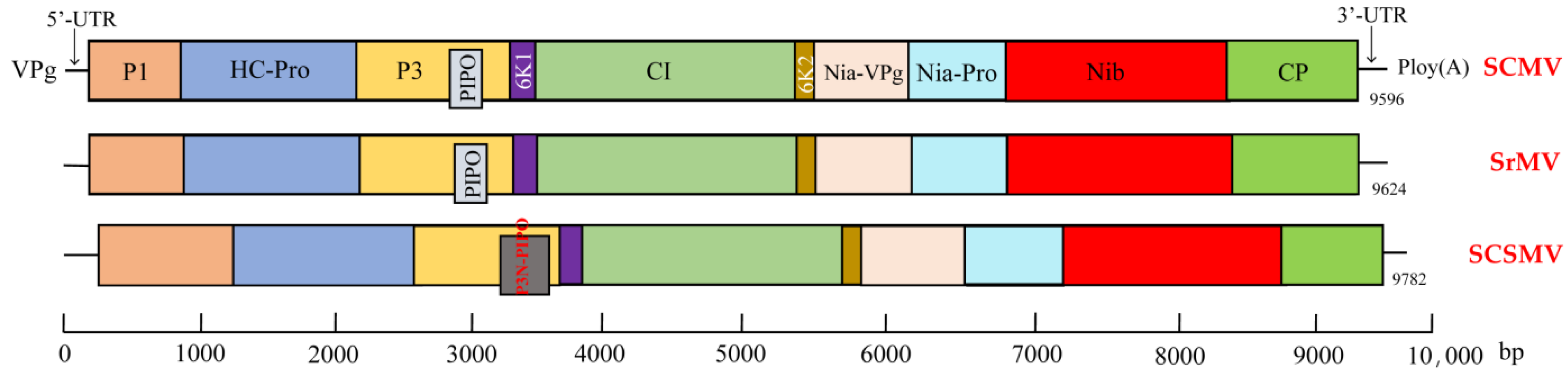
The genome of SCMV, SrMV, and SCSMV is represented by a positive-sense single-stranded RNA (+ssRNA) of about 10 Kb, consisting of untranslated regions (UTR) at both ends and a single open reading frame (ORF) encoding for a large polyprotein. The viral RNA harbour a genome-linked protein (VPg) at the RNA 5′-terminus and a poly (A) tract at the 3′-terminus [75]. The genome structure of the sugarcane mosaic virus is shown in Figure 3. The polyprotein is processed by the virus-encoded proteases P1-pro, HC-Pro and NIa-Pro into 10 mature functional proteins [73,76]. In addition, SCMV and SrMV encode an additional PIPO [77], and SCSMV encodes P3N-PIPO, which are expressed from the P3 ORF through a +2 or +1 frame-coding slippage mechanism, respectively [78,79].
Table 2 describes the main functions of the proteins encoded by viruses of the Potyviridae family. The PIPO and P3N-PIPO mainly affect the movement of the virus between cell-to-cell movement. P3N-PIPO binds to CI to recruit itself into plasmodesmata to promote intercellular movement of the virus [77,80]. Compared with SCMV and SrMV, a highly conserved motif of “Asp-Ala-Gly (DAG)” was absent in the CP sequence of SCSMV, which is necessary for aphid transmission [81,82]. It is worth mentioning that RNA silencing and RNA silencing repressors are mechanisms of defence and counter-defence interactions between host plants and viruses [83]. HC-Pro is the first strong RNA silencing inhibitor discovered [84], which has multiple targets in the RNA silencing pathway to regulate the accumulation of different siRNAs [85]. Moreover, when it fuses with P1, the expression of P1/HC-Pro and the inhibitory activity is enhanced [86]. The P1 protein of Poacevirus also has a silencing inhibitory function, which is more obvious than HC-Pro. However, when HC-Pro is present, the inhibitory activity of P1 on RNA silencing is reduced [87].
3.4. Genetic Diversity and Taxonomy
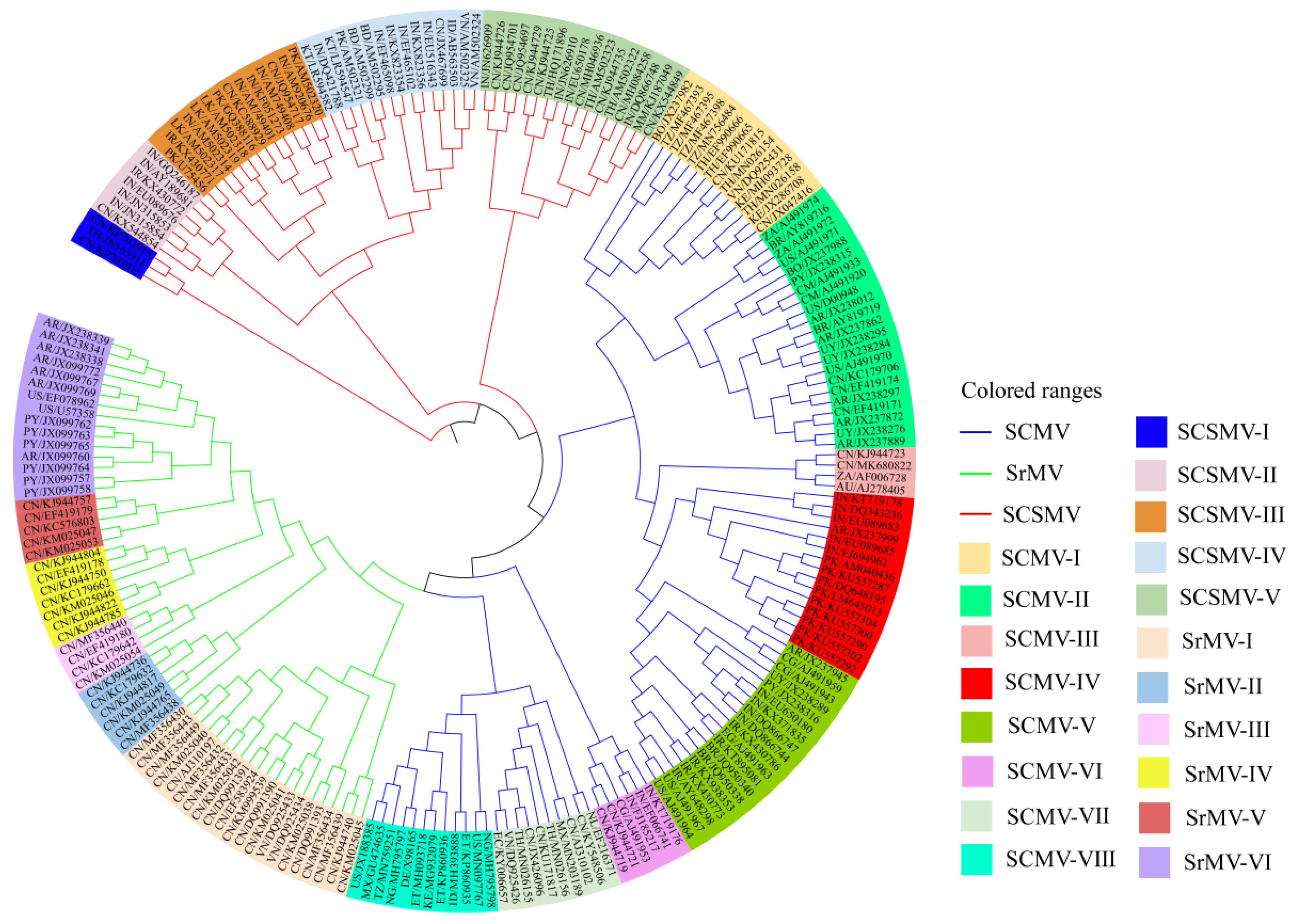
During evolution, SCMV, SrMV and SCSMV accumulated a rich pool of genetic diversity. Before 2000, Summers [106,107] and Summers et al. [11] divided SCMV into 10 strains and sub-strains according to the disease symptoms of sugarcane varieties CO281, CP29-291 and CP31-294. Tippett and Abbott [108] divided SCMV into five strains, namely, A, B, D, E and F, according to the mosaic symptoms of CP31-294, but two strains, A and H, according to CP31-588. According to serological cross-reaction, the evolutionary relationship and characteristics of CP nucleotide sequence based on the 3’-end sequence of the viral genome, Potyvirus isolates from Australia and the U.S. were divided into 11 SCMV strains, namely, MBD, A, B, D, E, SC, BC, Sabi, ISIS, Bris, and Bund, and three SrMV strains, namely H, I and M [15,43]. Since 2000, the genetic diversity of mosaic viruses isolates from China [109,110], South Africa [111], India [112,113], Mexico [114], Argentina [115] and Thailand [116,117], and others have also been reported based on the nucleotide and/or amino acid sequence variability of CP [81,118], HC-Pro [119] and P1 [79]. To sum up the results of genetic diversity, 216 relatively complete nucleotide sequences of the coat protein genes from102 SCMV isolates from 26 countries, 58 SrMV isolates from five countries, and 56 SCSMV isolates from 11 countries are downloaded from the NCBI database (accessed date 16 May 2021) and analysed using the maximum likelihood method (ML) of MEGA v6.0 software (Raynham MA, USA). The results are depicted in Figure 4.
Plant RNA viruses mutate in a variety of ways, including natural selection and substitution, transversion, deletion, insertion, recombination, reassortment, etc. [120,121]. Gell et al. [122] found that recombination was the main driving force for evolving SCMV subgroup populations. Recombination and strong selection pressure may accelerate the elimination process of deleterious mutations in the SCSMV genome P1 gene [79]. Strong purifying selection has been dominant in Indian SCMV populations, in which the CI and HC-Pro genes are prevalent [123]. He [64] reported that negative selection and genetic drift rather than recombination were the main driving force for the evolution of SrMV and SCSMV in China. In addition, natural selection, gene migration and geographical isolation may also affect the evolution of virus population in different regions [22,81,124].
4. Diagnosis/Identification
4.1. Visual Observation
Mosaic disease can be identified by visual inspection of sugarcane leaves for symptoms when evaluating germplasm resistance [38,39,125,126]. The method is simple and timesaving. However, it cannot determine the virus types. In addition, it requires a higher level of professional knowledge, skill, and ability to catch sugarcane leaf mosaic symptoms with the naked eye.
4.2. Biological Identification
Traditionally, the type of virus, host range and transmission mode are determined mainly based on the symptoms of the host plant after inoculation with the virus [11,127]. Before 1940, Summers EM artificially inoculated leaves of indicator plants of varieties CP31-294 and Co281 with press juice friction from a symptomatic plant and made a judgment based on whether the leaves showed any mosaic symptoms [11,106,107]. However, it is hard to do, labour intensive, and difficult to determine the specific virus strain. The amount of needed plant material can fill up the greenhouses and strict test conditions are required.
4.3. Microscopic Observation
The most prominent pathological feature of plant tissue cells infected by a virus of the Potyviridae family is the production of columnar inclusions. The feature has been used to distinguish among virus species. Edwardson [128] divided cylindrical inclusions into four types: scrolls body and pinwheels (Type Ⅰ); lamellar aggregates with arm extension and pinwheels (Type Ⅱ); scrolls body, pinwheels, and lamellar aggregates (Type Ⅲ); and scrolls body, pinwheels, and short and curved lamellar aggregates (Type Ⅳ). Studies have shown that the characteristics of columnar inclusions in SCMV-, SrMV- [129] and SCSMV-infected [53] sugarcane leaf cells were similar to Ⅲ, Ⅰ, and Ⅱ, respectively. However, this method is seldomly used in practice due to the complicated operation procedure of electron microscopy.
4.4. Serological Detection
Serological detection is a simple, rapid, and low-cost method for plant viruses, including agar gel immunodiffusion (AGID), electroblot immunoassay (EBIA), double antibody sandwich enzyme-linked immunosorbent assay (DAS-ELISA), direct antigen coating ELISA (DAC-ELISA), indirect ELISA, and dot enzyme-linked immunosorbent assay (Dot-ELISA) (also dot immunobinding assay, DIBA; or dot-bolt immunobinding assays, DBIA) [74,130,131]. Hema et al. [74] detected SCSMV by AGID, DAC-ELISA and EBIA. Mohammadi et al. [132] used DAS-ELISA and DIBA to detect SCMV and SrMV in infected samples. SrMV was first detected in Miscanthus by Grisham et al. [51] using indirect ELISA. Gaur et al. [133] used DAC-ELISA and DIBA to detect SCMV in infected cane juice samples even when the samples were diluted to 1/150. Wang et al. [134] established a high-throughput DAC-ELISA method for the detection of SrMV. However, serological detection requires specific antiserums, and its sensitivity is relatively lower than molecular detection techniques [130,131].
4.5. Molecular Detection
PCR-based molecular techniques have been developed and widely used in the detection of the mosaic virus, such as direct-binding polymerase chain reaction (DB-PCR), reverse transcription PCR (RT-PCR), immunocapture RT-PCR (IC-RT-PCR), duplex immunocapture RT-PCR (D-IC-RT-PCR), one-step multiplex RT-PCR, real-time quantitative RT-PCR (qRT-PCR) and loop-mediated isothermal amplification (LAMP) [83]. These molecular detection techniques have a high sensitivity and specificity and can identify virus species rapidly and accurately. Yang and Mirkov [135] used DNA restriction enzymes to digest RT-PCR fragments to produce SCMV- or SrMV-specific RFLP patterns without the need of using identification host plants. Xie et al. [136] established a one-step quadruplex RT-PCR method to simultaneously detect four viruses, namely, SrMV, SCMV, SCSMV and Sugarcane yellow leaf virus (SCYLV). Smith and Velde [137] were able to detect SCMV by RT-PCR in samples that were diluted 10,000-fold. Fu et al. [52] developed a qRT-PCR method for the detection of SCSMV, which was 100 times more sensitive than conventional RT-PCR. Hema et al. [138] reported that IC-RT-PCR was 5,000 times more sensitive than DBIA and 10 times more sensitive than DB-PCR to detect SCSMV. Chen et al. [139] demonstrated that the sensitivity of detecting SrMV by IC-RT-PCR 100-times higher than that of indirect ELISA and 1000-times higher than that of Dot-ELISA and therefore recommended IC-RT-PCR was more suitable for large volumes of samples. Subba et al. [140] developed a D-IC-RT-PCR method that combined both serological and molecular methods to simultaneously detect and distinguish SCMV and SCSMV and was more sensitive than DAC-ELISA. In addition, a new reverse transcription-LAMP (RT-LAMP) technology was also applied to detect SCMV and SrMV with a lower sensitivity than RT-PCR and qRT-PCR [141]. The primer sequence information of different molecular techniques for detecting sugarcane mosaic viruses is shown in Table 3.
5. Prevention and Control Strategy
5.1. Exploitation and Utilization of Resistant Germplasm
Selection and rational distribution of disease-resistant varieties are the most economic and effective prevention and control measures against these viruses. The genetic base of modern sugarcane cultivars is narrow, with about 80% from S. officinarum, 10–15% from S. spontaneum, and 5–10% from recombinant chromosomes [142]. During the long evolutionary process, the Saccharum and its related genera have formed extremely rich and valuable germplasm resources [68,143], which contain large numbers of disease resistance genes [144]. In recent years, some Saccharum hybrid clones with high resistance to mosaic disease have been identified by natural infection or artificial inoculation, such as SP70-1143, IACSP95-2078 [145], SWSwm1 [146], YG34, YG55 [147], ROC16 [38], GT03-2309, and LC03-1137 [39]. The current artificial inoculation methods are shown in Table 4.
5.2. Acceleration of Molecular Breeding
Molecular marker-assisted breeding and genetic engineering improvements have helped promote the development of resistant varieties. Molecular marker-assisted breeding is an effective method to accelerate the breeding process of multi-resistant varieties. However, the technology is limited by the lack of markers closely linked to disease resistance. Sugarcane (2 n = 12 x = 100−130; genome size = ~10 Gb) is a highly complex autopolyploid and aneuploid crop, and a complete reference genome of modern sugarcane cultivated species is still lacking up to now [154,155]. The development of molecular markers associated with economic target traits is an extremely slow process for sugarcane. Previous studies on the development of molecular markers for mosaic resistance were mainly focus on corn (2 n = 2 x = 20; genome size = ~2300 Mb) [156,157,158,159,160,161,162,163,164]. However, these studies may provide a good reference for developing molecular markers and related gene mining on sugarcane. In addition, genetic engineering is an effective way to obtain disease-resistant sugarcane varieties [165]. The most mature strategy is CP gene-mediated transfer since the first report of the introduction of the Tobacco mosaic virus (TMV) CP gene into tobacco in 1986 [166]. Smith et al. [167] used a gene gun to bombard a SCMV-CP gene into sugarcane meristem and obtained chimeric transformed plants. Subsequently, Joyce et al. [168], Ingelbrecht et al. [169], and Sooknandan et al. [170] also succeeded in obtaining transgenic sugarcane plants by using the same method. On the other hand, an RNA interference technology that targets sugarcane virus encoded RNA silencing inhibitors has also been successfully used to develop highly effective disease-resistant transgenic sugarcane plants [171,172,173,174,175]. Field experiments on transgenic sugarcane have shown improved resistance to mosaic disease with significantly increased yield and sucrose content [171,176,177,178]. However, none of the transgenic sugarcane has yet been applied in field production due to regulations.
5.3. Application of Virus-Free Plantlets
The application of virus-free plantlets through tissue culture can not only eliminate or slow down the incidence of mosaic disease, but also increase the sucrose content by more than 0.5% and the cane yield by 20–40% [179]. In one case, the yield increased by more than 100% [180]. The common methods of detoxification include heat treatment, stem buds tissue culture detoxification, and ultra-low temperature therapy. SCMV elimination can be achieved by cultivation at 52 °C, 57.3 °C, 57.3 °C, once every other day, 20 min each time, without harming cane buds [179]. SCMV can also be removed by cryo-treating micro shoot tips (~3 mm) [181] or by inserting 4~8 mm axillary bud explants into MS regeneration medium supplemented with a 25 mg/L of ribavirin [182]. Hot water treatment for 10 min at 55 ℃ was not effective for SCSMV elimination [183]. Detoxification by stem buds tissue culture could completely eliminate SCSMV [184,185]. A combination of heat treatment and axillary shoot tip culture had a better effect on virus removal [186].
To determine the technical specifications for the production and testing of sugarcane virus-free plantlets, leaves were collected from the virus-free plantlets produced by the heat treatment and axillary shoot tip culture technology in 2015, followed by tracking and PCR testing the leaves of 120 putative virus-free plantlets collected from different companies at different ecological demonstration sites in China [187]. The PCR results showed that SCMV was not detected in 100% of the samples, the rate of SrMV detection was greater than 35%, and the rate of SCSMV detection was more than 50%. Therefore, it was concluded that SrMV and SCSMV were more difficult to be eliminated from infected sugarcane plants than SCMV. Although the virus-free effect of sugarcane stalk can be improved by appropriately increasing treatment temperature and time (30 min at 59 °C), the germination rate, however, was significantly reduced to only about 20% and the water temperature was difficult to control accurately in an industrial setting (unpublished).
5.4. Strengthen Cultivation and Control
Good field management is another effective way to enhance plant resistance and reduce the spread of viruses. Specific measures include: (1) avoiding planting virus host crops around or in the sugarcane fields [188]; (2) the timely removal of infected plants and weeds; (3) improvement in soil structure, rational fertilization, and irrigation to promote plant growth and improve disease resistance [189,190]; (4) chemical and biological control of aphids [63]; and (5) fortifying cropping systems and paying attention to rotation with non-host crops, such as soybeans, sweet potatoes and peanuts [191].
6. Perspective
The cultivation and planting of resistant varieties are the most economic and effective methods to control sugarcane infecting viruses, and growers are most likely to adopt this technology. However, due to the diversity of pathogens, the highly complex genome of sugarcane, the wide segregation of traits among hybrid progenies, and the extremely low probability of excellent gene aggregation, sugarcane cross-breeding may have to rely on a huge population, for example, 1 to 1.2 million of plantlets to be planted annually in China. Therefore, it is very difficult to select good varieties that have both commercial value and mosaic resistance. In addition, field evaluation tests of virus-free plantlets from integrated detoxification technology in China have shown that the “virus-free” effect is not ideal, even if the plantlets were produced by a more effective method of combining heat treatment and axillary bud or shoot tip culture. In view of all these facts, the development of molecular markers associated with disease resistance genes should be the most effective way to breed sugarcane varieties resistant to different viruses. Sugarcane propagates asexually and F1 populations can be used for correlation analysis between genotypes and phenotypic traits. There are also successful cases on genetic engineering via gene gun bombardment or gene editing to improve disease resistance in sugarcane. Successfully edited alleles can be up to 49 copies simultaneously targeting the chlorophyll content gene, which draws a new blueprint for transforming the disease resistance pathway in sugarcane. We hope that the present review can provide scientific references and thoughts for the effective prevention and control of viruses in sugarcane.
Author Contributions
Conceptualization, G.L. and L.X.; methodology, G.L. and L.X.; validation, Z.W. and F.X.; writing—original draft preparation, G.L. and L.X.; writing—review and editing Y.-B.P., L.X. and M.P.G.; supervision, L.X.; project administration, L.X.; funding acquisition, L.X. All authors have read and agreed to the published version of the manuscript.
Funding
This research was funded by China Agriculture Research System of MOF and MARA [CARS-17]; Special Fund for Science and Technology Innovation, FAFU(KFA20083A); and a Non-Funded Cooperative Agreement (Accession Number: 428234) between the USDA-ARS and NRDCSIT on Sugarcane Breeding, Varietal Development, and Disease Diagnosis. The funders had no role in the design of the study; in the collection, analyses, or interpretation of data; in the writing of the manuscript, or in the decision to publish the results.
Institutional Review Board Statement
Not applicable.
Informed Consent Statement
Not applicable.
Data Availability Statement
All the data is present in the manuscript.
Acknowledgments
The authors are thankful to Youxiong Que from Fujian Agriculture and Forestry University in China, and Sushma Sood, Yuling Jia, and Perng-Kuang Chang for reviewing the manuscript with excellent comments. USDA is an equal opportunity provider and employer.
Conflicts of Interest
The authors declare no conflict of interest.
References
- Arruda, P. Perspective of the Sugarcane Industry in Brazil. Trop. Plant Biol. 2011, 4, 3–8. [Google Scholar] [CrossRef]
- Junior, J.F.; Palacio, J.E.; Leme, R.C.; Lora, E.S.; Eduardo, L.; Reyes, A.M.; Olmo, O.D. Biorefineries productive alternatives optimization in the brazilian sugar and alcohol industry. Appl. Energy 2019, 259, 113092. [Google Scholar] [CrossRef]
- Lu, G.; Wang, S.; Lian, Y.; Wei, Y. Development and utilization of sugarcane by-products in the sugar manufacturing process. Sugar Crop. China 2020, 42, 75–80. [Google Scholar]
- Sindhu, R.; Gnansounou, E.; Binod, P.; Pandey, A. Bioconversion of sugarcane crop residue for value added products—An overview. Renew. Energy 2016, 98, 203–215. [Google Scholar] [CrossRef]
- Li, Y.-R. Modern Sugarcane Cultivation; China Agriculture Press: Beijing, China, 2010; pp. 358–359. [Google Scholar]
- Musschenbroek, V.S.C. Beschrijving van twee tot dusverre in west-Java onbekende rietziekten. Soerabaiasche Ver. Suiker Fabr. 1893, 42, 113–118. [Google Scholar]
- Kelly, N.L. BSES cane pest and diseases report. Qld. Agric. J. 1927, 27, 82–83. [Google Scholar]
- Brandes, E.W. The Mosaic Disease of Sugar Cane and Other Grasses; Technical Bulletin No. 829; United States Department of Agriculture: Washington, DC, USA, 1919.
- Dastur, J.F. The mosaic disease of sugarcane in India. Agric. J. India 1923, 18, 505–509. [Google Scholar]
- Brandes, E.W. Artificial and Insect Transmission of Sugar-Cane Mosaic. J. Agric. Res. 1920, 19, 131. [Google Scholar]
- Summers, E.M.; Brandes, E.W.; Rands, R.D. Mosaic of Sugarcane in the United States, with Special Reference to Strains of the Virus; Technical Bulletin No. 955; United States Department of Agriculture: Washington, DC, USA, 1948.
- Koike, H.; Gillaspie, J.R. Mosaic. In Disease of Sugarcane: Major Disease; Ricaud, C., Egan, B.T., Gillaspie, A.G., Hughes, C.G., Eds.; Elsevier Science Publisher: Amsterdam, The Netherland, 1989; pp. 301–322. [Google Scholar]
- Grisham, M.P. Mosaic. In A Guide to Sugarcane Diseases; Rott, P., Bailey, R.A., Comstock, J.C., Croft, B.J., Eds.; CIRAD Publication Services: Montepellier, France, 2011; pp. 249–254. [Google Scholar]
- Wu, L.; Zu, X.; Wang, S.; Chen, Y. Sugarcane mosaic virus—Long history but still a threat to industry. Crop Prot. 2012, 42, 74–78. [Google Scholar] [CrossRef]
- Shukla, D.D.; Frenkel, M.J.; Mckern, N.M.; Ward, C.W.; Jilka, J.; Tosic, M.; Ford, R.E. Present status of the sugarcane mosaic subgroup of potyviruses. In Potyvirus Taxonomy; Springer: New York, NY, USA, 1992; pp. 363–373. [Google Scholar]
- Hall, J.S.; Adams, B.; Parsons, T.J.; French, R.; Lane, L.C.; Jensen, S.G. Molecular Cloning, Sequencing, and Phylogenetic Relationships of a New Potyvirus: Sugarcane Streak Mosaic Virus, and a Reevaluation of the Classification of the Potyviridae. Mol. Phylogenet. Evol. 1998, 10, 323–332. [Google Scholar] [CrossRef]
- Adams, M.J.; Carstens, E.B. Ratification vote on taxonomic proposals to the International Committee on Taxonomy of Viruses. Arch. Virol. 2012, 157, 1411–1422. [Google Scholar] [CrossRef] [Green Version]
- Perera, M.F.; Filippone, M.P.; Noguera, A.S.; Cuenya, M.I.; Castagnaro, A.P. An Overview of the Sugarcane Mosaic Disease in South America. Funct. Plant Sci. Biotechnol. 2012, 6, 98–107. [Google Scholar]
- Feng, X.Y.; Shen, L.B.; Zhao, T.T.; Xiong, G.R.; Wang, J.G.; Yang, B.P.; Wang, W.Z.; Feng, C.L.; Zhang, S.Z. Molecular identification of virus diseases in sugarcane in Yunnan. J. South. Agric. 2018, 49, 2198–2203. [Google Scholar]
- Putra, L.K.; Kristini, A.; Achadian, E.M.; Damayanti, T.A. Sugarcane streak mosaic virus in Indonesia: Distribution, Characterisation, Yield Losses and Management Approaches. Sugar Tech 2014, 16, 392–399. [Google Scholar] [CrossRef]
- Michèle, C.; Candy, M.; Jeanclaude, G.; Daniel, G.; Rao, G.P.; Monique, R.; Lockhart, B.; Philippe, R. Mosaic symptoms in sugarcane are caused by Sugarcane streak mosaic virus (SCSMV) in several Asian countries. In Proceedings of the Pathology Workshop of the International Society of Sugar Cane Technologists, Baton Rouge, LA, USA, 11–16 May 2003. [Google Scholar]
- Liang, S.S.; Alabi, O.J.; Damaj, M.B.; Fu, W.L.; Gao, S.J. Genomic variability and molecular evolution of Asian isolates of sugarcane streak mosaic virus. Arch. Virol. 2016, 161, 1493–1503. [Google Scholar] [CrossRef] [PubMed]
- Sorho, F.; Sereme, D.; Kouamé, K.; Koné, N.; Kone, D.; Yao, K.; Ouattara, M.; Tapsoba, W.; Ouattara, B.; Kone, D. First report of Sugarcane Streak Mosaic Virus (SCSMV) infecting sugarcane in Cote d’Ivoire. Plant Dis. 2021, 105, 519. [Google Scholar] [CrossRef]
- Wang, X.Y.; Li, W.F.; Huang, Y.K.; Zhang, R.Y.; Shan, H.L.; Yin, J.; Luo, Z.M. Molecular detection and phylogenetic analysis of viruses causing mosaic symptoms in new sugarcane varieties in China. Eur. J. Plant Pathol. 2017, 148, 931–940. [Google Scholar] [CrossRef]
- Viswanathan, R.; Balamuralikrishnanan, M.; Karuppaiah, R. Sugarcane mosaic complex in India: Cause of different viruses/strains. Indian J. Virol. 2008, 19, 94. [Google Scholar]
- Perera, M.F.; Filippone, M.P.; Ramallo, J.C.; Cuenya, M.I.; Castagnaro, A.P. Diversidad genética del complejo de virosis asociadas a la enfermedad del mosaico de la caña de azúcar en Tucumán, Argentina. Rev. Ind. Agríc. Tucumán 2009, 86, 1–6. [Google Scholar]
- Wang, W.; Ma, Z.; Zhang, S.; Yang, B.; Cai, W.; Gu, L.; Li, J. Research on Genetic Engineering of Sugarcane Mosaic Disease. Biotech. Bull. 2009, 30, 22–26. [Google Scholar]
- Chaves-Bedoya, G.; Espejel, F.; Alcalá-Briseño, R.; Hernández-Vela, J.; Silva-Rosales, L. Short distance movement of genomic negative strands in a host and nonhost for Sugarcane mosaic virus (SCMV). Virol. J. 2011, 8, 15. [Google Scholar] [CrossRef] [PubMed] [Green Version]
- Pokorny, R.; Porubova, M. Movement of Sugarcane mosaic virus in plants of resistant and susceptible maize lines. Cereal Res. Commun. 2006, 34, 1109–11116. [Google Scholar] [CrossRef]
- Bagyalakshmi, K.; Viswanathan, R.; Ravichandran, V. Impact of the viruses associated with mosaic and yellow leaf disease on varietal degeneration in sugarcane. Phytoparasitica 2019, 47, 591–604. [Google Scholar] [CrossRef]
- Irvine, J.E. Photosynthesis in Sugarcane Varieties Infected with Strains of Sugarcane Mosaic Virus. Physiol. Plant. 1971, 24, 51–54. [Google Scholar] [CrossRef]
- Pan, D.R.; Ping, X.L.; Luo, J.; Ying, F.H. Improvement of photosynthetic characteristics and yield of Sugarcane mosaic virus-free chewing cane. J. Fujian Agric. For. Univ. 2001, 30, 320–323. [Google Scholar]
- Singh, V.; Sinha, O.K.; Kumar, R. Progressive decline in yield and quality of sugarcane due to Sugarcane mosaic virus. Indian Phytopathol. 2003, 56, 500–502. [Google Scholar]
- Singh, S.P.; Rao, G.P.; Singh, J.; Singh, S.B. Effect of sugarcane mosaic potyvirus infection on metabolic activity, yield and juice quality. Sugar Cane 1997, 5, 19–23. [Google Scholar]
- He, Y.S.; Li, R.M. Research Status of Sugarcane Mosaic Virus Disease in China. Sugar Crop. China 2006, 28, 47–49. [Google Scholar]
- Viswanathan, R.; Balamuralikrishnan, M. Impact of mosaic infection on growth and yield of sugarcane. Sugar Tech 2005, 7, 61–65. [Google Scholar] [CrossRef]
- Jones, C. Mosaic disease at Isis. BSES Bull 1987, 20, 15–16. [Google Scholar]
- Li, Y.J.; Tang, S.Y.; Huang, Y.Z.; Duan, W.X.; Wang, Z.P.; Luo, T.; Lin, S.H. Occurrence and resistance analysis of sugarcane mosaic in Liuzhou and Laibin regions. China Plant Prot. 2017, 37, 51–55. [Google Scholar]
- Yang, R.Z.; Zhou, H.; Xiao, W.; Lü, D.; Liao, H.X.; Chen, D.D.; Liu, X.H.; Lei, J.C.; Lin, Y.F. Testing on sugarcane mosaic resistance of sugarcane major parents under field conditions. Sugar Crop. China 2020, 42, 47–52. [Google Scholar]
- Kumar, N.R.; Kumar, K.V.K.; Reddy, B.R. Characterization of Sugarcane Mosaic Disease and Its Management with PGPR. In Plant Growth Promoting Rhizobacteria (PGPR): Prospects for Sustainable Agriculture; Sayyed, R.Z., Reddy, M.S., Antonius, S., Eds.; Springer: New Delhi, India, 2019; pp. 145–155. [Google Scholar]
- Deshumkh, G.P.; Sawant, D.M. Studies on Symptomatology of Sorghum and Sugarcane Mosaic Virus. J. Plant Dis. Sci. 2008, 3, 116–117. [Google Scholar]
- Srinivas, K.P.; Reddy, C.V.; Ramesh, B.; Kumar, P.L.; Sreenivasulu, P. Identification of a virus naturally infecting sorghum in India as Sugarcane streak mosaic virus. Eur. J. Plant Pathol. 2010, 127, 13–19. [Google Scholar] [CrossRef] [Green Version]
- Shukla, D.D.; Ward, C.W. Structure of potyvirus coat proteins and its application in the taxonomy of the potyvirus group. Adv. Virus Res. 1989, 36, 273–314. [Google Scholar]
- Yang, B.P. Modern Sugarcane Cultivation Techniques; China Agriculture Press: Beijing, China, 2019; pp. 358–359. [Google Scholar]
- Rosenkranz, E. New hosts and taxonomic analysis of the mississippi native species tested for reaction to Maize dwarf mosaic and Sugarcane mosaic viruses. Phytopathology 1987, 77, 598–607. [Google Scholar] [CrossRef]
- Harmon, P. Mosaic Disease of St. Augustinegrass Caused by Sugarcane Mosaic Virus. Plant Pathol. 2015, 313–315. [Google Scholar]
- Mollov, D.; Tahir, M.N.; Wei, C.; Kaye, C.; Lockhart, B.; Comstock, J.C.; Rott, P. First Report of Sugarcane mosaic virus Infecting Columbus Grass (Sorghum almum) in the United States. Plant Dis. 2016, 100, 1510. [Google Scholar] [CrossRef]
- Zhao, H.; Zhang, H.; Yang, Z.; Wang, T.; Liu, Y.; Cheng, G.; Xu, J. First Report of Sugarcane Mosaic Virus on Pumpkin Plants Exhibiting Mosaic and Mottling Symptoms in China. Plant Dis. 2019, 103, 1802. [Google Scholar] [CrossRef]
- Baker, C.A.; Wilber, L.J.; Jones, L. A New Host Diagnosed with a Strain of Sugarcane mosaic virus in Florida: Red-Veined Prayer Plant (Maranta leuconeura erythroneura). Plant Dis. 2010, 94, 378–379. [Google Scholar] [CrossRef] [PubMed]
- Tang, W.; Xu, X.H.; Sun, H.W.; Li, F.; Gao, R.; Yang, S.K.; Li, X.D. First Report of Sugarcane mosaic virus Infecting Canna spp. in China. Plant Dis. 2016, 100, 2541. [Google Scholar] [CrossRef]
- Grisham, M.P.; Maroon-Lango, C.J.; Hale, A.L. First Report of Sorghum mosaic virus Causing Mosaic in Miscanthus sinensis. Plant Dis. 2012, 96, 150. [Google Scholar] [CrossRef] [PubMed]
- Fu, W.L.; Sun, S.R.; Fu, H.Y.; Chen, R.K.; Su, J.W.; Gao, S.J. A One-Step Real-Time RT-PCR Assay for the Detection and Quantitation of Sugarcane Streak Mosaic Virus. BioMed Res. Int. 2015, 2015, 569131. [Google Scholar] [CrossRef] [Green Version]
- Hema, M.; Savithri, H.S.; Sreenivasulu, P. Sugarcane streak mosaic virus: Occurrence, purification, characterization and detection. In Sugarcane Pathology. Virus and Phytoplasma Diseases; Rao, G.P., Ford, R.E., Tosic, M., Teakle, D.S., Eds.; Science Publishers: Enfield, NE, USA, 2001; Volume 2, pp. 37–70. [Google Scholar]
- Singh, D.; Rao, G.P. Sudan grass (Sorghum sudanense Stapf): A new sugarcane streak mosaic virus mechanical host. J. Guangxi Agric. Sci. 2010, 41, 436–438. [Google Scholar]
- Koike, H.; Gillaspie, J.A.G. Strain M, a new strain of sugarcane mosaic. Plant Dis. Rep. 1976, 60, 50–54. [Google Scholar]
- Kennedy, J.S.; Day, M.F.; Eastop, V.F. A Conspectus of Aphids as Vectors of Plant Viruses; Commonwealth Institute of Entomology: London, UK, 1962. [Google Scholar]
- Kondaiah, E.; Nayudu, M.V. Sugarcane mosaic virus strain H-a new record from India. Curr. Sci. India 1984, 53, 273–275. [Google Scholar]
- Singh, M.; Singh, A.; Upadhyaya, P.P.; Rao, G.P. Transmission studies on an Indian isolate of sugarcane mosaic potyvirus. Sugar Tech 2005, 7, 32–38. [Google Scholar] [CrossRef]
- Klein, P.; Smith, C.M. Host plant selection and virus transmission by Rhopalosiphum maidis are conditioned by potyvirus infection in Sorghum bicolor. Arthropod-Plant Interact. 2020, 14, 811–823. [Google Scholar] [CrossRef]
- Saladini, J.L.; Zettler, F.W. Resistance of St. Augustine grass to infection by sugarcane mosaic virus. Phytopathology 1973, 63, 162–166. [Google Scholar] [CrossRef]
- Brunt, A.A.; Crabtree, K.; Dallwitz, M.J.; Gibbs, A.J.; Watson, L.; Zurcher, E.J. (Eds.) Viruses of Plants. Descriptions and Lists from the VIDE Database; Cambridge University Press: Cambridge, UK, 1996. [Google Scholar]
- Lockhart, B.E.L.; Autrey, L.J.C. Mild mosaic. In A Guide to Sugarcane Diseases; Rott, P., Bailey, R.A., Comstock, J.C., Croft, B.J., Eds.; La Librairie du Cirad: Montpellier, France, 2000; pp. 245–248. [Google Scholar]
- Raza, A.; Farooq, T. Sugarcane mosaic virus in sugarcane. In Pest Management Decision Guides; CABI Publishers: Wallingford, UK, 2017; p. 1. [Google Scholar]
- He, Z. Molecular Evolution of Sugarcane Streak Mosaic and Sorghum mosaic virus. Ph.D. Thesis, Chinese Academy of Agricultural Sciences, Beijing, China, 2014. [Google Scholar]
- Huang, Y.K.; Li, W.F. Colored Atlas of Control on Diseases, Insect Pests and Weeds of Modern Sugarcane; China Agriculture Press: Beijing, China, 2016; pp. 118–122. [Google Scholar]
- Seifers, D.L.; Martin, T.J.; Harvey, T.L.; Fellers, J.P.; Michaud, J.P. Identification of the Wheat Curl Mite as the Vector of Triticum mosaic virus. Plant Dis. 2009, 93, 25–29. [Google Scholar] [CrossRef]
- Brandes, E.W.; Sartoris, G.B.; Grassl, C.O. Assembling and evaluating wild forms of sugarcane and closely related plants. Proc. Int. Soc. Sugar Cane Technol. 1939, 6, 128–153. [Google Scholar]
- Silva, M.F.; Goncalves, M.C.; Melloni, M.; Perecin, D.; Landell, M.; Xavier, M.A.; Pinto, L.R. Screening Sugarcane Wild Accessions for Resistance to Sugarcane Mosaic Virus (SCMV). Sugar Tech 2015, 17, 252–257. [Google Scholar] [CrossRef] [Green Version]
- Kuniata, L.S.; Magarey, R.C.; Rauka, G.; Price, T.V. Conservation of sugarcane germplasm: Survey of Papua New Guinea, Indonesia and northern Australia. Aciar Techn. Rep. 2006, 62, 36–42. [Google Scholar]
- Bagyalakshmi, K.; Viswanathan, R. Development of a Scoring System for Sugarcane Mosaic Disease and Genotyping of Sugarcane Germplasm for Mosaic Viruses. Sugar Tech 2021, 23, 1105–1117. [Google Scholar] [CrossRef]
- Putra, L.K.; Ogle, H.J.; James, A.P.; Whittle, P. Distribution of Sugarcane mosaic virus in sugarcane plants. Australas. Plant Pathol. 2003, 32, 305–307. [Google Scholar] [CrossRef]
- IPP; CAAS; CSPP. Crop Diseases and Insects in China, 3rd ed.; China Agriculture Press: Beijing, China, 2015; Volume 3, pp. 800–801. [Google Scholar]
- Adams, M.J.; Zerbini, F.M.; French, R.; Rabenstein, F.; Strenger, D.C.; Valkonen, J. Family Potyviridae. In Virus Taxonomy, Ninth Report of the International Committee on Taxonomy of Viruses; Oxford University Press: London, UK, 2012; pp. 1069–1089. [Google Scholar]
- Hema, M.; Joseph, J.; Gopinath, K.; Sreenivasulu, P.; Savithri, H.S. Molecular characterization and interviral relationships of a flexuous filamentous virus causing mosaic disease of sugarcane (Saccharum officinarum L.) in India. Arch. Virol. 1999, 144, 479–490. [Google Scholar] [CrossRef]
- Verma, R.K.; Mishra, R.; Sharma, P.; Gaur, R.K. Systemic infection of Potyvirus: A compatible interaction between host and viral proteins. In Approaches to Plant Stress and Their Management; Springer: New Delhi, India, 2014; pp. 353–363. [Google Scholar]
- Xu, D.L.; Zhou, G.H.; Xie, Y.J.; Mock, R.; Li, R. Complete nucleotide sequence and taxonomy of Sugarcane streak mosaic virus, member of a novel genus in the family Potyviridae. Virus Genes 2010, 40, 432–439. [Google Scholar] [CrossRef]
- Chung, Y.W.; Miller, W.A.; Atkins, J.F.; Firth, A.E. An overlapping essential gene in the Potyviridae. Proc. Natl. Acad. Sci. USA 2008, 105, 5897–5902. [Google Scholar] [CrossRef] [Green Version]
- Chandran, V.; Gajjeraman, P. Molecular Diversity Analysis of Pretty Interesting Potyviridae ORF (PIPO) Coding Region in Indian Isolates of Sugarcane streak mosaic virus. Sugar Tech 2016, 18, 214–221. [Google Scholar] [CrossRef]
- Parameswari, B.; Bagyalakshmi, K.; Chinnaraja, C.; Viswanathan, R. Molecular characterization of Indian Sugarcane streak mosaic virus isolates reveals recombination and negative selection in the P1 gene. Gene 2014, 552, 199–203. [Google Scholar] [CrossRef]
- Wei, T.; Zhang, C.; Hong, J.; Xiong, R.; Kasschau, K.D.; Zhou, X.; Carrington, J.C.; Wang, A. Formation of Complexes at Plasmodesmata for Potyvirus Intercellular Movement Is Mediated by the Viral Protein P3N-PIPO. PLoS Pathog. 2010, 6, e1000962. [Google Scholar] [CrossRef] [Green Version]
- He, Z.; Li, W.; Yasaka, R.; Huang, Y.; Li, S. Molecular variability of sugarcane streak mosaic virus in China based on an analysis of the P1 and CP protein coding regions. Arch. Virol. 2014, 159, 1149–1154. [Google Scholar] [CrossRef]
- López-Moya, J.; Pirone, T.P.; Wang, R.Y. Context of the coat protein DAG motif affects potyvirus transmissibility by aphids. J. Gen. Virol. 1999, 80, 3281–3288. [Google Scholar] [CrossRef] [PubMed]
- Liang, S.; Luo, Q.; Chen, R.; Gao, S. Advances in researches on molecular biology of viruses causing sugarcane mosaic. J. Plant Prot. 2017, 44, 363–370. [Google Scholar]
- Anandalakshmi, R.; Pruss, G.J.; Ge, X.; Marathe, R.; Mallory, A.C.; Smith, T.H.; Vance, V.B. A viral suppressor of gene silencing in plants. Proc. Natl. Acad. Sci. USA 1998, 95, 13079–13084. [Google Scholar] [CrossRef] [Green Version]
- Zhang, X.; Peng, D.; Lu, L.; Qi, X.; Wang, W.; Cao, X.; Bo, R.; Wei, C.; Yi, L. Contrasting effects of HC-Pro and 2b viral suppressors from Sugarcane mosaic virus and Tomato aspermy cucumovirus on the accumulation of siRNAs. Virology 2008, 374, 351–360. [Google Scholar] [CrossRef] [Green Version]
- Pruss, G.; Xin, G.; Xing, M.S.; Vance, C. Plant Viral Synergism: The Potyviral Genome Encodes a Broad-Range Pathogenicity Enhancer That Transactivates Replication of Heterologous Viruses. Plant Cell 1997, 9, 859–868. [Google Scholar] [CrossRef]
- Tatineni, S.; Qu, F.; Li, R.; Morris, T.J.; French, R. Triticum mosaic poacevirus enlists P1 rather than HC-Pro to suppress RNA silencing-mediated host defense. Virology 2012, 433, 104–115. [Google Scholar] [CrossRef] [Green Version]
- Valli, A.; Lopez-Moya, J.J.; Garcia, J.A. Recombination and gene duplication in the evolutionary diversification of P1 proteins in the family Potyviridae. J. Gen. Virol. 2007, 88, 1016–1028. [Google Scholar] [CrossRef] [PubMed]
- Kasschau, K.D.; Carrington, J.C. Long-Distance Movement and Replication Maintenance Functions Correlate with Silencing Suppression Activity of Potyviral HC-Pro. Virology 2001, 285, 71–81. [Google Scholar] [CrossRef] [Green Version]
- Atreya, C.D.; Atreya, P.L.; Thornbury, D.W.; Pirone, T.P. Site-directed mutations in the potyvirus HC-Pro gene affect helper component activity, virus accumulation, and symptom expression in infected tobacco plants. Virology 1992, 191, 106–111. [Google Scholar] [CrossRef]
- Eiamtanasate, S.; Juricek, M.; Yap, Y.K. C-terminal hydrophobic region leads PRSV P3 protein to endoplasmic reticulum. Virus Genes 2007, 35, 611–617. [Google Scholar] [CrossRef]
- Suehiro, N. An important determinant of the ability of Turnip mosaic virus to infect Brassica spp. and/or Raphanus sativus is in its P3 protein. J. Gen. Virol. 2004, 85, 2087–2098. [Google Scholar] [CrossRef]
- Hong, X.Y.; Chen, J.; Shi, Y.H.; Chen, J.P. The ‘6K1’ protein of a strain of Soybean mosaic virus localizes to the cell periphery. Arch. Virol. 2007, 152, 1547–1551. [Google Scholar] [CrossRef]
- Cui, H.; Wang, A. Plum Pox Virus 6K1 Protein Is Required for Viral Replication and Targets the Viral Replication Complex at the Early Stage of Infection. J. Virol. 2016, 90, 5119–5131. [Google Scholar] [CrossRef] [Green Version]
- Sorel, M.; Garcia, J.A.; German-Retana, S. The Potyviridae Cylindrical Inclusion Helicase: A Key Multipartner and Multifunctional Protein. Mol Plant Microbe Interact. 2014, 27, 215–226. [Google Scholar] [CrossRef] [Green Version]
- Romain, G.; Jiang, J.; Wan, J.; Maxime, A.; Zheng, H.; Jean-Francois, L. 6K2-induced vesicles can move cell to cell during turnip mosaic virus infection. Front. Microbiol. 2013, 4, 351. [Google Scholar]
- Spetz, C.; Valkonen, J. Potyviral 6K2 protein long-distance movement and symptom-induction functions are independent and host-specific. Mol. Plant Microbe Interact. 2004, 17, 502–510. [Google Scholar] [CrossRef] [Green Version]
- Jiang, J.; Patarroyo, C.; Cabanillas, D.G.; Zheng, H.; Laliberté, J. The Vesicle-Forming 6K2 Protein of Turnip Mosaic Virus Interacts with the COPII Coatomer Sec24a for Viral Systemic Infection. J. Virol. 2015, 89, 6695–6710. [Google Scholar] [CrossRef] [Green Version]
- Zhu, M.; Chen, Y.; Ding, X.; Webb, S.; Zhou, T.; Nelson, R.S.; Fan, Z. Maize Elongin C interacts with the viral genome-linked protein, VPg, of Sugarcane mosaic virus and facilitates virus infection. New Phytol. 2014, 203, 1291–1304. [Google Scholar] [CrossRef] [Green Version]
- Cui, X.Y. Functional Characterization of Potyvirus-Encoded Membrane-Associated Proteins. Ph.D. Thesis, Northwest Agricuture and Forest Science and Technology University, Yangling, China, 2010. [Google Scholar]
- Gao, L. The Screening and Functional Exploration of the Interactive Host Factors in Papaya to Papaya Ringspot Virus NIa-Pro. Ph.D. Thesis, Hainan University, Haikou, China, 2012. [Google Scholar]
- Anindya, R.; Savithri, H.S. Potyviral NIa Proteinase, a Proteinase with Novel Deoxyribonuclease Activity. J. Biol. Chem. 2004, 279, 32159–32169. [Google Scholar] [CrossRef] [Green Version]
- Han, J.; Zheng, J.; Yan, P.; Decai, T.; Li, X.; Shen, W.; Zhou, P. Prokaryotic Expression and Biological Activity of NIa-Pro Protein from Papaya Ringspot Virus. Chin. J. Trop. Crop. 2016, 37, 722–727. [Google Scholar]
- Liu, F.; Sukhacheva, E.; Erokhina, T.; Schubert, J. Detection of Potyviral Nuclear Inclusion b Proteins by Monoclonal Antibodies Raised to Synthetic Peptides. Eur. J. Plant Pathol. 1999, 105, 389–395. [Google Scholar] [CrossRef]
- Jebasingh, T.; Jacob, T.; Shah, M.; Das, D.; Usha, R. Optimized expression, solubilization and purification of nuclear inclusion protein b of Cardamom mosaic virus. Indian J. Biochem. Biophys. 2008, 45, 98–105. [Google Scholar]
- Summers, E.M. An Investigation of Types or Strains of the Mosaic Virus of Sugarcane in Louisiana; Iowa State University: Ames, IA, USA, 1935. [Google Scholar]
- Summers, E.M. A study of the common mosaic of sugarcane with special reference to strain of the virus. Int. Soc. Sugar Cane Technol. 1939, 6, 564–565. [Google Scholar]
- Tippett, R.L.; Abbott, E.V. A new strain of sugarcane mosaic virus in Louisiana. Plant Dis. Rep. 1968, 52, 449–451. [Google Scholar]
- Che, J.; Zheng, H.Y.; Chen, J.P.; Adams, M.J. Characterisation of a potyvirus and a potexvirus from Chinese scallion. Arch. Virol. 2002, 147, 683–693. [Google Scholar]
- Luo, Q.; Ahmad, K.; Fu, H.Y.; Wang, J.; Chen, R.K.; Gao, S.J. Genetic diversity and population structure of Sorghum mosaic virus infecting Saccharum spp. hybrids. Ann. Appl. Biol. 2016, 169, 398–407. [Google Scholar] [CrossRef]
- Alegria, O.M.; Royer, M.; Bousalem, M.; Chatenet, M.; Peterschmitt, M.; Girard, J.C.; Rott, P. Genetic diversity in the coat protein coding region of eighty-six sugarcane mosaic virus isolates from eight countries, particularly from Cameroon and Congo. Arch. Virol. 2003, 148, 357–372. [Google Scholar] [CrossRef]
- Gaur, R. Antigenic and biological diversity among sugarcane mosaic isolates from different geographical regions in India. Indian J. Biotechnol. 2004, 108, 538–554. [Google Scholar]
- Viswanathan, R.; Balamuralikrishnan, M.; Karuppaiah, R. Characterization and genetic diversity of sugarcane streak mosaic virus causing mosaic in sugarcane. Virus Genes 2008, 36, 553–564. [Google Scholar] [CrossRef] [PubMed]
- Espejel, F.; Jeffers, D.; Noa-Carrazana, J.C.; Ruiz-Castro, S.; Silva-Rosales, L. Coat protein gene sequence of a Mexican isolate of Sugarcane mosaic virus and its infectivity in maize and sugarcane plants. Arch. Virol. 2006, 151, 409–412. [Google Scholar] [CrossRef] [PubMed]
- Perera, M.F.; Filippone, M.P.; Ramallo, C.J.; Cuenya, M.I.; Castagnaro, A.P. Genetic Diversity Among Viruses Associated with Sugarcane Mosaic Disease in Tucumán, Argentina. Phytopathology 2009, 99, 38–49. [Google Scholar] [CrossRef]
- Gemechu, A.L.; Chiemsombat, P.; Attathom, S.; Reanwarakorn, K.; Lersrutaiyotin, R. Cloning and sequence analysis of coat protein gene for characterization of sugarcane mosaic virus isolated from sugarcane and maize in Thailand. Arch. Virol. 2006, 151, 167–172. [Google Scholar] [CrossRef]
- Paweena, K.; Pissawan, C.; Ratchanee, H. Characterization and Genetic Variation of Sugarcane Streak Mosaic Virus, a Poacevirus Infecting Sugarcane in Thailand. Mod. Appl. Sci. 2016, 10, 137–149. [Google Scholar]
- Hincapie, M.; Sood, S.; Mollov, D.; Odero, C.; Rott, P. Eight species of Poaceae are hosting different genetic and pathogenic strains of Sugarcane mosaic virus in the Everglades Agricultural Area. Phytopathology 2021. [Google Scholar] [CrossRef]
- Bagyalakshmi, K.; Parameswari, B.; Chinnaraja, C.; Karuppaiah, R.; Ganesh, V. Genetic variability and potential recombination events in the HC-Pro gene of sugarcane streak mosaic virus. Arch. Virol. 2012, 157, 1371–1375. [Google Scholar] [CrossRef] [PubMed]
- Aranda, M.A.; Fraile, A.; Dopazo, J.; Malpica, J.M.; García-Arenal, F. Contribution of Mutation and RNA Recombination to the Evolution of a Plant Pathogenic RNA. J. Mol. Evol. 1997, 44, 81–88. [Google Scholar] [CrossRef]
- Roossinck, M.J. Mechanisms of plant virus evolution. Annu. Rev. Rhytopathol. 1997, 35, 191. [Google Scholar] [CrossRef] [PubMed]
- Gyöngyvér, G.; Sebestyén, E.; Balázs, E. Recombination analysis of Maize dwarf mosaic virus (MDMV) in the Sugarcane mosaic virus (SCMV) subgroup of potyviruses. Virus Genes 2015, 50, 79–86. [Google Scholar]
- Bagyalakshmi, K.; Parameswari, B.; Viswanathan, R. Phylogenetic analysis and signature of recombination hotspots in sugarcane mosaic virus infecting sugarcane in India. Phytoparasitica 2019, 47, 275–291. [Google Scholar] [CrossRef]
- Zhen, H.; Dong, Z.; Gan, H. Genetic changes and host adaptability in sugarcane mosaic virus based on complete genome sequences. Mol. Phylogenet. Evol. 2020, 149, 106848. [Google Scholar]
- Lavín-Castaeda, J.; Sentíes-Herrera, H.; Trejo-Téllez, L.; Bello-Bello, J.J.; Gómez-Merino, F. Advances in the selection program of sugarcane (Saccharum spp.) varieties in the Colegio de Postgraduados. AGRO Productividad 2020, 13, 123–130. [Google Scholar]
- Rice, J.L.; Hoy, J.W.; Grisham, M.P. Sugarcane Mosaic Distribution, Incidence, Increase, and Spatial Pattern in Louisiana. Plant Dis. 2019, 103, 2051–2056. [Google Scholar] [CrossRef]
- Abbott, E.V.; Tippett, R.L. Strains of Sugarcane Mosaic Virus. USDA Techn. Bull. 1966, 1340, 25. [Google Scholar]
- Edwardson, J.R. Nclusion bodies. Arch. Virol. 1992, 5, 25–30. [Google Scholar]
- Wang, W.B.; Hong, J.; Zhou, X.P. Comparative studies on ultrastructural alteration of maize infected with Sorghum mosaic virus (SrMV) and Sugarcane mosaic virus (SCMV). J. Zhejiang Univ. 2004, 30, 215–220. [Google Scholar]
- Balamuralikrishnan, M.; Doraisamy, S.; Ganapathy, T.; Viswanathan, R. Note: Comparison of antibody- and genome-based diagnostic techniques for Sugarcane mosaic virus in sugarcane. Phytoparasitica 2004, 32, 52–56. [Google Scholar] [CrossRef]
- Thorat, A.S.; Pal, R.K.; Shingote, P.R.; Kharte, S.B.; Nalavade, V.M.; Dhumale, D.R.; Pawar, B.H.; Bahu, K.H. Detection of Sugarcane Mosaic Virus in Diseased Sugarcane using ELISA and RT-PCR Technique. J. Pure. Appl. Microbiol. 2015, 9, 319–327. [Google Scholar]
- Mohammadi, M.R.; Koohi-Habibi, M.; Mosahebi, G.; Hajieghrari, B. Identification of prevalent potyvirus on maize and johnsongrass in corn fields of Tehran province of Iran and a study on some of its properties. Comm. Appl. Biol. Sci. 2006, 71, 1311–1319. [Google Scholar]
- Gaur, R.K.; Singh, M.; Singh, A.P.; Singh, A.K.; Rao, G.P. Screening of sugarcane mosaic potyvirus (SCMV) from cane stalk juice. Sugar Tech 2002, 4, 169–171. [Google Scholar] [CrossRef]
- Wang, F.; Liao, Y.; Li, S.; Lin, Y.; Huang, Z.; Yuan, J.; Chen, B.; Wen, R. Establishment and Application of Indirect-ELISA for Detection of Sorghum mosaic virus in Sugarcane. Genom. Appl. Biol. 2017, 36, 4206–4211. [Google Scholar]
- Yang, Z.N.; Mirkov, T.E. Sequence and Relationships of Sugarcane Mosaic and Sorghum Mosaic Virus Strains and Development of RT-PCR-Based RFLPs for Strain Discrimination. Phytopathology 1997, 87, 932–939. [Google Scholar] [CrossRef] [Green Version]
- Xie, Y.; Wang, M.; Xu, D.; Li, R.; Zhou, G. Simultaneous detection and identification of four sugarcane viruses by one-step RT-PCR. J. Virol. Methods 2009, 162, 64–68. [Google Scholar] [CrossRef]
- Smith, G.R.; Velde, R. Detection of sugarcane mosaic virus and Fiji disease virus in diseased sugarcane using the polymerase chain reaction. Plant Dis. 1994, 78, 557–561. [Google Scholar] [CrossRef]
- Hema, M.; Savithri, H.S.; Sreenivasulu, P. Comparison of direct binding polymerase chain reaction with recombinant coat protein antibody based dot-blot immunobinding assay and immunocapture-reverse transcription-polymerase chain reaction for the detection of Sugarcane streak mosaic virus causing mosaic disease of sugarcane in India. Curr. Sci. India 2003, 85, 1774–1777. [Google Scholar]
- Chen, H.; Ali, N.; Lv, W.; Shen, Y.; Qing, Z.; Lin, Y.; Chen, B.; Wen, R. Comparison of IC-RT-PCR, Dot-ELISA and Indirect-ELISA for the Detection of Sorghum Mosaic Virus in Field-Grown Sugarcane Plants. Sugar Tech 2020, 22, 122–129. [Google Scholar] [CrossRef]
- Subba-Reddy, C.V.; Sreenivasulu, P.; Sekhar, G. Duplex-immunocapture-RT-PCR for detection and discrimination of two distinct potyviruses naturally infecting sugarcane (Saccharum spp. hybrid). Indian J. Exp. Biol. 2011, 49, 68–73. [Google Scholar]
- Keizerweerd, A.T.; Chandra, A.; Grisham, M.P. Development of a reverse transcription loop-mediated isothermal amplification (RT-LAMP) assay for the detection of Sugarcane mosaic virus and Sorghum mosaic virus in sugarcane. J. Virol. Methods 2015, 212, 23–29. [Google Scholar] [CrossRef] [Green Version]
- D’Hont, A. Unraveling the genome structure of polyploids using FISH and GISH; examples of sugarcane and banana. Cytogenet. Genome Res. 2005, 109, 27–33. [Google Scholar] [CrossRef]
- Heinz, D.J. Sugarcane Improvement Through Breeding; Elsevier Science Publishers: Amsterdam, The Netherlands, 1987. [Google Scholar]
- Grisham, M.P.; Burner, D.M.; Legendre, B.L. Resistance to the H Strain of Sugarcane Mosaic Virus Among Wild Forms of Sugarcane and Relatives. Plant Dis. 1992, 76, 360–362. [Google Scholar] [CrossRef]
- Da-Silva, M.F.; Goncalves, M.C.; Pinto, L.R.; Perecin, D.; Xavier, M.A.; Landell, M.G. Evaluation of Brazilian sugarcane genotypes for resistance to Sugarcane mosaic virus under greenhouse and field conditions. Crop Prot. 2015, 70, 15–20. [Google Scholar] [CrossRef]
- De-Souza, I.; Macêdo, G.; Barbosa, M.; Barros, B.; Gonalves, I. Reaction of sugarcane genotypes to strains of the Sugarcane mosaic virus. Int. J. Curr. Res. 2017, 9, 59112–59119. [Google Scholar]
- Li, W.F.; Shan, H.L.; Zhang, R.Y.; Wang, X.Y.; Luo, Z.M.; Yin, J.; Cui, X.Y.; Li, Y.J.; Huang, Y.K. Screening for resistance to Sugarcane streak mosaic virus and Sorghum mosaic virus in new elite sugarcane varieties/clones from China. Acta Phytopathol. Sinca 2018, 48, 389–394. [Google Scholar]
- Dean, J.L. A spray method for inoculating sugar-cane seedlings with the mosaic virus. Plant Dis. Rep. 1960, 44, 874–876. [Google Scholar]
- Giorda, L.M.; Toler, R.W. Effect of single and mixed infection of maize dwarf mosaic virus strain A, sugarcane mosaic virus strain H and an isolate of sugarcane mosaic virus strain H on 27 accessions of grain sorghum. Sorghum Newsl. 1985, 28, 103–105. [Google Scholar]
- Gan, D.; Zhang, J.; Jiang, H.; Jiang, T.; Zhu, S.; Cheng, B. Bacterially expressed dsRNA protects maize against SCMV infection. Plant Cell Rep. 2010, 29, 1261–1268. [Google Scholar] [CrossRef] [PubMed]
- Srisink, S.; Taylor, P.; Stringer, J.; Teakle, D. An abrasive pad rubbing method for inoculating sugarcane with sugarcane mosaic virus. Aus. J. Agric. Res. 1994, 45, 625–631. [Google Scholar] [CrossRef]
- Zhou, F.J. Molecular Detection and Physiological and Biochemical Changes of Sugarcane Mosaic Disease. Master’s Thesis, Guangxi University, Nanjing, China, 2015. [Google Scholar]
- Li, W.; Huang, Y.; Lu, W.; Luo, Z. Studies on a new method of inoculation for identification of resistance to sugarcane mosaic disease. Plant Prot. 2008, 34, 127–130. [Google Scholar]
- Zhang, J.; Zhang, X.; Tang, H.; Zhang, Q.; Hua, X.; Ma, X.; Zhu, F.; Jones, T.; Zhu, X.; Bowers, J.; et al. Allele-defined genome of the autopolyploid sugarcane Saccharum spontaneum L. Nat. Genet. 2018, 50, 1565–1573. [Google Scholar] [CrossRef] [Green Version]
- Garsmeur, O.; Droc, G.; Antonise, R.; Grimwood, J.; Potier, B.; Aitken, K.; Jenkins, J.; Martin, G.; Charron, C.; Hervouet, C.; et al. A mosaic monoploid reference sequence for the highly complex genome of sugarcane. Nat. Commun. 2018, 9, 2638. [Google Scholar] [CrossRef] [PubMed]
- Schnable, P.S.; Ware, D.; Fulton, R.S.; Stein, J.C.; Wei, F.; Pasternak, S.; Liang, C.; Zhang, J.; Fulton, L.; Graves, T.A.; et al. The B73 Maize Genome: Complexity, Diversity, and Dynamics. Science 2009, 326, 1112–1115. [Google Scholar] [CrossRef] [PubMed] [Green Version]
- Xia, X.; Melchinger, A.E.; Kuntze, L.; Lübberstedt, T. Quantitative Trait Loci Mapping of Resistance to Sugarcane Mosaic Virus in Maize. Phytopathology 1999, 89, 660–667. [Google Scholar] [CrossRef] [PubMed] [Green Version]
- Xu, M.L.; Melchinger, A.E.; Xia, X.C.; Lübberstedt, T. High-resolution mapping of loci conferring resistance to sugarcane mosaic virus in maize using RFLP, SSR, and AFLP markers. Mol. Gen. Genet. 1999, 261, 574–581. [Google Scholar] [CrossRef]
- Duble, C.M.; Melchinger, A.E.; Kuntze, L.; Stork, A.; Lubberstedt, T. Molecular mapping and gene action of Scm1 and Scm2, two major QTL contributing to SCMV resistance in maize. Plant Breed. 2010, 119, 299–303. [Google Scholar]
- Zhang, S.H.; Li, X.H.; Wang, Z.H.; George, M.L.; Jeffers, D.; Wang, X.D.; Liu, X.D.; Li, M.S.; Yuan, L.X. QTL mapping for resistance to SCMV in Chinese maize germplasm. Maydica 2003, 48, 307–312. [Google Scholar]
- De-Souza, I.; Schuelter, A.R.; Guimarães, C.; Schuster, I.; Oliveira, E.D.; Redinbaugh, M. Mapping QTL contributing to SCMV resistance in tropical maize. Hereditas 2010, 145, 167–173. [Google Scholar] [CrossRef]
- Liu, X.H.; Tan, Z.B.; Rong, T.Z. Molecular mapping of a major QTL conferring resistance to SCMV based on immortal RIL population in maize. Euphytica 2009, 167, 229–235. [Google Scholar] [CrossRef]
- Ding, J.; Li, H.; Wang, Y.; Zhao, R.; Zhang, X.; Chen, J.; Xia, Z.; Wu, J. Fine mapping of Rscmv2, a major gene for resistance to sugarcane mosaic virus in maize. Mol. Breed. 2012, 30, 1593–1600. [Google Scholar] [CrossRef]
- Soldanova, M.; Cholastova, T.; Polakova, M.; Piakova, Z.; Hajkova, P. Molecular mapping of quantitative trait loci (QTLs) determining resistance to Sugarcane mosaic virus in maize using simple sequence repeat (SSR) markers. Afr. J. Biotechnol. 2012, 11, 3496–3501. [Google Scholar]
- Xu, L.P.; Pan, D.R.; Chen, R.K. Genetic Engineering Sugarcane: Potential, Current Status and Prospects. Chin. J. Biotechnol. 2001, 17, 371–374. [Google Scholar]
- Abel, P.P.; Nelson, R.; De, B.; Hoffmann, N.; Beachy, R.N. Delay of disease development in transgenic plants that express the tobacco mosaic virus coat protein gene. Science 1986, 232, 738–743. [Google Scholar] [CrossRef]
- Smith, G.R.; Gambley, R.L.; Egan, B.T. Progress in development of a sugarcane meristem transformation system and production of SCMV-resistant transgenics. Proc. Aust. Soc. Sugar Cane Technol. 1994, 15, 237–243. [Google Scholar]
- Joyce, P.A.; Mcqualter, R.B.; Handley, J.A.; Dale, J.L.; Harding, R.M.; Smith, G.R. Transgenic sugarcane resistant to sugarcane mosaic virus. Proc. Aust. Soc. Sugar Cane Technol. 1998, 20, 204–210. [Google Scholar]
- Ingelbrecht, I.L.; Irvine, J.E.; Mirkov, T.E. Posttranscriptional Gene Silencing in Transgenic Sugarcane. Dissection of Homology-Dependent Virus Resistance in a Monocot That Has a Complex Polyploid Genome. Plant Physiol. 1999, 119, 1187–1197. [Google Scholar] [CrossRef] [PubMed] [Green Version]
- Sooknandan, S.; Snyman, S.J.; Potier, B.A.M.; Huckett, B. Progress in the Development of Mosaic Resistant Sugarcane via Transgenesis; South African Sugar Association Experiment Station: Mount Edgecombe, South Africa, 2003; Volume 77, pp. 624–627. [Google Scholar]
- Guo, Y.; Ruan, M.H.; Yao, W.; Chen, L.; Chen, R.K.; Zhang, M.Q. Difference of coat protein mediated resistance to sugarcane mosaic virus between Badila and Funong 91–4621. J. Fujian Agric. For. Univ. 2008, 37, 7–12. [Google Scholar]
- Xu, J.S.; Cheng, G.Y.; Xu, Q.; Tong, M.; Peng, L.; Yang, Y.Q.; Deng, Y.Q.; Gao, S.W.; Guo, J.L.; Xu, L.P. A Method for Breeding Transgenic Sugarcane Resistant to Mosaic Disease by Blocking Intercellular Migration of Virus with RNAi. Patent CN105821051A, 3 August 2016. [Google Scholar]
- Xu, J.S.; Yang, Y.Q.; Cheng, G.Y.; Gao, S.W.; Guo, J.L.; Zhai, Y.S.; Peng, L.; Deng, Y.Q.; Zheng, Y.R.; Xu, L.P. A Method for Breeding Sugarcane Resistant to Stripe Mosaic Disease by Silencing ScelF4E1 Gene with RNAi. Patent CN105039356A, 11 November 2015. [Google Scholar]
- Aslam, U.; Tabassum, B.; Nasir, I.A.; Khan, A.; Husnain, T. A virus-derived short hairpin RNA confers resistance against sugarcane mosaic virus in transgenic sugarcane. Transgenic Res. 2018, 27, 203–210. [Google Scholar] [CrossRef]
- Widyaningrum, S.; Pujiasih, D.R.; Sholeha, W.; Harmoko, R.; Sugiharto, B. Induction of resistance to sugarcane mosaic virus by RNA interference targeting coat protein gene silencing in transgenic sugarcane. Mol. Biol. Rep. 2021, 48, 3047–3054. [Google Scholar] [CrossRef]
- Yang, C.; Zhang, L.; Guo, Y.; Zheng, B.; Yuan, M.; Shi, X.; Chen, R. Evaluation on Yield and Sugar Characteristics in Transgenic Sugarcane Mediated with SrMV-P1 Gene from Sugarcane Mosaic Virus. Chin. J. Trop. Crop. 2012, 33, 843–847. [Google Scholar]
- Gilbert, R.A.; Gallo-Meagher, M.; Comstock, J.C.; Miller, J.D.; Jain, M.; Abouzid, A. Agronomic Evaluation of Sugarcane Lines Transformed for Resistance to Sugarcane Mosaic Virus Strain E. Crop Sci. 2005, 45, 2060–2067. [Google Scholar] [CrossRef] [Green Version]
- Yao, W.; Ruan, M.; Qin, L.; Yang, C.; Chen, R.; Chen, B.; Zhang, M. Field Performance of Transgenic Sugarcane Lines Resistant to Sugarcane Mosaic Virus. Front. Plant Sci. 2017, 8, 104. [Google Scholar] [CrossRef] [Green Version]
- Wang, X.Y.; Li, W.Y.; Huang, Y.K.; Lu, W.H.; Luo, Z.M. Research Progress on Sugarcane Mosaic Disease. Sugar Crop. China 2009, 31, 61–64. [Google Scholar]
- Viswanathan, R.; Malathi, P.; Neelamathi, D. Enhancing sugarcane yield per hectare through improved virus-free seed nursery programme. Icar News 2018, 24, 4–5. [Google Scholar]
- González-Arnao, M.; Banasiak, M.; Snyman, S.J. The Potential of Cryotherapy to Remove Sugarcane Mosaic Virus from Sugarcane (Saccharum spp. Hybrids) Shoot Tips. Cryoletters 2020, 41, 267–271. [Google Scholar] [PubMed]
- Barboza, A.; Júnior, H.; Souto, E.R.; Silva, C.M.; Marcuz, F.S.; Vieira, R.A. Detection of Sugarcane mosaic virus in Paraná state and virus elimination by tissue culture. Fitopatol. Bras. 2007, 32, 345–348. [Google Scholar] [CrossRef]
- Damayanti, T.A.; Putra, L.K. Hot water treatment of cutting-cane infected with sugarcane streak mosaic virus (SCSMV). J. ISSAAS 2010, 16, 17–25. [Google Scholar]
- Subba, R.; Sreenivasulu, P. Generation of Sugarcane streak mosaic virus-free sugarcane (Saccharum spp. hybrid) from infected plants by in vitro meristem tip culture. Eur. J. Plant Pathol. 2011, 130, 597–604. [Google Scholar]
- Suman, M.; Deepti, S.; Tiwari, A.; Lal, M.; Rao, G.P. Elimination of Sugarcane mosaic virus and Sugarcane streak mosaic virus by tissue culture. Int. Sugar J. 2010, 28, 119–122. [Google Scholar]
- Gonalves, M.C.; Pinto, L.R.; Souza, S.C.; Landell, M.G.A. Virus diseases of sugarcane A constant challenge to sugarcane breeding in Brazil. Funct. Plant Sci. Biotechnol. 2012, 6, 108–116. [Google Scholar]
- Xu, L.P.; Qu, Y.X.; Fang, X.D.; Zhou, Z.Y.; Zhang, S.Z.; Su, Y.C.; Chen, C.B.; Gao, S.W.; Yang, B.P.; Guo, J.L.; et al. Technical Specification for the Certification of Sugarcane Pathogen-Free Planting Stock; Standard NY/T 3179-2018; Ministry of Agriculture: Beijing, China, 2018.
- Nadif, A.; Madrance, A.; Hesse, W.F. Control of sugarcane mosaic virus in Morocco. In Proceedings of the XXI ISSCT Congress, Bangkok, Thailand, 5–14 March 1992. [Google Scholar]
- Pang, Z.; Tayyab, M.; Islam, W.; Tarin, M.; Sarfaraz, R.; Naveed, H.; Zaman, S.; Zhang, B.; Yuan, Z.; Zhang, H. Silicon-mediated improvement in tolerance of economically important crops under drought stress. Appl. Ecol. Environ. Res. 2019, 17, 6151–6170. [Google Scholar] [CrossRef]
- Pang, Z.; Tayyab, M.; Kong, C.; Hu, C.; Zhu, Z.; Wei, X.; Yuan, Z. Liming Positively Modulates Microbial Community Composition and Function of Sugarcane Fields. Agronomy 2019, 9, 808. [Google Scholar] [CrossRef] [Green Version]
- Pang, Z.; Dong, F.; Liu, Q.; Lin, W.; Hu, C.; Yuan, Z. Soil Metagenomics Reveals Effects of Continuous Sugarcane Cropping on the Structure and Functional Pathway of Rhizospheric Microbial Community. Front. Microbiol. 2021, 12, 369. [Google Scholar] [CrossRef] [PubMed]
Figure 1.
Symptoms of mosaic infection in sugarcane leaves. (a). healthy leaves; (b). infected leaves; (c). severely infected leaves; (d). abnormally twisted top leaves of infected plant.
Figure 1.
Symptoms of mosaic infection in sugarcane leaves. (a). healthy leaves; (b). infected leaves; (c). severely infected leaves; (d). abnormally twisted top leaves of infected plant.

Figure 2.
The transmission pathway of sugarcane mosaic disease.

Figure 3.
The genome structures of sugarcane mosaic viruses. Note: the reference genomes SCMV, SrMV, and SCSMV were assembled based on NC003398, NC004035 and GQ388116, respectively. P1, Protein 1; HC-Pro, Helper component proteinase; P3, Protein 3; PIPO, Pretty interesting Potyviridae ORF; 6K1, Protein 6K1; CI, Cylindrical inclusion protein; 6K2, Protein 6K2; VPg, Viral protein genome-linked; NIa-Pro, Nuclear inclusion a protein; NIb-Pro, Nuclear inclusion b protein; CP, Coat protein; P3N-PIPO, P3 N-terminus Pretty interesting Potyviridae ORF.

Figure 4.
Genetic diversity analysis of coat protein genes of three viruses causing sugarcane mosaic disease. Note: Red line represents SCSMV, blue line represents SCMV, and green line represents SrMV. Different colour boxes represent different subgroups.
Figure 4.
Genetic diversity analysis of coat protein genes of three viruses causing sugarcane mosaic disease. Note: Red line represents SCSMV, blue line represents SCMV, and green line represents SrMV. Different colour boxes represent different subgroups.


Table 1.
Basic pathogenic characteristics of sugarcane mosaic virus.
| Virus Species | Virion Size | Inactivation Temperature | Survival Time | Dilution Limit | Standard Sedimentation Constant and Buoyancy Density | References |
|---|---|---|---|---|---|---|
| SCMV | 630–770 nm × 13–15 nm | 53–57 ℃ | in vitro survival time is 17–24 h at 27 °C and 27 d at −6 °C | 10−3−10−5 | 160–175 S, 1.285–1.342 g/mL | [45] |
| SrMV | 620 nm × 15 nm | 53–55 ℃ | in vitro survival time is 1–2 d at 20 °C | 10−2–10−3 | - | [13] |
| SCSMV | 890 nm × 15 nm | 55–60 ℃ | in vitro survival time is 1–2 d and 8–9 d at room temperature and 4 ℃ | 10−4–10−5 | - | [53,74] |
Note: “-” means undetermined.
Table 2.
The main role of 12 functional proteins of Potato virus Y.
| Protein Name | Role | References |
|---|---|---|
| P1 |
| [88] [87] |
| HC-Pro |
| [89] [90] [86] |
| P3 |
| [91] [92] |
| PIPO |
| [77] |
| P3N-PIPO |
| [80] |
| 6K1 |
| [93] [94] |
| CI |
| [95] |
| 6K2 |
| [96] [97] [98] |
| VPg |
| [99] [100] |
| NIa-Pro |
| [101] [102] [103] |
| NIb-Pro |
| [104] [105] |
| CP |
| [73] |
Table 3.
The primer sequence information of different molecular detection techniques for sugarcane mosaic viruses.
Table 3.
The primer sequence information of different molecular detection techniques for sugarcane mosaic viruses.
| Technology Name | Detection Virus | Primer Sequence (5′→3′) | Sequence Position | Amplification Size (bp) | Annealing/ Incubated Temperature (°C) | Reference |
|---|---|---|---|---|---|---|
| RT-PCR | SCMV | F: TTTYCACCAAGCTGGAA R: AGCTGTGTGTCTCTCTGTATTCTC | NIb-CP | 873(-A), 885(-B/-D), 897(-E) | 60 | [135] |
| SrMV | F: AAGCAACAGCACAAGCAC R: TGACTCTCACCGACATTCC | NIb-CP | 871(-SCH/-SCI/-SCM) | 60 | ||
| One-step RT-PCR | SCMV | F: CAATCTTGAGGAATGCGGAAAAC R: ATCGATAGGCCCACAAATGAGTCT | HC-pro | 720 | 54 | [136] |
| SrMV | F: ACAGCAGAWGCAACRGCACAAGC R: CTCWCCGACATTCCCATCCAAGCC | CP | 860 | |||
| SCSMV | F: ATTTTGCCGTCCCGTTTTACATC R: AGCGCGTTGTCTTTCTTCTTCAGTCA | NIa-NIb | 1160 | |||
| qRT-PCR | SCSMV | F: FAM-TGCTGCATTGATTTCGTGATGGTG-TAMRA R: FAM-TGCTGCATTGATTTTGTGATGGTG-TAMRA | CP | 115 | 60 | [52] |
| IC-RT-PCR | SCSMV | F: GGACAAGGAACGCAGCCACCTCAG R: TTTTTTCCTCCTCACGGGGCAGGTTGATTG | CP | 1047 | 55 | [138] |
| SrMV | F: ATCGCCATGGCTGCAGGGGTTGGAACGGTGG R: ATCGCTCGAGGTGGTGCTGTTGCACCCCAAG | CP | ~1000 | 54 | [139] | |
| D-IC-RT-PCR | SCMV | F: ATGTC(GA)AAGAA(GA)ATGCGCTTGC R: -d(T)18(AGC)- | CP | ~900 | 56 | [140] |
| SCSMV | F: AAGTGGTTAAACGCCTGTGG R: -d(T)18(AGC)- | NIb-CP | ~1400 | 56 | ||
| RT-LAMP | SCMV | F3-4: GTGGTCTAATGGTATGGTGTATT B3-4: TCTAGCTGGTGTCCTTGAA FIP-4: CCGGAATGTTGGAGATGCGTGTTGGACAATGATGGATGGA BIP-4: TTCAGTGATGCAGCTGAAGCACGCTGAAGTCCATATCGTG | CP | - | 63 | [141] |
| SrMV | F3-4: ACAACAACAAGACATTTCAAACA B3-1: GTTCCGATACTCTATGTACGC FIP-4: CATTAATATTAGGTGAGCATCCGTTCTCTAGATGATACGCAGATGACAG BIP-4: TTCAGTGATGCAGCTGAAGCACGCTGAAGTCCATATCGTG | CP | - | 63 |
Note: M = A/C, Y = C/T, W = A/T, R = A/G, and W = A/T in primer sequences. FAM: 6-carboxyfluorescein, TAMRA: 6-carboxy tetramethyl rhodamine. -A, -B, -D and -E indicate different SCMV strains; -SCH, -SCI and -SCM indicate different SrMV strains. “-” means uncertained.
Table 4.
Comparison of artificial inoculation methods for mosaic resistance evaluation.
| Type | Methods | Characteristics | Application | References |
|---|---|---|---|---|
| Airbrush inoculation | The venom was uniformly sprayed on the leaves of sugarcane under high pressure | The operation is simple; but the inoculation efficiency is not high | Larger group material | [148,149,150] |
| Mechanical inoculation | Use fingers dipped in a little quartz sand containing the disease venom, through the young leaf scratch infection | Strict inoculation conditions; but the work efficiency is not high, the wound is not uniform, the effect of vaccination is not stable | Small group material | [145,146,151] |
| Pricking or inject inoculation | Use a micro syringe to absorb a small amount of venom and inject it into the axillary buds and subcutaneous tissue of sugarcane species | Venom dosage controllable, standardized use, less damage to plants, the effect of inoculation was higher; but inoculation efficiency mediocre | Moderate group material | [151,152] |
| Stalk cutting inoculation | Cut off the above ground part of the cane plant with a sharp blade or branch shears. Immediately drop quantitative of virus liquid into the wound and shade it for 24 h | Simple and efficient operation method, the effect of inoculation was higher; but dark treatment environment is difficult in the field, serious damage to plants | Large group material | [147,153] |
Publisher’s Note: MDPI stays neutral with regard to jurisdictional claims in published maps and institutional affiliations. |
© 2021 by the authors. Licensee MDPI, Basel, Switzerland. This article is an open access article distributed under the terms and conditions of the Creative Commons Attribution (CC BY) license (https://creativecommons.org/licenses/by/4.0/).
Share and Cite
MDPI and ACS Style
Lu, G.; Wang, Z.; Xu, F.; Pan, Y.-B.; Grisham, M.P.; Xu, L. Sugarcane Mosaic Disease: Characteristics, Identification and Control. Microorganisms 2021, 9, 1984. https://0-doi-org.brum.beds.ac.uk/10.3390/microorganisms9091984
AMA Style
Lu G, Wang Z, Xu F, Pan Y-B, Grisham MP, Xu L. Sugarcane Mosaic Disease: Characteristics, Identification and Control. Microorganisms. 2021; 9(9):1984. https://0-doi-org.brum.beds.ac.uk/10.3390/microorganisms9091984
Chicago/Turabian StyleLu, Guilong, Zhoutao Wang, Fu Xu, Yong-Bao Pan, Michael P. Grisham, and Liping Xu. 2021. "Sugarcane Mosaic Disease: Characteristics, Identification and Control" Microorganisms 9, no. 9: 1984. https://0-doi-org.brum.beds.ac.uk/10.3390/microorganisms9091984
Note that from the first issue of 2016, this journal uses article numbers instead of page numbers. See further details here.