
Fatal Infection in an Alpaca (Vicugna pacos) Caused by Pathogenic Rhodococcus equi


1
Chemical and Veterinary Analysis Agency Stuttgart (CVUAS), Schaflandstr. 3/3, 70736 Fellbach, Germany
2
Consiliary Laboratory for Corynebacterium pseudotuberculosis (DVG), Schaflandstr. 3/3, 70736 Fellbach, Germany
*
Author to whom correspondence should be addressed.
Animals 2022, 12(10), 1303; https://0-doi-org.brum.beds.ac.uk/10.3390/ani12101303
Submission received: 20 March 2022
/
Revised: 5 May 2022
/
Accepted: 13 May 2022
/
Published: 19 May 2022
(This article belongs to the Special Issue Trends in Camel Health and Production)
Abstract
:Simple Summary
Serious consequences of septicemic bacterial infections include the formation of purulent and pyogranulomatous inflammation resulting in abscesses in inner organs. Different bacteria are known to cause these infections in livestock. In this study, we report in detail on a case of a fatal Rhodococcus (R.) equi infection in an alpaca (Vicugna pacos), to our knowledge, for the first time. R. equi is a member of the actinomycetes, a bacterial group known to contain several pathogenic bacteria. R. equi primarily affects equine foals and other domestic animals, but also humans, which renders this bacterium a zoonotic agent. The rhodococcal infection of the alpaca reported herein caused septicemia, resulting in emaciation and severe lesions in the lungs and heart. The onset of infection was presumably caused by aspiration pneumonia, resulting in abscesses exclusively in the lungs. The R. equi isolate proved to be pathogenic, based on the virulence gene vapA encoding the virulence-associated protein A. Antibiotic susceptibility testing revealed a susceptibility to doxycycline, erythromycin, gentamycin, neomycin, rifampicin, trimethoprim/sulfamethoxazole, tetracycline and vancomycin. This report of an R. equi infection in an alpaca makes clear that we still have knowledge gaps about bacterial infectious diseases in alpacas and potential zoonotic impacts. Therefore, the determination of pathogenic, zoonotic bacteria in alpacas is essential for treatment and preventive measures with respect to sustaining the health, welfare and productivity of this camelid species.
Abstract
Rhodococcus (R.) equi is a pathogen primarily known for infections in equine foals, but is also present in numerous livestock species including New World camelids. Moreover, R. equi is considered an emerging zoonotic pathogen. In this report, we describe in detail a fatal rhodococcal infection in an alpaca (Vicugna pacos), to our best knowledge, for the first time. The alpaca died due to a septicemic course of an R. equi infection resulting in emaciation and severe lesions including pyogranulomas in the lungs and pericardial effusion. The onset of the infection was presumably caused by aspiration pneumonia. R. equi could be isolated from the pyogranulomas in the lung and unequivocally identified by MALDI-TOF MS analysis and partial sequencing of the 16S rRNA gene, the 16S-23S internal transcribed spacer (ITS) region and the rpoB gene. The isolate proved to possess the vapA gene in accordance with tested isolates originating from the lungs of infected horses. The R. equi isolates revealed low minimal inhibitory concentrations (MIC values) for doxycycline, erythromycin, gentamycin, neomycin, rifampicin, trimethoprim/sulfamethoxazole, tetracycline and vancomycin in antibiotic susceptibility testing. Investigations on the cause of bacterial, especially fatal, septicemic infections in alpacas are essential for adequately addressing the requirements for health and welfare issues of this New World camelid species. Furthermore, the zoonotic potential of R. equi has to be considered with regard to the One Health approach.
1. Introduction
Rhodococcus (R.) equi is a well-known pathogen primarily affecting foals. Rhodococcal infections cause serious pyogranulomatous bronchopneumonia, lymphadenitis, enteritis and lesions in internal organs as a consequence of septicemia [1,2]. However, besides horses, several other livestock and companion animals such as pigs, cattle, goats, cats and dogs are susceptible to R. equi, leading to a purulent infection especially in the lungs and lymph nodes, but also in other inner organs [3,4,5,6,7,8,9]. Case reports on R. equi infections in camelids have been presented for dromedaries [5] and llamas [10,11], but not yet, to our best knowledge, for alpacas.
Increasing numbers of alpacas kept in Europe for hobby, animal-aided therapy and commercial reasons require adequate diagnostic, therapeutic and preventive regimes to address health and welfare issues. Infections causing abscesses or pyogranulomas in inner organs are serious and often entail a debilitating and fatal end. Corynebacterium (C.) pseudotuberculosis is the most important pathogen associated with multiple external and internal abscesses in New World camelids [12,13,14]. However, several other pathogenic aerobic and anaerobic bacteria have been encountered in purulent infections and abscesses of New World camelids. Reports cover a broad spectrum of bacteria that include Actinomyces sp. [15,16], Bibersteinia trehalosi, Schaalia sp., Trueperella pyogens [16], Mannheimia haemolyica [17], Actinobacillus sp. [18], Streptococcus agalactiae [19], the Streptococcus equi subspecies zooepidemicus [20,21] and Mycobacterium bovis [22,23]. Therefore, investigations considering a broad spectrum of bacteria causing purulent infections and abscesses are essential for therapeutic options, and the prognosis and assessment of health risks.
R. equi is classified as a member of the Nocardiaceae family in the order of Actinomycetales. Due to confusion regarding the nomenclature of Corynebacterium hoagie, R. equi, and R. hoagii, we follow the recommendation of Vázquez-Boland et al. (2020) [24] by using the name R. equi.
Pathogenic R. equi can be found in pathogen-shedding animals and in soil considered to be a source of infection [25]. Hence, animals acquire rhodococcal infections by animal-to-animal transmission or from the soil, primarily via inhalation or ingestion [25,26,27,28]. In pathogenic R. equi, three virulence plasmid types have been described. The circular plasmids of the type pVAPA and pVAPB are associated with equine and porcine isolates, while the circular plasmid of the type pVAPN is associated with ruminants [29]. These plasmids carry the virulence genes vapA, vapB and vapN, respectively. These virulence-associated proteins (Vap) are essential for intramacrophage replication and disease development [30]. The vapA gene could be detected in R. equi isolates responsible for clinical diseases in equines and camelids [5,11,31]. However, human R. equi isolates carry each of these host-associated plasmid types [32], which suggests that animals are a source of infection in humans, rendering R. equi a zoonotic pathogen [33]. Therefore, the assessment of the pathogenicity of equine and camelid R. equi isolates is possible by testing the virulence-associated protein gene vapA. All strains of R. equi isolated from diseased horses [31] and camelids [5,11] possess a large plasmid carrying the vapA gene.
Furthermore, it has to be noted that R. equi is considered an emerging zoonotic pathogen. Rhodococcal infections mainly affect immunocompromised humans, due to HIV-infection, chemotherapy or organ transplantation, mainly affecting the lungs [34,35,36,37]. The zoonotic potential of this pathogen should particularly be noted in connection with animal associated infections, as a consequence of occupational or recreational exposure to farming and livestock or dry soil fertilized with manure from herbivores [29,38].
2. Materials and Methods
The 11-year-old male alpaca in question was born on 24 November 2010 and lived on a farm in Germany, in a group of 23 stallions that were part of a herd comprising about 77 animals. The alpaca stallion fell ill for the first time on 10 March 2021 with nasal discharge and purulent sinusitis and was treated with tetracycline. This alpaca was the only one to fall ill with those symptoms. About two weeks later, the stallion had a fever (39.8 °C) and was treated another time with enrofloxacin and meloxicam on 25 and 29 March 2021. Due to the deterioration of the animal’s condition, a treatment with amoxicillin, tulathromycin, meloxicam and dexamethasone and an inhalation therapy with a saline solution followed in a veterinary hospital from 30 March to 11 April 2021. Since the animal did not recover, it was released from the veterinary hospital. Later, the alpaca suffered from neurological symptoms resulting in dysphagia, cachexia and emaciation and eventually died on 16 May 2021. The carcass was submitted for pathological-anatomical and histopathological examinations on the same day. For the histopathological analysis, tissue samples were stained with hematoxylin and eosin.
Routine bacteriological examinations including incubation under aerobic and anaerobic conditions were carried out on organ specimens from the lung, liver, spleen, kidney, mesenteric lymph nodes and heart. The aseptically cut surfaces of the organ specimens were streaked directly onto Columbia agar supplemented with 5% sheep blood and onto MacConkey agar (BD, Heidelberg, Germany) and water-blue metachrome-yellow lactose agar according to Gassner (Oxoid, Wesel, Germany). In addition, specimens of the lungs were smeared on Pasteurella selective agar (Oxoid, Wesel, Germany). In addition, contents of the lung abscesses were streaked on CNA agar, a selective agar for Gram-positive bacteria (Columbia sheep blood agar supplemented with colistin and nalidixic acid; BD Heidelberg, Germany). The agar plates were incubated at 37 °C under aerobic and anaerobic (lung abscesses) conditions for at least 48 h. For cultivation of anaerobic bacteria, Schaedler agar and Wilkins–Chalgren agar with amikacin (BD, Heidelberg, Germany) were inoculated. Bacteria isolated in pure cultures were analyzed by MALDI-TOF mass-spectra generated by the microflex LT System (Bruker Daltonik, Bremen, Germany) using the Bruker Biotyper software Version 3.1. Spectral data were evaluated with the Bruker Taxonomy database (DB 9.604 entries; Bruker Daltonik) and an in-house database extension (Status: 1010 entries) as previously described [39]. The in-house extension includes additional MALDI-TOF mass-spectra for the R. equi reference strain ATCC 33701 (horse lung) and four R. equi German field isolates, CVUAS 2998 and CVUAS 952 (each from horse lungs), CVUAS 4057 (pig placenta), CVUAS 5384.2 (cattle lymph nodes) and three further R. equi field isolates from horses, isolated in Japan [40]. MALDI TOF mass-spectra of these R. equi, including the spectra isolated from the diseased alpaca, are available on our information exchange site via the MALDI-TOF user platform (https://www.maldi-up.ua-bw.de (accessed on 14 March 2022) [39]. Cluster analysis was done by the Biotyper OC software (Bruker) with setting correlation for distance measure to build a score-oriented dendrogram in average linkage mode.
The spectroscopic identification of the pathogen was verified by molecular identification. For this, DNA was extracted from bacterial cell suspensions by heat and the cell free supernatant used for PCR. PCR products for partial sequencing of the 16S rRNA gene were generated by using the broad-spectrum primers 27-F (AGAGTTTGATCMTGGCTCAG) [41] and 1492-R_modified (TASGGHTACCTTGTTACGACTT), which had been designed on the basis of sequences provided by Fredriksson et al. (2013) [42]. The DNA sequence of the 16S-23S rRNA internal transcribed spacer (ITS) region was obtained by decoding the PCR product obtained by using the 16S primer 1492-F_modified (reverse complement sequence of the primer 1492-R_modified) and the 23S primer 189-R_modified (GGSTACTDAGATGTTTCASTTC) designed on the basis of data published by Hunt et al. (2006) [43]. PCR products for sequencing the rpoB gene were generated by using the primers C2700F and C3130R [44]. DNA sequencing was performed on demand by Microsynth (Balgach, Switzerland) using the PCR primers. The R. equi isolates were identified by comparison of the DNA sequences with sequence entries deposited in the GenBank (National Center for Biotechnology Information [NCBI] [45]) and the EzBioCloud databases [46].
Furthermore, the alpaca and German R. equi isolates were tested for urease and nitrate reductase activity, synergistic hemolytic activity with Staphylococcus aureus (DSM 1104), Listeria monocytogenes (ATCC 19115), Corynebacterium pseudotuberculosis (field isolate CVUAS 10232) [47] and Listeria ivanovii (field isolate CVUAS 3382) [48]. The R. equi reference strain ATCC 33701 served as a positive control.
Detection of the virulence gene vapA was performed using the real-time PCR described by Harrington et al. (2005) [49], employing the R. equi reference strain ATCC 33701 as a positive control.
Minimum inhibitory concentrations (MIC) of antimicrobial drugs for the R. equi isolates were determined by the microdilution method, using the antibiotic microdilution plates MICRONAUT S Large animal and MICRONAUT-S Lifestock/Equines (GP) (MERLIN a Bruker Company, Bornheim-Hersel, Germany), which includes a layout designed for livestock and Gram-positive bacteria. Antimicrobial susceptibility testing was carried out according to the manufacturer’s instructions and recommendations of Riesenberg et al. (2013) [50] and evaluated using the MICRONAUT Software MCN 6 (MERLIN, Bornheim-Hersel, Germany).
3. Results and Discussion
3.1. Postmortem Examination
The carcass of the alpaca stallion had a low body weight of 49 kg compared to a normal average weight of about 80 kg. The pericardial and perirenal fat showed serious atrophy during the post mortem examination.
Further evident changes were found in the cranioventral part of the lungs, which showed a deep red to purple color and a solid consistency. Aspiration pneumonia and multifocal pea-sized pyogranulomas in the right lung became apparent. The tracheal mucosa showed a high degree of redness and the lymph nodes were very swollen. The pericardium contained a low-grade, clear, slightly reddish effusion. Moreover, strands of fibrin were noted in the thoracic cavity and pericardium.
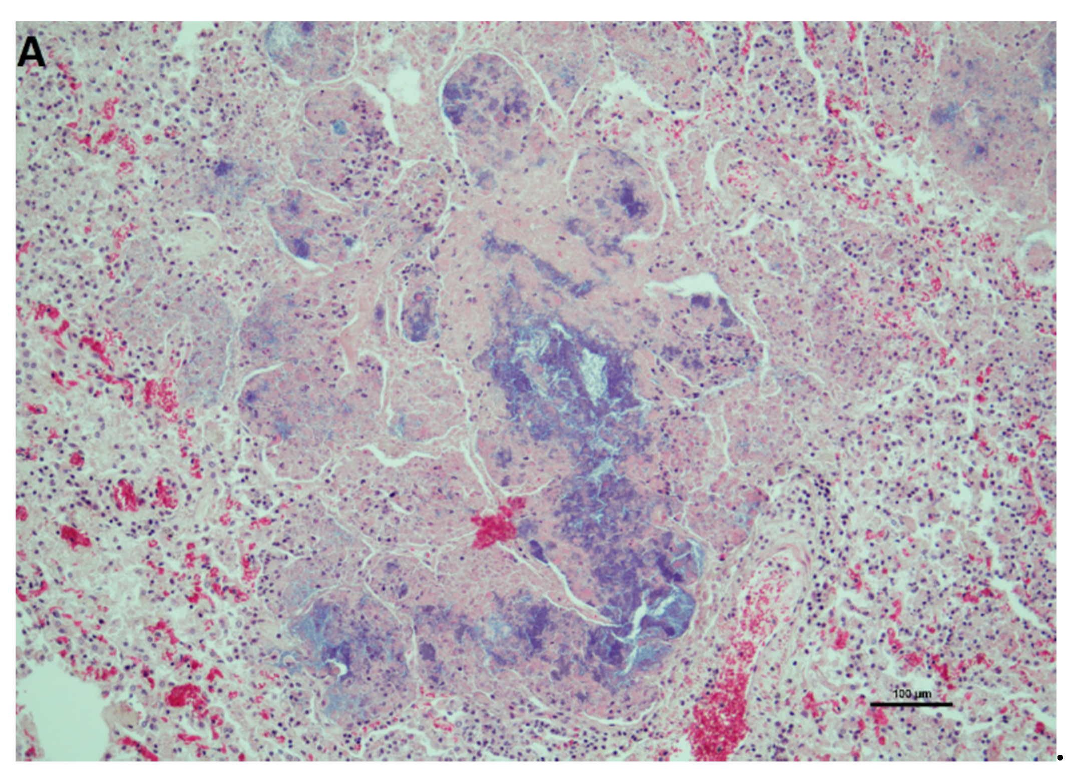
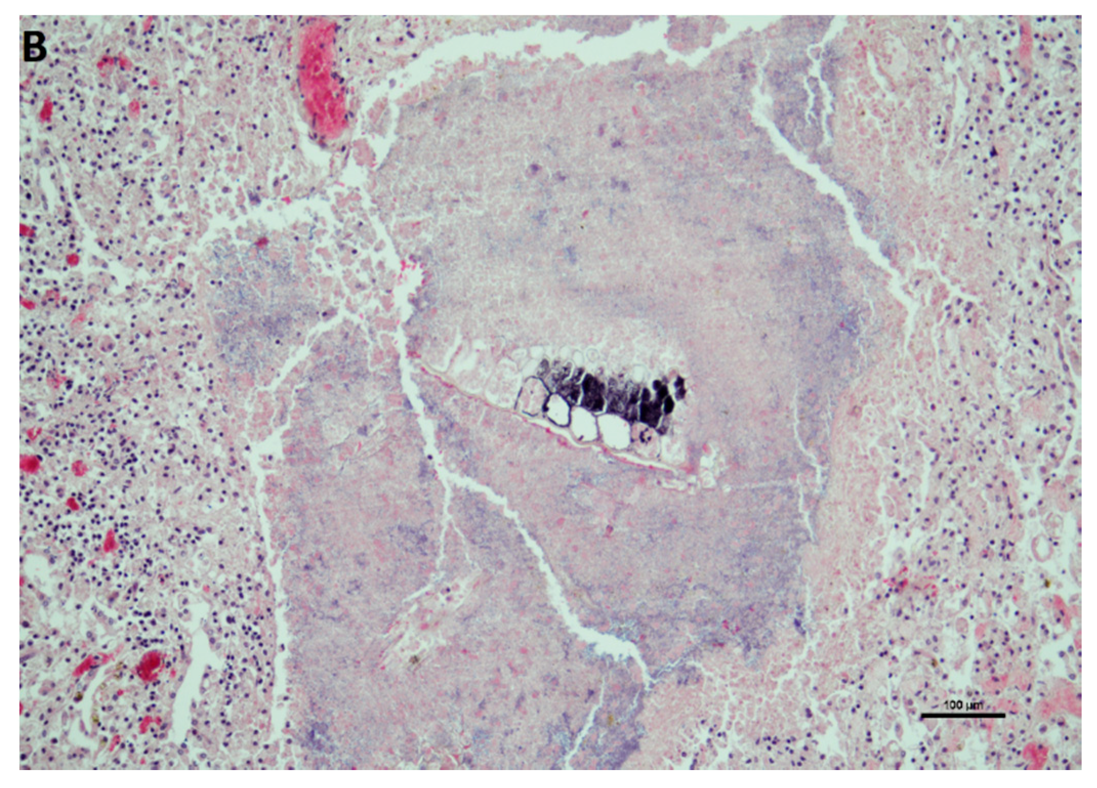
Histopathological examinations of the lung showed high-grade, acute to subacute, multifocal to diffuse pyogranulomatous pneumonia. High numbers of alveolar macrophages, multifocal neutrophil granulocytes and individual plasma cells could be seen. Furthermore, numerous pyogranulomas with central cell detritus and numerous intralesional bacteria and partially enclosing plant material were visible (Figure 1).
The hepatocytes revealed dystrophy and a chronic proliferation of the bile tract with periportal fibrosis and acute hemorrhages. In those areas, the hepatocytes showed fibro-necrotizing changes. In the histological preparation of the gut, single cell necrosis in the epithelium of the villi and crypts and proliferation of the Peyer’s patches became evident.
Likewise, serious pathological-anatomical and histopathological changes and lesions have been found in the lungs of a llama [10] and in dromedaries [5] during post mortem examinations. However, Löhr et al. (2019) [11] reported on an R. equi infection in an 11-year-old llama severely affecting the small intestine (jejunum). The mesenteric lymph nodes, the liver and peritoneum were also involved. In all of the reported cases of R. equi infections in camelids, the macroscopic and microscopic (histological) changes were characterized by prominent pyogranulomatous to necrogranulomatous lesions and the formation of abscesses in the lungs and intestines. Lymph nodes, the pleura and peritoneum, and internal organs such as the liver and spleen were also affected [5,10,11]. There is evidence that survival is significantly reduced in equine foals infected with R. equi that develop extrapulmonary disorders in multiple organs, compared to foals that suffer from infections only affecting the lungs. However, it has to be considered that a clinical diagnosis of extrapulmonary manifestations is difficult and in many cases is not recognized until post mortem examination [51].
3.2. Bacteriological Examination
A bacteriological examination of material obtained from the lung pyogranulomas of the alpaca yielded a heavy growth of the R. equi isolate. In addition to the growth of R. equi, Helcococcus ovis and the obligate anaerobic bacteria Fusobacterium necrophorum, Bacteroides fragilis and Prevotella heparinolytica could be co-cultured. The concomitant growth of bacteria of the order Actinomycetales and the Gram-negative anaerobic bacteria Fusobacterium necrophorum, Bacteroides spp. and Prevotella spp. has also been previously encountered in bacteriological examinations of abscesses in camelids and ruminants [5,11,16,52].
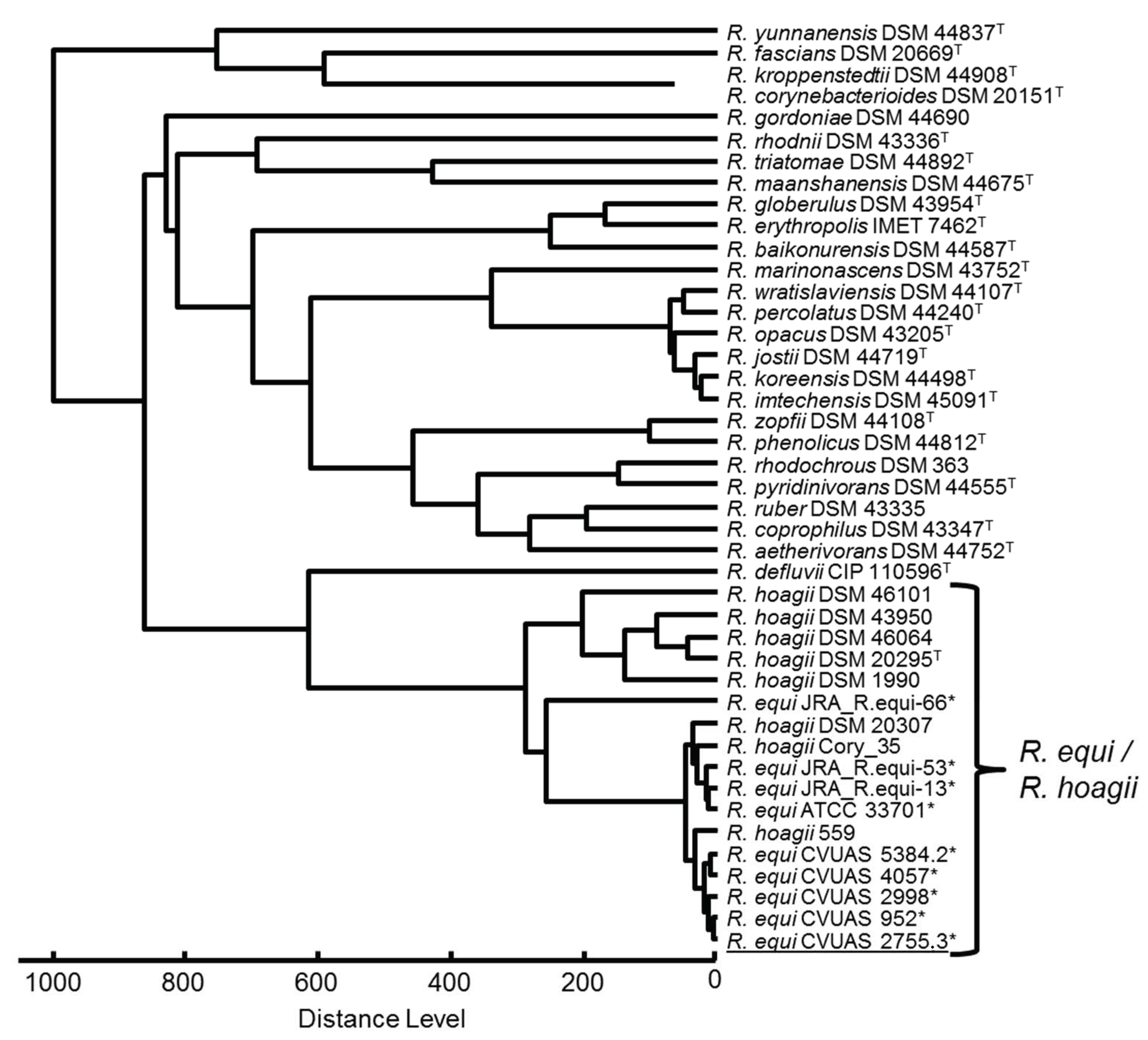
R. equi could be identified by conventional methods. All R. equi isolates and the R. equi reference strain showed urease and nitrate reduction activity and synergistic hemolytic activities with Staphylococcus aureus, Listeria monocytogenes, Corynebacterium pseudotuberculosis and Listeria ivanovii. The identification based on classical methods was confirmed by MALDI-TOF MS, 16S rRNA gene, 16S-23S ITS and rpoB gene sequencing. In the MALDI-TOF MS analysis, a slightly higher score value was achieved for the R. equi isolate CVUAS 2755.3 from the alpaca due to the use of the supplemented database entries, in comparison to the commercial database (2.399 vs. 2.295). The isolates are reported as R. hoagii using the Bruker database. The MALDI-TOF MS dendrogram created by cluster analysis of spectra obtained by MALDI-TOF mass spectrometry shows a clear separation of the Rhodococcus species and a clear assignment of the isolate CVUAS 2755.3 to R. equi (Figure 2).
Partial sequencing of the 16S rRNA gene resulted in R. equi with a percentage identity difference of 0.6 (NCBI GenBank) or 0.7 (EzBioCloud database) to R. soli. However, 16S-23S ITS and rpoB sequencing revealed percent identity differences of 12.2% and 7.7%, respectively. Homology values of 100% and a minimum distance of differences in nucleotide sequences (SNPs) to the next best matching species of a 0.8% identity threshold allow the allocation of the bacterial species R. equi [53]. The partial 16S rRNA gene, 16S-23S ITS, and rpoB gene DNA sequences have been deposited into the NCBI GenBank with the accession numbers OM996150, ON003979 and ON009447, respectively.
The vapA gene, a characteristic feature of virulent R. equi strains, could be detected by PCR in accordance with previous findings for all clinical diseases in horses [24] and camelids [5,11]. While detection of the vapA gene in our study was successful in isolates originating in the alpaca and equine foals, this was not the case for the porcine and bovine isolate. Porcine and bovine isolates usually carry the vapB and vapN genes [8], which we did not test for in this study.
Conspicuously, fatal R. equi infections in camelids have been reported in current and previous cases exclusively for adult animals aged 2 to 11 years [5,10,11]. In contrast, adult horses are considered largely resistant to rhodococcosis [54]. However, from time to time fatal respiratory and intestinal R. equi infections have also been reported for adult horses in different countries [55,56,57].
The infection route and source of the pathogen remain puzzling. However, the pronounced and uniform changes in the respiratory and digestive tract of adult horses [55,56,57] and adult camelids [5,10,11] indicate an infection route via inhalation and/or ingestion. Remarkably, in our and another case of a llama fatality [11], plant material was detected in pyogranulomas in the lungs and intestines, respectively. This observation suggests an environmental origin of the pathogen of this air- and soil-borne pathogen, absorbed by aspiration or ingestion [28].
A comprehensive study performed in Australia revealed various factors favoring the pathogen load and infection in foals [58]. This study, performed on foals in stud farms comes to the conclusion that the main infection route of R. equi is via the respiratory tract. The inhalation of contaminated dust or direct foal to foal transmission through aerosolized virulent R. equi in high concentrations proved to be the most relevant factors for rhodococcal infections. This is especially the case in the stable, which is the most dangerous environment for the foals. It has been shown that the R. equi concentration inside the stables is ten times higher than in dusty paddock pastures [59]. Nevertheless, environmental conditions such as dry areas with low pasture cover are concomitant to significantly higher airborne concentrations of virulent R. equi and, as a consequence, increased infections of the respiratory tract of foals. As a result, clinically and subclinically infected foals exhale high concentrations of virulent R. equi into the air and in turn infect other animals.
Nevertheless, the primary source of infection in our case also remains unclear, all the more so because no direct contact with horses had occurred. However, the hay meadows had been fertilized with horse manure and horses and cows had been kept in the stables until they began, and have continued, to be used for the alpaca keeping in 2013.
3.3. Antibiotic Susceptibility
Antibiotic susceptibility testing of the R. equi isolates in vitro using the microdilution method revealed susceptibility to doxycycline, erythromycin, gentamycin, neomycin, rifampicin, trimethoprim/sulfamethoxazole, tetracycline and vancomycin. However, resistance or intermediate susceptibility was found to the penicillines ampicillin, oxacillin, and penicillin G, the cephalosporines cefquinome, ceftiofur, cefazolin, and cefoxitin, clindamycin, enrofloxacin, florfenicol and nitrofurantoin (Table 1). MIC values (MIC90) in a range comparable to our results have been previously reported by Riesenberg et al. (2014) [60]. In our study, we could not detect significant differences between the MIC values of the alpaca, equine, porcine or bovine R. equi strains. However, resistance to the antibiotics used for standard therapy and the emergence of multi-drug resistant R. equi isolates have been recently noted, thus limiting the available spectrum of effective antibiotics [61,62]. Due to infections caused by isolates resistant to macrolides and rifampicin, an alternative use of antibiotics for the treatment of rhodococcal infections has been critically reviewed by Cisek et al. (2014) [63]. The authors recommend treating rhodococcosis with a combination of at least two antibiotics including macrolides, rifampicin, fluoroquinolones, aminoglycosides, glycopeptides and carbapenems. Furthermore, the macrolides can cause life-threatening hyperthermia caused by drug-induced anhidrosis and diarrhea in equine foals and fatal colitis in mares [64].
In this respect, guidelines for the prudent use of antibiotics in veterinary medicine should be taken into account to avoid ineffective antimicrobial treatment. A recent study on the treatment of foals suffering from pneumonia caused by R. equi concluded that foals with smaller lesions could even be spared from antibiotic treatment without a significant increase in mortality [65]. Whether and to what extent this treatment regime also applies to alpacas still needs to be investigated.
4. Conclusions
This is the first detailed report of a fatal R. equi infection in an adult alpaca, to our best knowledge. The present report extends our knowledge about R. equi infection in alpacas, the most important New World camelid species kept under human care. Thus, this report should be of particular concern for the health and husbandry of this New World camelid species living in Europe. Furthermore, this bacterium is a zoonotic agent. The source of infections of this soil-and-airborne pathogen is suspected to be environmental and its transmission caused by aspiration or ingestion. However, transmission via close contact between animals and from animal to humans might also be of relevance. Therefore, reports on R. equi infections are important for assessing the relevance of pathogen reservoirs and transmission routes as a basis of effective measures in order to sustain the health, productivity and welfare of alpacas.
Author Contributions
Conceptualization R.S.; methodology R.S., I.S., M.K., M.M. and J.R.; validation R.S., I.S., M.K., M.M. and J.R.; formal analysis R.S., I.S., M.K., M.M. and J.R.; investigation R.S., I.S., M.K., M.M. and J.R.; resources R.S., I.S., M.K., M.M. and J.R.; data curation R.S and J.R.; writing— original draft preparation R.S.; writing—review and editing R.S., I.S., M.K., M.M. and J.R.; visualization R.S.; supervision, R.S.; project administration, R.S. All authors have read and agreed to the published version of the manuscript.
Funding
This research received no external funding.
Institutional Review Board Statement
Ethical review and approval are waived for this study because the investigations were carried out on samples taken from the carcass of an animal that had died on a farm. The carcass of the alpaca was subsequently submitted to a post mortem examination in our institute in order to investigate the cause of death.
Informed Consent Statement
Informed consent was obtained from all subjects involved in the study.
Data Availability Statement
Partial 16S rRNA gene, 16S-23S ITS and rpoB gene DNA sequences have been deposited into the NCBI GenBank with the accession numbers OM996150, ON003979 and ON009447, respectively. MALDI-TOF MS reference entries of R. equi isolates generated in the present study, as well as single spectra for validation purposes, are available for exchange via the MALDI-UP homepage (https://www.maldi-up.ua-bw.de (accessed on 14 March 2022) [39].
Acknowledgments
The authors would like to thank Maja Hrubenja for excellently carrying out the molecular examinations. We would also like to express our thanks to Martin Dyk for his diligent MALDI-TOF MS analysis and Matthias Contzen for submitting DNA sequence data to the NCBI GenBank.
Conflicts of Interest
The authors declare no conflict of interest.
References
- Rakowska, A.; Cywinska, A.; Witkowski, L. Current trends in understanding and managing equine rhodococcosis. Animals 2020, 10, 1910. [Google Scholar] [CrossRef] [PubMed]
- Ruocco, N.A.; Luedke, L.K.; Fortier, L.A.; Ducharme, N.G.; Reesink, H.L. Rhodococcus equi joint sepsis and osteomyelitis is associated with a grave prognosis in foals. Front. Vet. Sci. 2020, 6, 503. [Google Scholar] [CrossRef] [PubMed]
- Aslam, M.W.; Lau, S.F. A review on Rhodococcus equi: Horse, cat and human. J. Vet. Malays. 2019, 31, 1–12. [Google Scholar]
- Bryan, L.K.; Clark, S.D.; Díaz-Delgado, J.; Lawhon, S.D.; Edwards, J.F. Rhodococcus equi infections in dogs. Vet. Pathol. 2017, 54, 159–163. [Google Scholar] [CrossRef] [Green Version]
- Kinne, J.; Madarame, H.; Takai, S.; Jose, S.; Wernery, U. Disseminated Rhodococcus equi infection in dromedary camels (Camelus dromedarius). Vet. Microbiol. 2011, 149, 269–272. [Google Scholar] [CrossRef]
- Nakagawa, R.; Moki, H.; Hayashi, K.; Ooniwa, K.; Tokuyama, K.; Kakuda, T.; Yoshioka, K.; Takai, S. A case report on disseminated Rhodococcus equi infection in a Japanese black heifer. J. Vet. Med. Sci. 2018, 80, 819–822. [Google Scholar] [CrossRef] [Green Version]
- Stranahan, L.W.; Plumlee, Q.D.; Lawhon, S.D.; Cohen, N.D.; Bryan, L.K. Rhodococcus equi infections in goats: Characterization of virulence plasmids. Vet. Pathol. 2018, 55, 273–276. [Google Scholar] [CrossRef] [Green Version]
- Witkowski, L.; Rzewuska, M.; Takai, S.; Kizerwetter-Świda, M.; Kita, J. Molecular epidemiology of Rhodococcus equi in slaughtered swine, cattle and horses in Poland. BMC Microbiol. 2016, 16, 98. [Google Scholar] [CrossRef] [Green Version]
- Zychska, M.; Witkowski, L.; Klementowska, A.; Rzewuska, M.; Kwiecién, E.; Stefánska, I.; Czopowicz, M.; Szalus-Jordanow, O.; Mickiewicz, M.; Moroz, A.; et al. Rhodococcus equi—Occurrence in goats and clinical case report. Pathogens 2021, 10, 1141. [Google Scholar] [CrossRef]
- Hong, C.B.; Donahue, J.M. Rhodococcus equi-associated necrotizing lymphadenitis in a llama. J. Comp. Pathol. 1995, 113, 85–88. [Google Scholar] [CrossRef]
- Löhr, C.V.; O’Neill, T.W.; Daw, D.N.; Pitel, M.O.; Schlipf, J.W. Pyogranulomatous enteritis and mesenteric lymphadenitis in an adult llama caused by Rhodococcus equi carrying virulence-associated protein A gene. J. Vet. Diagn. Investig. 2019, 31, 747–751. [Google Scholar] [CrossRef]
- Braga, W.U.; Chavera, A.; Gonzalez, A. Corynebacterium pseudotuberculosis infection in highland alpacas (Lama pacos) in Peru. Vet. Rec. 2006, 159, 23–24. [Google Scholar] [CrossRef]
- Sprake, P.; Gold, J.R. Corynebacterium pseudotuberculosis liver abscess in a mature alpaca (Lama pacos). Can. Vet. J. 2012, 53, 387–390. [Google Scholar]
- Wernery, U. Caseous lymphadenitis (Pseudotuberculosis) in camelids. J. Camel Pract. Res. 2012, 19, 21–27. [Google Scholar]
- Ramos-Vara, J.A.; Kopcha, M.; Richter, E.; Watson, G.L.; Patterson, J.S.; Juan-Sallés, C.; Yamini, B. Actinomycotic splenitis and intestinal volvulus in an alpaca (Lama pacos). J. Zoo Wildl. Med. 1998, 29, 228–232. [Google Scholar]
- Sting, R.; Schwalm, A.K.; Contzen, M.; Roller, M.; Rau, J. Actinomycetes associated with abscess formation in a goat, a llama and two alpacas. Berl. Munch. Tieraerztl. Wschr. 2020, 133. [Google Scholar] [CrossRef]
- Dwan, L.W.; Thompson, H.; Taylor, D.J.; Philbey, A.W. Laryngeal abscessation due to Mannheimia haemolytica in an alpaca (Vicugna pacos) cria. Vet. Rec. 2008, 163, 124–125. [Google Scholar] [CrossRef]
- Hill, F.I.; Johnstone, A.C. Actinobacillosis in an alpaca (Lama pacos). N. Z. Vet. J. 1992, 40, 28–30. [Google Scholar] [CrossRef]
- Tavella, A.; Bettini, A.; Cocchi, M.; Idrizi, I.; Colorio, S.; Viel, L.; Zanardello, C.; Patrik Zanolari, P. Isolation of Streptococcus agalactiae in a female llama (Lama glama) in South Tyrol (Italy). BMC Vet. Res. 2018, 14, 343. [Google Scholar] [CrossRef]
- Corpa, J.M.; Carvallo, F.; Anderson, M.L.; Nyaoke, A.C.; Moore, J.D.; Uzal, F.A. Streptococcus equi subspecies zooepidemicus septicemia in alpacas: Three cases and review of the literature. J. Vet. Diagn. Investig. 2018, 30, 598–602. [Google Scholar] [CrossRef] [Green Version]
- Wernery, U.; Kinne, J.; Jose, S.; Das Gupta, A.; Taha, A.A.; Ismail, A.A.; Joseph, M.; Nagy, P.; Juhasz, J. ‘Alpaca Fever’ in Dromedary Camel Calves–A Case Report. J. Camel Pract. Res. 2021, 28, 291–295. [Google Scholar] [CrossRef]
- Krajewska-Wędzina, M.; Didkowska, A.; Sridhara, A.A.; Elahi, R.; Johnathan-Lee, A.; Radulski, L.; Lipiec, M.; Anusz, K.; Lyashchenko, K.P.; Miller, M.A.; et al. Transboundary tuberculosis: Importation of alpacas infected with Mycobacterium bovis from the United Kingdom to Poland and potential for serodiagnostic assays in detecting tuberculin skin test false-negative animals. Transbound. Emerg. Dis. 2020, 67, 1306–1314. [Google Scholar] [CrossRef]
- Krajewska-Wędzina, M.; Miller, M.A.; Didkowska, A.; Kycko, A.; Radulski, L.; Lipiec, M.; Weiner, M. The potential risk of international spread of Mycobacterium bovis associated with movement of alpacas. J. Vet. Res. 2022, 66, 53–59. [Google Scholar] [CrossRef]
- Vázquez-Boland, J.A.; Scortti, M.; Meijer, W.G. Conservation of Rhodococcus equi (Magnusson 1923) Goodfellow and Alderson 1977 and rejection of Rhodococcus hoagii (Morse 1912) Kämpfer et al. 2014. Int. J. Syst. Evol. Microbiol. 2020, 70, 3572–3576. [Google Scholar] [CrossRef] [PubMed]
- Cohen, N.D.; Carter, C.N.; Scott, H.M.; Chan, M.K.; Smith, J.L.; Grimm, M.B.; Kuskie, K.R.; Takai, S.; Martens, R.J. Association of soil concentrations of Rhodococcus equi and incidence of pneumonia attributable to Rhodococcus equi in foals on farms in central Kentucky. Am. J. Vet. Res. 2008, 69, 385–395. [Google Scholar] [CrossRef] [PubMed]
- Cohen, N.D.; Chan, M.K.; Kuskie, K.R.; Syndergaard, M.K.; Blodgett, G.P.; Takai, S. Association of perinatal exposure to airborne Rhodococcus equi with risk of pneumonia caused by R. equi in foals. Am. J. Vet. Res. 2013, 74, 102–109. [Google Scholar] [CrossRef] [PubMed]
- Giguère, S.; Cohen, N.D.; Chaffin, M.K.; Hines, S.A.; Hondalus, M.K.; Prescott, J.F.; Slovis, N.M. Rhodococcus equi: Clinical manifestations, virulence, and immunity. J. Vet. Intern. Med. 2011, 25, 1221–1230. [Google Scholar] [CrossRef]
- Muscatello, G.; Lowe, J.M.; Flash, M.L.; McBride, K.L.; Browning, G.L.; Gilkerson, J.R. Review of the epidemiology and ecology of Rhodococcus equi. AAEP Proc. 2007, 53, 214–217. [Google Scholar]
- Ocampo-Sosa, A.A.; Lewis, D.A.; Avas, J.; Quigley, F.; Callejo, R.; Scortti, M.; Leadon, D.P.; Fogarty, U.; Vázquez-Bolan, J.A. Molecular Epidemiology of Rhodococcus equi Based on traA, vapA, and vapB virulence plasmid markers. J. Infect. Dis. 2007, 196, 763–769. [Google Scholar] [CrossRef] [Green Version]
- Willingham-Lane, J.M.; Berghaus, L.J.; Giguère, S.; Hondalus, M.K. Influence of plasmid type on the replication of Rhodococcus equi in host macrophages. mSphere 2016, 1, e00186-16. [Google Scholar] [CrossRef] [Green Version]
- Hondalus, M.K. Pathogenesis and virulence of Rhodococus equi. Vet. Microbiol. 1997, 56, 257–268. [Google Scholar] [CrossRef]
- Salazar-Rodríguez, D.; Aleaga-Santiesteban, Y.; Iglesias, E.; Plascencia-Hernández, A.; Pérez-Gómez, H.R.; Calderón, E.J.; Vázquez-Boland, J.A.; de Armas, Y. Virulence plasmids of Rhodococcus equi isolates from Cuban patients with AIDS. Front. Vet. Sci. 2021, 8, 628239. [Google Scholar] [CrossRef]
- Vázquez-Boland, J.A.; Meijer, W.G. The pathogenic actinobacterium Rhodococcus equi: What’s in a name? Mol. Microbiol. 2019, 112, 1–15. [Google Scholar] [CrossRef] [Green Version]
- Bryan, L.K.; Alexander, E.R.; Lawhon, S.D.; Cohen, N.D. Detection of vapN in Rhodococcus equi isolates cultured from humans. PLoS ONE 2018, 13, e0190829. [Google Scholar] [CrossRef] [Green Version]
- Takai, S.; Sawada, N.; Nakayama, Y.; Ishizuka, S.; Nakagawa, R.; Kawashima, G.; Angkanjanavanich, N.; Sasaki, Y.; Kakuda, T.; Suzuki, Y. Reinvestigation of the virulence of Rhodococcus equi isolates from patients with and without AIDS. Lett. Appl. Microbiol. 2020, 71, 679–683. [Google Scholar] [CrossRef]
- Vázquez-Boland, J.A.; Giguère, S.; Hapeshi, A.; MacArthur, I.; Anastasi, E.; Valero-Rello, A. Rhodococcus equi: The many facets of a pathogenic actinomycete. Vet. Microbiol. 2013, 167, 9–33. [Google Scholar] [CrossRef]
- Yamshchikov, A.V.; Schuetz, A.; Lyon, G.M. Rhodococcus equi infection. Lancet Infect. Dis. 2010, 10, 350–359. [Google Scholar] [CrossRef]
- Shah, R.; Klumpp, L.C.; Nerella, N.; Liu-Young, G.; Jordan, J. Infective endocarditis caused by Rhodococcus equi in an immunocompetent patient. Cureus 2020, 12, e7829. [Google Scholar] [CrossRef] [Green Version]
- Rau, J.; Eisenberg, T.; Männig, A.; Wind, C.; Lasch, P.; Sting, R. MALDI-UP—An Internet Platform for the Exchange of MALDI-TOF mass spectra—User guide for http://maldi-up.ua-bw.de. Asp. Food Control Anim. Health 2016, 1, 1–17. [Google Scholar]
- Uchida-Fujii, E.; Niwa, H.; Kinoshita, Y.; Nukada, T. Construction and application of an in-house matrix-assisted laser desorption ionization-time of flight mass spectrometry (MALDI-TOF MS) database specific to bacteria from horses. J. Equine Vet. Sci. 2021, 103, 103664. [Google Scholar] [CrossRef]
- Lane, D.J. 16S/23S rRNA sequencing. In Nucleic Acid Techniques in Bacterial Systematics; Stackebrandt, E., Goodfellow, M., Eds.; John Wiley & Sons Inc.: New York, NY, USA, 1991; pp. 115–175. [Google Scholar]
- Fredriksson, N.J.; Hermansson, M.; Wilén, B.M. The choice of PCR primers has great impact on assessments of bacterial community diversity and dynamics in a wastewater treatment plant. PLoS ONE 2013, 8, e76431. [Google Scholar] [CrossRef]
- Hunt, D.E.; Klepac-Ceraj, V.; Acinas, S.G.; Gautier, C.; Bertilsson, S.; Polz, M.F. Evaluation of 23S rRNA PCR primers for use in phylogenetic studies of bacterial diversity. Appl. Environ. Microbiol. 2006, 72, 2221–2225. [Google Scholar] [CrossRef] [Green Version]
- Khamis, A.; Raoult, D.; La Scola, B. rpoB gene sequencing for identification of Corynebacterium species. J. Clin. Microbiol. 2004, 42, 3925–3931. [Google Scholar] [CrossRef] [Green Version]
- Pruitt, K.; Brown, G.; Tatusova, T.; Maglott, D. The Reference Sequence (RefSeq) Database. In The NCBI Handbook; McEntyre, J., Ostell, J., Eds.; National Center for Biotechnology Information (US): Bethesda, MD, USA, 2002; pp. 307–328. [Google Scholar] [CrossRef] [Green Version]
- Yoon, S.H.; Ha, S.M.; Kwon, S.; Lim, J.; Kim, Y.; Seo, H.; Chun, J. Introducing EzBioCloud: A taxonomically united database of 16S rRNA and whole genome assemblies. Int. J. Syst. Evol. 2017, 67, 1613–1617. [Google Scholar] [CrossRef]
- Quinn, P.J.; Carter, M.E.; Markey, B.K.; Carter, G.R. Clinical Veterinary Microbiology; Mosby: London, UK, 1998. [Google Scholar]
- Navas, J.; González-Zorn, B.; Ladrón, N.; Garrido, P.; Vázquez-Boland, J.A. Identification and mutagenesis by allelic exchange of choE, encoding a cholesterol oxidase from the intracellular pathogen Rhodococcus equi. J. Bacteriol. 2001, 183, 4796–4805. [Google Scholar] [CrossRef] [Green Version]
- Harrington, J.R.; Golding, M.C.; Martens, R.J.; Halbert, N.D.; Cohen, N.D. Evaluation of a real-time quantitative polymerase chain reaction assay for detection and quantitation of virulent Rhodococcus equi. Am. J. Vet. Res. 2005, 66, 755–761. [Google Scholar] [CrossRef] [Green Version]
- Riesenberg, A.; Feßler, A.T.; Frömke, C.; Kadlec, K.; Klarmann, D.; Kreienbrock, L.; Werckenthin, C.; Schwarz, S. Harmonization of antimicrobial susceptibility testing by broth microdilution for Rhodococcus equi of animal origin. J. Antimicrob. Chemother. 2013, 68, 2173–2175. [Google Scholar] [CrossRef] [Green Version]
- Reuss, S.M.; Chaffin, M.K.; Cohen, N.D. Extrapulmonary disorders associated with Rhodococcus equi infection in foals: 150 cases (1987–2007). J. Am. Vet. Med. Assoc. 2009, 235, 855–863. [Google Scholar] [CrossRef] [PubMed]
- Stalb, S.; Hiller, E.; Rau, J.; Eisenberg, T.; Sting, R. Pathological significance of Helcococcus ovis and Helcococcus kunzii in veterinary medicine. Berl. Muench. Tieraerztl. Wschr. 2018, 131, 482–488. [Google Scholar] [CrossRef]
- Sting, R.; Eisenberg, T.; Hrubenja, M. Rapid and reasonable molecular identification of bacteria and fungi in microbiological diagnostics using rapid real-time PCR and Sanger sequencing. J. Microbiol. Meth. 2019, 159, 148–156. [Google Scholar] [CrossRef] [PubMed]
- Giguère, S.; Prescott, J.F. Clinical manifestations, diagnosis, treatment, and prevention of Rhodococcus equi infections in foals. Vet. Microbiol. 1997, 56, 313–334. [Google Scholar] [CrossRef]
- Staufenbiel, C.; Schulze, C.; Winterhoff, N.; Brandt, K. Intestinale Rhodokokkose bei einem dreijährigen Warmblutwallach. Prakt. Tierarzt 2021, 102, 486–496. [Google Scholar] [CrossRef]
- Vengust, M.; Stæmpfli, H.; Prescott, J.F. Rhodococcus equi pleuropneumonia in an adult horse. Can. Vet. J. 2002, 43, 706–708. [Google Scholar]
- Waldridge, B.M.; Morresey, P.R.; Loynachan, A.T.; Reimer, J.; Riddle, W.T.; Williams, N.M.; Bras, R. Rhodococcus equi pneumonia in an adult horse. Equine Vet. Educ. 2008, 20, 67–71. [Google Scholar] [CrossRef]
- Muscatello, G.; Gilkerson, J.R.; Browning, G.F. Rattles in Horses: Effects of stud management on ecology of virulent Rhodococcus equi. In A Report for the Rural Industries Research and Development Corporation; RIRDC Publication: Canberra, Australia, 2006; No. 06/097; ISSN 1440-6845. [Google Scholar]
- Bordin, A.I.; Huber, L.; Sanz, M.G.; Cohen, N.D. Rhodococcus equi foal pneumonia: Update on epidemiology, immunity, treatment and prevention. Equine Vet. J. 2022, 54, 481–494. [Google Scholar] [CrossRef]
- Riesenberg, A.; Feßler, A.T.; Erol, E.; Prenger-Berninghoff, E.; Stamm, I.; Böse, R.; Heusinger, A.; Klarmann, D.; Werckenthin, C.; Schwarz, S. MICs of 32 antimicrobial agents for Rhodococcus equi isolates of animal origin. J. Antimicrob. Chemother. 2014, 69, 1045–1049. [Google Scholar] [CrossRef]
- Álvarez-Narváez, S.; Berghaus, L.J.; Morris, E.R.A.; Willingham-Lane, J.M.; Slovis, N.M.; Giguère, S.; Cohen, N.D. A common practice of widespread antimicrobial use in horse production promotes multi-drug resistance. Sci. Rep. 2020, 10, 911. [Google Scholar] [CrossRef]
- Álvarez-Narváez, S.; Giguère, S.; Cohen, N.; Slovis, N.; Vázquez-Boland, J.A. Spread of multidrug-resistant Rhodococcus equi, United States. Emerg. Infect. Dis. 2021, 27, 529–537. [Google Scholar] [CrossRef]
- Cisek, A.A.; Rzewuska, M.; Witkowski, L.; Binek, M. Antimicrobial resistance in Rhodococcus equi. Act. Biochim. Pol. 2014, 61, 633–638. [Google Scholar] [CrossRef] [Green Version]
- Stieler, A.L.; Sanchez, L.C.; Mallicote, M.F.; Martabano, B.B.; Burrow, J.A.; MacKay, R.J. Macrolide-induced hyperthermia in foals: Role of impaired sweat responses. Equine Vet. J. 2016, 48, 590–594. [Google Scholar] [CrossRef]
- Arnold-Lehna, D.; Venner, M.; Berghaus, L.J.; Berghaus, R.; Giguère, S. Changing policy to treat foals with Rhodococcus equi pneumonia in the later course of disease decreases antimicrobial usage without increasing mortality rate. Equine Vet. J. 2020, 52, 531–537. [Google Scholar] [CrossRef]
- Kahn, S.K.; Cywes-Bentley, C.; Blodgett, G.P.; Canaday, N.M.; Turner-Garcia, C.E.; Flores-Ahlschwede, P.; Metcalfe, L.L.; Nevill, M.; Vinacur, M.; Sutter, P.J.; et al. Randomized, controlled trial comparing Rhodococcus equi and poly-N-acetyl glucosamine hyperimmune plasma to prevent R equi pneumonia in foals. J. Vet. Intern. Med. 2021, 35, 2912–2919. [Google Scholar] [CrossRef]
Figure 1.
Histological section of a focal pyogranuloma in the lung of the alpaca with central cell debris as well as peripheral neutrophil granulocytes and alveolar macrophages (A), and plant material (B). Numerous bacteria, both extra- and intracellular in neutrophils and macrophages are also pictured (magnification 100×).


Figure 2.
MSP Dendrogram created by cluster analysis of reference main spectra (MSP) obtained by MALDI-TOF mass spectrometry (MALDI Biotyper, Version 3.1, Bruker Daltonik) of a Rhodococcus (R.) equi isolate from an alpaca (underlined) in comparison to a selection of available MSPs of strains of the genus Rhodococcus from the commercial Bruker Biotyper database and external reference entries, documented in the MALDI-UP catalogue on https://maldi-up.ua-bw.de (accessed on 14 March 2022) [39]. Type strains (T) and external reference spectra (*) are indicated. In context of this study, the following self-created MSPs were used for comparison: R. equi reference strain ATCC 33701 (horse lung) and the four R. equi German field isolates, CVUAS 2998 and CVUAS 952 (each from horse lungs), CVUAS 4057 (pig placenta), and CVUAS 5384.2 (cattle lymph nodes).


Table 1.
Determination of minimal inhibitory concentration (MIC) values (mg/L) for R. equi isolates using the micro-dilution method. Assessment of the MIC values was carried out using the MCN6 software (Merlin, Germany). R = resistance, I = intermediary susceptibility, S = susceptibility. n.d.p. = no data provided.
| Antibiotic | CVUAS 2755.3 (Alpaca, Lung) | CVUAS 246 (ATCC 33701) | CVUAS 2998 (Horse, Lung) | CVUAS 952 (Horse, Lung) | CVUAS 4057 (Pig, Placenta) | CVUAS 5384-2 (Cattle, Lymph Node) | MIC50 1 | MIC90 1 |
|---|---|---|---|---|---|---|---|---|
| Ampicillin | =8 (R) | =8 (R) | =8 (R) | =8 (R) | =8 (R) | =8 (R) | 4 | 8 |
| Cefquinom | >4 (R) | >4 (R) | =4 (I) | =4 (I) | =4 (I) | >4 (R) | 2 | 4 |
| Ceftiofur | >4 (R) | >4 (R) | >4 (R) | >4 (R) | >4 (R) | >4 (R) | 8 | 16 |
| Cefazolin | >8 (R) | >8 (R) | >8 (R) | >8 (R) | >8 (R) | >8 (R) | n.d.p. | n.d.p. |
| Clindamycin | >2 (R) | >2 (R) | >2 (R) | >2 (R) | >2 (R) | >2 (R) | 4 | 8 |
| Cefoxitin | >4 (R) | =4 (S) | >4 (R) | >4 (R) | >4 (R) | >4 (R) | n.d.p. | n.d.p. |
| Doxycyclin | =0.5 (S) | =0.5 (S) | =0.5 (S) | =0.5 (S) | =0.5 (S) | =0.5 (S) | 1 | 1 |
| Enrofloxacin | =1 (I) | =0.5 (I) | =0.5 (I) | =0.5 (I) | =0.5 (I) | =0.5 (I) | 1 | 1 |
| Erythromycin | =0.5 (S) | =0.5 (S) | =0.5 (S) | =0.5 (S) | =0.5 (S) | =0.5 (S) | 0.5 | 0.5 |
| Florfenicol | =8 (R) | >8 (R) | >8 (R) | >8 (R) | =8 (R) | >8 (R) | 16 | 16 |
| Gentamicin | ≤1 (S) | ≤1 (S) | ≤1 (S) | ≤1 (S) | ≤1 (S) | ≤1 (S) | 0.5 | 0.5 |
| Neomycin | ≤8 (S) | ≤8 (S) | ≤8 (S) | ≤8 (S) | ≤8 (S) | ≤8 (S) | n.d.p. | n.d.p.. |
| Nitrofurantoin | =64 (I) | =64 (I) | =64 (I) | =64 (I) | =64 (I) | =64 (I) | n.d.p. | n.d.p.. |
| Oxacillin | >4 (R) | >4 (R) | >4 (R) | >4 (R) | >4 (R) | >4 (R) | n.d.p. | n.d.p. |
| Penicillin G | >8 (R) | >8 (R) | >8 (R) | =8 (R) | =8 (R) | =8 (R) | 4 | 4 |
| Rifampicin | =0.5 (S) | =0.5 (S) | =0.5 (S) | =0.25 (S) | =0.5 (S) | =0.25 (S) | 0.06 | 0.12 |
| Trimethoprim/Sulfamethoxazole 2 | =1 (S) | =1 (S) | =1 (S) | =2 (S) | =1 (S) | =1 (S) | 0.25 | 0.5 |
| Tetracycline | =2 (S) | =4 (S) | =2 (S) | =2 (S) | =2 (S) | =2 (S) | 4 | 8 |
| Vancomycin | ≤0.5 (S) | ≤0.5 (S) | ≤0.5 (S) | ≤0.5 (S) | ≤0.5 (S) | ≤0.5 (S) | 0.5 | 0.5 |
1 MIC50 and MIC90 values adapted from Riesenberg et al. (2014) [60]. 2 The MIC values of trimethoprim/sulfamethoxazole (1:19) are expressed as the MIC values of trimethoprim.
Publisher’s Note: MDPI stays neutral with regard to jurisdictional claims in published maps and institutional affiliations. |
© 2022 by the authors. Licensee MDPI, Basel, Switzerland. This article is an open access article distributed under the terms and conditions of the Creative Commons Attribution (CC BY) license (https://creativecommons.org/licenses/by/4.0/).
Share and Cite
MDPI and ACS Style
Sting, R.; Schwabe, I.; Kieferle, M.; Münch, M.; Rau, J. Fatal Infection in an Alpaca (Vicugna pacos) Caused by Pathogenic Rhodococcus equi. Animals 2022, 12, 1303. https://0-doi-org.brum.beds.ac.uk/10.3390/ani12101303
AMA Style
Sting R, Schwabe I, Kieferle M, Münch M, Rau J. Fatal Infection in an Alpaca (Vicugna pacos) Caused by Pathogenic Rhodococcus equi. Animals. 2022; 12(10):1303. https://0-doi-org.brum.beds.ac.uk/10.3390/ani12101303
Chicago/Turabian StyleSting, Reinhard, Ingo Schwabe, Melissa Kieferle, Maren Münch, and Jörg Rau. 2022. "Fatal Infection in an Alpaca (Vicugna pacos) Caused by Pathogenic Rhodococcus equi" Animals 12, no. 10: 1303. https://0-doi-org.brum.beds.ac.uk/10.3390/ani12101303
Note that from the first issue of 2016, this journal uses article numbers instead of page numbers. See further details here.