Article

Clinical Impact of Immune Checkpoint Inhibitor (ICI) Response, DNA Damage Repair (DDR) Gene Mutations and Immune-Cell Infiltration in Metastatic Melanoma Subtypes

1
National Institute for Cellular Biotechnology, School of Biotechnology, Dublin City University, Glasnevin, D09 E432 Dublin, Ireland
2
Department of Life Sciences, Institute of Technology Sligo, F91 YW50 Sligo, Ireland
3
Siemens Healthcare Diagnostics Products GmbH, 50667 Cologne, Germany
4
Royal Victoria Eye and Ear Hospital, Adelaide Road, D02 XK51 Dublin, Ireland
5
Department of Medical Oncology, St. Vincent’s University Hospital, D04 T6F4 Dublin, Ireland
*
Author to whom correspondence should be addressed.
These authors contributed equally to this work.
Submission received: 8 March 2022 / Revised: 13 May 2022 / Accepted: 17 May 2022 / Published: 24 May 2022
(This article belongs to the Section Cancer and Cancer-Related Research)

Abstract

:
Molecular and histopathological analysis of melanoma subtypes has revealed distinct epidemiological, genetic, and clinical features. However, immunotherapy for advanced metastatic melanoma patients does not differ based on subtype. Response to immune checkpoint inhibitors (ICI) has been shown to vary, therefore, predictive biomarkers are needed in the design of precision treatments. Targeted sequencing and histopathological analysis (CD8 and CD20 immunohistochemistry) were performed on subtypes of metastatic melanoma (cutaneous melanoma (CM, n = 10); head and neck melanoma (HNM, n = 7); uveal melanoma (UM, n = 4); acral lentiginous melanoma (AM, n = 1) and mucosal melanoma (MM, n = 1) treated with ICI). Progression-free survival (PFS) was significantly associated with high CD8 expression (p = 0.025) and mutations in DNA damage repair (DDR) pathway genes (p = 0.012) in all subtypes but not with CD20 expression. Our study identified that immune cell infiltration and DDR gene mutations may have an impact in response to ICI treatment in metastatic melanoma but differs among subtypes. Therefore, a comprehensive understanding of the immune infiltration cells’ role and DDR gene mutations in metastatic melanoma may identify prognostic biomarkers.

1. Introduction

Immune checkpoint inhibitor (ICI) therapies which target the programmed cell death protein 1 (PD-1)/programmed death ligand 1 (PD-L1) or the cytotoxic T-lymphocyte-associated protein 4 (CTLA-4) alone or in combination are used in the treatment of multiple advanced solid tumours including metastatic melanoma. ICI has changed the treatment landscape for metastatic cutaneous melanoma (CM), with ICI combinations displaying objective response rates (ORR) of greater than 60%; moreover, compared to traditional chemotherapy, patients can achieve complete response and long-term survival with novel combination strategies of ICI [1,2,3].
Rare subtypes of non-cutaneous melanoma such as acral, mucosal and uveal melanomas have different clinical, histopathological and genomic features [4,5]. Acral lentiginous melanomas (AM) tend to arise on the non-hair skin areas such as the palms of the hands and soles of the feet; mucosal melanomas (MM) arise from melanocytes of the mucosal epithelium of the respiratory, alimentary and genitourinary tract regions; and uveal melanomas (UM) arise from melanocytes in the iris, ciliary body, or choroid [6]. Head and neck cutaneous melanomas (HNM) are commonly found on the occipital scalp and skin of the cheek, and represent a distinct entity of skin cancer [7,8,9]. Despite the advances in CM treatment and management, ICI therapy has shown varied and limited benefits for other subtypes of metastatic melanoma. These subtypes of metastatic melanoma tend to be less susceptible to ICI therapies compared to CM, potentially due to the low presence of tumour-infiltrating lymphocytes, low somatic mutational burden, and the lack of a UV-mutational signature [10]. Nevertheless, post-hoc analysis of clinical trials and small retrospective studies have shown disparities in response rates to ICI [11,12]. A further retrospective study of 428 metastatic melanoma patients treated with ICI observed the median OS as 45 months for CM, 17 months for AM, 18 months for MM, and 12 months for UM. Although long-term survival was observed, complete responses were rare in metastatic melanoma subtypes [13]. These studies suggest that ICI is a valuable treatment option for metastatic melanoma subtypes, but attention is needed in considering the predictive clinical and biological characteristics to identify patients who will benefit most from ICI. Clinical predictors of ICI response, such as tumour mutational burden (TMB), DNA damage response pathways, neo-antigen load, and tumour immune microenvironment (TME) biomarkers have been extensively studied [reviewed in Bai et al. [14]]. However, predictive biomarkers of ICIs efficacy involve complex interactions between the different subtypes of metastatic melanoma and the regulation of the immune system network. Therefore, we performed targeted exome sequencing and assessed the expression of TILs (CD8+) and tumour infiltrating B-cells (CD20+) in subtypes of 23 metastatic melanoma patients treated with ICI in order to explore their clinical impact as prognostic factors.

2. Materials and Methods

2.1. Patient Characteristics

This is a retrospective clinical study consisting of 23 patients diagnosed with metastatic melanoma and treated with ICI in St. Vincent’s University Hospital, Dublin between 2014–2017. The median age was 62 years (range 42–83). The 23 metastatic melanoma subtypes consisted of primary tumours derived from cutaneous melanoma (CM, n = 10); head and neck melanoma (HNM, n = 7); uveal melanoma (UM, n = 4); acral lentiginous (AM, n = 1) and mucosal melanoma (MM, n = 1). All 23 patients were treated with ICI (pembrolizumab, ipilimumab or nivolumab), 12/23 (52%) with one ICI and 11/23 (48%) with two or more ICIs. The majority of patients did not receive chemotherapy/targeted therapy 17/23 (74%) (Supplementary Table S1). This study was approved by the Institutional Review Board/Ethics Committee of St. Vincent’s University Hospital, Dublin.

2.2. Immunohistochemistry Staining and Scoring

Representative 4 μm sections of formalin-fixed paraffin-embedded metastatic melanoma tumour tissues were cut using a microtome, mounted onto poly-l-lysine coated slides and dried overnight at 37 °C. Slides were stored at room temperature until required. Immunohistochemistry (IHC) staining was performed using an automated staining apparatus for IHC (Autostainer, DakoCytomation) according to the manufacturer’s guidelines. Optimum primary antibody dilutions were predetermined using known positive control tissues. Negative and known positive control sections were included in each run. Deparaffinisation and heat-induced epitope antigen retrieval (HIER) consisted of 40-min incubation in pH 9.0 buffer (Target Retrieval, DakoCytomation) in a 95 °C water bath followed by cooling to room temperature. In the Dako Autostainer, sections were treated with 3% H2O2 for 10 min to quench endogenous peroxidase and then rinsed. Quenched sections were incubated with antibodies, CD8 (clone C8/144B; Dako, Agilent, Santa Clara, CA, USA) and CD20cy (clone L26; Dako) for 30 min, followed by incubation with Dako REAL™ detection system, alkaline phosphatase/RED reagent for 30 min. The antigen-antibody complex was visualised using AP-Fast Red-type chromogen system. Sections were then counterstained with Dako REAL haematoxylin (S3301, Dako), and mounted using VectaMount AQ aqueous mounting medium (h-5501-60). Slides were reviewed by light microscopy. The presence of CD8+ TIL was scored as 0 (negative), low (1, positive cells at edge of tumour, <5% of area), moderate (2, tumour infiltration of TILs, 5–50% of area), or high (3, strong diffuse infiltration by TIL, >50% of area). Scores of 0, 1, 2 were collated and compared against score 3 for evaluation purposes.

2.3. Targeted Sequencing and Analysis

Retrospectively, formalin-fixed paraffin-embedded (FFPE) tissue was analysed by using a comprehensive hybrid capture–based next-generation sequencing assay as previously described [15]. Genomic DNA was extracted from FFPE, sheared (Covaris, Woburn, MA, USA), and subjected to hybrid capture–based next-generation sequencing to detect point mutations, small insertions and deletions, copy number alterations and rearrangement/gene fusions in a single assay. In brief, after shearing, adapters were ligated and individual genomic regions of interest were enriched using complementary bait sequences (hybrid capture procedure). The selected baits ensure optimal coverage of all relevant genomic regions. After enrichment, targeted fragments were amplified (clonal amplification) and sequenced in parallel at high sequencing depth. Reads were trimmed and aligned to the hg38 reference genome using BWA and duplicate reads were marked. Base recalibration was conducted with GATK. Variant calling was performed using Mutect2 from GATK (4.1.3). The Copy Number Aberrations (CNA) burden was estimated for each sample and enrichment analysis was performed using EnrichR [16,17,18].

2.4. Statistical Analysis

Progression-free survival (PFS) was calculated from the start date of ICI for metastatic disease to systemic relapse/death. Overall survival (OS) was calculated from date of diagnosis for metastatic disease to death or latest follow-up. Comparisons between groups were performed using Fisher’s exact test. A p-value < 0.05 was considered statistically significant. Univariate and Cox regression analysis was used to determine independent prognostic predictors of PFS and OS. The Kaplan–Meier survival estimator method was applied to calculate PFS and OS, and the log-rank test was used for assessment of statistical significance. Statistical analysis was performed using STATA v17.0 (StataCorp, LLC, College Station, TX, USA).

3. Results

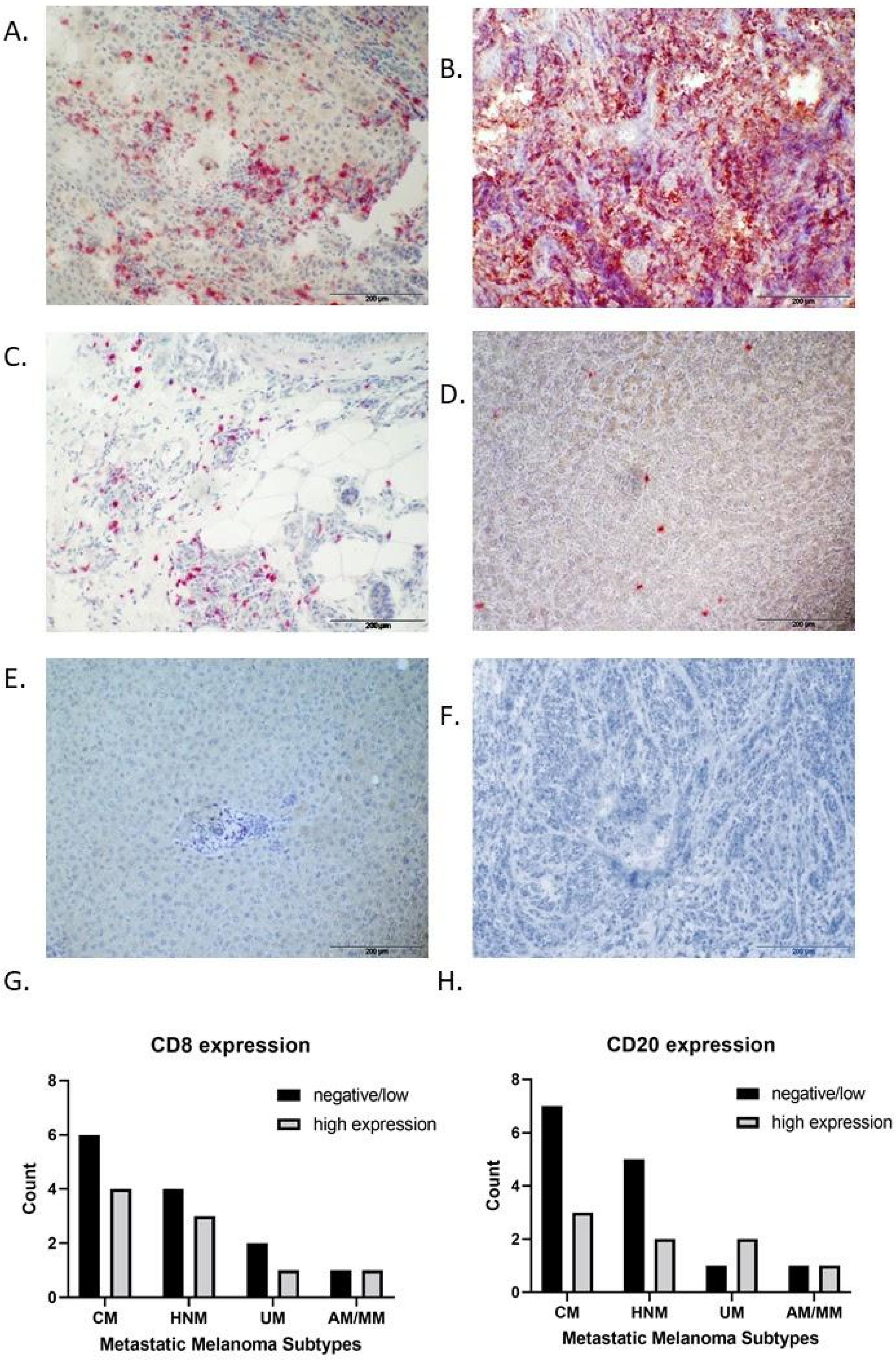
3.1. IHC Expression of CD8 and CD20 in Metastatic Melanoma Subtypes

Of the 23 patients, only 22 had available specimens for IHC (one UM subtype contained too much melanin for analysis). Among the 22 patients, 13/22 (59%) displayed low IHC staining for CD8 with peritumour and/or interstitial TIL infiltration (score 0, 1, 2) and 9/22 (41%) displayed high CD8 staining with strong diffuse immune cell infiltration (score 3). CD20 low IHC staining (score 0, 1, 2) was observed in 15/22 (68%) of tumours and 7/22 (32%) displayed (score 3) high staining (Figure 1A–F). The majority of CM and HNM patients displayed low CD8 and CD20 staining, compared to UM subtype samples which displayed high CD8 and CD20 staining (Figure 1G–H). There was no significant correlation with CD8 expression and clinical variables, whereas CD20 high expression was significantly associated with UM (p = 0.023) (Table 1).

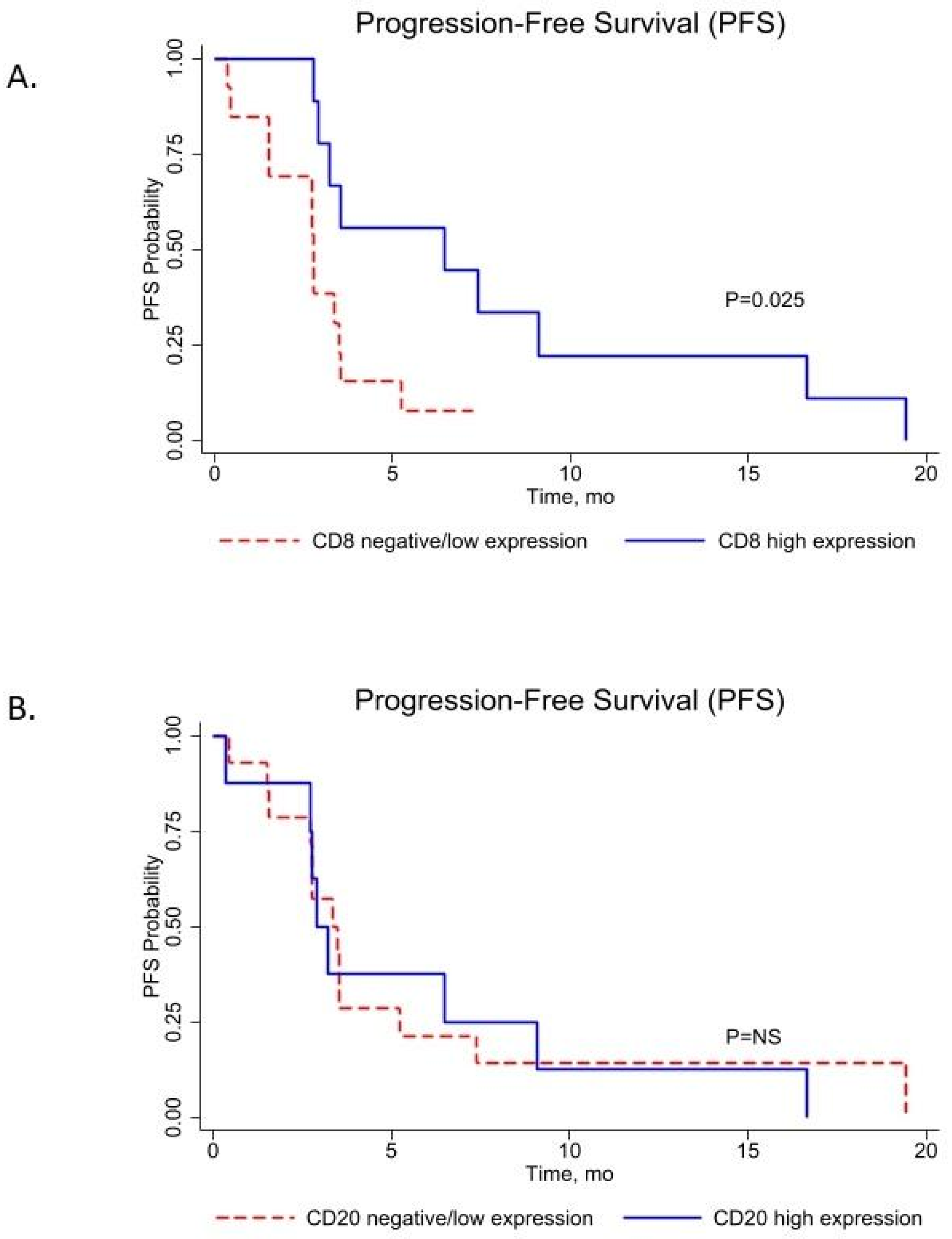
3.2. CD8 and CD20 Cell Infiltration Score Associated with Progression-Free Survival (PFS) in Metastatic Melanoma Subtypes

In the entire patient group (n = 23), the median OS and PFS times were 27.72 months (95% CI: 1.81–177.74) and 3.35 months (95% CI: 0.36–39.65), respectively. A high CD8 score was significantly associated with better progression-free survival compared to a low CD8 score (median PFS, 6.93 months (95% CI: 2.75–19.44) vs. 2.77 months (95% CI: 0.36–7.45), p = 0.025, log rank) (Figure 2A). CD20 B-cell infiltration score did not significantly improve PFS (median PFS, 3.07 months (95% CI: 0.36–16.65) vs. 3.41 months (95% CI: 0.45–19.44), p = NS, log rank) in metastatic melanoma (Figure 2B).

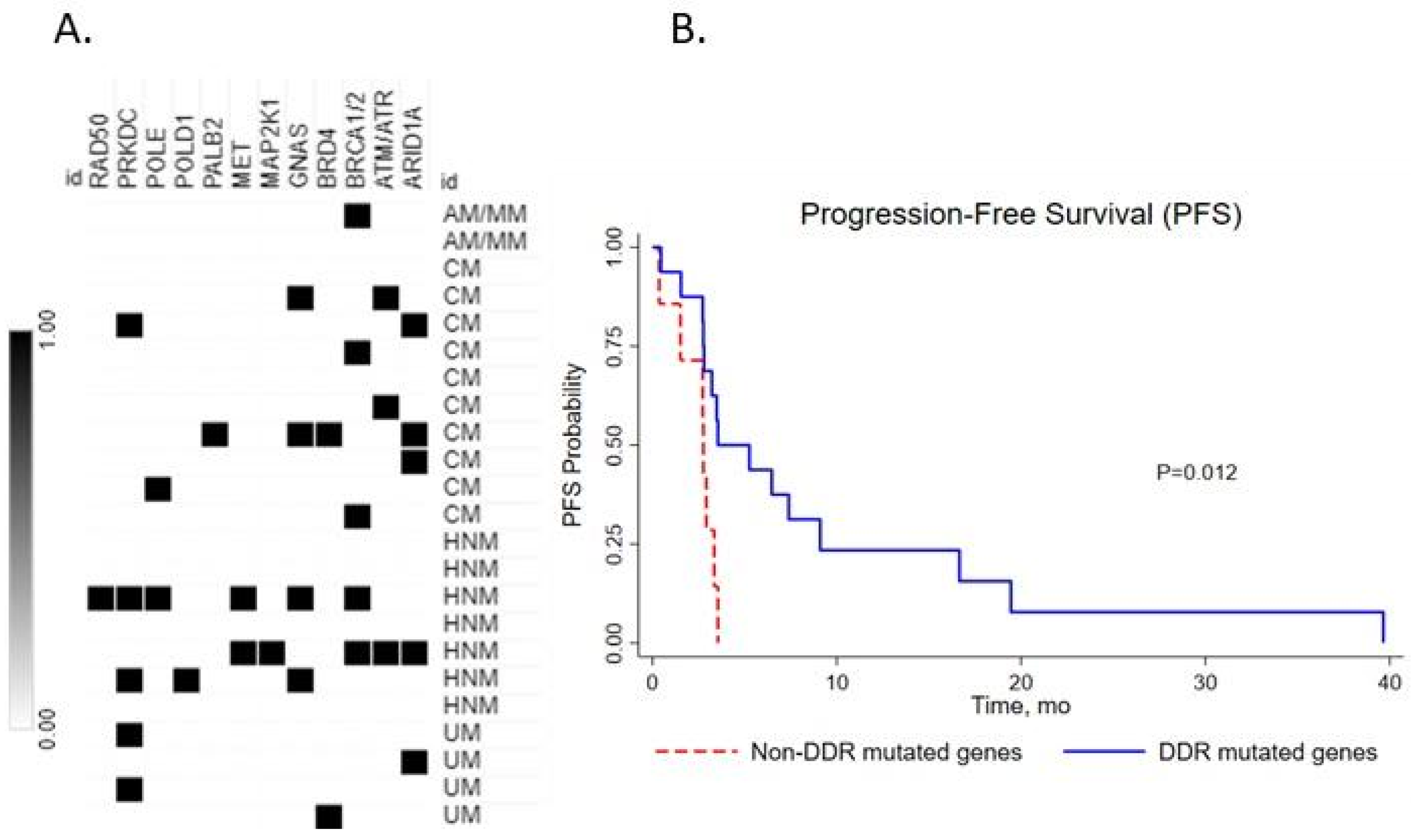
3.3. Impact of DNA Damage Repair (DDR) Gene Mutation on Clinical Outcome

We next investigated the impact of the number of DDR gene mutations on the clinical outcome of metastatic melanoma subtypes after ICI therapy. We found that 83% of metastatic melanoma cases had at least one mutation in a DDR pathway gene (Figure 3A). DDR gene mutations were significantly associated with better PFS (3.54 months (95% CI: 2.75–9.10) vs. 2.75 months (95% CI: 0.36–3.35), p = 0.012, log rank) in the full cohort (n = 23) (Figure 3B).
There was a significant association observed with DDR gene mutation and age (p = 0.012, Fisher’s exact test), but no significant correlation with CD8/CD20 expression and clinical variables (Table 1).

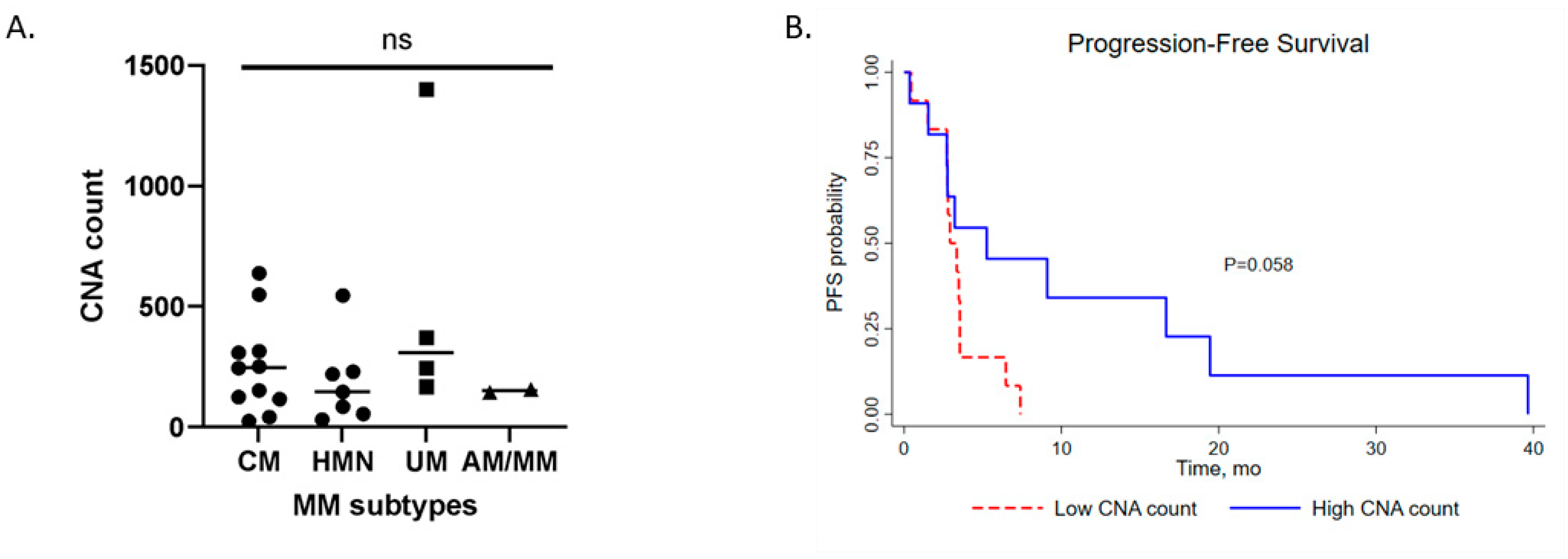
3.4. Copy Number Aberrations (CNA) of Metastatic Melanoma Subtypes and Clinical Impact

The median CNA count for the full metastatic melanoma cohort was 168 (range: 26–1402). Sub-analyses based on subtype showed the median CNA count in CM, 202.5 (range: 26–639); HNM, 147 (range: 32–547); UM, 308.5 (range: 168–1402); AM/MM, 151 (range: 145–157). UM has the highest CNA count compared to the other tumours, but not significantly (Figure 4A). Metastatic melanoma tumours with a high CNA count showed better PFS (HR: 0.39; median PFS: 5.25 months vs 3.54 months; p = 0.058, log rank) (Figure 4B).
Univariate analyses for PFS and OS are shown in Table 2. Univariate analysis revealed that high CD8 expression (HR 0.32; 95% CI: 0.11–0.93, p = 0.039) and the presence of DDR gene mutations (HR 0.28; 95% CI: 0.10–0.81, p = 0.019) are favourable prognostic factors for PFS; no variable was found to be a prognostics factor for OS. The variables remained as independent favourable prognostic factors in multivariate analysis (p = 0.018).

4. Discussion

Clinical markers of response to ICI therapies have been extensively studied. Comprehensive predictive and prognostic models have been developed integrating the expression of intermolecular interactions within the tumour, its microenvironment as well as the correlation with tumour genome mutational, neo-antigen burden, and genetic variations in DNA mismatch repair genes [14]. However, conflicting evidence into the clinical predictive efficacy of ICI biomarkers in different cancer subtypes exists [19]. PD-L1 expression measured by immunohistochemistry (IHC) has emerged as a widely used biomarker of response to ICI; however, patients with PD-L1 negative tumours also have shown efficacy to ICI treatment. In a clinical trial of refractory or metastatic cervical cancer patients treated with nivolumab, the overall response rates (ORR) were 2/10 (20%) in PD-L1 positive patients and 1/6 (16.7%) in PD-L1 negative patients. Indeed, a patient with a PD-L1 negative tumour had a durable partial response that exceeded 24 months [20].
Responses in other cancer types such as small cell lung cancer (SCLC), squamous cell carcinoma of the head and neck (SCCHN) and cutaneous melanoma led to an accelerated all-comer approval for pembrolizumab regardless of PD-L1 status [21,22]. However, a recent follow-up of the cervical cancer KEYNOTE-158 study found that all 14 responses were in patients with PD-L1–positive tumours (ORR 17.1%) and with no responses observed in the PD-L1–negative cohort (ORR 0.0%) [23].
Metastatic melanoma is a heterogeneous group of cancers characterised by site of origin, subtypes based on the cumulative levels of exposure to ultraviolet (UV) radiation [10,11,24]. Melanomas found in sun damaged areas of the body tend to have a higher mutational burden than tumours arising from non-sun exposed areas correlating with somatic mutational profile of the tumour and high response rates after ICI therapy [25]. In addition, many studies have found that melanoma is characterised by high immunogenicity and patients with high TIL cells have shown favourable therapeutic outcomes and prognosis. However, as ICI only shows efficiency in some metastatic melanoma patients (majority of cutaneous melanoma), this study examined the clinical relevance of CD8+ TIL and CD20+ tumour infiltrating B cells and the genomic mutational landscape in different subtypes of 23 metastatic melanoma patients treated with immune checkpoint inhibitors (ICI). TILs have previously been identified as prognostic and predictive biomarkers in many cancers, including melanoma. However, complex tumour-immune interactions exist whereby some cancers with similar TIL cell expression respond differently to immunotherapy [26]. We observed lower CD8+ and CD20+ expression in subtypes CM and HNM. CD8+ TILs have been detected in metastatic uveal melanoma (MUM) [27], however, no difference in CD8+ infiltrating T cells between metastatic CM and MUM was observed [28]. However, the number of UM in our study was very low (n = 4). Location of the tumour in head and neck melanoma has a significant impact on prognosis, with the scalp displaying the worst prognosis, followed by the ear, cheek, and neck [9]. A rare variant of HNM is desmoplastic melanoma (DM), which is associated with old age, chronic sun exposure, and location on the head and neck. HNM DM has shown a great response rate to ICI therapy [29]. However, Frydenlund et al. [30] found that the presence of CD8+ lymphocytes correlated significantly with depth of invasion > 1 mm, and PD-L1 expression in DM. Additionally, CD8+ TILs correlated with PD-L1 expression, which was associated with tumour aggressiveness and progression in DM [31]. However, in our study, HNM did not harbour a higher mutational burden comparable to other metastatic melanoma subtypes. High tumour mutation burden (TMB) has been associated with response to ICI in several cancers. Several studies of multiple cancers have shown that ICI mean response rate positively correlates with TMB [32]. The FDA have approved TMB (TMB-High defined as ≥10 mutations/megabase of DNA (mut/Mb), as determined by the targeted sequencing FoundationOne CDx (F1CDx) assay) as a companion diagnostic biomarker for pembrolizumab [33]. However, some clinical studies also showed that high TMB does not predict clinically relevant responses to ICI in all cancer types. Specifically, cancer types such as breast, prostate and glioma displayed no relationship between high CD8 TIL expression and neo-antigen load. Designated TMB-H tumours failed to achieve a 20% ORR (15.3%) and displayed significantly lower ORR relative to TMB-Low tumours [34].
In agreement with many studies, DDR gene mutations correlated with better PFS in the metastatic melanoma cohort. Additionally, metastatic melanoma tumours with a high CNA count showed better PFS (HR: 0.39; median PFS: 5.25 months vs. 2.92 months; p = 0.058, logrank) but this was not significant. In univariate analysis DDR gene mutations and CD8 high were prognostic indicators of better PFS. Therefore, this study highlights the differential expression of TILs and genomic variation (DDR gene mutations and CNV counts) in subsets of metastatic melanoma. The variability of these biomarkers highlights the need for additional predictive and prognostic specific to metastatic melanoma subtypes. However, this study is retrospective in nature, and is limited by the small sample and subtype size, therefore further analysis on larger cohorts would be required.

Supplementary Materials

The following supporting information can be downloaded at: https://0-www-mdpi-com.brum.beds.ac.uk/article/10.3390/medsci10020026/s1, Table S1: Patient information and detected mutations.

Author Contributions

Conceptualization, G.G., J.C. and N.W.; data curation, C.A., A.-M.L., R.M., J.M.-E. and N.W.; formal analysis, C.A., N.M., A.-M.L., R.M., J.M.-E. and N.W.; funding acquisition, G.G. and J.C.; methodology, N.M., A.-M.L., A.M., S.K. and N.W.; resources, N.H., S.K., G.G. and J.C.; supervision, N.W.; writing—original draft, C.A. and N.W.; writing—review & editing, C.A., R.M., J.M.-E. and N.W. All authors have read and agreed to the published version of the manuscript.

Funding

This research was supported by Cancer Clinical Research Trust (CCRT), and by a HRB Summer Scholarship Ref: SS-2016-1806 (NMcN).

Institutional Review Board Statement

The study was conducted according to the guidelines of the Declaration of Helsinki and approved by the Research Ethics Committee of St Vincent’s University Hospital, Dublin 4, Ireland and Dublin City University (DCUREC/2015/192).

Informed Consent Statement

Informed consent was obtained from living subjects involved in the study and for deceased patients informed consent was waived by the local Ethics Committee due to the retrospective nature of the study.

Data Availability Statement

The sequencing and clinical variables for this study are available from EGA European Genome-Phenome Archive accession number: EGAD00001008336.

Conflicts of Interest

Authors R.M. and J.M. were employees of NEO New Oncology GmbH, a Siemens Healthineers company, at the time of sequencing. All other authors declare no conflict of interests.

References

  1. Larkin, J.; Chiarion-Sileni, V.; Gonzalez, R.; Grob, J.-J.; Rutkowski, P.; Lao, C.D.; Cowey, C.L.; Schadendorf, D.; Wagstaff, J.; Dummer, R.; et al. Five-Year Survival with Combined Nivolumab and Ipilimumab in Advanced Melanoma. N. Engl. J. Med. 2019, 381, 1535–1546. [Google Scholar] [CrossRef] [PubMed] [Green Version]
  2. Robert, C.; Thomas, L.; Bondarenko, I.; O’Day, S.; Weber, J.; Garbe, C.; Lebbe, C.; Baurain, J.-F.; Testori, A.; Grob, J.-J.; et al. Ipilimumab plus Dacarbazine for Previously Untreated Metastatic Melanoma. N. Engl. J. Med. 2011, 364, 2517–2526. [Google Scholar] [CrossRef] [PubMed] [Green Version]
  3. Prieto, P.A.; Yang, J.C.; Sherry, R.M.; Hughes, M.S.; Kammula, U.S.; White, D.E.; Levy, C.L.; Rosenberg, S.A.; Phan, G.Q. CTLA-4 Blockade with Ipilimumab: Long-Term Follow-up of 177 Patients with Metastatic Melanoma. Clin. Cancer Res. 2012, 18, 2039–2047. [Google Scholar] [CrossRef] [PubMed] [Green Version]
  4. Chin, L.; Garraway, L.A.; Fisher, D.E. Malignant Melanoma: Genetics and Therapeutics in the Genomic Era. Genes Dev. 2006, 20, 2149–2182. [Google Scholar] [CrossRef] [Green Version]
  5. Tas, F.; Keskin, S.; Karadeniz, A.; Daǧoǧlu, N.; Sen, F.; Kilic, L.; Yildiz, I. Noncutaneous Melanoma Have Distinct Features from Each Other and Cutaneous Melanoma. Oncology 2012, 81, 353–358. [Google Scholar] [CrossRef]
  6. Whiteman, D.C.; Pavan, W.J.; Bastian, B.C. The Melanomas: A Synthesis of Epidemiological, Clinical, Histopathological, Genetic, and Biological Aspects, Supporting Distinct Subtypes, Causal Pathways, and Cells of Origin. Pigment Cell Melanoma Res. 2011, 24, 879–897. [Google Scholar] [CrossRef] [Green Version]
  7. Shashanka, R.; Smitha, B.R. Head and Neck Melanoma. ISRN Surg. 2012, 2012, 143–152. [Google Scholar] [CrossRef] [Green Version]
  8. Al-Qurayshi, Z.; Hassan, M.; Srivastav, S.; Sperry, S.; Pagedar, N.; Hamner, J.; Kandil, E. Risk and Survival of Patients with Head and Neck Cutaneous Melanoma: National Perspective. Oncology 2017, 93, 18–28. [Google Scholar] [CrossRef]
  9. Zito, P.M.; Scharf, R. Melanoma of the Head and Neck. In StatPearls; StatPearls Publishing: Treasure Island, FL, USA, 2021. [Google Scholar]
  10. Rabbie, R.; Ferguson, P.; Molina-Aguilar, C.; Adams, D.J.; Robles-Espinoza, C.D. Melanoma Subtypes: Genomic Profiles, Prognostic Molecular Markers and Therapeutic Possibilities. J. Pathol. 2019, 247, 539–551. [Google Scholar] [CrossRef]
  11. Chacón, M.; Pfluger, Y.; Angel, M.; Waisberg, F.; Enrico, D. Uncommon Subtypes of Malignant Melanomas: A Review Based on Clinical and Molecular Perspectives. Cancers 2020, 12, 2362. [Google Scholar] [CrossRef]
  12. Johnson, D.B.; Peng, C.; Abramson, R.G.; Ye, F.; Zhao, S.; Wolchok, J.D.; Sosman, J.A.; Carvajal, R.D.; Ariyan, C.E. Clinical Activity of Ipilimumab in Acral Melanoma: A Retrospective Review. Oncologist 2015, 20, 648–652. [Google Scholar] [CrossRef] [PubMed] [Green Version]
  13. Klemen, N.D.; Wang, M.; Rubinstein, J.C.; Olino, K.; Clune, J.; Ariyan, S.; Cha, C.; Weiss, S.A.; Kluger, H.M.; Sznol, M. Survival after Checkpoint Inhibitors for Metastatic Acral, Mucosal and Uveal Melanoma. J. Immunother. Cancer 2020, 8, e000341. [Google Scholar] [CrossRef] [PubMed]
  14. Bai, R.; Lv, Z.; Xu, D.; Cui, J. Predictive Biomarkers for Cancer Immunotherapy with Immune Checkpoint Inhibitors. Biomark. Res. 2020, 8, 1–17. [Google Scholar] [CrossRef] [PubMed]
  15. Schatz, S.; Falk, M.; Jóri, B.; Ramdani, H.O.; Schmidt, S.; Willing, E.M.; Menon, R.; Groen, H.J.M.; Diehl, L.; Kröger, M.; et al. Integration of Tumor Mutation Burden and PD-L1 Testing in Routine Laboratory Diagnostics in Non-Small Cell Lung Cancer. Cancers 2020, 12, 1685. [Google Scholar] [CrossRef]
  16. Chen, E.Y.; Tan, C.M.; Kou, Y.; Duan, Q.; Wang, Z.; Meirelles, G.V.; Clark, N.R.; Ma’ayan, A. Enrichr: Interactive and Collaborative HTML5 Gene List Enrichment Analysis Tool. BMC Bioinform. 2013, 14, 128. [Google Scholar] [CrossRef] [Green Version]
  17. Kuleshov, M.V.; Jones, M.R.; Rouillard, A.D.; Fernandez, N.F.; Duan, Q.; Wang, Z.; Koplev, S.; Jenkins, S.L.; Jagodnik, K.M.; Lachmann, A.; et al. Enrichr: A Comprehensive Gene Set Enrichment Analysis Web Server 2016 Update. Nucleic Acids Res. 2016, 44, W90–W97. [Google Scholar] [CrossRef] [Green Version]
  18. Xie, Z.; Bailey, A.; Kuleshov, M.V.; Clarke, D.J.B.; Evangelista, J.E.; Jenkins, S.L.; Lachmann, A.; Wojciechowicz, M.L.; Kropiwnicki, E.; Jagodnik, K.M.; et al. Gene Set Knowledge Discovery with Enrichr. Curr. Protoc. 2021, 1, e90. [Google Scholar] [CrossRef]
  19. Grossman, J.E.; Vasudevan, D.; Joyce, C.E.; Hildago, M. Is PD-L1 a Consistent Biomarker for Anti-PD-1 Therapy? The Model of Balstilimab in a Virally-Driven Tumor. Oncogene 2021, 40, 1393–1395. [Google Scholar] [CrossRef]
  20. Naumann, R.W.; Hollebecque, A.; Meyer, T.; Devlin, M.-J.; Oaknin, A.; Kerger, J.; López-Picazo, J.M.; Machiels, J.-P.; Delord, J.-P.; Evans, T.R.J.; et al. Safety and Efficacy of Nivolumab Monotherapy in Recurrent or Metastatic Cervical, Vaginal, or Vulvar Carcinoma: Results From the Phase I/II CheckMate 358 Trial. J. Clin. Oncol. 2019, 37, 2825–2834. [Google Scholar] [CrossRef]
  21. Kefford, R.; Ribas, A.; Hamid, O.; Robert, C.; Daud, A.; Wolchok, J.D.; Joshua, A.M.; Hodi, F.S.; Gangadhar, T.C.; Hersey, P.; et al. Clinical Efficacy and Correlation with Tumor PD-L1 Expression in Patients (Pts) with Melanoma (MEL) Treated with the Anti-PD-1 Monoclonal Antibody MK-3475. J. Clin. Oncol. 2014, 32 (Suppl. 15), 3005. [Google Scholar] [CrossRef]
  22. Mehra, R.; Seiwert, T.Y.; Gupta, S.; Weiss, J.; Gluck, I.; Eder, J.P.; Burtness, B.; Tahara, M.; Keam, B.; Kang, H.; et al. Efficacy and Safety of Pembrolizumab in Recurrent/Metastatic Head and Neck Squamous Cell Carcinoma: Pooled Analyses after Long-Term Follow-up in KEYNOTE-012. Br. J. Cancer 2018, 119, 153–159. [Google Scholar] [CrossRef] [PubMed]
  23. Chung, H.; Delord, J.-P.; Perets, R.; Italiano, A.; Shapira-Frommer, R.; Manzuk, L.; Piha-Paul, S.; Xu, L.; Jin, F.; Norwood, K.; et al. Pembrolizumab Treatment of Advanced Cervical Cancer: Updated Results from the Phase II KEYNOTE-158 Study. Gynecol. Oncol. 2021, 162, S27. [Google Scholar] [CrossRef]
  24. Shain, A.H.; Bastian, B.C. From Melanocytes to Melanomas. Nature Reviews Cancer. Nat. Rev. Cancer 2016, 16, 345–358. [Google Scholar] [CrossRef]
  25. Pham, T.V.; Boichard, A.; Goodman, A.; Riviere, P.; Yeerna, H.; Tamayo, P.; Kurzrock, R. Role of Ultraviolet Mutational Signature versus Tumor Mutation Burden in Predicting Response to Immunotherapy. Mol. Oncol. 2020, 14, 1680–1694. [Google Scholar] [CrossRef]
  26. Paijens, S.T.; Vledder, A.; de Bruyn, M.; Nijman, H.W. Tumor-Infiltrating Lymphocytes in the Immunotherapy Era. Cell. Mol. Immunol. 2020, 18, 842–859. [Google Scholar] [CrossRef] [PubMed]
  27. Qin, Y.; Bollin, K.; de Macedo, M.P.; Carapeto, F.; Kim, K.B.; Roszik, J.; Wani, K.M.; Reuben, A.; Reddy, S.T.; Williams, M.D.; et al. Immune Profiling of Uveal Melanoma Identifies a Potential Signature Associated with Response to Immunotherapy. J. Immunother. Cancer 2020, 8, e000960. [Google Scholar] [CrossRef] [PubMed]
  28. Qin, Y.; de Macedo, M.P.; Reuben, A.; Forget, M.-A.; Haymaker, C.; Bernatchez, C.; Spencer, C.N.; Gopalakrishnan, V.; Reddy, S.; Cooper, Z.A.; et al. Parallel Profiling of Immune Infiltrate Subsets in Uveal Melanoma versus Cutaneous Melanoma Unveils Similarities and Differences: A Pilot Study. Oncoimmunology 2017, 6, e1321187. [Google Scholar] [CrossRef] [Green Version]
  29. Eroglu, Z.; Zaretsky, J.M.; Hu-Lieskovan, S.; Kim, D.W.; Algazi, A.; Johnson, D.B.; Liniker, E.; Kong, B.; Munhoz, R.; Rapisuwon, S.; et al. High Response Rate to PD-1 Blockade in Desmoplastic Melanomas. Nature 2018, 553, 347–350. [Google Scholar] [CrossRef]
  30. Frydenlund, N.; Leone, D.; Yang, S.; Hoang, M.P.; Deng, A.; Hernandez-Perez, M.; Singh, R.; Biswas, A.; Yaar, R.; Mahalingam, M. Tumoral PD-L1 Expression in Desmoplastic Melanoma Is Associated with Depth of Invasion, Tumor-Infiltrating CD8 Cytotoxic Lymphocytes and the Mixed Cytomorphological Variant. Mod. Pathol. 2017, 30, 357–369. [Google Scholar] [CrossRef]
  31. Kraft, S.; Fernandez-Figueras, M.-T.; Richarz, N.A.; Flaherty, K.T.; Hoang, M.P. PDL1 Expression in Desmoplastic Melanoma Is Associated with Tumor Aggressiveness and Progression. J. Am. Acad. Dermatol. 2017, 77, 534–542. [Google Scholar] [CrossRef]
  32. Yarchoan, M.; Hopkins, A.; Jaffee, E.M. Tumor Mutational Burden and Response Rate to PD-1 Inhibition. N. Engl. J. Med. 2017, 377, 2500. [Google Scholar] [CrossRef] [PubMed]
  33. FDA. Approves Pembrolizumab for Adults and Children with TMB-H Solid Tumors FDA. Available online: https://www.fda.gov/drugs/drug-approvals-and-databases/fda-approves-pembrolizumab-adults-and-children-tmb-h-solid-tumors (accessed on 4 October 2021).
  34. McGrail, D.J.; Pilié, P.G.; Rashid, N.U.; Voorwerk, L.; Slagter, M.; Kok, M.; Jonasch, E.; Khasraw, M.; Heimberger, A.B.; Lim, B.; et al. High Tumor Mutation Burden Fails to Predict Immune Checkpoint Blockade Response across All Cancer Types. Ann. Oncol. 2021, 32, 661–672. [Google Scholar] [CrossRef] [PubMed]
Figure 1. Immunohistochemical staining of tumour samples at ×200 magnification. Representative high expression (strong diffuse immune cell infiltration) for (A). CD8 in HNM, (B). CD20 staining in HNM. Representative low expression (peritumour and/or interstitial immune infiltration) for (C). CD8 in MM, (D). CD20 expression in CM. Representative negative expression for (E). CD8 and (F). CD20 expression in HNM. Overall expression of (G). CD8 and (H). CD20 in metastatic melanoma subtypes.
Medsci 10 00026 g001
Figure 2. Kaplan–Meier plots of progression-free survival (PFS) between (A). CD8 high and CD8 low, and (B). CD20 high and CD20 low TIL score groups with log-rank p value.
Medsci 10 00026 g002
Figure 3. (A) Map of metastatic melanoma subtypes with DDR gene mutations. (B) Kaplan–Meier plots of progression-free survival (PFS) for cohorts with DDR mutated genes and without DDR mutated genes with log-rank p-value.
Medsci 10 00026 g003
Figure 4. (A). Profile of CNA count on subtypes of metastatic melanoma (CM Medsci 10 00026 i001; HNM Medsci 10 00026 i001; UM Medsci 10 00026 i002; AM/MM Medsci 10 00026 i003). (B). Progression-free survival (PFS) of CNA low (red line) and high (blue line) in the total metastatic melanoma cohort. Kaplan–Meier survival curves with a log-rank test were used for the analysis. ns, not significant.
Medsci 10 00026 g004
Table 1. Association between the expression of CD8, CD20 expression, DDR gene mutations, CNA count and clinical features.
CD8 (n = 22)CD20 (n = 22)DDR Mutation (n = 23)
Neg/LowHighp-ValueNeg/LowHighp-ValueNegPosp-Value
(n = 14)(n = 8)(n = 14)(n = 8)(n = 7)(n = 16)
Age<62550.221460.048640.012
≥6293102112
Melanoma subtypeCM730.917730.720280.171
HNM435243
UM211204
AM/MM111111
ChemotherapyYes240.096330.369150.382
No124115611
Immunotherapy1 ICI740.670740.670570.222
2+ ICI747429
CD20High350.072 350.510
Neg/Low113410
DDR mutationPositive960.490
Negative52
CNA countHigh550.221550.221380.556
Low939348
Table 2. Univariate Cox Regression analysis of clinical variables associated with PFS and OS.
PFSUnivariate AnalysisOSUnivariate Analysis
Median PFS, Months (Range)HR (95% CI)p-ValueMedian OS, Months (Range)HR (95% CI)p-Value
3.35 (0.36–39.65) 27.72 (1.80–177.74)
Categories (n = 23)
Age<62103.23 (0.36–16.65) Ref 44.13 (5.12–100.66)Ref
≥62133.35 (0.45–39.65)0.74 (0.30–1.78)0.50227.36 (1.80–177.74)1.27 (0.415–3.92)0.670
Melanoma SubtypeCM103.54 (0.36–19.45)Ref 51.51 (1.8–100.67)Ref
HNM72.92 (1.51–7.46)1.38 (0.48–3.98)0.54827.73 (5.12–177.74)1.66 (0.36–7.66)0.513
UM43.01 (1.54–39.65)0.82 (0.22–3.07)0.77719.52 (10.74–50.13)4.86 (1.12–21.05)0.034
AM/MM25.05 (2.72–7.39)1.27 (0.27–6.01)0.75620.90 (14.06–27.72)5.23 (0.86–31.73)0.072
Chemo-TherapyYes64.40 (2.92–7.39) Ref 38.04 (23.52–100.66)Ref
No172.79 (0.36–39.65)1.00 (0.37–2.69)0.99727.30 (1.80–177.74)0.69 (1.87–2.55)0.582
Immuno-Therapy1 ICI122.85 (0.36–39.65)Ref 25.74 (1.80–80.91)Ref
2+ ICI113.48 (2.72–19.44)0.99 (0.41–2.35)0.98435.58 (14.06–177.74)0.36 (0.11–1.20)0.098
DDR MutationNeg162.75 (0.36–3.54)Ref 40.50 (5.12–177.74)Ref
Pos74.40 (0.45–39.65)0.28 (0.10–0.81)0.01927.54 (1.80–100.66)0.98 (0.29–3.25)0.984
CNA CountLow122.92 (1.51–3.54)Ref 31.65 (1.80–177.74)Ref
High115.25 (1.54–19.4)0.39 (0.14–1.08)0.07023.75 (5.12–100.66)1.24 (0.417–3.74)0.690
Categories (n = 22)
CD8 ExpressionNeg/Low142.78 (0.36–7.45)Ref 27.72 (1.81–177.74)Ref
High86.93 (2.75–19.44)0.32 (0.11–0.94)0.03934.11 (5.12–53.61)0.83 (0.24–2.81)0.773
CD20 ExpressionNeg/Low143.41 (0.45–19.44)Ref 27.54 (1.81–177.74)Ref
High83.07 (0.36–16.65)1.03 (0.41–2.57)0.93532.01 (10.74–80.91)0.91 (0.27–3.07)0.888
Publisher’s Note: MDPI stays neutral with regard to jurisdictional claims in published maps and institutional affiliations.

