Article

Commercial Red Food Dyes Preparations Modulate the Oxidative State in Three Model Organisms (Cucumis sativus, Artemia salina, and Danio rerio)

1
Department of Science and Technology, University of Naples Parthenope, Via Acton n. 38-I, 80133 Napoli, NA, Italy
2
Department of Biology, University of Naples Federico II, 80126 Naples, NA, Italy
3
BAT Center-Interuniversity Center for Studies on Bioinspired Agro-Environmental Technology, 80055 Portici, NA, Italy
*
Authors to whom correspondence should be addressed.
Submission received: 8 April 2022 / Revised: 4 May 2022 / Accepted: 18 May 2022 / Published: 21 May 2022

Abstract

:
The growing environmental spreading of food synthetic dyes and bio-colors have the potential for altering organisms’ redox states. Here, three model species for aquatic pollution trials, Cucumis sativus seeds, Artemia salina cysts, and Danio rerio embryos, were short-term exposed to a fixed concentration of the artificial red E124, and two red bio-colors, cochineal E120, and vegan red (VEGR). In the animal models, we evaluated the total reactive oxygen species (ROS) and the susceptibility to in vitro oxidative stress, and in C. sativus, H2O2 production and antioxidant capacity. We also measured organismal performance indices (routine oxygen consumption in the animal models, dark oxygen consumption, and photosynthetic efficiency in C. sativus). In C. sativus, only E124 increased ROS and affected dark oxygen consumption and photosynthetic efficiency, while all dyes enhanced the antioxidant defenses. In the A. salina nauplii, all dyes increased ROS, while E120 and E124 reduced the susceptibility to oxidative stress. In D. rerio, treatments did not affect ROS content, and reduced oxidative stress susceptibility. Our data show that red food dyes affect the redox state of the developing organisms, in which ROS plays a significant role. We suggest a potentially toxic role for red food dyes with environmentally relevant consequences.

1. Introduction

The addition of dyes to food is an established practice introduced to overcome the loss of original appearance and appeal upon food processing and preservation. Artificial and animal-based dyes have dominated food color production for the last 100 years. Currently, human and animal health concerns are attracting the attention of the food industry on ‘bio-colors’ of vegetal origin such as betacyanins, anthocyanins, carotenoids, and chlorophylls [1]. Vegetal bio-colors are usually fundamental compounds in plants for their role as photosynthetic pigments or for preserving plant health [2]. They are commonly extracted from fruits and vegetables and are considered safer and healthier than the artificial dyes for their antioxidant and anti-inflammatory properties [3].
Independently from their origin, the enormous and growing utilization of food dyes [4] is increasing their release into the environment. The spreading of food dyes in water bodies and watered soils is particularly worrying [5], as both artificial dyes and animal-based bio-colors have a proven direct toxicity on aquatic or water-dependent organisms [6,7]. Recently, it has been reported that also vegetal bio-colors may be dangerous due to the possible formation of hazardous by-products [8].
A putative major source of damaging effects of food dye on living organisms is the alteration of their redox state. Sub-lethal concentrations of synthetic and natural dyes may stimulate the production of reactive oxygen species (ROS) in aquatic organisms, inducing oxidative stress [6], a condition that can cause a wide spectrum of functional alterations, from molecular (e.g., damaged proteins, lipids, and DNA) to tissue (e.g., enzyme activity and substrate availability), and whole-body (e.g., metabolic rate, motility, etc.) levels [6,9,10,11,12]. On the other hand, these pollutants might also interact with the detoxification processes by modulating the antioxidant defenses [12,13].
In this context, it is relevant to evaluate the putative effects of artificial and natural food dyes on the redox state of different species. The most suitable approach to assessing the potential hazards of food dyes spreading in the environment is the simultaneous analysis of model organisms belonging to different water-related environments, which may exhibit a diverse sensitivity. We recently reported that a given concentration of specific food dyes can be detrimental for one species and not others [7]. In the present study, we aimed to compare two aquatic animals Artemia salina (marine invertebrate) and Danio rerio (freshwater fish) with a plant species Cucumis sativus reared with dye-treated water. Young, fast-growing developmental stages were used (nauplii, embryos, and seedlings, respectively), which can be considered the most sensitive. The nauplii of Artemia salina are a useful and accurate invertebrate model for applied toxicology, especially for their resistance to manipulation and fast development [14,15]. Danio rerio, commonly known as the zebrafish, is a widely used model organism for screening the eco-toxicological effects induced by environmental pollutants [16,17,18,19] including artificial dyes [7,9,20]. Finally, Cucumis sativus L. is a plant model organism, widely used to evaluate the toxic effects of different types of pollutants, i.e., dyes, heavy metals, and polycyclic aromatic hydrocarbons [7,21,22]. Cucumber seedlings are sensitive indicators of soil pollution, including watering contamination [23].
The aim of this work was to compare three models for ROS levels, antioxidant capacity, and organismal performance parameters (routine oxygen consumption and, for C. sativus, also the photosynthetic efficiency) in response to food dyes. We focused the attention on three red dyes widely used not only for food dyeing but also from other industrial activities (cosmetics and textile) [24]. In particular, we tested the artificial dye Ponceau Red (E124, [25]), a commercial bio-color containing pulverized radish, black currant, and apple (Red Vegan, VEGR), and the natural dye Cochineal Red (E120, [26]) extracted from the insects Dactylopius coccus and Kermes vermilio. There are indications that the azo dye E124 may induce free radical production, but results are so far contrasting [27,28]. Compared with the synthetic E124, the two natural dyes may display different effects due to the complex composition of commercial products. VEGR likely contains elevated levels of polyphenols [29]. These compounds are usually regarded as antioxidants, but they may also accelerate free radical formation under specific conditions, and so may act as pro-oxidant [30]. Commercial E120 may pose an oxidative stress risk due to the formation of some derivatives of carminic acid [31,32,33].

2. Materials and Methods

2.1. Preparation of Food Dye Solutions

The three food dyes used in this study are commercially available products used to prepare domestic foods. Ponceau red E124 (IPAFOOD srl, Italy) and cochineal red E120 (IPAFOOD srl, Italy) are sold in 3 mL vials, containing 20% dye in water. The red bio-color VEGR (Rebecchi Fratelli Valtrebbia, Italy), sold in powder, contains extracts of radish, black currant, and apple; it also contains unknown amounts of maltodextrin and citric acid.
A single vial of E124 or E120 was diluted in mineral water for human consumption at a final concentration of 1.2 g L−1, according to Motta et al. [7]. The red bio-color powder was dissolved in mineral water at the same final concentration. The dye concentrations utilized in this study are two-fold higher than those usually found in natural environments (up to 0.5 g L−1) [34], because we wanted to obtain a positive control for the three models and assess their capacity to counteract the oxidative stress imposed by these substances. The solutions were used as such to water Cucumis sativus seeds and as breeding solutions for Danio rerio. Water salinity was adjusted according to the specific requirements of Artemia salina nauplii [35] and Danio rerio embryos [36]. Controls were exposed to pure mineral water or seawater.

2.2. The Plant Model: Cucumis sativus L. Seedlings

2.2.1. Growth and Experimental Design

C. sativus L. seeds were surface-sterilized [7] and arranged randomly in groups of 13, in 90 mm Petri dishes containing tissue paper wetted with 8 mL of mineral water (CONT) or water added with the three food dyes (VEGR, E120, and E124). The Petri dishes, set in triplicate, were incubated in the dark in a climatic chamber at the Department of Biology of the University of Naples Federico II (General Impianti S.A.S., Naples, Italy), at a fixed temperature of 25 ± 2 °C and relative humidity (RH) of 50 ± 5%.
After germination, sprouts were transferred in a growth chamber in vials added with mineral water (CONT) or different dye solutions (VEGR, E120, or E124) and exposed to a Photosynthetic Photon Flux Density (PPFD) of 190 ± 10 µmol photons m−2 s−1, provided by white LEDs (light emitting diodes), under a photoperiod of 12 h. The cucumber sprouts were let to grow until 15 days after germination (DAG).
Seedlings were sprinkled with distilled water during the growth to reintegrate the water lost by evapotranspiration. The photosynthetic apparatus functionality was assessed at 15 DAG by measuring in vivo the PSII maximal photochemical efficiency (Fv/Fm) on seedlings exposed to different dyes.
Two weeks were needed to allow the full expansion of cotyledons to perform the physiological measurements in vivo. Leaves from different plants were collected to determine the endogenous hydrogen peroxide (H2O2) level, the total photosynthetic pigment content (chlorophyll a, chlorophyll b, and carotenoids), the antioxidant capacity, and total polyphenols. The seedling total biomass and the maximal photochemical efficiency of photosystem II (Fv/Fm) were also measured as proxies of plant growth and photosynthetic apparatus functionality, respectively. Finally, these data were compared with the dark oxygen consumption of 48 h seedlings, as an early stress index, when the photosynthetic apparatus was not yet developed.

2.2.2. Photosynthetic Pigment Content, Chlorophyll Fluorescence Emission Measurements, and Dark Oxygen Consumption Determination

The photosynthetic pigment content and the fluorescence emission measurements were carried out on n = 10 leaves for each treatment at 15 DAG.
The concentrations of chlorophylls and carotenoids were determined according to Lichtenthaler [37]. Briefly, pigments were extracted in ice-cold 100% acetone and centrifuged (Labofuge GL, Heraeus Sepatech, Hanau, Germany) at 3000× g for 5 min. The absorbance was measured by a spectrophotometer (Cary 100 UV-VIS, Agilent Technologies, Santa Clara, CA, USA) at wavelengths of 470, 645, and 662 nm and pigment concentration was expressed in μg cm−2.
Fluorescence emission analysis of chlorophyll a, which allows a non-invasive evaluation of the healthy status of photosystems in plants, was performed with a portable pulse amplitude modulated fluorometer equipped with a light sensor (FluorPen FP100, Photon System Instruments, Brno, Czech Republic). The ground fluorescence F0 was induced on 30′ dark-adapted leaves, by a blue LED internal light of about 1–2 μmol photons m−2 s−1. The maximal fluorescence level in the dark-adapted state, Fm was induced by a saturating light pulse of 3.000 μmol photons m−2 s−1. The maximum PSII photochemical efficiency, Fv/Fm, was calculated as (Fm − F0)/Fm. Measurements were carried out on n = 10 samples and repeated twice [38].
In a dark chamber, the dark oxygen consumption in seedlings was determined at 2 DAG using an Oxygen Monitor System (YSI 5300 A) [39]. Measurements were conducted from imbibed seeds at 25 °C, over a 4 h period. Cumulative consumption of 15 germinated seeds was measured for each replicate of control or treated groups and expressed as g O2 h−1 seed−1. The oxygen uptake rates were calculated from linear regressions of pO2-time-course data (R2 > 0.90). Four replicates for each group were performed.

2.2.3. Endogenous H2O2 Determination and Plant Antioxidant Response

The occurrence of possible oxidative stress and recovery in C. sativus strategies were assessed by determining the endogenous H2O2 content, total antioxidant capacity, and total polyphenol levels. All the analyses were carried out on n = 10 seedlings for each treatment and repeated twice.
Endogenous H2O2, a marker of ROS, is a constitutive product within the cells and variations of this compound can suggest possible stress conditions. The levels of H2O2 were determined in leaves and roots of cucumber seedlings according to Sergiev et al. [40]. Briefly, samples (0.250 g) were homogenized in ice with 0.1% (w:v) TCA. The homogenate was centrifuged at 12,000× g for 15 min and the supernatant was added to 10 mM potassium phosphate buffer pH 7.0 (1:1, v/v) and 1 M KI (1:2, v/v). The content of H2O2 in samples was evaluated by comparing the absorbance values at 390 nm with an H2O2 standard calibration curve and expressed as µmol H2O2 g−1 fresh weight (µmol H2O2 g−1 FW).
The antioxidant capacity was evaluated in leaves and roots of cucumber seedlings by the ferric reducing/antioxidant power assay (FRAP) according to the method reported in George et al. [41] and modified by Costanzo et al. [42]. Fresh samples (0.250 g) were ground in liquid nitrogen, treated with 60:40 (v/v) methanol/water solution, and centrifuged at 14,000× g for 15 min at 4 °C. The extracts were then mixed with FRAP reagents (300 mM Acetate Buffer pH 3.6; 10 mM tripyridyltriazine (TPTZ); 40 mM HCl and 12 mM FeCl3 added to samples in the following ratio 16.6:1.6:1.6 (v/v)) and incubated in darkness for 1 h. The sample absorbance was measured at 593 nm by a spectrophotometer (UV-VIS Cary 100, Agilent Technologies, Palo Alto, CA, USA) and Trolox (6-hydroxy-2,5,7,8- tetramethylchroman-2-carboxylic acid) was used to obtain a Trolox standard calibration curve. The total antioxidant capacity was quantified and expressed as μmol Trolox equivalents g−1 fresh weight (μmol Trolox eq g−1 FW).
Total polyphenols were determined as reported in Arena et al. [43]. Samples of root and leaf (0.02 g) were ground in liquid nitrogen, incubated with methanol at 4 °C for 30′, and then centrifuged at 11,000× g for 5 min. Supernatant was mixed with 1:1 (v/v) 10% Folin–Ciocalteu solution, and after 3 min, the 700 mM Na2CO3 solution was added to the resulting mixture in 5:1 (v/v). After an incubation of 2 h in darkness, the absorbance at 765 nm was measured by a spectrophotometer (UV-VIS Cary 100, Agilent Technologies, Palo Alto, CA, USA). The polyphenol concentration was calculated by a gallic acid standard curve and expressed as mg Gallic acid equivalents g−1 FW (mg GAE g−1 FW).

2.3. The Artemia salina Nauplii

2.3.1. Preparation of Nauplii

Artemia salina cysts (0.5 g) were incubated for 48 h at 22 °C, under constant aeration with a 16 h light photoperiod, in 500 mL of artificial seawater (36‰ Instant Ocean, Instant Ocean Spectrum Brands, St. Blacksburg, VA, USA) (CONT) or artificial seawater containing 1.2 g L−1 of the food dye, VEGR, E120 or E124. Hatching occurred within a few hours from incubation, and nauplii were collected after 48 h incubation. Mortality in control never exceeded the 10% at 48 h [7,44] and was not affected by treatments. All determinations on nauplii were carried out on four different batches of cysts for each treatment group.

2.3.2. Oxygen Consumption, Total ROS Determination, and Susceptibility to In Vitro Oxidative Stress

Samples of nauplii, separated from cysts by exploiting their phototactic response, were collected with a filter net. Aliquots of nauplii from each treatment group were resuspended in 1 mL of sea water and used for in vivo routine oxygen consumption measures.
Oxygen consumption of each aliquot was monitored at 28 °C by an Hansatech Oxygraph (Hansatech Instruments Ltd., Norfolk, UK).
Oxygen consumption rates were expressed in nmol O/min for individual. The number of nauplii in each aliquot was determined by counting on a stereomicroscope. A few drops of 4% formaldehyde were added to 150 μL of a 1:100 diluted nauplii suspension to fix the animals. The nauplii number was determined in 50 µL of the diluted suspension, and the mean of three replicate counts was calculated. This procedure was preliminarily proved to be a reliable evaluation of the number of individuals collected.
Other nauplii were resuspended in ice-cold homogenization medium (HM) (220 mM mannitol, 70 mM sucrose, 1 mM EDTA, 10 mM Tris, pH 7.4) and homogenized with a glass Potter-Elvehjem homogeniser set at a standard velocity (500 rpm) for 1 min. The protein concentration in homogenates was measured by the biuret method [45].
The total level of ROS in the homogenates was measured following the ROS-induced conversion of 2′,7′-dichlorodihydrofluorescin diacetate (DCFH-DA, non-fluorescent compound) in dichlorofluorescein (DCF, fluorescent compound), according to Driver et al. [46]. In brief, homogenate aliquots, containing 12.5 µg of proteins and diluted in a 0.1 M monobasic phosphate buffer, pH 7.4, were incubated for 15 min with DCFH-DA 10 µM. Then, FeCl3 100 µM was added and the mixture was incubated for 30 min. The conversion of DCFH-DA to the fluorescent product DCF was measured using a multi-mode microplate reader (Synergy™ HTX Multi-Mode Microplate Reader, BioTek, Santa Clara, CA USA) with excitation at 485 nm and emission at 530 nm. Background fluorescence (conversion of DCFH to DCF in the absence of homogenate) was corrected with parallel blanks.
The homogenate susceptibility to in vitro oxidative stress, which furnishes information on the total antioxidant capacity, was evaluated by the change in the hydroperoxide levels in nauplii homogenates (0.01 mg of proteins) treated with 100/1000 μM iron/ascorbate (Fe/As), for 10 min at room temperature [47]. The reaction was stopped by adding 0.2% 2,6-di-t-butyl-p-cresol (BHT) and the hydroperoxide levels were evaluated according to Heath and Tappel (1976) [48].

2.4. The Danio rerio Embryos

2.4.1. Animal Maintenance and Treatments

Embryos were generated from healthy Danio rerio adults housed in well-oxygenated tanks, with a photoperiod of 12 h:12 h light/dark, at a temperature of 28.5 °C and a pH of 7.6. Fish had fed twice a day with a commercial diet supplemented with Artemia sp. Nauplii [36]. Spawning was stimulated by the onset of the first light in the morning. Eggs were collected with a siphon from the fish tanks and housed in dishes containing E3 medium (5 mM NaCl, 0.17 mM KCl, 0.33 mM CaCl2·2H2O, and 0.33 mM MgSO4) in a water bath at 28.5 °C [36]. At the shield stage (6 h post-fertilization, hpf), embryos were selected for experimental procedures. These were in accordance with the guidelines and policies dictated by European regulations on the wellness of animals employed for experimental purposes (Directive 2010/63/EU) [49]. Embryos at the shield stage were randomly distributed into six-well plates, ten embryos per well. Each well contained 10 mL of control water (only mineral water) or treatment dye solutions [7]. Embryos were exposed to dyes at 28.5 °C for 72 h, and the solutions were renewed daily.

2.4.2. In Vivo Routine Oxygen Consumption, Total ROS Levels, and Susceptibility to In Vitro Oxidative Stress

After 72 h of treatment, three larvae of each treatment group were collected, suspended in 1 mL of control water or treatment dye solutions and used for in vivo oxygen consumption measures. In vivo routine oxygen consumption was monitored at 28 °C by a Hansatech respirometer, and oxygen consumption rates were expressed in nmol O/min for individual. The measures were repeated in triplicate for each batch of control and treated embryos. Four batches of embryos were used for control and each dye treatment. Oxygen consumption of control or treatment water was undetectable (5 replicas).
About 80 larvae from each treatment group were homogenized into 1 mL of ice-cold homogenization medium (HM) (220 mM mannitol, 70 mM sucrose, 1 mM EDTA, 10 mM Tris, pH 7.4) using a glass Potter-Elvehjem homogenizer set at a standard velocity (500 rpm) for 1 min. Protein concentration in homogenates was measured by the biuret method [45]. Homogenate aliquots were used to measure ROS levels and in vitro susceptibility to oxidative stress as described for Artemia salina.

2.5. Statistical Analyses

Statistically significant differences among all treatments were assessed by Graph Pad Prism 8 (GraphPad Software, La Jolla, CA USA), using the one-way ANOVA. The Tukey’s test was applied for all pairwise comparison tests with a significance level of p < 0.05. All data were expressed as mean ± SEM. Different letters were used to indicate statistically significant differences among means, assigning the letter a to the highest value in each Graph or Table.

3. Results

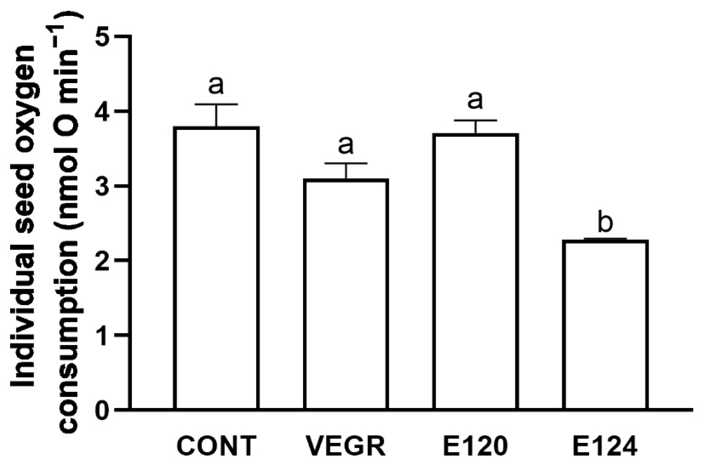
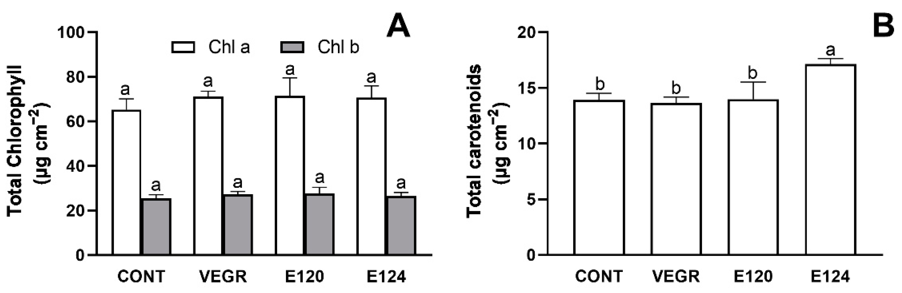
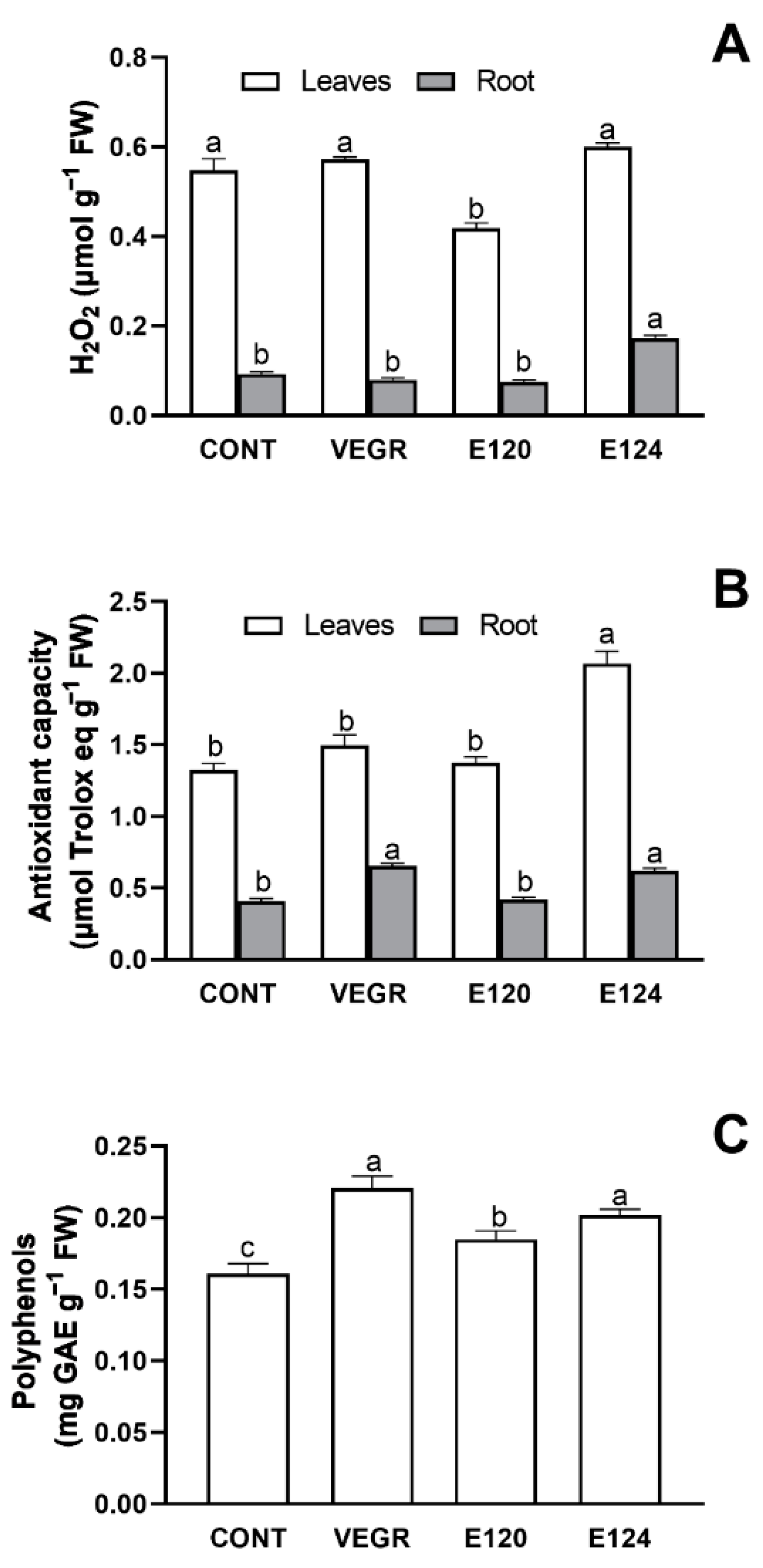
3.1. Growth and Antioxidant Response in C. sativus Seedlings Exposed to Different Red Food Dyes

At two DAG, before photosynthesis started, the artificial dye E124 significantly reduced the seeds oxygen consumption (p < 0.05) compared to CONT. Conversely, no differences were observed among CONT and the two natural dyes VEGR and E120 (Figure 1).
At 15 DAG, E124 increased (p < 0.05) the levels of carotenoids compared to the other treatments (Figure 2B), while the levels of chlorophylls a and b were not affected (Figure 2A). On the other hand, chlorophylls and carotenoid levels were not affected by both VEGR and E120 (Figure 2A,B).
Apart from this relatively small effect on carotenoids by E124, the seedling exposure to different food dyes did not induce changes in photosystem functionality, as indicated by the PSII maximal photochemical efficiency (Fv/Fm), which was comparable among all seedlings (Table 1). It is noteworthy that at 15 DAG, the treatment with VEGR induced a significant reduction (p < 0.05) of seedlings’ total fresh biomass compared to the other treatments (Table 1).
The three tested dyes differently affected the redox state in leaves and roots of 15 DAG seedlings of C. sativus (Figure 3). Compared to CONT, E124 induced a significant increase in H2O2 levels (Figure 3A, p < 0.001) and antioxidant capacity (Figure 3B, p < 0.05) in roots, while stimulating antioxidant capacity (Figure 3B, p < 0.001) and polyphenols (Figure 3C, p < 0.05) in leaves. E120 induced a significant reduction of leaves’ H2O2 levels (Figure 3A, p < 0.05), associated with an increase in the leaves’ polyphenols levels compared to the control (Figure 3C, p < 0.05). Finally, VEGR did not affect H2O2 levels in both leaves and roots, but significantly stimulated the antioxidant capacity in roots (Figure 3B, p < 0.001) and polyphenols (Figure 3C, p < 0.05) in leaves. Root polyphenols were not affected by any of the dyes (data not shown).

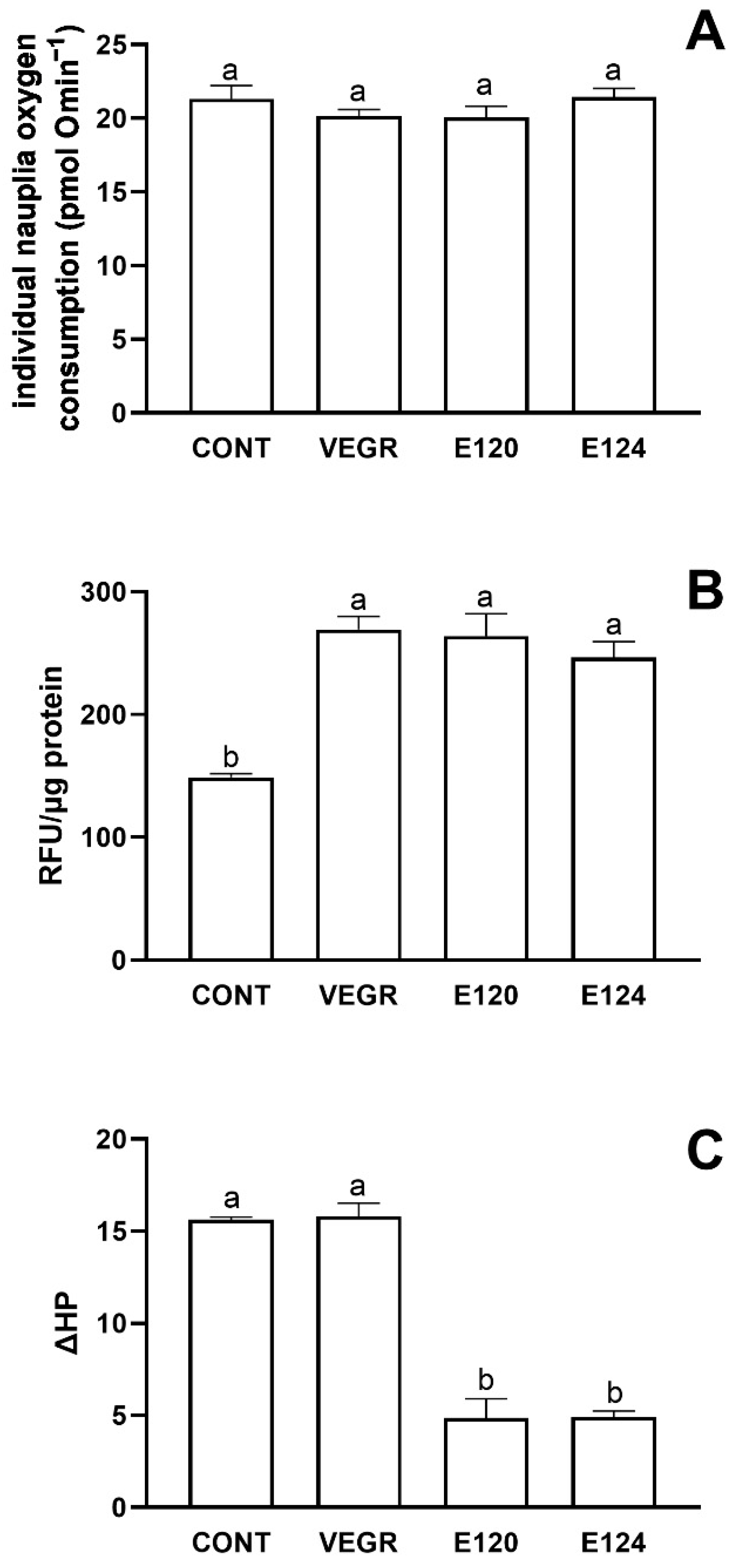
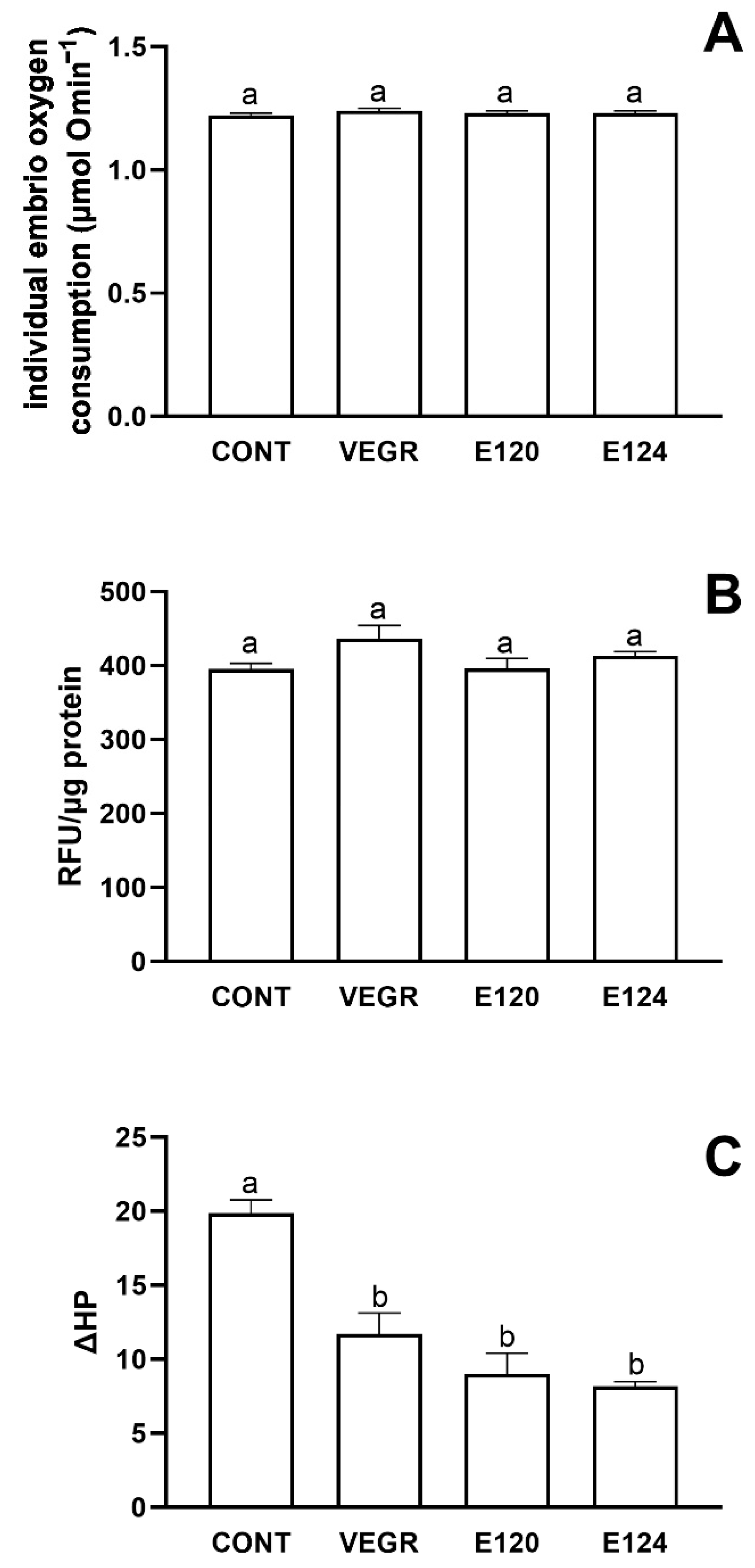
3.2. Artemia salina and Danio rerio Responses to Red Food Colors Exposure

In A. salina nauplii, the tested dyes did not affect the in vivo routine oxygen consumption (Figure 4A), but significantly increased the levels of ROS (Figure 4B). The hatching of the cysts in the presence of and the successive exposition of the nauplii up to 48 h to VEGR, E120, and E124 almost doubled ROS levels with respect to the CONT group (p < 0.0001, p = 0.0001, and p = 0.0006, respectively). The effects of the three dyes were not different. However, differences were observed in the in vitro susceptibility to oxidative stress, measured as Fe2+-ascorbate-induced changes in the lipid-bound hydroperoxide (ΔHP). While ΔHP in the VEGR treated nauplii was similar to that of CONT, and that of nauplii treated with the E120 and E124 was strongly reduced (p < 0.0001, Figure 4C). The basal HP levels were 29.44 ± 0.96, 24.22 ± 0.96, 5.13 ± 0.21, and 6.46 ± 0.80 nmol NADPH min−1 per mg protein, for CONT, VEGR, E120, and E124, respectively, and were significantly lower in E120 (p < 0.0001) and E124 (p < 0.0001) treated animals, compared to CTRL (One-Way ANOVA with Tukey post-hoc test).
In Danio rerio, the treatment with the food dyes also did not affect the in vivo routine oxygen consumption (Figure 5A). Differently from A. salina, there was no change in the levels of ROS in the homogenates (Figure 5B), while ΔHP was significantly reduced not only by E120 (p < 0.0001) and E124 (p < 0.0001), but also by VEGR (p = 0.011) (Figure 5C). The initial HP levels were 28.78 ± 0.94, 13.69 ± 1.29, 28.05 ± 0.99, and 32.19 ± 3.61 nmol NADPH min−1 per mg protein, for CONT, VEGR, E120, and E124, respectively. The VEGR value was significantly lower than CONT (p = 0.01), E120 (p = 0.0015), and E124 (p = 0.0002) ((one-way ANOVA with Tukey’s post-hoc test).

4. Discussion

4.1. The Functional and Antioxidant Responses of the C. sativus Induced by Different Food Dyes

The antioxidant capacity of plants under stress conditions either increases or decreases depending on species, stress factors [50,51,52,53,54], and exposure time [55]. We recently showed that after one week of treatment with E120 and E124, leaves’ antioxidant capacity of cucumber sprouts was reduced by consuming the scavengers’ compounds to counteract the oxidative stress [7]. Here we report that after two weeks of exposure to E120, cucumber seedlings appear to respond well to the oxidative stress, as shown by the reduced levels of H2O2 in leaves. On the contrary, E124 treatment increased root H2O2 levels and the antioxidant capacity of roots and leaves, indicating an elevated oxidative stress risk. It is worth noting that already at two DAG (i.e., before the development of the photosynthetic system), the seedlings exposed to E124 showed the lowest value of oxygen consumption, suggesting the occurrence of an early stress condition elicited by this specific dye on seed post-metabolism after germination. This result agrees with the known tendency of plants to decrease their oxygen consumption in response to stress conditions [56]. The different responses observed in seedlings exposed to E120 and E124 may be linked to their different origin (animal extract and synthetic, respectively). Interestingly, the redox state of seedlings exposed to the other natural dye tested, VEGR, was scarcely affected apart from a significant increase in the roots’ antioxidant capacity.
With their antioxidant properties, polyphenols might protect from oxidative stress by red dyes. Polyphenols are considered the main components involved in cellular reducing capability and free radical scavenging and are known to enhance the plant–environment interaction and act as a strong defense against biotic and abiotic stresses [57,58,59]. Their level is a predictor of the general response of plant antioxidant capacity [60]. In the present study, the leaf total polyphenol content considerably increased in seedlings exposed to all tested red dyes, and this increase proceeds together with the rise of antioxidant capacity. The rise of polyphenols in leaves treated with VEGR may be ascribed to the dye composition [29], whereas what was observed in leaves treated with E120 and E124 possibly reflects a general response to stress conditions [61].
The photosynthetic apparatus, and in particular the photosystem II (PSII) is a potential target of oxidative stress due to biotic and abiotic stress [62,63]. Only the treatment with E124 affected the photosynthetic system in the present study. The ratio of Fv/Fm was comparable to the control in all treatments, indicating the absence of damage to PSII [64]. Contrary to the effects shown at seven DAG [7], the levels of chlorophylls and carotenoids in seedlings treated with E120 were comparable to the control. Again, the prolonged exposure to food dyes may offset the response induced during the first week of development.
Conversely, E124 caused an increase in carotenoid production, likely enhancing the action of the antenna complexes implicated in light-harvesting for photosynthesis. The accumulation of carotenoids is also observed after exposure to metals or dyes from industrial wastewater [65,66,67]. These pigments play a key role in the photoprotection of the photosynthetic apparatus against the formation of reactive oxygen species [68]. They dissipate excess energy in the PSII antenna complexes as heat, thus avoiding the photooxidation of photosynthetic membranes [69].
Overall, when compared with one-week sprouts [7], two-week seedlings were more effective in facing oxidative stress, especially from E120, by increasing antioxidant capacity and carotenoids as a defense strategy.

4.2. The Responses to Red Food Dyes Exposure in Artemia salina and Danio rerio

The marine invertebrate A. salina nauplii appears to be more sensitive to the oxidative stress induced by the tested red food dyes than the freshwater vertebrate Danio rerio larvae. All treatments stimulated ROS levels in the 48 h nauplii of Artemia salina hatched in their presence, but not in the 72 h Danio rerio larvae. The difference may imply a higher efficacy of the innate antioxidant system of the developing zebrafish [70], as confirmed by the lower susceptibility to Fe2+-ascorbate-induced oxidative stress. The susceptibility to Fe2+-ascorbate-induced oxidative stress depends on the balance between hydroperoxides content and the capacity to scavenge radicals. t is supposed to be directly related to oxidative damage and inversely related to antioxidant capacity [71]. Interestingly, this parameter was reduced in the E120 and E124-treated A. salina nauplii, notwithstanding the increased ROS content. In this species, the initial levels of hydroperoxides were lower in E120 and E124 than in CONT and VEGR nauplii suggesting stimulation of the antioxidant capacity, possibly due to ROS-dependent, Nrf2-mediated antioxidant enzymes induction [72,73]. Therefore, the reduction in the in vitro susceptibility to Fe2+-ascorbate-induced oxidative stress in E120 and E124 nauplii depends on an increased capacity to scavenge ROS. Notably, this effect is not elicited by VEGR.
It is worth noting that, despite the concordance of effects, there are reasons to believe that ROS stimulation in A. salina may involve different mechanisms with different dyes. E124 may directly induce ROS, as reported for many azo dyes [74,75]. The ROS stimulation by VEGR can involve polyphenols [29], which may in animals either protect against oxidative stress [76,77] or exert prooxidant or both anti- and prooxidant effects [30]. Finally, the prooxidant effect of E120 can depend on 4-aminocarminic acid (ACA), a primary amine produced in low amounts during the E120 productive process [33,78]. ACA might degrade to aromatic amines [31], inducing ROS via redox cycling [32].
The effects of treatments on the redox state did not affect the routine oxygen consumption in both species. This result agrees with previous results reporting the lack of effect of red dyes on parameters directly related with the mitochondria-dependent oxygen consumption, i.e., the mobility in A. salina nauplii (E2120 and E124) and the heart rate in the D. rerio embryos(E120) [7]. Consequently, the lack of effects of treatments on the total oxygen consumption also suggest that there is no change in the non-mitochondrial respiration, known to occur at high levels during early embryogenesis and acting as a protection from oxidative stress [79].

4.3. Effects of Red Food Dyes: Global Comparison and Conclusion

Besides the specific parameters required to test responses in plant and animal models, the effect of food dyes can be qualitatively grouped as pro-oxidant effects (H2O2 levels, in C. sativus, leaves, root or both, or total ROS levels, in A. salina and D. rerio), anti-oxidant effects (total antioxidant activity and/or polyphenols, in C. sativus -leaves, root or both- or oxidative stress susceptibility in A. salina and D. rerio), and indices of organismal performance (oxygen consumption, in all three species, or photosynthetic system functionality, in C. sativus, leaves, root or both). Table 2 shows this qualitative resume of our results. Effects were defined in term of stimulation (+), reduction (−), or no effect (0).
Only E124 stimulated both the ROS levels and the antioxidant capacity, suggesting a higher oxidative stress risk posed by this food dye on living organisms. Compared to E124, E120 and VEGR are less effective in stimulating ROS production. An exception is the effect of VEGR on A. salina, with a significant prooxidant response, without the stimulation of antioxidant defenses. Apparently, the nauplii of this species are particularly sensitive to VEGR. All species seem to be able to protect themselves from the potential harmful pro-oxidant effects induced by the short exposure to red food dyes, by modulating the antioxidant defenses in one way or the other.
Although the results here reported seem to suggest that food dyes induce an adaptive reinforcement of the antioxidant system, it should be emphasized that ROS participate in cell signaling processes. It has been reported that ROS play a pivotal role in the network that influences pathways defining the final fate of developing cells, and the perturbations of ROS content require constant adjustments for proper development [80]. Thus, an imbalance between ROS production and the antioxidant system can affect organismal development and the morphological alterations previously reported can be the result of such an imbalance [7].
In conclusion, the reported results provide novel insights into the toxicity of red food dyes and may be relevant in the context of human and environmental health. The tested model species respond to the exposure to red food dyes with a remodulation of the redox state, which might be functional in maintaining organismal performance. Compared to the artificial red dye E124, the two alternative natural dyes, VEGR and E120, have a lower capacity to stimulate ROS production. The three tested dyes are similar in stimulating the antioxidant capacity, the only exception being VEGR on Artemia salina. The overall results show that the different food dyes used in this study induce a stress response involving the modulation of redox homeostasis. Though the tested model species appear capable of facing the oxidative stress induced by red food dyes under the experimental exposure conditions utilized, the observed remodulation of the redox state, a costly process, could represent a potential health hazard in nature. This conclusion stimulates further investigation, in particular on the dose-response relation and the activity of specific components of redox homeostasis (e.g., SOD, CAT, GSH-Px and GSH-R, or MDH), outlining the mechanisms of the organismal response.

Author Contributions

Conceptualization, Writing—original draft, Writing—review and editing, Data curation, Visualization, Investigation: G.N., P.V. and C.A. (Carmen Arena). Supervision, Conceptualization, Writing—original draft, Writing—review and editing, Data curation, Visualization, Investigation: C.A. (Claudio Agnisola). Conceptualization, Writing—review and editing: C.M.M., I.F., B.A. and P.S. Methodology, Formal analysis, Investigation: G.F., T.C., G.C. and E.V. All authors have read and agreed to the published version of the manuscript.

Funding

This research received no external funding.

Institutional Review Board Statement

Not applicable.

Informed Consent Statement

Not applicable.

Data Availability Statement

Data will be provided by corresponding authors upon reasonable request.

Conflicts of Interest

The authors declare no conflict of interest.

References

  1. Rymbai, H.; Sharma, R.R.; Srivastav, M. Biocolorants and its implications in Health and Food Industry—A Review. Int. J. Pharmacol. Res. 2011, 3, 2228–2244. [Google Scholar]
  2. Xu, D.P.; Li, Y.; Meng, X.; Zhou, T.; Zhou, Y.; Zheng, J.; Zhang, J.J.; Li, H.B. Natural Antioxidants in Foods and Medicinal Plants: Extraction, Assessment and Resources. Int. J. Mol. Sci. 2017, 18, 96. [Google Scholar] [CrossRef] [PubMed]
  3. Schweiggert, R.M. Perspective on the Ongoing Replacement of Artificial and Animal-Based Dyes with Alternative Natural Pigments in Foods and Beverages. J. Agric. Food Chem. 2018, 66, 3074–3081. [Google Scholar] [CrossRef] [PubMed]
  4. Grand View Research. Global Natural Food Colors Market Size & Share, Industry Report, 2025: Natural Food Colors Market Size, Share & Trends Analysis Report by Product (Curcumin, Carotenoids, Anthocyanin, Carmine, Chlorophyllin), by Application, by Region, and Segment Forecasts 2018–2025. 2018. Available online: https://www.marketsandmarkets.com/Market-Reports/natural-colors-flavors-market-676.html?gclid=Cj0KCQjw1ZeUBhDyARIsAOzAqQKqRiXCfQn-TaRl5lmnRhjrBR-v3hwoaAYRP5ybns58jbHueqKYgtcaAu31EALw_wcB (accessed on 2 December 2021).
  5. Hussain, S.; Khan, N.; Gul, S.; Khan, S.; Khan, H. Contamination of Water Resources by Food Dyes and Its Removal Technologies. In Water Chemistry; Eyvaz, M., Yüksel, E., Eds.; IntechOpen: London, UK, 2019. [Google Scholar] [CrossRef] [Green Version]
  6. Abe, F.R.; Gravato, C.; Soares, A.M.V.M.; de Oliveira, D.P. Biochemical approaches to assess oxidative stress induced by exposure to natural and synthetic dyes in early life stages in zebrafish. J. Toxicol. Environ. Health A 2017, 80, 1259–1268. [Google Scholar] [CrossRef] [PubMed]
  7. Motta, C.M.; Simoniello, P.; Arena, C.; Capriello, T.; Panzuto, R.; Vitale, E.; Agnisola, C.; Tizzano, M.; Avallone, B.; Ferrandino, I. Effects of four food dyes on development of three model species, Cucumis sativus, Artemia salina and Danio rerio: Assessment of potential risk for the environment. Environ. Pollut. 2019, 253, 1126–1135. [Google Scholar] [CrossRef]
  8. Räisänen, R.; Primetta, A.; Nikunen, S.; Honkalampi, U.; Nygren, H.; Pihlava, J.M.; Vanden Berghe, I.; von Wright, A. Examining Safety of Biocolourants from Fungal and Plant Sources-Examples from Cortinarius and Tapinella, Salix and Tanacetum spp. and Dyed Woollen Fabrics. Antibiotics 2020, 9, 266. [Google Scholar] [CrossRef]
  9. Abe, F.R.; Soares, A.M.V.M.; de Oliveira, D.P.; Gravato, C. Toxicity of dyes to zebrafish at the biochemical level: Cellular energy allocation and neurotoxicity. Environ. Pollut. 2018, 235, 255–262. [Google Scholar] [CrossRef]
  10. Napolitano, G.; Venditti, P.; Fasciolo, G.; Esposito, D.; Uliano, E.; Agnisola, C. Acute hypoxia/reoxygenation affects muscle mitochondrial respiration and redox state as well as swimming endurance in zebrafish. J. Comp. Physiol. B 2019, 189, 97–108. [Google Scholar] [CrossRef]
  11. Cikcikoglu Yildirim, N.; Yaman, M. The usability of oxidative stress and detoxification biomarkers in Gammarus pulex for ecological risk assessment of textile dye methyl orange. Chem. Ecol. 2019, 35, 319–329. [Google Scholar] [CrossRef]
  12. Valavanidis, A.; Vlahogianni, T.; Dassenakis, M.; Scoullos, M. Molecular biomarkers of oxidative stress in aquatic organisms in relation to toxic environmental pollutants. Ecotoxicol. Environ. Saf. 2006, 64, 178–189. [Google Scholar] [CrossRef]
  13. Venditti, P.; Napolitano, G.; Di Meo, S. Role of Mitochondria and Other ROS Sources in Hyperthyroidism-Linked Oxidative Stress. Immunol. Endocr. Metab. Agents Med. Chem. 2015, 15, 5–36. [Google Scholar] [CrossRef] [Green Version]
  14. Persoone, G.; Wells, P.G. Artemia in aquatic toxicology: A review. In Artemia. Research and Its Applications; Sorgeloos, P., Bengston, B.A., Decleir, W., Jaspers, E., Eds.; Universa Press: Wetteren, Belgium, 1987; pp. 259–275. [Google Scholar]
  15. Motta, C.; Cerciello, R.; De Bonis, S.; Mazzella, V.; Cirino, P.; Panzuto, R.; Ciaravolo, M.; Simoniello, P.; Toscanesi, M.; Trifuoggi, M.; et al. Potential toxicity of improperly discarded exhausted photovoltaic cells. Environ. Pollut. 2016, 216, 786–792. [Google Scholar] [CrossRef] [PubMed]
  16. Capriello, T.; Grimaldi, M.C.; Cofone, R.; D’Aniello, S.; Ferrandino, I. Effects of aluminium and cadmium on hatching and swimming ability in developing zebrafish. Chemosphere 2019, 222, 243–249. [Google Scholar] [CrossRef] [PubMed]
  17. Favorito, R.; Chiarelli, G.; Grimaldi, M.C.; Bonis, S.D.; Lancieri, M.; Ferrandino, I. Bioaccumulation of cadmium and its cytotoxic effect on zebrafish brain. Chem. Ecol. 2011, 27, 39–46. [Google Scholar] [CrossRef]
  18. Monaco, A.; Capriello, T.; Grimaldi, M.C.; Schiano, V.; Ferrandino, I. Neurodegeneration in zebrafish embryos and adults after cadmium exposure. J. List Eur. J. Histochem. 2017, 61, 2833. [Google Scholar] [CrossRef] [PubMed] [Green Version]
  19. Monaco, A.; Grimaldi, M.C.; Ferrandino, I. Aluminium chloride-induced toxicity in zebrafish larvae. J. Fish Dis. 2017, 40, 629–635. [Google Scholar] [CrossRef] [Green Version]
  20. Capriello, T.; Visone, I.M.; Motta, C.; Ferrandino, I. Adverse effects of E150d on zebrafish development. Food Chem. Toxicol. 2021, 147, 111877. [Google Scholar] [CrossRef]
  21. Wang, X.; Sun, C.; Gao, S.; Wang, L.; Shuokui, H. Validation of germination rate and root elongation as indicator to assess phytotoxicity with Cucumis sativus. Chemosphere 2001, 44, 1711–1721. [Google Scholar] [CrossRef]
  22. Wu, X.; Wang, W.; Zhu, L. Enhanced organic contaminants accumulation in crops: Mechanisms, interactions with engineered nanomaterials in soil. Environ. Pollut. 2018, 240, 51–59. [Google Scholar] [CrossRef]
  23. Shakoor, S.; Nasar, A. Adsorptive treatment of hazardous methylene blue dye from artificially contaminated water using Cucumis sativus peel waste as a low-cost adsorbent. Groundw. Sustain. Dev. 2017, 5, 152–159. [Google Scholar] [CrossRef]
  24. Tkaczyk, A.; Mitrowska, K.; Posyniak, A. Synthetic organic dyes as contaminants of the aquatic environment and their implications for ecosystems: A review. Sci. Total Environ. 2020, 717, 137222. [Google Scholar] [CrossRef] [PubMed]
  25. EFSA Panel on Food Additives and Nutrient Sources Added to Food. Scientific Opinion on the re-evaluation of Ponceau 4R (E 124) as a food additive. EFSA J. 2009, 7, 1328. [Google Scholar] [CrossRef] [Green Version]
  26. EFSA Panel on Food Additives and Nutrient Sources added to Food (ANS). Scientific Opinion on the re-evaluation of cochineal, carminic acid, carmines (E 120) as a food additive. EFSA J. 2015, 13, 4288. [Google Scholar]
  27. ANS. Statement on Allura Red AC and other sulphonated mono azo dyes authorised as food and feed additives. EFSA J. 2013, 11, 3234. [Google Scholar] [CrossRef]
  28. Leo, L.; Loong, C.; Ho, X.L.; Raman, M.F.B.; Suan, M.Y.T.; Loke, W.M. Occurrence of azo food dyes and their effects on cellular inflammatory responses. Nutrition 2018, 46, 36–40. [Google Scholar] [CrossRef]
  29. Koley, T.K.; Khan, Z.; Oulkar, D.; Singh, B.K.; Maurya, A.; Singh, B.; Banerjee, K. High resolution LC-MS characterization of phenolic compounds and the evaluation of antioxidant properties of a tropical purple radish genotype. Arab. J. Chem. 2020, 13, 1355–1366. [Google Scholar] [CrossRef]
  30. Lambert, J.D.; Elias, R.J. The antioxidant and pro-oxidant activities of green tea polyphenols: A role in cancer prevention. Arch. Biochem. Biophys. 2010, 501, 65–72. [Google Scholar] [CrossRef] [Green Version]
  31. Gosetti, F.; Chiuminatto, U.; Mazzucco, E.; Calabrese, G.; Gennaro, M.C.; Marengo, E. Identification of photodegradation products of Allura Red AC (E129) in a beverage by ultra-high-performance liquid chromatography–quadrupole-time-of-flight mass spectrometry. Anal. Chim. Acta 2012, 746, 84–89. [Google Scholar] [CrossRef]
  32. Kovacic, P.; Somanathan, R. Novel, unifying mechanism for aromatic primary- amines (therapeutics, carcinogens and toxins): Electron transfer, reactive oxygen species, oxidative stress and metabolites. Med. Chem. Comm. 2011, 2, 106–112. [Google Scholar] [CrossRef]
  33. Sabatino, L.; Scordino, M.; Gargano, M.; Lazzaro, F.; Borzì, M.A.; Traulo, P.; Gagliano, G. Aminocarminic acid in E120-labelled food additives and beverages. Food Addit. Contam. Part B 2012, 5, 295–300. [Google Scholar] [CrossRef]
  34. Nojavan, S.; Tahmasebi, Z.; Bidarmanesh, T.; Behdad, H.; Nasiri-Aghdam, M.; Mansori, S.; Pourahadi, A. Electrically enhanced liquid-phase microextraction of three textile azo dyes from wastewater and plant samples. J. Sep. Sci. 2013, 36, 3256–3263. [Google Scholar] [CrossRef] [PubMed]
  35. Krishnaraju, A.V.; Rao, T.V.N.; Sundararaju, D.; Vanisree, M.; Tsay, H.S.; Subbaraju, G.V. Assessment of bioactivity of Indian medicinal plants using brine shrimp (Artemia salina) lethality assay. Int. J. Appl. Sci. Eng. 2005, 3, 125–134. [Google Scholar]
  36. Westerfield, M. The Zebrafish Book. A Guide for the Laboratory Use of Zebrafish (Danio rerio), 4th ed.; University of Oregon Press: Eugene, OR, USA, 2020. [Google Scholar]
  37. Lichtenthaler, H.K. Chlorophylls and carotenoids: Pigments of photosynthetic biomembranes. In Methods in Enzymology, Plant Cell Membranes; Academic Press: Cambridge, MA, USA, 1987; pp. 350–382. [Google Scholar] [CrossRef]
  38. Maxwell, K.; Johnson, G.N. Chlorophyll fluorescence—A practical guide. J. Exp. Bot. 2000, 51, 659–668. [Google Scholar] [CrossRef] [PubMed]
  39. Li, X.; Zhang, G.; Sun, B.; Zhang, S.; Zhang, Y.; Liao, Y.; Zhou, Y.; Xia, X.; Shi, K.; Yu, J. Stimulated Leaf Dark Respiration in Tomato in an Elevated Carbon Dioxide Atmosphere. Sci. Rep. 2013, 3, 3433. [Google Scholar] [CrossRef] [Green Version]
  40. Sergiev, I.; Alexieva, V.; Karanov, E. Effect of spermine, atrazine and combination between them on some endogenous protective systems and stress markers in plants. Compt. Rend. Acad. Bulg. Sci. 1997, 51, 121–124. [Google Scholar]
  41. George, B.; Kaur, C.; Khurdiya, D.S.; Kapoor, H.C. Antioxidants in tomato (Lycopersium esculentum) as a function of genotype. Food Chem. 2004, 84, 45–51. [Google Scholar] [CrossRef]
  42. Costanzo, G.; Iesce, M.R.; Naviglio, D.; Ciaravolo, M.; Vitale, E.; Arena, C. Comparative Studies on Different Citrus Cultivars: A Revaluation of Waste Mandarin Components. Antioxidants 2020, 9, 517. [Google Scholar] [CrossRef]
  43. Arena, C.; Vitale, E.; Mele, B.H.; Cataletto, P.R.; Turano, M.; Simoniello, P.; Micco, V.D. Suitability of Solanum lycopersicum L. ‘Microtom’ for growth in Bioregenerative Life Support Systems: Exploring the effect of high-LET ionising radiation on photosynthesis, leaf structure and fruit traits. Plant Bio 2019, 21, 615–626. [Google Scholar] [CrossRef]
  44. Vanhaecke, P.; Persoone, G.; Claus, C.; Sorgeloos, P. Proposal for a short-term toxicity test with Artemia nauplii. Ecotoxicol. Environ. Saf. 1981, 5, 382–387. [Google Scholar] [CrossRef]
  45. Gornall, A.G.; Bardawill, C.J.; David, M.M. Determination of serum proteins by means of the biuret reaction. J. Biol. Chem. 1949, 177, 751–766. [Google Scholar] [CrossRef]
  46. Driver, A.S.; Kodavanti, P.R.S.; Mundy, W.R. Age-related changes in reactive oxygen species production in rat brain homogenates. Neurotoxicol. Teratol. 2000, 22, 175–181. [Google Scholar] [CrossRef]
  47. Napolitano, G.; Fasciolo, G.; Salbitani, G.; Venditti, P. Chlorella sorokiniana Dietary Supplementation Increases Antioxidant Capacities and Reduces Ros Release in Mitochondria of Hyperthyroid Rat Liver. Antioxidants 2020, 9, 883. [Google Scholar] [CrossRef] [PubMed]
  48. Heath, R.L.; Tappel, A.L. A new sensitive assay for the measurement of hydroperoxides. Anal. Biochem. 1976, 76, 184–191. [Google Scholar] [CrossRef]
  49. European Parliament, Council of the European Union. Directive 2010/63/EU of the European Parliament and of the Council of 22 September 2010 on the Protection of Animals Used for Scientific Purposes. Off. J. Eur. Union 2010, 276, 33–79. [Google Scholar]
  50. Arena, C.; De Maio, A.; De Nicola, F.; Santorufo, L.; Vitale, L.; Maisto, G. Assessment of eco-physiological performance of Quercus ilex L. leaves in urban area by an integrated approach. Water Air Soil Pollut. 2014, 225, 1824. [Google Scholar] [CrossRef]
  51. Navabpour, S.; Yamchi, A.; Bagherikia, S.; Kafi, H. Lead-induced oxidative stress and role of antioxidant defense in wheat (Triticum aestivum L.). Physiol. Mol. Biol. Plants 2020, 26, 793–802. [Google Scholar] [CrossRef]
  52. Gonçalves, J.F.; Becker, A.G.; Cargnelutti, D.; Tabaldi, L.A.; Pereira, L.B.; Battisti, V.; Spanevello, R.M.; Morsch, V.M.; Nicoloso, F.T.; Schetinger, M.R.C. Cadmium toxicity causes oxidative stress and induces response of the antioxidant system in cucumber seedlings. Braz. J. Plant Physiol. 2007, 19, 223–232. [Google Scholar] [CrossRef]
  53. Jadhav, S.B.; Surwase, S.N.; Kalyani, D.C.; Gurav, R.G.; Jadhav, J.P. Biodecolorization of azo dye remazol orange by Pseudomonas aeruginosa BCH and toxicity (oxidative stress) reduction in Allium cepa root cells. Appl. Biochem. Biotechnol. 2012, 168, 1319–1334. [Google Scholar] [CrossRef]
  54. Nagajyoti, P.C.; Lee, K.D.; Sreekanth, T.V.M. Heavy metals, occurrence and toxicity for plants: A review. Environ. Chem. Lett. 2010, 8, 199–216. [Google Scholar] [CrossRef]
  55. Vitale, L.; Vitale, E.; Costanzo, G.; De Maio, A.; Arena, C. Photo-Protective mechanisms and the role of poly (ADP-Ribose) polymerase activity in a facultative CAM plant exposed to long-term water deprivation. Plants 2020, 9, 1192. [Google Scholar] [CrossRef]
  56. Geigenberger, P. Response of plant metabolism to too little oxygen. Curr. Opin. Plant Biol. 2003, 6, 247–256. [Google Scholar] [CrossRef]
  57. Hyson, D.A. A Comprehensive Review of apples and apple components and their relationship to human health. Adv. Nutr. 2011, 2, 408–420. [Google Scholar] [CrossRef] [PubMed]
  58. Karjalainen, R.; Stewart, D.; McDougall, G.J.; Hilz, H.; Anttonen, M.; Saviranta, N.; Mattila, P.; Törrönen, R. Bioactive compounds in black currants (Ribes nigrum L.) and their potential health-promoting properties. In Proceedings of the International Symposium on Biotechnology of Fruit Species, Dresden, Germany, 1–5 September 2008. [Google Scholar]
  59. Manivannan, A.; Kim, J.H.; Kim, D.S.; Lee, E.S.; Lee, H.E. Deciphering the Nutraceutical Potential of Raphanus sativus—A Comprehensive Overview. Nutrients 2019, 11, 402. [Google Scholar] [CrossRef] [PubMed] [Green Version]
  60. Racchi, M.L. Antioxidant Defenses in Plants with Attention to Prunus and Citrus spp. Antioxidants 2013, 2, 340–369. [Google Scholar] [CrossRef]
  61. Kumar, A.; Prasad, M.N.V.; Sytar, O. Lead toxicity, defense strategies and associated indicative biomarkers in Talinum triangulare grown hydroponically. Chemosphere 2012, 89, 1056–1065. [Google Scholar] [CrossRef]
  62. Pospíšil, P. Production of Reactive Oxygen Species by Photosystem II as a Response to Light and Temperature Stress. Front. Plant Sci. 2016, 7, 1950. [Google Scholar] [CrossRef]
  63. Pospíšil, P.; Yamamoto, Y. Damage to photosystem II by lipid peroxidation products. Biochim. Biophys. Acta Gen. Subj. 2017, 1861, 457–466. [Google Scholar] [CrossRef]
  64. Demmig, B.; Björkman, O. Comparison of the effect of excessive light on chlorophyll fluorescence (77K) and photon yield of O2 evolution in leaves of higher plants. Planta 1987, 171, 171–184. [Google Scholar] [CrossRef]
  65. Arena, C.; Santorufo, L.; Cataletto, P.R.; Memoli, V.; Scudiero, R.; Maisto, G. Eco-physiological and antioxidant responses of holm oak (Quercus ilex L.) leaves to Cd and Pb. Water Air Soil Pollut. 2017, 228, 459. [Google Scholar] [CrossRef]
  66. Puvaneswari, N.; Muthukrishnan, J.; Gunasekaran, P. Toxicity assessment and microbial degradation of azo dyes. Indian. J. Exp. Biol. 2006, 44, 618–626. [Google Scholar]
  67. Tauqeer, H.M.; Ali, S.; Rizwan, M.; Ali, Q.; Saeed, R.; Iftikhar, U.; Ahmad, R.; Farid, M.; Abbasi, G.H. Phytoremediation of heavy metals by Alternanthera bettzickiana: Growth and physiological response. Ecotoxicol. Environ. Saf. 2016, 126, 138–146. [Google Scholar] [CrossRef]
  68. Halliwell, B. Reactive species and antioxidants. Redox biology is a fundamental theme of aerobic life. Plant Physiol. 2006, 141, 312–322. [Google Scholar] [CrossRef] [PubMed] [Green Version]
  69. Demmig-Adams, B.; Adams, W.W. The role of xanthophyll cycle carotenoids in the protection of photosynthesis. Trends Plant Sci. 1996, 1, 21–26. [Google Scholar] [CrossRef]
  70. Sant, K.E.; Sinno, P.P.; Jacobs, H.M.; Timme-Laragy, A.R. Nrf2a modulates the embryonic antioxidant response to perfluorooctanesulfonic acid (PFOS) in the zebrafish, Danio rerio. Aquat. Toxicol. 2018, 198, 92–102. [Google Scholar] [CrossRef] [PubMed]
  71. Laszczyca, P.; Kawka-Serwecińska, E.; Witas, I.; Dolezych, B.; Migula, P. Iron ascorbate-stimulated lipid peroxidation in vitro. Why is the method controversial? Gen. Physiol. Biophys. 1995, 14, 3–18. [Google Scholar] [PubMed]
  72. Shaw, P.; Chattopadhyay, A. Nrf2–ARE signaling in cellular protection: Mechanism of action and the regulatory mechanisms. J. Cell. Physiol. 2020, 235, 3119–3130. [Google Scholar] [CrossRef] [PubMed]
  73. Kasai, S.; Shimizu, S.; Tatara, Y.; Mimura, J.; Itoh, K. Regulation of Nrf2 by Mitochondrial Reactive Oxygen Species in Physiology and Pathology. Biomolecules 2020, 10, 320. [Google Scholar] [CrossRef] [Green Version]
  74. Pinheiro, H.M.; Touraud, R.E.; Thomas, O. Aromatic amines from azo dye reduction: Status review with emphasis on direct UV spectrophotometric detection in textile industry wastewaters. Dye Pigment. 2004, 61, 121–139. [Google Scholar] [CrossRef]
  75. Seesuriyachan, P.; Takenaka, S.; Kuntiya, A.; Klayraung, S.; Murakami, S.; Aoki, K. Metabolism of azo dyes by Lactobacillus casei TISTR 1500 and effects of various factors on decolorization. Water Res. 2007, 41, 985–992. [Google Scholar] [CrossRef] [Green Version]
  76. Baldin, J.C.; Michelin, E.C.; Polizer, Y.J.; Rodrigues, I.; de Godoy, S.H.S.; Fregonesi, R.P.; Pires, M.A.; Carvalho, L.T.; Fávaro-Trindade, C.S.; de Lima, C.G.; et al. Microencapsulated jabuticaba (Myrciaria cauliflora) extract added to fresh sausage as natural dye with antioxidant and antimicrobial activity. Meat Sci. 2016, 118, 15–21. [Google Scholar] [CrossRef]
  77. Silva, M.C.; Souza, V.; Thomazini, M.; da Silva, E.R.; Smaniotto, T.; de Carvalho, R.A.; Genovese, M.I.; Favaro-Trindade, C.S. Use of the jabuticaba (Myrciaria cauliflora) depulping residue to produce a natural pigment powder with functional properties. LWT—Food Sci. Technol. 2014, 55, 203–209. [Google Scholar] [CrossRef]
  78. LCI. Is ‘Red’ Always ‘Red’? 4-aminocarminic acid as an accompanying substance in ‘carmine’. LCI Focus 2017, 4, 5. [Google Scholar]
  79. Stackley, K.D.; Beeson, C.C.; Rahn, J.J.; Chan, S.S.L. Bioenergetic Profiling of Zebrafish Embryonic Development. PLoS ONE 2011, 6, e25652. [Google Scholar] [CrossRef] [PubMed] [Green Version]
  80. Hernández-García, D.; Wood, C.D.; Castro-Obregón, S.; Covarrubias, L. Reactive oxygen species: A radical role in development? Free Radic. Biol. Med. 2010, 49, 130–143. [Google Scholar] [CrossRef] [PubMed]
Figure 1. Individual seed dark oxygen consumption in C. sativus control seedlings (CONT) and seedlings treated with different food dyes (VEGR, E120, and E124). Values are means ± SEM (n = 10). Different letters indicate statistically significant differences among treatments.
Environments 09 00063 g001
Figure 2. Concentration of chlorophyll a (Chl a) and b (Chl b) (A) and total carotenoids (B) in C. sativus control seedlings (CONT) and seedlings treated with different food dyes (VEGR, E120, and E124). Values are means ± SEM (n = 10). Different letters indicate statistically significant differences among treatments.
Environments 09 00063 g002
Figure 3. H2O2 concentration (A) and antioxidant capacity (B) in leaf and root, and total polyphenols in leaf (C) in C. sativus control seedlings (CONT) and seedlings treated with different food dyes (VEGR, E120, and E124). Values are means ± SEM (n = 10). Different letters indicate statistically significant differences among treatments.
Environments 09 00063 g003
Figure 4. Effect of food dyes (VEGR, E120, and E124) exposition on: (A) in vivo routine oxygen consumption of 48 h Artemia salina nauplii; (B) ROS levels (expressed as Relative Fluorescent Units, RFU, per µg protein) of homogenates of 48 h Artemia salina nauplii; (C) in vitro susceptibility to Fe2+-ascorbate-induced oxidative stress (measured as the level changes on the lipid-bound hydroperoxide, ΔHP, expressed as nmol NADPH min−1 per mg protein) of homogenates of 48 h Artemia salina nauplii. Values are mean ± SEM (n = 4). Different letters indicate statistically significant differences among treatments.
Environments 09 00063 g004
Figure 5. Effect of food dyes (VEGR, E120, and E124) exposition on: (A) in vivo routine oxygen consumption of Danio rerio larvae; (B) ROS levels (expressed as Relative Fluorescent Units, RFU, per µg protein) of homogenates of D. rerio larvae. Data are expressed as mean ± SEM (n = 4); and (C) in vitro susceptibility to Fe2+-ascorbate-induced oxidative stress (measured as the level changes on the lipid-bound hydroperoxide, ΔHP, expressed as nmol NADPH min−1 per mg protein) of homogenates of D. rerio larvae. Different letters indicate statistically significant differences among treatments.
Environments 09 00063 g005
Table 1. Total fresh biomass and maximal PSII photochemical efficiency (Fv/Fm) in C. sativus control seedlings (CONT) and seedlings treated with different food dyes (VEGR, E120, E124). Values are means ± SEM (n = 20). Different letters indicate statistically significant differences among treatments.
CONTVEGRE120E124
Total biomass
(g FW plant−1)
0.410 ± 0.01 a0.352 ± 0.012 b0.451 ± 0.02 a0.414 ± 0.01 a
Fv/Fm0.810 ± 0.01 a0.814 ± 0.02 a0.812 ± 0.01 a0.814 ± 0.02 a
FW = fresh weight.
Table 2. Qualitative synthesis of the effects of E124, E120, and VEGR on the functional performance (oxygen consumption, in all three species, or photosynthetic system, in C. sativus, leaves, root or both), pro-oxidant levels (H2O2 levels, in C. sativus, leaves, root or both, or Total ROS levels, in A. salina and D. rerio), and antioxidant activity (total antioxidant activity and/or polyphenols, in C. sativus -leaves, root or both- or oxidative stress susceptibility in A. salina and D. rerio). Note that an increased oxidative stress susceptibility implies a reduced antioxidant activity and vice versa [71]. The effects were classified as stimulatory (+), inhibitory (−), or no effect (0).
VEGRE120E124
EffectCucumis sativusArtemia salinaDanio rerioCucumis sativusArtemia salinaDanio rerioCucumis sativusArtemia salinaDanio rerio
Functional performance 00000000
Pro-oxidant 0+0+0++0
Antioxidant +0+++++++
Publisher’s Note: MDPI stays neutral with regard to jurisdictional claims in published maps and institutional affiliations.

