Review

Root Canal Morphology of the Permanent Mandibular Incisors by Cone Beam Computed Tomography: A Systematic Review

by
Silvia Herrero-Hernández
1,
Nansi López-Valverde
1,*,
Manuel Bravo
2,
Óliver Valencia de Pablo
3,
Manuel Peix-Sánchez
1,
Javier Flores-Fraile
1,
Juan Manuel Ramírez
4,
Bruno Macedo de Sousa
5 and
Antonio López-Valverde
1
1
Department of Surgery, University of Salamanca, Instituto de Investigación Biomédica de Salamanca (IBSAL), 37007 Salamanca, Spain
2
Department of Preventive and Community Dentistry, Facultad de Odontología, Campus de Cartuja s/n, 18071 Granada, Spain
3
Department of Endodontics, Universidad Europea, 28670 Madrid, Spain
4
Department of Morphological Sciences, University of Cordoba, Avenida Menéndez Pidal s/n, 14071 Cordoba, Spain
5
Institute for Occlusion and Orofacial Pain Faculty of Medicine, University of Coimbra, Polo I-Edifício Central Rua Larga, 3004-504 Coimbra, Portugal
*
Author to whom correspondence should be addressed.
Submission received: 24 June 2020 / Revised: 6 July 2020 / Accepted: 13 July 2020 / Published: 17 July 2020

Abstract

:
Knowledge of dental anatomy through the assessment of the anatomic variations of each tooth’s root canal system is essential to undertake endodontic therapy. The aim of this systematic review was to analyze the different studies on the internal morphology of permanent mandibular incisors where Cone-Beam Computed Tomography (CBCT) X-ray imaging is used. Pubmed, CENTRAL, Wiley Library and Web of Science electronic databases were searched for scientific studies included until March 2020. The terms used in the search were: “permanent mandibular incisors”, “root canal morphology” and “cone-beam computed tomography”. The search was limited to studies whose aim was the analysis of the morphology of the root canal system evaluating the parameters of methodology, population, sample, number and configuration. A total of 19 studies met the inclusion criteria. There was a noticeable lack of unanimity in the setting adjustments of each of the CBCT devices used. The presence of two root canals varied from 0.4% to 45%. The most frequent configurations were Vertucci’s Types I, III, II, V, IV, VII and VI. Type VIII configuration was non-existent. CBCT revealed the existence of anatomical symmetry patterns, and there was no unanimity of criteria regarding the presence of a second root canal. Results concerning the presence of a second root canal in the mandibular incisors differ widely, with a possible influence of the geographic area where the study was conducted. The prevalence of a second canal is higher in mandibular lateral incisors than in mandibular central incisors. There was no direct relationship between voxel size (0.125–0.3 mm) and increased prevalence of a second canal.

1. Introduction

Good knowledge of the root canal anatomy is essential for the success of any endodontic treatment. The development of new materials and techniques has contributed to increasing the chances of ensuring good results. Nevertheless, anatomical knowledge is still the most valuable tool when it comes to addressing each separate case and deciding which materials and tools to use [1].
In 1984, Vertucci drew up a classification based on the different root canal morphologies [2]. Figure 1 shows Vertucci’s configuration for the lower incisor. The purpose of this figure, created by the authors, is to provide a better understanding of the classification.
Over time, the study of internal root canal morphology has been approached using different methodologies: traditional radiography, radiography using radiopaque contrast agents, sectioning, scanning electron microscopy, clearing technique, Cone-Beam Computed Tomography (CBCT) and micro-Cone-Beam Computed Tomography (µ-CT) [3,4,5,6,7,8]. Besides, in recent years, there has been an increase in the number of published studies on the morphology of the root canal system (Figure 2).
All of them have contributed to creating a detailed anatomical map of permanent mandibular incisors, conveying the fact that the internal morphology of such tooth is not simple and that it is necessary to invoke variations of Vertucci’s classification [9].
CBCT offers the advantage of being a highly accurate technique that provides three-dimensional images in a non-invasive way, exposing patients to lower doses of radiation than other techniques such as traditional computed tomography. Its use in the assessment of root canal morphology has been endorsed by the European Society of Endodontology and provides clinicians with evidence-based criteria. The greatest disadvantage of CBCT in the study of dental anatomy is the presence of image artifacts caused by the presence of highly radiopaque materials from the filling and restoration of the tooth or resulting from other factors such as the patient’s movements [10]. Likewise, in in vivo studies, the strong impact of the use of CBCT as a method to study the root canal system is reflected in the high number of samples obtained in some clinical trials, allowing adequate statistical analysis in prevalence studies [11].
Failure of endodontic treatment is usually a cause for concern for both clinicians and patients. Von Arx identified the presence of isthmuses or untreated canals as the main cause of endodontic therapy failure [12]. Lack of knowledge of anatomy and, consequently, missing untreated canals is highly associated with the presence of periapical lesions. Specifically, as regards such lesions, the prevalence rate of missed canals ranges between 12.2% and 17.4% in central and lateral mandibular incisors, respectively [13]. These data should encourage clinicians to search for a more complex internal anatomy and reveal the importance of chemo-mechanical preparation of the root canal as the main strategy for its disinfection [14].
On the other hand, there are widely differing data on the existence of a second root canal. Reported percentages range from 0.4% [15] to 70% [16] when none of the methodologies were excluded. This renders it necessary to eliminate the heterogeneity as regards the methodology used.
The purpose of this study was to carry out a systematic review on the morphology of the root canal system in permanent mandibular incisors, assessed using CBCT in human clinical studies, with voxel sizes of up to 0.3 mm. We believe that this systematic review will contribute to the understanding of the widely differing results expressed in the literature as regards the presence of a second root canal and to the finding of possible explanations, as well as to the search for the lowest radiation dose possible for the study of internal anatomy.

2. Methods

The study was planned and structured according to the PRISMA guidelines (Preferred Reporting Items for Systematic Review and Meta-analysis) [17].

2.1. Protocols

The search strategy was conducted using the condition, context and population framework (CoCoPop), based on the following question: “What is the prevalence of root canal configuration of the permanent mandibular incisors?”. To answer this question, the condition was the morphology of the root canal system in the mandibular incisors. Only studies that used an in vivo CBCT methodology were included. The context included all the in vivo studies carried out using CBCT, without excluding any country in the world. The population consisted of patients who had been subjected to CBCT, regardless of its diagnostic purposes.

2.2. Search Method for the Identification of Studies

A bibliographic search of the Pubmed, CENTRAL, Wiley Library and Web of Science electronic databases was conducted in order to identify the most relevant studies available until 28 March 2020. The purpose was to identify in vivo prevalence studies where the canal system of the permanent mandibular incisors was analyzed using CBCT. The last update was made on 1 July 2020. The search terms were “permanent mandibular incisors”, “root canal morphology”, “cone-beam computed tomography” and “CBCT”. The keywords were used individually or in combination, using the boolean operators “AND”, “OR”, “NOT” to independently search for the term “cone-beam computed tomography” or its abbreviation (“CBCT”), include all terms, or exclude “permanent mandibular incisor” because of the possibility of finding studies that evaluated all dental groups. The search was completed manually by reading two endodontic journals: Journal of Endodontics and International Journal of Endodontics.

2.3. Inclusion and Exclusion Criteria

The inclusion criteria covered studies that evaluated the configuration of the root system of the permanent mandibular incisors and that were published in English. Furthermore, the evaluation of the canals had to indicate not only the presence of a second canal but also its internal configuration using Vertucci’s classification. Exclusion criteria were studies that did not use CBCT as a diagnostic tool, in vitro studies, systemic reviews, case reports, and duplicate studies. Besides, those that did not indicate the number of samples and patients who participated in the study, or whose samples had been partially analyzed in other included studies, were excluded.

2.4. Data Extraction and Analysis

First, two reviewers (SH-H and NL-V) undertook the reading of all the titles and abstracts, and those that did not refer to the research question were removed. Subsequently, the aforementioned reviewers conducted an independent evaluation, following the inclusion/exclusion criteria, until reaching a consensus on the studies to be included in the study. Finally, a total of 19 studies were included and the full texts of the selected studies were obtained for review. The extraction of data from each study was performed using Excel spreadsheets, completing the following categories: year of publication, sample size, country, percentage of a second canal, Vertucci’s configuration, other relevant results, CBCT model, voxel size, FOV, CBCT settings and software visualization.

2.5. Quality of the Reports of the Included Studies

We used the STROBE recommendations checklist adapted by Martins et al. for cross-sectional studies on root and root canal anatomy using CBCT, as a proxy indicator of quality [18]. The objective of the checklist is to assess limitations and risk of bias in the studies, which could lead to an erroneous reading of the results. In addition, it increases the validity and strength of the findings and the reproducibility of the method, and it is an indicator that improves the overall quality of the prevalence studies. Each item was assessed by reviewers S.H.-H. and N.L.-V., who attributed scores of 0 (not reported) or 1 (reported), carrying out a complete count of all the studies included.

3. Results

3.1. Characteristics of the Studies

A total of 2290 studies were gathered and analyzed. Of these, 2219 were removed because of their being duplicates or not related to the study. Of the remaining 71 studies, 52 were removed due to lack of relevance, not using CBCT as their diagnostic methodology, or being in vitro trials, case studies or systematic reviews, leaving a total of 19 studies [8,14,19,20,21,22,23,24,25,26,27,28,29,30,31,32,33,34,35] (Figure 3. Flowchart). Table 1 includes the information corresponding to each of the assessed studies including their items: population, sample, second root canal percentage, Vertucci classification (%) and other results. Table 2 shows the details of each study regarding the parameters used in the CBCT imaging process.

3.2. Quality of the Reports of the Included Studies

The evaluation of “specific preferred reporting items for cross-sectional studies on root and root canal anatomy using cone-beam computed tomography (CBCT)” [18] demonstrated that 50% of the items were reported in all of the published studies. The items that were most frequently disregarded by the researchers were “future research” (not indicated in 78.9% of the studies), “strengths and limitations” (68.4% of the studies failed to assess limitations) and “reliability” (52.6% did not perform the intra and interrater reliability test). Furthermore, it was interesting to note how all the studies used the word “Cone-beam computed tomography or CBCT” in the title, but none of them mentioned the type of study (Table 3).

3.3. Synthesis of Studies Included

There is unanimity across the studies regarding the fact that type I is the most frequent configuration. The rest of Vertucci’s configurations were considered incisors with two canals, regardless of their path in the canal system. When there were two canals, the most frequent classification was type III, with the exception of four studies [8,22,23,35]. There was no heterogeneity as to the frequency of the rest of configurations, with studies where the next most frequent configurations were type II [14,23,26,27,30,33,34], type V [8,19,20,21,24,25,28,29,32], or type IV [31]. The remaining types [VI, VII and others] were generally present in very low or non-existent percentages. Five studies reported symmetry between the incisors on the right and left side of the patient [8,19,20,23,26,33]. Lateral incisors showed a higher prevalence in the existence of a second canal compared to central incisors in fourteen studies [7,14,2021,22,25,27,28,29,31,32,33,34,35]. The central incisor had a higher prevalence in only three [19,23,26]. Likewise, we calculated the weighted average of all the percentages in the existence of a second canal, obtaining 17.2% in the central and 23.7% in the lateral incisors. Only two studies reported a relationship between age and the presence of a second canal [24,29]. The remaining studies did not assess patient age or, if so, did not find any significant differences [8,30]. Regarding gender influence, there was no unanimity as to a higher prevalence of second canals in men [19,20,24,31] than in women [27,29], and there were even certain studies where no significant differences were found [8,22,25,30,34,35]. Geographical distribution indicated a lower incidence of a second canal in the Chinese population [14,29,32,33,34]. There was no direct relationship between voxel size and the increased prevalence of a second canal.

4. Discussion

Historically, studies on the configuration of the root canal system of incisors were mainly in vitro, using methodologies such as x-rays [3], clearing technique [6] or, more recently, CBCT [36] and µ-CT [8]. However, in vitro methodologies involve tooth extraction, which means that the number of samples is limited. For the purposes of this systematic review, articles were screened to include only human studies using CBCT. This decision was based on two reasons: first, because the marked disparity in results concerning the existence of a second root canal led to the rejection of the use of different methodologies as a potential source of bias; and secondly, because of the high number of samples included in these studies as a consequence of their being extracted from clinical databases created for other diagnostic purposes.
After assessing the studies included, we noticed that, even when the same methodology was used, the differences in second root canal percentages (0.4%–45%) remained [14,23]. These results are in contrast with the findings of other authors [4], who attribute the disparity of results to the methodology used. On the other hand, our findings are consistent with those where the use of a variety of methodologies offered the same precision in the study of the internal morphology of incisors [37,38].
The clinical application of CBCT has yielded promising results in the identification of morphological relationships among the different teeth of the same patient. The findings of Wu and colleagues [24] show a correlation between the presence of a distolingual root in permanent mandibular molars and the presence of complicated configurations in central incisors. Moreover, Monsarrat and colleagues [39], report that the presence of a second canal in lower incisors could be related to the presence of an additional canal in lower premolars.
Likewise, CBCT also confirmed the existence of an anatomical symmetry pattern in the morphology of the root canal system between the right- and left-side incisors of the same patient. Before this, anatomical symmetry had only been anecdotally described in published case reports [40]. Nevertheless, a thorough and careful reading of the analyzed studies revealed that some of them reported high percentages of anatomical symmetry [20,26,33]. However, such percentages include symmetry data corresponding to Vertucci’s type I configuration and, given its high incidence, it is typical for such significant symmetries to be reported. Baxter and colleagues [7] claim that findings regarding symmetry in mandibular incisors are not significant for the purpose of making a diagnosis based on contralateral tooth anatomy and insist on the importance of performing an accurate clinical and radiographic diagnosis of the tooth to be treated. In their study, symmetry when there were two root canals (excluding Vertucci’s classification type I) was of 17.5% and 20.5% in central and lateral incisors, respectively. In conclusion, CBCT is a very useful tool in the study of internal anatomy, allowing clinicians to obtain three-dimensional images of the canal system and contributing to the identification of new anatomical relationships among the different teeth of a given patient and a given population.
Another finding of our systematic review is that not all the studies reviewed used the same CBCT devices and that they even used different imaging parameters (Table 2). The reason for this is that none of the CBCT assessed [8,14,19,20,21,22,23,24,25,26,27,28,29,30,31,32,33,34,35] were specifically performed for the study of root canal configuration but were aimed at maintaining minimal exposures to radiation for a particular diagnosis. In this regard, Bauman and colleagues [41] reported that the detection of the mesio-buccal canal in maxillary molars was more accurate (93.3%) when CBCT parameters were set at small voxel sizes than when larger sizes were used (60.1%).
Martins and colleagues [42] performed a systematic review on the influence of demographic factors on the prevalence of a second root canal in the mandibular incisors. Their results are consistent with ours, confirming a lower prevalence in the Asian population. However, one of their inclusion criteria was the use of an equal to or lower than 0.2 mm voxel size. In our study, we included larger voxel sizes in order to assess whether they also identify the presence of a second canal. This has an important clinical application in guiding the clinician in the decision to expose the patient to radiation as low as reasonably achievable (A.L.A.R.A.) [43].
According to our assessment of the CBCT parameters used in the studies (Table 2), there is no direct relationship between voxel size and the detection of a second root canal (when voxel sizes between 0.125 mm and 0.3 mm were used). The most relevant result was that using the same voxel size (0.2 mm) the presence of a second root canal varied between 2.7% [14] and 44% [23]. A reasonable explanation for this would be that the first and second studies were conducted in different geographical areas (China and Italy). Zhengyan and colleagues, in a study based on Chinese patients [29], also reported a low prevalence of second root canals (7.2%), despite using a smaller voxel size [44]. Thus, this finding would be consistent with those of other authors [45,46,47] who claim that the morphology of the root canal system is directly related to the geographical area where the study is conducted. Except for the study by Martins and colleagues [14], who clearly specify the inclusion of a particular ethnic group, in all the remaining studies CBCT was performed in local healthcare facilities without clarifying whether other ethnic groups had been excluded. Therefore, our systematic review mentions a geographical area variable rather than an ethnic variable. In addition, studies using a 0.3 mm voxel size [8,31] revealed higher second canal incidence rates than others where smaller voxel sizes were used [14,20,23,25,28]. Hence, as regards clinical application, our study proves the lack of justification for overexposing a patient to radiation if the purpose of the CBCT is to confirm the presence of a root canal. However, this systematic review cannot clarify which particular voxel size is appropriate in the study of internal anatomy. In the future, it would be necessary to carry out new studies that can confirm it.
Another of the aspects assessed in the reviewed studies was the influence of tooth position. Out of a total of 19 studies, 2 made no differentiation between central and lateral incisors; of the remaining studies, 14 reported a greater prevalence of second root canals in lateral incisors. With the purpose of illustrating the existing trend in the number of canals, we performed an analysis of the weighted average. The results showed a higher prevalence of a second canal in the lateral incisors (23.7%) than in the central incisors (17.2%). The literature revealed other studies with consistent results [48,49], although most of the in vitro studies conducted do not consider said variable [7,9], possibly because of the difficulties that are inherent to this technique. Accordingly, this is an extremely important finding because of its clinical applications.
As for age, very few studies addressed this variable, which leads to confusion and contradictory results concerning its influence [24,29] in root canal system morphology. Regarding the influence of gender, there was no unanimity as to the results, finding studies that reported a higher prevalence of a second canal in men [19,20,24,31], women [27,29], and also some where no significant differences were found [8,22,25,30,34,35].
The studies reviewed reveal broad consensus in that the most frequent configuration is Vertucci’s type I, followed by type III, II, V, IV, VII and VI, respectively (Table 1). However, there are certain studies that do not report type III configuration as the most frequent. Baxter and colleagues [7] explain this variation as the result of using a too large voxel size (0.3 mm), which allows the visualization of the number of canals, but does not provide an accurate display of their configuration. Nevertheless, Valenti-Obino and colleagues [23] report a very high prevalence of type II configuration using a small voxel size (0.2 mm), so that it is possible that a critical discrepancy among the different studies reviewed could lie in how clinicians understand Vertucci’s configuration; thus, different interpretations of the morphology configurations are indeed possible depending on whether the assessment has been performed by oral and maxillofacial radiologists [25] or by dentists [7], probably due to differences in their expertise.
One of our limitations was the exclusion of unpublished studies in English that could lead to a bias in the interpretation of the results. Besides, there is a lack of homogeneity across the data reported in the studies concerning position, patient gender, patient age or other data on CBCT acquisition parameters.
This systematic review helped us to understand the clinical relevance of analyzing clinical studies conducted under very similar conditions. Nevertheless, future research needs to develop guidelines for the greater standardization of studies where CBCT is used.
Additionally, the inclusion of patient demographics, especially when addressing countries with low rates of multiracial representation, would provide a better understanding of the distribution of root canal morphologies. The strong demographic influence revealed suggests the need for more studies in different populations, since the results on the prevalence of a second canal are not generalizable across populations.

5. Conclusions

The findings of our systematic review reveal wide disparities concerning the existence of a second canal in permanent mandibular incisors, which could perhaps derive from the geographical area studied. The most frequent canal system configuration is Vertucci’s type I, followed by type III. The percentage of second canals is higher in lateral than in central incisors. No direct relationship was found between the voxel size used in the CBCT scan and the presence of a second canal when voxel sizes smaller than 0.3 mm were used. The difficulty of standardizing clinicians’ interpretation of Vertucci’s configuration poses limitations for the comparison of studies. The existence of a pattern of anatomical symmetry in the morphology of the root canal system of mandibular incisors was confirmed, although this finding does not obviate the obligation to thoroughly assess each tooth.

Author Contributions

Study concept and design: S.H.-H., N.L.-V., M.B.; acquisition of data (literature search and study selection): S.H.-H., N.L.-V., Ó.V.d.P., J.F.-F.; analysis and interpretation of data (literature): B.M.d.S., J.M.R., M.P.-S.; drafting of the manuscript: S.H.-H., A.L.-V.; critical revision of the manuscript for important intellectual content: A.L.-V., M.B. All authors have read and agreed to the published version of the manuscript.

Funding

This research received no external funding.

Conflicts of Interest

The authors declare no conflict of interest.

Abbreviations

CBCTCone-Beam Computed Tomography
µ-CTMicro-Computed Tomography
FOVField of View

References

  1. Kucher, M.; Dannemann, M.; Modler, N.; Haim, D.; Hannig, C.; Weber, M.-T. Continuous Measurement of Three-Dimensional Root Canal Curvature Using Cone-Beam Computed and Micro-Computed Tomography: A Comparative Study. Dent. J. 2020, 8, 16. [Google Scholar] [CrossRef] [PubMed] [Green Version]
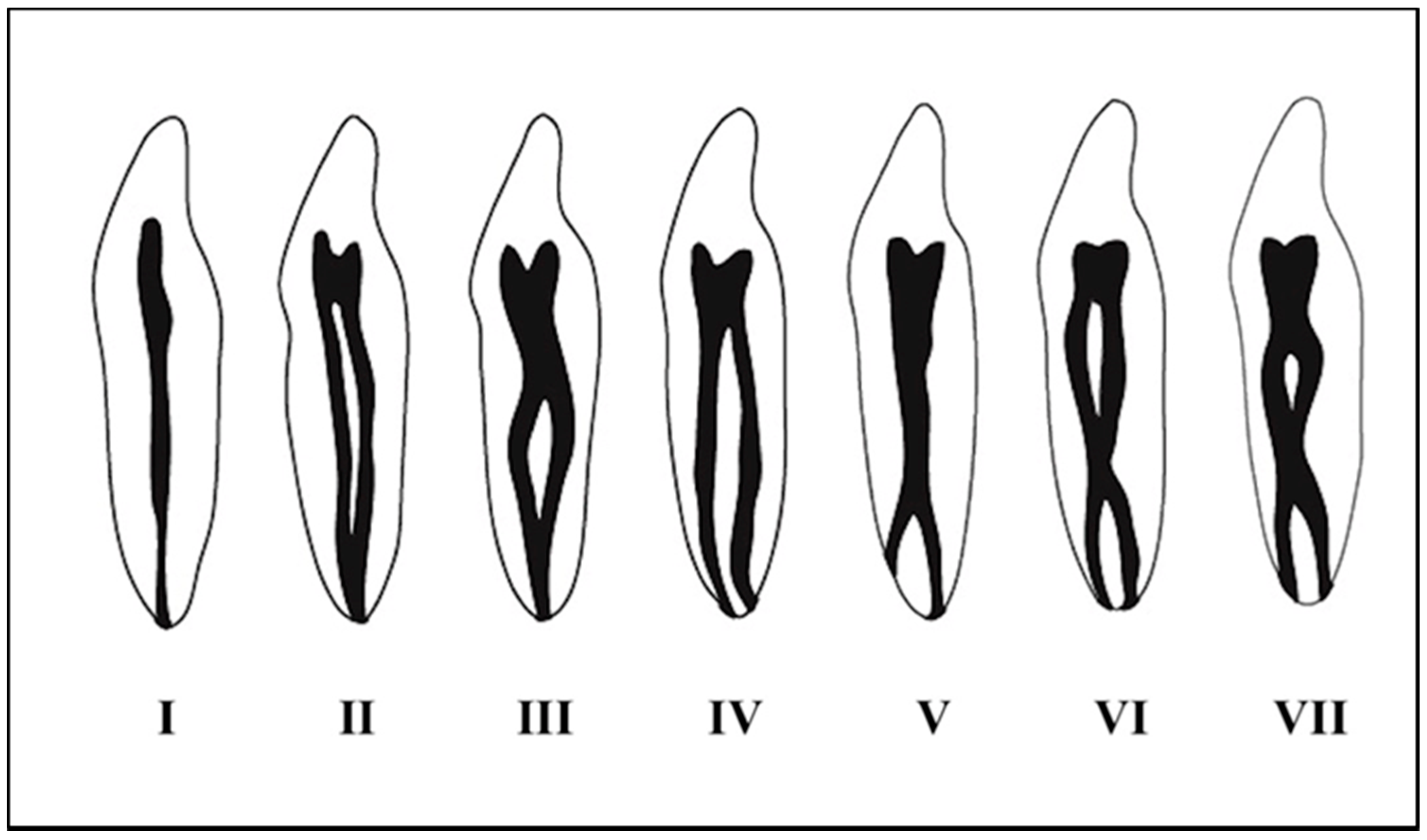
  2. Vertucci, F.J. Root canal anatomy of the human permanent teeth. Oral Surg. Oral Med. Oral Pathol. 1984, 58, 589–599. [Google Scholar] [CrossRef]
  3. Naoum, H.J.; Chandler, N.P.; Love, R. Conventional versus Storage Phosphor-Plate Digital Images to Visualize the Root Canal System Contrasted with a Radiopaque Medium. J. Endod. 2003, 29, 349–352. [Google Scholar] [CrossRef]
  4. Assadian, H.; Dabbaghi, A.; Gooran, M.; Eftekhar, B.; Sharifi, S.; Shams, N.; Najvani, A.D.; Tabesh, H. Accuracy of CBCT, Digital Radiography and Cross-Sectioning for the Evaluation of Mandibular Incisor Root Canals. Iran. Endod. J. 2016, 11, 106–110. [Google Scholar] [PubMed]
  5. Gilles, J.; Reader, A. An SEM investigation of the mesiolingual canal in human maxillary first and second molars. Oral Surg. Oral Med. Oral Pathol. 1990, 70, 638–643. [Google Scholar] [CrossRef]
  6. Nogueira, B.M.L.; Fagundes, N.C.F.; Menezes, T.O.D.A.; Lima, R.R.; Brandão, J.M.D.S. Root and Canal Morphology of Permanent Mandibular Incisors. Int. J. Odontostomatol. 2017, 11, 95–100. [Google Scholar] [CrossRef] [Green Version]
  7. Baxter, S.; Jablonski, M.; Hülsmann, M. Cone-beam-computed-tomography of the symmetry of root canal anatomy in mandibular incisors. J. Oral Sci. 2020, 62, 180–183. [Google Scholar] [CrossRef] [PubMed] [Green Version]
  8. Wolf, T.G.; Stiebritz, M.; Boemke, N.; Elsayed, I.; Paqué, F.; Wierichs, R.J.; Briseño-Marroquín, B. 3-dimensional Analysis and Literature Review of the Root Canal Morphology and Physiological Foramen Geometry of 125 Mandibular Incisors by Means of Micro–Computed Tomography in a German Population. J. Endod. 2020, 46, 184–191. [Google Scholar] [CrossRef]
  9. Ahmed, H.M.A.; Versiani, M.A.; De-Deus, G.; Dummer, P.M.H. A new system for classifying root and root canal morphology. Int. Endod. J. 2016, 50, 761–770. [Google Scholar] [CrossRef] [Green Version]
  10. Patel, S.; Brown, J.; Semper, M.; Abella, F.; Mannocci, F.; Dummer, P.M.H. European Society of Endodontology position statement: Use of cone beam computed tomography in Endodontics. Int. Endod. J. 2019, 52, 1675–1678. [Google Scholar] [CrossRef] [Green Version]
  11. Martins, J.N.; Marques, D.; Silva, E.J.N.L.; Caramês, J.; Versiani, M.A. Prevalence Studies on Root Canal Anatomy Using Cone-beam Computed Tomographic Imaging: A Systematic Review. J. Endod. 2019, 45, 372–386.e4. [Google Scholar] [CrossRef] [PubMed]
  12. Von Arx, T. Frequency and type of canal isthmuses in first molars detected by endoscopic inspection during periradicular surgery. Int. Endod. J. 2005, 38, 160–168. [Google Scholar] [CrossRef] [PubMed]
  13. Baruwa, A.O.; Martins, J.N.; Meirinhos, J.; Pereira, B.; Gouveia, J.; Quaresma, S.A.; Monroe, A.; Ginjeira, A. The Influence of Missed Canals on the Prevalence of Periapical Lesions in Endodontically Treated Teeth: A Cross-sectional Study. J. Endod. 2019, 46, 34–39. [Google Scholar] [CrossRef] [PubMed]
  14. Carvalho, A.P.L.; Nardello, L.C.L.; Fernandes, F.S.; Bruno, F.P.; Paz, L.R.; Iglecias, E.F.; Honório, H.M.; Mayer, M.P.A.; Gavini, G.; Pinheiro, E.T. Effects of Contemporary Irrigant Activation Schemes and Subsequent Placement of an Interim Dressing on Bacterial Presence and Activity in Root Canals Associated with Asymptomatic Apical Periodontitis. J. Clin. Med. 2020, 9, 854. [Google Scholar] [CrossRef] [PubMed] [Green Version]
  15. Martins, J.N.; Gu, Y.; Marques, D.; Francisco, H.; Caramês, J. Differences on the Root and Root Canal Morphologies between Asian and White Ethnic Groups Analyzed by Cone-beam Computed Tomography. J. Endod. 2018, 44, 1096–1104. [Google Scholar] [CrossRef] [Green Version]
  16. Sert, S.; Bayirli, G.S. Evaluation of the Root Canal Configurations of the Mandibular and Maxillary Permanent Teeth by Gender in the Turkish Population. J. Endod. 2004, 30, 391–398. [Google Scholar] [CrossRef]
  17. Moher, D.; Liberati, A.; Tetzlaff, J.; Altman, D.G. PRISMA Group. Preferred reporting items for systematic reviews and meta-analyses: The PRISMA statement. PLoS Med. 2009, 6, e1000097. [Google Scholar] [CrossRef] [Green Version]
  18. Martins, J.N.; Kishen, A.; Marques, D.; Silva, E.J.N.L.; Caramês, J.; Mata, A.; Versiani, M.A. Preferred Reporting Items for Epidemiologic Cross-sectional Studies on Root and Root Canal Anatomy Using Cone-beam Computed Tomographic Technology: A Systematized Assessment. J. Endod. 2020, 46, 915–935. [Google Scholar] [CrossRef]
  19. Sroczyk-Jaszczyńska, M.; Kołecki, J.; Lipski, M.; Puciło, M.; Wilk, G.; Falkowski, A.; Kot, K.; Nowicka, A. A study of the symmetry of roots and root canal morphology in mandibular anterior teeth using cone-beam computed tomographic imaging in a Polish population. Folia Morphol. 2015. [Google Scholar] [CrossRef] [Green Version]
  20. Mashyakhy, M. Anatomical analysis of permanent mandibular incisors in a Saudi Arabian population: An in vivo cone-beam computed tomography study. Niger. J. Clin. Pr. 2019, 22, 1611–1616. [Google Scholar] [CrossRef]
  21. Mirhosseini, F.; Tabrizizadeh, M.; Nateghi, N.; Rad, E.S.; Derafshi, A.; Ahmadi, B.; Daneshvar, M. Evaluation of Root Canal Anatomy in Mandibular Incisors Using CBCT Imaging Technique in an Iranian Population. J. Dent. 2019, 20, 24–29. [Google Scholar]
  22. Pan, J.Y.Y.; Parolia, A.; Chuah, S.R.; Bhatia, S.; Mutalik, S.; Pau, A. Root canal morphology of permanent teeth in a Malaysian subpopulation using cone-beam computed tomography. BMC Oral Health 2019, 19, 14. [Google Scholar] [CrossRef] [PubMed]
  23. Valenti-Obino, F.; Di Nardo, D.; Quero, L.; Miccoli, G.; Gambarini, G.; Testarelli, L.; Galli, M. Symmetry of root and root canal morphology of mandibular incisors: A cone-beam computed tomography study in vivo. J. Clin. Exp. Dent. 2019, 11, e527–e533. [Google Scholar] [CrossRef] [PubMed]
  24. Wu, Y.-C.; Cheng, W.-C.; Weng, P.-W.; Chung, M.-P.; Su, C.-C.; Chiang, H.-S.; Tsai, Y.-W.C.; Chung, C.-H.; Shieh, Y.-S.; Huang, R.-Y. The Presence of Distolingual Root in Mandibular First Molars Is Correlated with Complicated Root Canal Morphology of Mandibular Central Incisors: A Cone-beam Computed Tomographic Study in a Taiwanese Population. J. Endod. 2018, 44, 711–716.e1. [Google Scholar] [CrossRef]
  25. Saati, S.; Shokri, A.; Foroozandeh, M.; Poorolajal, J.; Mosleh, N. Root Morphology and Number of Canals in Mandibular Central and Lateral Incisors Using Cone Beam Computed Tomography. Braz. Dent. J. 2018, 29, 239–244. [Google Scholar] [CrossRef]
  26. Shemesh, A.; Kavalerchik, E.; Levin, A.; Ben Itzhak, J.; Levinson, O.; Lvovsky, A.; Solomonov, M. Root canal morphology evaluation of central and lateral mandibular incisors using cone-beam computed tomography in a Israeli population. J. Endod. 2018, 44, 51–55. [Google Scholar] [CrossRef] [Green Version]
  27. Verma, G.R.; Bhadage, C.; Bhoosreddy, A.R.; Vedpathak, P.R.; Mehrotra, G.P.; Nerkar, A.C.; Bhandari, A.; Chaubey, S. Cone Beam Computed Tomography Study of Root Canal Morphology of Permanent Mandibular Incisors in Indian Subpopulation. Pol. J. Radiol. 2017, 82, 371–375. [Google Scholar] [CrossRef] [Green Version]
  28. Da Silva, E.J.N.L.; De Castro, R.W.Q.; Nejaim, Y.; Silva, A.I.V.; Haiter-Neto, F.; Silberman, A.; Cohenca, N. Evaluation of root canal configuration of maxillary and mandibular anterior teeth using cone beam computed tomography: An in-vivo study. Quintessence Int. 2016, 47, 19–24. [Google Scholar]
  29. Zhou, Z.; Yang, Z.; Lu, K.; Wang, F.; Li, Y.; Zhengyan, Y.; Keke, L.; Fei, W.; Yueheng, L.; Zhi, Z. Cone-beam computed tomography study of the root and canal morphology of mandibular permanent anterior teeth in a Chongqing population. Ther. Clin. Risk Manag. 2015, 12, 19–25. [Google Scholar] [CrossRef] [Green Version]
  30. Geduk, G.; Deniz, Y.; Eroğlu, E.; Zengin, A. Cone-beam computed tomography study of root canal morphology of permanent mandibular incisors in a Turkish sub-population. J. Oral Maxillofac. Radiol. 2015, 3, 7. [Google Scholar] [CrossRef]
  31. Altunsoy, M.; Ok, E.; Nur, B.G.; Aglarci, O.S.; Gungor, E.; Colak, M. A cone-beam computed tomography study of the root canal morphology of anterior teeth in a Turkish population. Eur. J. Dent. 2014, 8, 302–306. [Google Scholar] [CrossRef] [PubMed]
  32. Han, T.; Ma, Y.; Yang, L.; Chen, X.; Zhang, X.; Wang, Y. A Study of the Root Canal Morphology of Mandibular Anterior Teeth Using Cone-beam Computed Tomography in a Chinese Subpopulation. J. Endod. 2014, 40, 1309–1314. [Google Scholar] [CrossRef]
  33. Lin, Z.; Hu, Q.; Wang, T.; Ge, J.; Liu, S.; Zhu, M.; Wen, S. Use of CBCT to investigate the root canal morphology of mandibular incisors. Surg. Radiol. Anat. 2014, 36, 877–882. [Google Scholar] [CrossRef]
  34. Liu, J.; Luo, J.; Dou, L.; Yang, D. CBCT study of root and canal morphology of permanent mandibular incisors in a Chinese population. Acta Odontol. Scand. 2013, 72, 26–30. [Google Scholar] [CrossRef]
  35. Aminsobhani, M.; Sadegh, M.; Meraji, N.; Razmi, H.; Kharazifard, M.J. Evaluation of the root and canal morphology of mandibular permanent anterior teeth in a Iranian population by cone-beam computed tomography. J. Dent. 2013, 10, 358–366. [Google Scholar]
  36. Ghamari, M.; Mollashahi, N.F.; Salarpour, M.; Mousavi, E.; Kazemian, K.; Moudi, E.; Arab, S. Evaluation of the relationship between crown size and root canal morphology of mandibular incisors by cone beam computed tomography (CBCT). Electron. Physician 2017, 9, 5001–5007. [Google Scholar] [CrossRef] [PubMed] [Green Version]
  37. Neelakantan, P.; Subbarao, C.; Subbarao, C.V. Comparative Evaluation of Modified Canal Staining and Clearing Technique, Cone-Beam Computed Tomography, Peripheral Quantitative Computed Tomography, Spiral Computed Tomography, and Plain and Contrast Medium-enhanced Digital Radiography in Studying Root Canal Morphology. J. Endod. 2010, 36, 1547–1551. [Google Scholar] [CrossRef]
  38. Kajan, Z.D.; Taramsari, M.; Khosravifard, N.; Kanani, M. Accuracy of Cone-beam Computed Tomography in Comparison with Standard Method in Evaluating Root Canal Morphology: An In Vitro Study. Iran. Endod. J. 2018, 13, 181–187. [Google Scholar]
  39. Monsarrat, P.; Arcaute, B.; Peters, O.A.; Maury, E.; Telmon, N.; Georgelin- Gurgel, M.; Maret, D. Internationalships in the variability of root canal anatomy among the permanent teeth: A full- mouth approach by cone-beam CT. PLoS ONE 2016, 20, 11. [Google Scholar]
  40. Tiku, A.M.; Kalaskar, R.R.; Damle, S.G. An unusual presentation of all the mandibular anterior teeth with two root canals—A case report. J. Indian Soc. Pedod. Prev. Dent. 2005, 23, 204. [Google Scholar] [CrossRef]
  41. Bauman, R.; Scarfe, W.; Clark, S.; Morelli, J.; Scheetz, J.; Farman, A. Ex vivo detection of mesiobuccal canals in maxillary molars using CBCT at four different isotropic voxel dimensions. Int. Endod. J. 2011, 44, 752–758. [Google Scholar] [CrossRef]
  42. Martins, J.N.R.; Marques, D.; Leal Silva, E.J.N.; Caramês, J.; Mata, A.; Versiani, M.A. In Influence of demographic factors on the prevalence of a second root canal in mandibular anterior teeth—A systematic review and meta-analysis of cross-sectional studies using cone beam computed tomography. Arch. Oral Biol. 2020, 116, 104749. [Google Scholar] [CrossRef] [PubMed]
  43. Patel, S.; Dawood, A.; Ford, T.P.; Whaites, E. The potential applications of cone beam computed tomography in the management of endodontic problems. Int. Endod. J. 2007, 40, 818–830. [Google Scholar] [CrossRef]
  44. Brown, J.; Jacobs, R.; Levring Jäghagen, E.; Lindh, C.; Baksi, G.; Schulze, D. Basic training requirements for the use of dental CBCT by dentists: A position paperprepared by the European Academy of Dentomaxillofacial Radiology. Dentomaxillofac. Radiol. 2014, 43, 1–7. [Google Scholar] [CrossRef] [Green Version]
  45. Martins, J.N.; Alkhawas, M.-B.A.; Altaki, Z.; Bellardini, G.; Berti, L.; Boveda, C.; Chaniotis, A.; Flynn, D.; Gonzalez, J.A.; Kottoor, J.; et al. Worldwide Analyses of Maxillary First Molar Second Mesiobuccal Prevalence: A Multicenter Cone-beam Computed Tomographic Study. J. Endod. 2018, 44, 1641–1649. [Google Scholar] [CrossRef]
  46. De Pablo, O.V.; Estevez, R.; Sánchez, M.P.; Heilborn, C.; Cohenca, N. Root Anatomy and Canal Configuration of the Permanent Mandibular First Molar: A Systematic Review. J. Endod. 2010, 36, 1919–1931. [Google Scholar] [CrossRef]
  47. Martins, J.N.R.; Marques, D.; Silva, E.J.N.L.; Caramês, J.; Mata, A.; Versiani, M.A. Prevalence of C-shaped canal morphology using cone beam computed tomography—A systematic review with meta-analysis. Int. Endod. J. 2019, 52, 1556–1572. [Google Scholar] [CrossRef]
  48. Milani, A.S.; Shahi, S.; Sergiz, Y.; Nezafati, S.; Lotfi, M.; Rahimi, S. Prevalence of two root canals in human mandibular anterior teeth in an Iranian population. Indian J. Dent. Res. 2013, 24, 234. [Google Scholar] [CrossRef] [PubMed]
  49. De Oliveira, S.H.G.; De Moraes, L.C.; Faig-Leite, H.; Camargo, S.E.A.; Camargo, C.H.R. In vitro incidence of root canal bifurcation in mandibular incisors by radiovisiography. J. Appl. Oral Sci. 2009, 17, 234–239. [Google Scholar] [CrossRef] [PubMed] [Green Version]
Figure 1. Vertucci’s configuration adapted to the permanent mandibular incisor.
Applsci 10 04914 g001
Figure 2. Studies according to the U.S. National Library of Medicine database, using “root canals” and “anatomy morphology” as the keywords. Source: U.S. National Library of Medicine. US National Library of Medicine. https://www.nlm.nih.gov (accessed on 28 February 2020).
Applsci 10 04914 g002
Figure 3. Flowchart of the study selection process. PRISMA (Preferred Reporting Items for Systematic Review and Meta-Analyses) [17].
Applsci 10 04914 g003
Table 1. Root canal configuration of permanent mandibular incisors.
Study/YearnCountry2nd Root CanalVertucciOther Outcomes
IIIIIIIVVVIVIIOthers
Baxter et al./2020 [7]604 CI
604 LI
Germany22.6%
24.3%
76.1%
76.6%
22%
21.3%
-0.6%
1%
1.1%
1%
--0.2%
0.1%
Symmetry: Type I: 77% (CI), 77% (LI); Rest of types: 17.5% (CI), 20.5% (LI).
Sroczyk et al./2019 [19]212 CI
208 LI
Poland34.1%
31.8%
65.4%
67.2%
1%
0.9%
26.4%
25%
-
0.5%
5.3%
3.9%
-
1%
-
0.9%
2.5%
Symmetry: Type I: 54.86% (CI), 56.12% (LI). Rest of types: 23.53% (CI), 19,19% (LI).
Mashyakhy et al./2019 [20]410 CI
412 LI
Saudi Arabia26.3%
30.8%
73.7%
69.2%
-26.3%
29.8%
--
1%
---Symmetry: 91.2% (CI), 85.8% (LI).
Mirhosseini et al./2019 [21]330 CI
351 LI
Iran23.9%
35%
76.1%
65%
0%
0.6%
15.8%
15.7%
0.6%
0.9%
7.6%
17.8%
---% 2nd root canal: LI > CI
Pan et al./2019 [22]408 CI
400 LI
Malaysia5.1%
12.3%
94.9%
87.8%
-1%
3.7%

0.3%
4.1%
8.2%
----
Valenti-Obino et al./2019 [23]487 CI
491 LI
Italy45%
43%
55%
57%
34.3%
35.7%
9.3%
6.9%
0.6%
-
--0.8%
0.4%
-Symmetry: 44.6% (CI), 44.8% (LI).
Martins et al./2018 [15]240 CI
240 LI
1203 CI
1234 LI
China
Portugal
0.4%
5%
27.4%
29.9%
99.6%
95%
72.6%
70.1%
-
2.9%
2.4%
6.1%
0.4%
0.8%
24%
23.1%
-
-
0.1%
-
-
1.3%
0.3%
0.2%
-
-
-
-
-
-
0.5%
0.2%
-
-
0.1%
0.3%
The Asian group have a higher prevalence of Vertucci type I configuration compared to the white ethnic group.
Wu et al./2018 [24]800 CITaiwan15.6%84.4%-13.5% 2.1%---Correlation between complicated root canal CI-DLR in PMFM.
Saati et al./2018 [25]207 CI
207 LI
Iran15.5%
21.8%
54.5%
56.5%
-34.2%
26.1%
-11.3%
17.4%
----
Shemesh et al./2018 [26]1472 CI
1508 LI
Israel40.5%
37.9%
59.5%
62.1%
4%
4.3%
33.7%
31.9%
0.8%
0.4%
0.5%
0.5%
--1.5%
0.8%
Symmetry: 69.8% (CI), 68.7% (LI).
Verma et al./2017 [27]400 CI
400 LI
India31.8%
35%
68.3%
65%
11%
13.2%
15.3%
15.2%
1.7%
3.0%
3.7%
3.6%
---% 2nd root canal: LI > CI
Da Silva et al./2016 [28]200 CI
200 LI
Brazil35.5%
39.5%
64.5%
60.5%
-
0.5%
18%
25.5%
-
-
14.5%
12%
0.5%
-
2.5%
1.5%
--
Zhengyan et al./2016 [29]3375 CI
3257 LI
China3.8%
10.6%
96.2%
89.4%
0.1%
1%
2.7%
7.7%
0.1%
0.3%
0.7%
1.2%
--0.2%
0.4%
2nd root canal > LI women
Geduk et al./2015 [30]1438 ITurkey3.6%64.4%15.2%19.4%0.2%0.8%---2nd root canal > 41–50 years
Altunsoy et al./2014 [31]1582 CI
1603 LI
Turkey15.3%
19.2%
84.4%
80.2%
0.4%
1.3%
0.8%
1.0%
4.3%
5.4%
10.1%
12.1%
---2nd root canal: men > women
Han et al./2014 [32]1286 CI
1294 LI
China15.7%
27.4%
84.3%
72.6%
3.4%
4.0%
6.5%
15.5%
1.2%
2.3%
3.9%
5.1%
-
0.2%
0.3%
0.2%
0.4%
0.1%
IL > IC. Distance apex-root canal bifurcation: 6–12 mm.
Lin et al./2014 [33]706 CI
706 LI
China10.9%
25.5%
89.1%
74.5%
2.4%
3.7%
6.2%
19.3%
1.7%
2.1%
0.6%
0.4%
---Symmetry: 95.2% (CI), 93.8% (LI).2nd root canal: LI > CI
Liu et al./2014 [34]786 CI
785 LI
China8.9%
17.5%
91.1%
82.5%
2.0%
3.9%
5.3%
10.4%
1.3%
2.8%
0.3%
0.3%
----
Aminsobhani et al./2013 [35]632 CI
614 LI
Iran27.3%
29.4%
72.7%
70.6%
11.3%
7.1%
4.7%
3.7%
7.7%
15.4%
3.6%
3.2%
---AV: 21.3 ± 0.10 (CI), 21.9 ± 0.13 (LI). No gender difference
n (number of incisors); CI (central incisor); LI (lateral incisor); DLR (distolingual root); PMFM (permanent mandibular first molar); I (incisors); AV (average length).
Table 2. Cone-beam computed tomography parameter values of each study.
Study/YearCountry% 2nd Root CanalCBCT ModelVoxel SizeFOVSettings CBCTSoftware Visualization
Baxter et al./2020 [7]Germany23.45%Galaxis Galileo (Sirona, Bensheim, Germany)0.3 mm15 cm385 Kv/5–7 mA/-
Sroczyk et al./2019 [19]Poland32.9%Cranex 3D (Soredex, Tuusula, Finland)---Horos
Mashyakhy/2019 [20]Saudi
Arabia
28.55%3D Accuitomo 170 (Morita, Kyoto, Japan)0.25 mm-90 Kv/5–8 mA/17.5 s.Morita’s i-Dixel 3D
Mirhosseini et al./2019 [21]Iran23.9%Planmeca ProMax 3D (Planmeca, Helsinki, Finland)0.2 mm100 × 70 × 50 mm90 Kv/10 mA/14 s.Planmeca Romexis
Pan et al./2019 [22]Malaysia17.4%KaVo 3D eXam (Imaging Sciences International, Hatfield, PA, USA)0.25 mm-120 kV/5 mA/26.9 s.eXam Vision
Valenti-obino et al./2019 [23]Italy44%GXDP-500 system (Gendex Dental, Biberach, Germany)0.2 mm13 × 9 × 13 cm90 kV/7 mA/23 s.Horos
Martins et al./2018 [15]China2.7%Kodak 9500 (Carestream, Atlanta, GA)0.2 mmFull arch.90 kV/10 mA/10.8 s.CS 900 3D imaging
Martins et al./2018 [15]Portugal28.6%Planmeca ProMax 3D (Planmeca, Helsinki, Finland)0.2 mmFull arch 80 kV/15 mA/12.0 s.Planmeca Romexis
Wu et al./2018 [24]Taiwan15.6%NewTom 5G (QR, Verona, Italy)-Full arch110 kV/11.94 mA/7 s.-
Shemesh et al./2018 [25]Israel39.2%Asahi Alioth (Asahi Roentgen IND, Kyoto, Japan)0.155 mm80 × 80 mm85 kV/6 mAOnDemand 3D
Saati et al./2018 [26]Iran18.6%NewTom 5G (QR, Verona, Italy)0.25 mm-110 kV/2.5–6.7 mA/12 s.NNT Viewer
Verma et al./2017 [27]India33.5%Galaxis Galileo (Sirona, Bensheim, Germany)--98 kV/5–15 mA-
Da Silva et al./2016 [28]Brazil37.5%i-CAT (Imaging Sciences International, Hatfield, PA, USA)0.2 mm-120 kV/7 mA/40 s.i-CAT
Zhengyan et al./2016 [29]China7.2%-0.125 mm-120 kV/5 mA/9–18 s.i-CAT
Geduk et al./2015 [30]Turkey3.6%Galaxis Galileo (Sirona, Bensheim, Germany)--98 kV/15–30 mASIDEXIS XG
Altunsoy et al./2014 [31]Turkey17.3%i-CAT (Imaging Sciences International, Hatfield, PA, USA)0.3 mm-120 kV/9–14 mA/6 s.-
Han et al./2014 [32]China21.7%Galaxis Galileo (Sirona, Bensheim, Germany)0.125 mm-85 kV/35.0 mA/2–6 s.SIDEXIS XG
Lin et al./2014 [33]- -----
Liu et al./2014 [34]China13.2%i-CAT (Imaging Sciences International, Hatfield, PA, USA)--120 kV/5 mA/9–18 s.eXam Vision
Aminsobhani et al./2013 [35]Irán28.35%Planmeca ProMax 3D (Planmeca, Helsinki, Finland)---Planmeca Romexis
FOV (Field of View).
Table 3. Specific Preferred Reporting Items for Cross-sectional Studies on Root and Root Canal Anatomy Using Cone-beam Computed Tomographic (CBCT).
Section and Itemn(%)
1. Title19100
Introduction
2. Keywords19100
3. Aim19100
Methods
4. Participants (in vivo assessment)19100
5. CBCT19100
6. Morphology concept and assessed teeth (variables)19100
7. Assessment19100
8. Observers1368.4
9. Potential sources of bias1578.9
10. Final simple size1578.9
11. Reliability947.4
12. Statistical analysis1368.4
13. Ethics Committee1378.9
Results
14. Primary outcomes19100
15. Other analysis1578.9
16. Visual documentation support1684.2
Discussion
17. Outcomes interpretation19100
18. Strength and limitations631.5
19. Generalizability19100
20. Future research421.0

