Article

Effect of Low and High Viscosity Composites on Temperature Rise of Premolars Restored through the Bulk-Fill and the Incremental Layering Techniques

1
Institute of Polymers, Composites and Biomaterials—National Research Council of Italy, V.le J.F. Kennedy 54—Mostra d’Oltremare Pad. 20, 80125 Naples, Italy
2
Department of Neurosciences, Reproductive and Odontostomatological Sciences, University “Federico II” of Naples, via S. Pansini 5, 80131 Naples, Italy
3
Department of Prosthodontics and Dental Materials, University of Siena, 53100 Siena, Italy
*
Author to whom correspondence should be addressed.
These authors share co-first authorship.
Submission received: 20 October 2020 / Revised: 6 November 2020 / Accepted: 11 November 2020 / Published: 13 November 2020

Abstract

:

Featured Application

The study provides guide lines for clinicians in operative protocols to avoid thermal stress during restorations.

Abstract

Background: Deep dental cavities can be restored through a single step according to the bulk-fill technique. Due to the great amount of resin to be cured, a main concern is the temperature rise occurring in the pulp chamber, potentially higher than that developed through the incremental layering technique. Temperature rise of bulk-fill composites have been evaluated. Methods: Bulk-fill composites, differing in material composition and viscosity, were used. Maximum temperature and temperature rate occurring in the composites were measured. Mesio-occlusal-distal cavities of human premolars were restored through the bulk-fill or the incremental layering techniques, and peak temperature and temperature rate occurring in the dentin, 1 mm below the cavity floor, were evaluated. Results: Temperature peak and temperature rise of flowable composites were significantly higher (p < 0.05) than packable composites. For both the techniques, higher temperature peaks were recorded in the dentin for flowable composites. Peak temperatures higher than 42 °C were recorded for the incremental layering technique considering flowable composites. Conclusions: For all the composites, the light curing modality of 1000 mW/cm2 for 20 s can be considered safe if the bulk-fill technique is performed. Instead, for the incremental layering technique, potentially dangerous temperature peaks have been recorded for flowable composites.

1. Introduction

Resin based composites (RBCs) have been widely studied and developed in the last decades for improving functional and esthetic performances [1,2,3,4,5,6,7]. The enhanced translucency in conjunction with a powerful initiator system provides a significant increase in the depth of cure of bulk-fill RBCs [8,9]. Thanks to the bulk-fill technique, cavities with a depth even higher than 4 mm can be restored, and the chair time required to fill a cavity is reduced if compared with the traditional incremental filling techniques [6,10]. Bulk-fill RBCs can be distinguished according to rheological properties [11]. Low-viscosity RBCs, also known as flowable composites, allows a self-adaption to the cavity walls. This class of RBCs is directly injected into the cavity through a needle. Historically, SDR (Smart Dentin Replacement) bulk-fill (Sirona Dentsply, Konstantz, Germany) represents the first flowable bulk-fill material on the market. However, hardness of low-viscosity bulk-fill composites is lower than that of conventional nanohybrid and microhybrid RBCs. Therefore, to avoid wear, capping with a conventional RBC is recommended [7,12,13,14]. On the other hand, high viscosity bulk-fill RBCs show mechanical properties similar to conventional nanohybrid and microhybrid RBCs. This class of bulk-fill RBCs is applied into the cavity with a traditional spatula as for Tetric EvoCeram (EVC) Bulk-fill (Ivoclar Vivadent, Schaan, Liechtenstein); the exception is SonicFill 2 (Kerr Corporation, Orange, CA, USA) that is applied through a sonic vibration hand piece that reduces viscosity during material placement [8]. Similar to other restorative composites for posterior teeth, SonicFill 2 shows an amount of inorganic filler higher than other bulk-fill resin composites [15]. SDR and other flowable bulk-fill composites, such as Tetric EvoFlow (EVF) Bulk-fill (Ivoclar Vivadent, Schaan, Liechtenstein), have an elastic modulus higher than flowable RBCs; however, mechanical properties are lower than packable nanohybrid or microhybrid RBCs [7]. The viscosity of SDR and EVF is similar to flowable restorative composites (viscosity lower than 1 kPa·s), while the viscosity of EVC and SFL is similar to that of highly filled RBCs (viscosity higher than 100 kPa·s) [11].
An important concern of light cured bulk-fill RBCs is related to temperature rise occurring through the polymerization process, as the heat developed through the radical polymerization process is proportional to the amount of resin to be cured. Temperature increase in the pulp cavity is of great importance, and thermal injury thresholds represent the main limit to the power level which can be safely delivered [2,16]. Curing of restorative material causes a pulp temperature rise that may result in pulp inflammation [17]. This temperature increase is related to both the exothermic polymerization reaction and to the heat irradiated by the light-curing unit [18]. To prevent tissue damage, temperature rise should be constrained.
It is proved that temperature rise in experiments with active pulp fluid circulation is lower than the temperature rise in absence of the pulp fluid [19,20,21].
Thermocouple have been used to measure temperature changes in the oral cavity from several decades [22] and they still remain a valuable tool to evaluate temperature variation in vitro [2]. In fact, the small dimension of these temperature sensors allows to measure temperature variation occurring in the bulk of both the composite and dental tissues [23,24]. Hamze et al., using thermocouples, have demonstrated that pulp temperature was significantly increased during photo-polymerization [25]. However, using a similar measurement system, it has been suggested that temperature rise in the pulp chamber after composite light curing is below the critical temperature value above which irreversible pulp damage occurs [26].
The aim of this investigation was to evaluate the effect of a variety of bulk-fill composites and the effect of the layering technique on temperature rise occurring in the restoration of mesio-occlusal-distal (MOD) cavities of human premolars. The null hypothesis is that different layering techniques and different types of composites would affect the temperature rise.

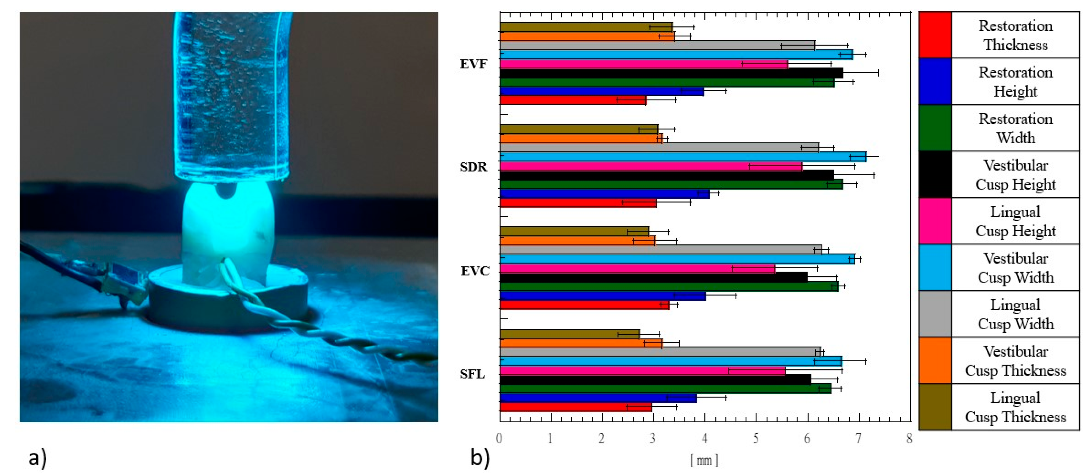
2. Materials and Methods

Four bulk-fill RBCs listed in Table 1 were used. Maximum temperature rise occurring in the composites was preliminarily measured. Mesio-occlusal-distal cavities of human premolars were restored through the bulk-fill or the incremental layering techniques, and temperature rise occurring in dentin, 1 mm below the cavity floor, was evaluated.

2.1. Restorative Materials

Four bulk-fill RBCs (Table 1) largely differing in material composition and rheological properties have been investigated. Low-viscosity flowable SDR bulk-fill was provided in capsules, each containing 0.25 g of composite. Each capsule was equipped with a stainless steel needle from which the composite was injected through a plastic manual gun. Tetric EvoFlow Bulk-fill and Tetric EvoCeram Bulk-fill were provided in syringe. High viscosity Sonic Fill 2 was provided in capsules, each containing 0.25 g of composite. Each capsule was equipped with a plastic needle from which the composite was injected through a sonic vibration hand piece that reduces viscosity during material placement [8]. The polymeric matrix of the investigated composite materials were based on bisphenol A-glycidyl methacrylate (Bis-GMA), ethoxylated bisphenol A dimethacrylate (EBPADMA), urethane dimethacrylate (UDMA), and triethylene glycol dimethacrylate (TEGDMA). Table 1 depicts the material composition of the four bulk-fill RBCs.

2.2. Maximum Temperature Rise

Maximum temperature rise occurring in bulk-fill composites was investigated up to 60 s through a previously described protocol [2]. Briefly, bulk composites were injected into prismatic PTFE moulds of 5.0 mm × 5.0 mm × 1.5 mm. Disposable K-type thermocouples (RS components, Corby, United Kingdom) were used to measure temperature profiles. A micromanipulator was adopted to facilitate the gentle and precise positioning of the thermocouple into the composites. A filtered photocell obtained from the Demetron LED radiometer (Kerr Corporation) was employed to monitor light power level. The light curing unit Swiss Master Light (EMS, Nyon, Switzerland) at an intensity level of 1000 mW/cm2 and exposure time of 10 s was used to light cure composite materials. The halogen bulb of the curing unit was cooled through a water-pumped system that allows to keep the bulb at a safety temperature level even for prolonged exposure time. Light intensity and temperature data were simultaneously acquired at a speed of 50 p/s up to 60 s using the National Instrument DAC (National Instruments, Austin, Texas) driven by Signal Express software (National Instruments). Ten specimens for each bulk-fill composite were used. Temperature peak, temperature rate, and the time to reach the temperature peak were measured. Data were analysed using two-way ANOVA followed by Tukey’s test at a critical value of 0.05.

2.3. Selection of Teeth

Eighty premolars were obtained by orthodontic extractions from Caucasian ethnicity patients aged between 18 and 30 years (approved by the Ethics Committee of the University Naples Federico II, protocol number 137/2017). Teeth were selected according to an average length of 22 ± 1 mm, a buccal-lingual dimension of 7 ± 1 mm, and a disto-mesial distance of 9 ± 1 mm.
Selection criteria were: tooth vitality, absence of conservative reconstructions and/or prosthesis, absence of local gingivitis, no periodontal damage, no periodontal pockets involving root and alveolus, no loss of attachment and no loss of alveolar bone support, no erosion, no enamel infringement (the absence of infringement has been evaluated with a transillumination test), and physiological distance between CEJ (Cement Enamel Junction) and alveolar ridge.
All geometrical details of teeth (Figure 1b) were measured with a digital caliper (Mitutoyo, Takatsuku, Japan). The dental elements have been fixed with acrylic resin in 16-mm diameter aluminium hollow cylinders, and each tooth was X-ray scanned with the Partner 70 equipment (Anthos, Bologna, Italy) in the mesial-distal projection, and bucco-lingual and occlusal-apical at 70 kV for 0.08 s. Dental radiographs were imaged with the MicroDicom viewer v3.0.1 software (MicroDicom, Sofia, Bulgaria). Before the test, the teeth were immersed and kept in a 0.5% thymol solution at 8 °C. Each tooth was tested within 2 months after extraction.

2.4. MOD Cavity Preparation

Teeth were subjected to Class II MOD cavity preparation, and dimensions were 3-mm intercuspal width and 4-mm depth in occlusal box. Cavities were realized with a diamond bur mounted on the turbine Fona8080 (Fonadental, Assago, Italy) on a high-speed contra-angle. Teeth cusps and restoration MOD cavity dimensions are reported in Figure 1b.
Teeth were randomly divided into 4 groups according to the bulk-fill composites employed for the restoration (Figure 1b). Each group, consisting of 20 specimens, was randomly divided into two subgroups according to the type of the layering technique adopted for the MOD restoration (i.e., bulk-fill and incremental layering techniques).

2.5. Adhesive Protocol and Composite Restoration

After cavity preparation, dental substrates were subjected to the following adhesive protocol: the etching gel (37% phosphoric acid) (Gerhò, Bolzano, Italy) was applied for 30 s on enamel and for 15 s on dentin. Subsequently, the cavity was rinsed and dried for 5 s. Then, the adhesive system Optibond SE (Kerr Corporation) was applied in two steps: the primer was applied for 20 s using the microbrush, and then, the cavity was dried with a gentle stream of air for 5 s; then, the bond was applied with a microbrush to create a thin layer of adhesive, and subsequently the cavity was dried with gentle air stream for 5 s. The last step consisted of the adhesive polymerization for 20 s by using the Swiss Master Light curing unit (EMS) at an intensity of 1000 mW/cm2. For each bulk-fill RBC, MOD cavities were restored according to the bulk-fill (one single increment of 4 mm) or the incremental layering (two increments each of 2 mm) techniques. SDR and EVF were injected using the manual gun and the syringe, respectively. EVC was applied using the spatula, while SFL was applied using the sonic vibration hand piece driven by air pressure at 2.5 bar.

2.6. Temperature Measurements

Disposable K-type thermocouples (RS components) were adopted to measure the temperature variation. Thermocouples were placed into a standardized hole created 1 mm below the cavity floor (Figure 1a). A filtered photocell, obtained from Demetron LED radiometer (Kerr Corporation), was employed to monitor the light power level. The light curing unit EMS Swiss Master Light at an intensity level of 1000 mW/cm2 and exposure time of 20 s was employed to cure bulk-fill composites. In the case of MOD restoration, the rationale for choosing an exposure time of 20 s relies on the distance between the light guide of the curing unit and the composite (Figure 1a). In fact, dental cusps prevent the positioning of the light guide of the curing unit close to the composite to be cured. Thus, prolonged exposition time has been considered to compensate for the light intensity attenuation due to the distance between the light guide and the composite. The rationale for choosing a halogen-based curing unit relies on the initiator systems characterizing the bulk-fill composites that have been investigated. In fact, some of these composites (see Table 1) contain both camphorquinone (CQ, absorption peak around 460 nm) and ivocerin (IV, absorption peak around 400 nm). Compared to blue LED (Light Emitting Diode) curing units having a very narrow emission spectrum centred around 460 nm, the halogen curing unit has a broad emission spectrum covering the wavelengths between 400 nm and 480 nm [27]. Thus, this curing unit is more appropriate for curing all the bulk-fill composites that have been investigated. To simulate pulp temperature levels, a Thermoblock system (Falc, Genova, Italy) [23] set at 37 °C was used to keep each sample heated during the test. Before each test, the ThorLab energy meter console PM100D (ThorLabs, Newton, NJ, USA), equipped with a S121C sensor (ThorLabs) and connected to the PMD100D software running under LabView (National Instruments), was employed to measure the power output of the light curing unit.
Temperature and light intensity data were simultaneously acquired at a speed of 50 p/s up to 300 s using the National Instruments DAC (National Instruments) driven by the Signal Express software (National Instruments).
Ten specimens for each bulk-fill composite and for each restorative technique were used. Peak temperature data were analysed using ANOVA, followed by Tukey’s test at a critical value of 0.05.

3. Results

Figure 1b shows that the four samples, displayed in Table 1, can be considered roughly equivalent, from a geometrical point of view. The reported geometrical details are essential for the boundary conditions and may be important for modeling the heat developed through MOD cavities restored with composite materials.
Figure 2a depicts typical temperature profiles recorded for the four bulk-fill composites. Maximum temperature rise levels are reported in Figure 2b. The time required to reach the temperature peak is reported in Figure 2c. A steep temperature profile can be observed for each composite as the light is turned on. The slope of the temperature profile (i.e., temperature rate) is shown in Figure 2d. Flowable bulk-fill RBCs show a significantly higher temperature peak (p < 0.05) than packable bulk-fill RBCs. The peak temperature measured for EVF, SDR, EVC, and SFL were 49.9 °C ± 1.8 °C, 47.5 °C ± 1.7 °C, 44.3 °C ± 1.6 °C, and 41.4 °C ± 1.5 °C, respectively. Flowable bulk-fill RBCs show a significantly higher temperature rate (p < 0.05) than packable bulk-fill RBC (Figure 2d), while the time required to reach the temperature peak (Figure 2c) was significantly lower (p < 0.05).
Simultaneous time measurements of temperature and photo-diode signals for MOD cavities restored with the four bulk-fill RBCs according to the layering technique are reported in Figure 3. For both the bulk-fill (Figure 3a) and incremental layering (Figure 3b) techniques, a steep temperature profile is detected as the light is turned on. For flowable RBCs, the temperature decreases as the light is switched off. Instead, for packable RBCs, a delay is observed in the peak temperature for the bulk-fill technique (Figure 3a), and in the second peak temperature for the incremental layering technique. Maximum peak temperature values were recorded for the flowable EVF and SDR composites (43.9 °C ± 1.8 °C and 42.9°C ± 1.7 °C, respectively) as the incremental layering technique was performed to restore the MOD cavities.
Temperature rise and temperature rate measured for the investigated composite according to the different MOD cavity layering technique are reported in Table 2.

4. Discussion

Dental pulp is highly vascularized with arterial vessels entering from the apex radicular apex and reaching the dental crown through the tubules; the microcirculation of the blood in the dental pulp plays an important role in tooth physiology, also providing the nutrition of the peripheral tissues. Thermal injury represents an important issue related to temperature increase that may result in pulp inflammation [16,17,23]. Radiation and polymerization exothermal reaction simultaneously provide the heat that increases the composite temperature. However, while the radiant light released by the curing unit is almost constant through the whole light curing cycle, the heat due to the exothermal polymerization reaction reaches a peak as the gel-point (the transition of the composite material from the gel state to the glassy state) is approached. The polymerization kinetics is therefore the main heat mechanism shaping the temperature profile occurring around the peak temperature shown in Figure 2.
Figure 2a clearly shows a steep temperature profile as the light is turned on. The temperature rise and the slope measured for the flowable bulk-fill RBCs are significantly higher (p < 0.05) than those measured for the packable composites (Figure 2a,d), and this result is obviously ascribed to the higher amount of resin characterizing flowable composites (Table 1). The type of initiator system also plays a fundamental role, as the temperature rate measured for EVF is significantly higher than SDR (p < 0.05), although the amount of filler of the two flowable RBCs is almost similar (Table 1). The higher temperature rate suggests a faster kinetics; accordingly, the temperature rise of EVF is higher than SDR (Figure 2d), but the difference between the means is not significant (p = 0.59). On the other hand, the significantly lower temperature rate recorded for SFL (p < 0.05) can be ascribed to the high filler amount (Table 1). The recorded temperature increase for SDR (Figure 2b) is consistent with that of 26 °C measured on the surface of the specimen, with similar thickness, using a thermal imaging camera [27], while a higher temperature increase has been measured on thicker specimens using thermocouples [6] and Bragg grating sensors [28]. These temperature rise values would be detrimental for the pulp tissue. Fortunately, thermal conductivity of dentin [29,30] effectively reduces temperature rise occurring into the pulp. In fact, temperature rise recorded by the thermocouples placed 1 mm below the MOD cavity floor shows that temperature increase values are lower than 13 °C (Figure 3 and Table 2).
Concerning the bulk-fill technique (Figure 3a), temperature rise mean values spanned from 8.2 °C to 10.6 °C, and higher temperature levels have been recorded for the flowable RBCs (Table 2). However, the difference between the mean values recorded for the different composites is not significant. Instead, the temperature rate measured for EVF (Table 2) is significantly higher than that measured for the packable RBCs (p < 0.05). Again, this result can be ascribed to the higher resin content of flowable RBCs and to the type of initiator system (Table 1). Karacan and Ozyurt have recorded similar temperature increase values with thermocouples placed 1 mm below the MOD cavity and using a high viscosity bulk-fill composite [24]. Since dentin acts as a thermal insulator [27], the thickness of occlusal dentin, spacing the restorative material from the pulp chamber, is of great importance. In fact, higher temperature increase levels (12.8 °C) have been recorded for SDR as the thickness of occlusal dentin is reduced to 0.5 mm [31]. For both flowable and packable composites the light curing modality of 1000 mW/cm2 for 20 s can be considered safe, if the bulk-fill technique is performed and an appropriate thickness of occlusal dentin is preserved. However, it must be pointed out that, for all the investigated RBCs, the starting temperature level (Figure 3) is about 31 °C, although the Thermoblock system used to mimic temperature levels occurring in the oral environment has been set at 37 °C. The reason is related to the rinsing and drying processes involved in the adhesive procedure that drastically reduces temperature level of the coronal tissues. Therefore, it is very important that the RBCs restorative procedure is performed soon after the adhesive procedure; otherwise, the temperature base level of dental tissues may increase and the temperature rise occurring during photo-polymerization (Table 2) may largely exceed the temperature level of 42 °C, thus representing a potential thermal injury for the pulp tissue.
Concerning the incremental layering technique (Figure 3b), temperature rise values recorded through the first increment spanned from 9.5 °C to 12.9 °C, and higher temperature levels have been recorded for the flowable RBCs (Table 2). The Lambert’s law can be used to explain the higher temperature levels recorded for the incremental layer technique. It is important to distinguish between light attenuation occurring through the composite height (bulk-fill technique) and light attenuation due to the distance between the light source and the composite (incremental layer technique). The coefficient of attenuation of the composite is much higher than the coefficient of attenuation occurring through the air medium [27]. For this reason, for the bulk-fill technique, the bottom layer (close to the pulp chamber) receives an attenuated light intensity due to the shading effect of the top layer. Instead, for the incremental layer technique, light intensity polymerizing the first layer increment (close to the pulp chamber) has a lower attenuation. As a consequence of the different light attenuation, higher temperature levels have been recorded for the incremental layer technique.
It is worth to note that the base line for temperature measurements of the composites (Figure 2) is close to room temperature, while that related to MOD restoration is about 31 °C (Figure 3). Even if there are some clinicians who prefer to store the dental composite in the fridge, or other clinicians who prefer to heat the composite before applying into the dental cavity, most clinicians store the dental composites at room temperature. As the composite is applied, light curing is suddenly performed (heat transfer from the dental cavity walls to the composite is prevented, as there is no sufficient time for heat to be transferred). Therefore, the base line of the applied composite is room temperature (Figure 2). Instead, in Figure 3, the base temperature line is close to that of the pulp cavity. To simulate pulp temperature levels, in our experiments, we used a Thermoblock system [23] set at 37 °C. As shown in Figure 1a, the dental root can be assumed to be at the set temperature of 37 °C, but the dental crown is at a lower temperature level. Before applying the composite, an adhesive interface is realized, and the clinical procedure involves abundant rising, thus cooling the crown. For this reason, the base temperature line of the dentine layer hosting the thermocouple drops to 31 °C. Since this dental region is located in the tooth crown (see Figure 1a), and the composite restoration is realized immediately after the creation of the adhesive interface, the short time prevents heat transfer from the root heated at 37 °C to the cooled crown. For this reason, the temperature base line for the experiments of Figure 3 is located at about 31°C. Our set-up approach tries to simulate clinical conditions as close as possible. Therefore, the reported temperature levels and profiles should be very close to those occurring in clinical conditions.
Temperature profiles recorded for the investigated composites show that the temperature rise recorded for EVF is significantly higher than that recorded for SFL (p < 0.05). Again, this difference can be ascribed to the higher resin content of EVF and to the type of initiator system (Table 1). Temperature rates measured for the flowable RBCs (Table 2) are significantly higher than those measured for the packable RBCs (p < 0.05). Temperature peak levels higher than 42 °C have been observed for the flowable RBCs, thus representing a potential thermal injury for the pulp tissue. Therefore, as flowable bulk-fill RBCs in conjunction with the incremental layering technique is concerned, a curing modality lower than 1000 mW/cm2 is recommended, especially if the thickness of the occlusal dentin, spacing the restorative material from the pulp chamber, is lower than 1 mm.

5. Conclusions

Based on the reported results and within the limitation of this investigation, the following conclusions can be reported:
  • Temperature peaks and temperature rates measured in the core of flowable bulk-fill composites are significantly higher (p < 0.05) than those measured for packable bulk-fill composites.
  • The type of initiator system affects both the temperature rise and rate; a significantly higher temperature rate (p < 0.05) has been measured for EVF, suggesting faster polymerization kinetics.
  • For the bulk-fill technique, temperature rise levels are below 11 °C; however, no significant difference in the mean values of temperature rise has been observed among the investigated bulk-fill RBCs.
  • For the bulk-fill techniques, the light curing modality (1000 mW/cm2 for 20 s) can be considered safe for the integrity of the pulp tissue, if the thickness of the occlusal dentin, spacing the restorative material from the pulp chamber, is not lower than 1 mm.
  • For the bulk-fill technique, temperature rise levels are below 13 °C. Temperature rates measured for the flowable RBCs are significantly higher (p < 0.05) than those measured for the packable RBCs. The temperature rise measured for EVF is significantly higher (p < 0.05) than SFL.
  • As far as flowable bulk-fill RBCs in conjunction with the incremental layering technique are concerned, a curing modality lower than 1000 mW/cm2 is recommended, especially if the thickness of the occlusal dentin, spacing the restorative material from the pulp chamber, is lower than 1 mm.
  • Finally, the results that we have observed with the halogen curing unit may be extendable to the LED curing units incorporating a variety of semiconductors: a blue LED and a violet LED. This approach will be implemented in our future investigations.
  • The clinical operator should avoid the use of high level of light intensity for curing flow composites applied through the incremental technique in very deep cavities close to the pulp chamber. In these cases, a reduced light intensity in conjunction with an increased exposure time of the light source is recommended. The bulk-fill technique can be used safely without fear of causing thermal damage to the dental pulp.

Author Contributions

Conceptualization, R.D.S. and S.R.; methodology, V.L.; software, C.R.; validation, S.R., R.D.S. and A.V.; formal analysis, C.R.; investigation, V.G.; resources, S.R.; data curation, R.D.S.; writing—original draft preparation, R.D.S., V.G. and V.L.; writing—review and editing, R.D.S., V.G. and V.L.; visualization, A.V.; supervision, S.R.; project administration, A.V.; funding acquisition, S.R. All authors have read and agreed to the published version of the manuscript.

Funding

This work was financially supported by the PON 03 PE_00110_1/ptd1_000410. Title: Sviluppo dinanotecnologie orientate alla rigenerazione e ricostruzione tissutale, implantologia e sensoristica inodontoiatria/oculistica (SORRISO).

Acknowledgments

We thank Antonio Gallo and Antonio Franco for providing Kerr and Ivoclar composite materials.

Conflicts of Interest

The authors do not have any financial interest in the companies whose materials are included in this article. This study was approved by the Ethics Committee of the University of Naples Federico II, with protocol number 137–2017.

References

  1. Braga, R.R.; Ballester, R.Y.; Ferracane, J.L. Factors involved in the development of polymerization shrinkage stress in resin-composites: A systematic review. Dent. Mater. 2005, 21, 962–970. [Google Scholar] [CrossRef]
  2. De Santis, R.; Gloria, A.; Prisco, D.; Amendola, E.; Puppulin, L.; Pezzotti, G.; Rengo, S.; Ambrosio, L.; Nicolais, L. Fast curing of restorative materials through the soft light energy release. Dent. Mater. 2010, 26, 891–900. [Google Scholar] [CrossRef] [PubMed]
  3. Pashley, D.H. Clinical considerations of microleakage. J. Endod. 1990, 16, 70–77. [Google Scholar] [CrossRef]
  4. De Santis, R.; Mollica, F.; Prisco, D.; Rengo, S.; Ambrosio, L.; Nicolais, L. A 3D analysis of mechanically stressed dentin–adhesive–composite interfaces using X-ray micro-CT. Biomaterials 2005, 26, 257–270. [Google Scholar] [CrossRef] [PubMed]
  5. Kwon, Y.; Ferracane, J.; Lee, I.B. Effect of layering methods, composite type, and flowable liner on the polymerization shrinkage stress of light cured composites. Dent. Mater. 2012, 28, 801–809. [Google Scholar] [CrossRef]
  6. Kim, R.J.; Son, S.A.; Hwang, J.Y.; Lee, I.B.; Seo, D.G. Comparison of photopolymerization temperature increases in internal and external positions of composite and tooth cavities in real time: Incremental fillings of microhybrid composite vs. bulk filling of bulk fill composite. J. Dent. 2015, 43, 1093–1098. [Google Scholar] [CrossRef]
  7. Ilie, N.; Bucuta, S.; Draenert, M. Bulk-fill resin-based composites: An in vitro assessment of their mechanical performance. Oper. Dent. 2013, 38, 618–625. [Google Scholar] [CrossRef]
  8. Chesterman, J.; Jowett, A.; Gallacher, A.; Nixon, P. Bulk-fill resin-based composite restorative materials: A review. Br. Dent. J. 2017, 222, 337. [Google Scholar] [CrossRef]
  9. Lima, R.B.; Troconis, C.C.; Moreno, M.B.; Murillo-Gomez, F.; De Goes, M.F. Depth of cure of bulk fill resin composites: A systematic review. J. Esthet. Restor. Dent. 2018, 30, 492–501. [Google Scholar] [CrossRef]
  10. Hirata, R.; Kabbach, W.; De Andrade, O.S.; Bonfante, E.A.; Giannini, M.; Coelho, P.G. Bulk fill composites: An anatomic sculpting technique. J. Esthet. Restor. Dent. 2015, 27, 335–343. [Google Scholar] [CrossRef]
  11. Al-Ahdal, K.; Silikas, N.; Watts, D.C. Rheological properties of resin composites according to variations in composition and temperature. Dent. Mater. 2014, 30, 517–524. [Google Scholar] [CrossRef] [PubMed]
  12. El-Safty, S.; Akhtar, R.; Silikas, N.; Watts, D.C. Nanomechanical properties of dental resin-composites. Dent. Mater. 2012, 28, 1292–1300. [Google Scholar] [CrossRef] [PubMed]
  13. Swift, E.J., Jr. Bulk-fill Composites, Part, I. J. Esthet. Restor. Dent. 2015, 27, 176–179. [Google Scholar] [CrossRef] [PubMed]
  14. Kim, R.J.; Kim, Y.J.; Choi, N.; Lee, I. Polymerization shrinkage, modulus, and shrinkage stress related to tooth-restoration interfacial debonding in bulk-fill composites. J. Dent. 2015, 43, 430–439. [Google Scholar] [CrossRef]
  15. Ibarra, E.; Lien, W.; Jeffery Casey, J.; Dixon, S.A.; Vandewalle, K. Physical properties of a new sonically placed composite resin restorative material. Unif. Serv. Univ. Health Sci. Bethesda U. S. 2015, 63, 51–56. [Google Scholar]
  16. Bouillaguet, S.; Caillot, G.; Forchelet, J.; Cattani-Lorente, M.; Wataha, J.C.; Krejci, I. Thermal risks from LED-and high-intensity QTH-curing units during polymerization of dental resins. J. Biomed. Mater. Res. Part B Appl. Biomater. 2005, 72, 260–267. [Google Scholar] [CrossRef] [Green Version]
  17. Soares, C.J.; Ferreira, M.S.; Bicalho, A.; Rodrigues, M.; Braga, S.S.L.; Versluis, A. Effect of light activation of pulp-capping materials and resin composite on dentin deformation and the pulp temperature change. Oper. Dent. 2018, 43, 71–80. [Google Scholar] [CrossRef]
  18. De Santis, R.; Gloria, A.; Maietta, S.; Martorelli, M.; De Luca, A.; Spagnuolo, G.; Riccitiello, F.; Rengo, S. Mechanical and thermal properties of dental composites cured with CAD/CAM assisted solid-state laser. Materials 2018, 11, 504. [Google Scholar] [CrossRef] [Green Version]
  19. Braga, S.; Oliveira, L.; Ribeiro, M.; Vilela, A.B.F.; da Silva, G.R.; Price, R.B.; Soares, C.J. Effect of Simulated Pulpal Microcirculation on Temperature When Light Curing Bulk Fill Composites. Oper. Dent. 2019, 44, 289–301. [Google Scholar] [CrossRef]
  20. Runnacles, P.; Arrais, C.A.; Pochapski, M.T.; Dos Santos, F.A.; Coelho, U.; Gomes, J.C.; De Goes, M.F.; Mongruel Gomes, O.M.; Rueggeberg, F.A. In vivo temperature rise in anesthetized human pulp during exposure to a polywave LED light curing unit. Dent. Mater. 2015, 31, 505–513. [Google Scholar] [CrossRef]
  21. Zarpellon, D.C.; Runnacles, P.; Maucoski, C.; Coelho, U.; Rueggeberg, F.A.; Arrais, C. Controlling In Vivo, Human Pulp Temperature Rise Caused by LED Curing Light Exposure. Oper. Dent. 2019, 44, 235–241. [Google Scholar] [CrossRef] [PubMed]
  22. Airoldi, G.; Riva, G.; Vanelli, M.; Filippi, V.; Garattini, G. Oral environment temperature changes induced by cold/hot liquid intake. Am. J. Orthod. Dentofac. Orthop. 1997, 112, 58–63. [Google Scholar] [CrossRef]
  23. Simeone, M.; De Santis, R.; Ametrano, G.; Prisco, D.; Borrelli, M.; Paduano, S.; Riccitiello, F.; Spagnuolo, G. Temperature Profiles Along the Root with Gutta-percha Warmed through Different Heat Sources. Open Dent. J. 2014, 8, 229–235. [Google Scholar] [CrossRef] [PubMed] [Green Version]
  24. Karacan, A.O.; Ozyurt, P. Effect of preheated bulk-fill composite temperature on intrapulpal temperature increase in vitro. J. Esthet. Restor. Dent. 2019, 31, 583–588. [Google Scholar] [CrossRef]
  25. Hamze, F.; Nasab, S.A.G.; Eskandarizadeh, A.; Shahravan, A.; Fard, F.A.; Sinaee, N. Thermal Scanning of Dental Pulp Chamber by Thermocouple System and Infrared Camera during Photo Curing of Resin Composites. Iran. Endod. J. 2018, 13, 195. [Google Scholar] [CrossRef]
  26. Santini, A.; Watterson, C.; Miletic, V. Temperature rise within the pulp chamber during composite resin polymerisation using three different light sources. Open Dent. J. 2008, 2, 137. [Google Scholar] [CrossRef]
  27. De Santis, R.; Gloria, A.; Sano, H.; Amendola, E.; Prisco, D.; Mangani, F.; Rengo, S.; Ambrosio, L.; Nicolais, L. Effect of light curing and dark reaction phases on the thermomechanical properties of a Bis-GMA based dental restorative material. J. Appl. Biomater. Biomech. 2009, 7, 132–140. [Google Scholar]
  28. Vinagre, A.; Ramos, J.; Alves, S.; Messias, A.; Alberto, N.; Nogueira, R. Cuspal displacement induced by bulk fill resin composite polymerization: Biomechanical evaluation using fiber Bragg grating sensors. Int. J. Biomater. 2016, 1–9. [Google Scholar] [CrossRef] [Green Version]
  29. Craig, R.G.; Peyton, F.A. Thermal conductivity of tooth structure, dental cements, and amalgam. J. Dent. Res. 1961, 40, 411–418. [Google Scholar] [CrossRef]
  30. de Magalhaes, M.F.; Ferreira, R.A.; Grossi, P.A.; de Andrade, R.M. Measurement of thermophysical properties of human dentin: Effect of open porosity. J. Dent. 2008, 36, 588–594. [Google Scholar] [CrossRef]
  31. Yasa, E.; Atalayin, C.; Karacolak, G.; Sari, T.; Turkun, L.S. Intrapulpal temperature changes during curing of different bulk-fill restorative materials. Dent. Mater. J. 2017, 36, 566–572. [Google Scholar] [CrossRef] [PubMed] [Green Version]
Figure 1. (a) Experimental set-up for the simultaneous measurement of light intensity and temperature levels occurring in mesio-occlusal-distal (MOD) cavities; (b) geometrical details of human premolars and MOD cavities (b).
Applsci 10 08041 g001
Figure 2. (a) Temperature profiles developing into bulk-fill RBC; (b) maximum temperature rise; (c) time to temperature peak; and (d) temperature rate. Error bars represent the standard deviation. Different letters reported beside the bars indicate a significant difference between the means (p < 0.05).
Applsci 10 08041 g002
Figure 3. Simultaneous time measurements of temperature and photo-diode signals for MOD cavities restored with the investigated bulk-fill RBCs according to: (a) the bulk-fill technique; (b) the incremental technique.
Applsci 10 08041 g003
Table 1. Bulk-fill resin based composite (RBC) composition. The initiator system is based on camphorquinone (CQ) and ivocerin (IV).
MaterialManufacturerMatrixFillerInitiatorShadeAcronym
Tetric EvoFlow Bulk-fillIvoclar Vivadent, Schaan,
Liechtenstein
Bis-GMA, EBPADMA68.2 wt%: Barium glass, ytterbium trifluorideCQ, IVUniversal
A
EVF
SDR flow +Sirona Dentsply, Konstantz, GermanyUDMA,
TEGDMA, EBPDMA
68 wt%: Ba-Al-F-B-Si glass and St-Al-F-Si glassCQUniversalSDR
Tetric EvoCeram Bulk-fillIvoclar Vivadent, Schaan,
Liechtenstein
bis-GMA, UDMA79.5 wt%: Barium glass filler, Ytterbium trifluoride, Mixed oxide, prepolymerCQ, IVUniversal
A
EVC
SonicFill 2Kerr Corporation, Orange, CaliforniaBis-GMA, TEGDMA,
EBPDMA
83.5 wt%: SiO2, glass, oxide, prepolymerCQA2SFL
Table 2. Temperature rise and temperature rate measured for the investigated bulk-fill RBCs according to the different restoration techniques. Numbers in brackets represent the standard deviation. For each column, different superscript letters denote significant difference (p < 0.05).
Bulk-Fill TechniqueIncremental Layering Technique
First IncrementSecond Increment
∆T [°C]Temperature Rate [°C/s]∆T [°C]Temperature Rate [°C/s]∆T [°C]Temperature Rate [°C/s]
EVF10.6 (1.9) a0.72 (0.09) a12.9 (1.8) a1.69 (0.18) a7.0 (1.3) a0.33 (0.07) a
SDR9.5 (1.7) a0.57 (0.07) a,b11.9 (1.7) a,b1.54 (0.15) a6.5 (1.1) a0.27 (0.05) a,b
EVC8.7 (1.3) a0.53 (0.06) b10.1 (1.4) a,b0.72 (0.09) b5.3 (1.0) a0.23 (0.05) a,b
SFL8.2 (1.6) a0.45 (0.07) b9.5 (1.4) b0.69 (0.10) b5.1 (1.1) a0.21 (0.04) b
Publisher’s Note: MDPI stays neutral with regard to jurisdictional claims in published maps and institutional affiliations.

